Hypothermia is defined as a drop in the core body temperature below 35°C (95°F), and is classified into mild, moderate, severe, and profound forms based on the degree of temperature decrease. Individuals who are more vulnerable to accidental hypothermia include those at the extremes of age, the homeless, and those who have psychiatric or substance abuse disorders. Symptoms progress from shivering and lethargy to confusion, coma, and death. Mild hypothermia requires a warm environment and insulating blankets (passive rewarming). Severe or profound hypothermia requires active rewarming of the core (e.g., extracorporeal blood rewarming, heated infusion and lavage, inhalation), and the body surface (e.g., with forced-air warming systems, radiant sources). Evaluation should include assessment for associated trauma and contributing medical conditions.This monograph is limited to a discussion of accidental hypothermia, but therapeutic hypothermia is recommended in order to reduce the risk of neurologic injury in a post-cardiac arrest adult patient who is not awake (i.e., does not follow verbal commands).
HypothermiaHypothermiaHypothermia can be defined as a drop in the core body temperature below 35°C (95°F) and is classified into mild, moderate, severe, and profound forms based on the degree of temperature decrease. Hypothermia is a decrease in core body temperatureBody TemperatureThe measure of the level of heat of a human or animal.Heatstroke to below 35°C (95°F). The focus of this concept page is primarily on accidental hypothermiaAccidental HypothermiaHypothermia.
Epidemiology[4,6,7]
More common in cold climates, but occurs throughout the world
Alcohol intoxicationAlcohol intoxicationAn acute brain syndrome which results from the excessive ingestion of ethanol or alcoholic beverages.Alcohol Use Disorder
Substance abuse
Advanced age
Psychiatric disease
Etiology[1‒3]
Primary → prolonged exposure to cold environmental temperatures:
Adrenal insufficiencyAdrenal InsufficiencyConditions in which the production of adrenal corticosteroids falls below the requirement of the body. Adrenal insufficiency can be caused by defects in the adrenal glands, the pituitary gland, or the hypothalamus.Adrenal Insufficiency and Addison Disease
HypothyroidismHypothyroidismHypothyroidism is a condition characterized by a deficiency of thyroid hormones. Iodine deficiency is the most common cause worldwide, but Hashimoto’s disease (autoimmune thyroiditis) is the leading cause in non-iodine-deficient regions. Hypothyroidism
HypoglycemiaHypoglycemiaHypoglycemia is an emergency condition defined as a serum glucose level ≤ 70 mg/dL (≤ 3.9 mmol/L) in diabetic patients. In nondiabetic patients, there is no specific or defined limit for normal serum glucose levels, and hypoglycemia is defined mainly by its clinical features. Hypoglycemia
SkinSkinThe skin, also referred to as the integumentary system, is the largest organ of the body. The skin is primarily composed of the epidermis (outer layer) and dermis (deep layer). The epidermis is primarily composed of keratinocytes that undergo rapid turnover, while the dermis contains dense layers of connective tissue.Skin: Structure and Functions diseases (burnsBurnsA burn is a type of injury to the skin and deeper tissues caused by exposure to heat, electricity, chemicals, friction, or radiation. Burns are classified according to their depth as superficial (1st-degree), partial-thickness (2nd-degree), full-thickness (3rd-degree), and 4th-degree burns. Burns)
EthanolEthanolA clear, colorless liquid rapidly absorbed from the gastrointestinal tract and distributed throughout the body. It has bactericidal activity and is used often as a topical disinfectant. It is widely used as a solvent and preservative in pharmaceutical preparations as well as serving as the primary ingredient in alcoholic beverages.Ethanol Metabolism (impaired responses to cold/shiveringShiveringInvoluntary contraction or twitching of the muscles. It is a physiologic method of heat production in man and other mammals.Body Temperature Regulation, vasodilationVasodilationThe physiological widening of blood vessels by relaxing the underlying vascular smooth muscle.Pulmonary Hypertension Drugs)
Impaired thermoregulationThermoregulationBody temperature can be divided into external temperature, which involves the skin, and core temperature, which involves the CNS and viscera. While external temperature can be variable, the core temperature is maintained within a narrow range of 36.5-37.5ºC (97.7-99.5ºF).Body Temperature Regulation:
Spinal cord injuriesSpinal cord injuriesSpinal cord injuries are complex injuries that involve damage to the neural tissue within the spinal canal. Spinal cord injuries are commonly the result of trauma. Clinical presentation varies depending on the site of injury and on whether the injury is complete or incomplete. Spinal Cord Injuries
PolytraumaPolytraumaMultitrauma occurs when 2 or more traumatic injuries occur in at least 2 areas of the body. A systematic management approach is necessary for individuals who have undergone trauma to maximize outcomes and reduce the risk of undiscovered injuries.Multitrauma
Accidental hypothermia results from exposure to cold temperatures.
HeatHeatInflammation is lost mainly through the skinSkinThe skin, also referred to as the integumentary system, is the largest organ of the body. The skin is primarily composed of the epidermis (outer layer) and dermis (deep layer). The epidermis is primarily composed of keratinocytes that undergo rapid turnover, while the dermis contains dense layers of connective tissue.Skin: Structure and Functions and lungsLungsLungs are the main organs of the respiratory system. Lungs are paired viscera located in the thoracic cavity and are composed of spongy tissue. The primary function of the lungs is to oxygenate blood and eliminate CO2. Lungs: Anatomy:
Evaporation
RadiationRadiationEmission or propagation of acoustic waves (sound), electromagnetic energy waves (such as light; radio waves; gamma rays; or x-rays), or a stream of subatomic particles (such as electrons; neutrons; protons; or alpha particles).Osteosarcoma
The mean normal set point for human core temperature is 37 ± 0.5°C (98.6 ± 0.9°F).
The body uses autonomic mechanisms to conserve heatHeatInflammation in response to a cold environment.
The hypothalamusHypothalamusThe hypothalamus is a collection of various nuclei within the diencephalon in the center of the brain. The hypothalamus plays a vital role in endocrine regulation as the primary regulator of the pituitary gland, and it is the major point of integration between the central nervous and endocrine systems.Hypothalamus stimulates heat productionHeat ProductionFever through:
ShiveringShiveringInvoluntary contraction or twitching of the muscles. It is a physiologic method of heat production in man and other mammals.Body Temperature Regulation:
↑ Cardiac outputCardiac outputThe volume of blood passing through the heart per unit of time. It is usually expressed as liters (volume) per minute so as not to be confused with stroke volume (volume per beat).Cardiac Mechanics
↑ ThyroidThyroidThe thyroid gland is one of the largest endocrine glands in the human body. The thyroid gland is a highly vascular, brownish-red gland located in the visceral compartment of the anterior region of the neck.Thyroid Gland: Anatomy hormone
↑ CatecholaminesCatecholaminesA general class of ortho-dihydroxyphenylalkylamines derived from tyrosine.Adrenal Hormones
Once the core temperature reaches 32°C (90°F), shiveringShiveringInvoluntary contraction or twitching of the muscles. It is a physiologic method of heat production in man and other mammals.Body Temperature Regulation becomes less effective and usually stops.
Metabolism, ventilationVentilationThe total volume of gas inspired or expired per unit of time, usually measured in liters per minute.Ventilation: Mechanics of Breathing, cardiac outputCardiac outputThe volume of blood passing through the heart per unit of time. It is usually expressed as liters (volume) per minute so as not to be confused with stroke volume (volume per beat).Cardiac Mechanics, and mental status/level of consciousness begin to decline below a core temperature of 35°C (95°F).
The following features may be present, depending on the severity of hypothermiaHypothermiaHypothermia can be defined as a drop in the core body temperature below 35°C (95°F) and is classified into mild, moderate, severe, and profound forms based on the degree of temperature decrease. Hypothermia:
Cardiac:
Dysrhythmias (e.g., atrial fibrillationAtrial fibrillationAtrial fibrillation (AF or Afib) is a supraventricular tachyarrhythmia and the most common kind of arrhythmia. It is caused by rapid, uncontrolled atrial contractions and uncoordinated ventricular responses. Atrial Fibrillation, ventricular fibrillationVentricular fibrillationVentricular fibrillation (VF or V-fib) is a type of ventricular tachyarrhythmia (> 300/min) often preceded by ventricular tachycardia. In this arrhythmia, the ventricle beats rapidly and sporadically. The ventricular contraction is uncoordinated, leading to a decrease in cardiac output and immediate hemodynamic collapse. Ventricular Fibrillation (V-fib))
BradycardiaBradycardiaBradyarrhythmia is a rhythm in which the heart rate is less than 60/min. Bradyarrhythmia can be physiologic, without symptoms or hemodynamic change. Pathologic bradyarrhythmia results in reduced cardiac output and hemodynamic instability causing syncope, dizziness, or dyspnea.Bradyarrhythmias
Atrioventricular blockAtrioventricular blockAtrioventricular (AV) block is a bradyarrhythmia caused by delay, or interruption, in the electrical conduction between the atria and the ventricles. Atrioventricular block occurs due to either anatomic or functional impairment, and is classified into 3 types. Atrioventricular block (AV block)
AsystoleAsystoleNo discernible electrical activity, flatline on electrocardiogram (P waves and QRS complexes are not present).Cardiac Arrest
Vascular
Peripheral vasoconstrictionVasoconstrictionThe physiological narrowing of blood vessels by contraction of the vascular smooth muscle.Vascular Resistance, Flow, and Mean Arterial Pressure → cool, pale skinSkinThe skin, also referred to as the integumentary system, is the largest organ of the body. The skin is primarily composed of the epidermis (outer layer) and dermis (deep layer). The epidermis is primarily composed of keratinocytes that undergo rapid turnover, while the dermis contains dense layers of connective tissue.Skin: Structure and Functions
LethargyLethargyA general state of sluggishness, listless, or uninterested, with being tired, and having difficulty concentrating and doing simple tasks. It may be related to depression or drug addiction.Hyponatremia
ComaComaComa is defined as a deep state of unarousable unresponsiveness, characterized by a score of 3 points on the GCS. A comatose state can be caused by a multitude of conditions, making the precise epidemiology and prognosis of coma difficult to determine. Coma
Stages of hypothermiaHypothermiaHypothermia can be defined as a drop in the core body temperature below 35°C (95°F) and is classified into mild, moderate, severe, and profound forms based on the degree of temperature decrease. Hypothermia (by core temperature)[3‒6,12]
The following stages are based on core temperature, determined by placement of a low-reading thermometer (< 34°C [93°F]) in the rectumRectumThe rectum and anal canal are the most terminal parts of the lower GI tract/large intestine that form a functional unit and control defecation. Fecal continence is maintained by several important anatomic structures including rectal folds, anal valves, the sling-like puborectalis muscle, and internal and external anal sphincters. Rectum and Anal Canal: Anatomy (if patient conscious), or by probeProbeA device placed on the patient’s body to visualize a targetUltrasound (Sonography) in the lower one-third of the esophagusEsophagusThe esophagus is a muscular tube-shaped organ of around 25 centimeters in length that connects the pharynx to the stomach. The organ extends from approximately the 6th cervical vertebra to the 11th thoracic vertebra and can be divided grossly into 3 parts: the cervical part, the thoracic part, and the abdominal part. Esophagus: Anatomy.
Note: Clinical signs are more important for stagingStagingMethods which attempt to express in replicable terms the extent of the neoplasm in the patient.Grading, Staging, and Metastasis than temperature readings, since individual responses vary, and overlap exists. Other causes of abnormal findings besides hypothermiaHypothermiaHypothermia can be defined as a drop in the core body temperature below 35°C (95°F) and is classified into mild, moderate, severe, and profound forms based on the degree of temperature decrease. Hypothermia must be excluded.
ShiveringShiveringInvoluntary contraction or twitching of the muscles. It is a physiologic method of heat production in man and other mammals.Body Temperature Regulation is maximal.
DysarthriaDysarthriaDisorders of speech articulation caused by imperfect coordination of pharynx, larynx, tongue, or face muscles. This may result from cranial nerve diseases; neuromuscular diseases; cerebellar diseases; basal ganglia diseases; brain stem diseases; or diseases of the corticobulbar tracts. The cortical language centers are intact in this condition.Wilson Disease and ataxiaAtaxiaImpairment of the ability to perform smoothly coordinated voluntary movements. This condition may affect the limbs, trunk, eyes, pharynx, larynx, and other structures. Ataxia may result from impaired sensory or motor function. Sensory ataxia may result from posterior column injury or peripheral nerve diseases. Motor ataxia may be associated with cerebellar diseases; cerebral cortex diseases; thalamic diseases; basal ganglia diseases; injury to the red nucleus; and other conditions.Ataxia-telangiectasia develop.
Patient is apathetic.
TachycardiaTachycardiaAbnormally rapid heartbeat, usually with a heart rate above 100 beats per minute for adults. Tachycardia accompanied by disturbance in the cardiac depolarization (cardiac arrhythmia) is called tachyarrhythmia.Sepsis in Children
ShiveringShiveringInvoluntary contraction or twitching of the muscles. It is a physiologic method of heat production in man and other mammals.Body Temperature Regulation slows around 32°C, ceases at approximately 30°C.
BradycardiaBradycardiaBradyarrhythmia is a rhythm in which the heart rate is less than 60/min. Bradyarrhythmia can be physiologic, without symptoms or hemodynamic change. Pathologic bradyarrhythmia results in reduced cardiac output and hemodynamic instability causing syncope, dizziness, or dyspnea.Bradyarrhythmias usually occurs below 30°C.
ComaComaComa is defined as a deep state of unarousable unresponsiveness, characterized by a score of 3 points on the GCS. A comatose state can be caused by a multitude of conditions, making the precise epidemiology and prognosis of coma difficult to determine. Coma develops, and reflexes and voluntary motion are absent.
Ventricular dysrhythmias (ventricular fibrillationVentricular fibrillationVentricular fibrillation (VF or V-fib) is a type of ventricular tachyarrhythmia (> 300/min) often preceded by ventricular tachycardia. In this arrhythmia, the ventricle beats rapidly and sporadically. The ventricular contraction is uncoordinated, leading to a decrease in cardiac output and immediate hemodynamic collapse. Ventricular Fibrillation (V-fib))
Respiratory depression or apnea
HypotensionHypotensionHypotension is defined as low blood pressure, specifically < 90/60 mm Hg, and is most commonly a physiologic response. Hypotension may be mild, serious, or life threatening, depending on the cause. Hypotension is expected.
Non-cardiogenic pulmonary edemaPulmonary edemaPulmonary edema is a condition caused by excess fluid within the lung parenchyma and alveoli as a consequence of a disease process. Based on etiology, pulmonary edema is classified as cardiogenic or noncardiogenic. Patients may present with progressive dyspnea, orthopnea, cough, or respiratory failure.Pulmonary Edema
Profound bradycardiaBradycardiaBradyarrhythmia is a rhythm in which the heart rate is less than 60/min. Bradyarrhythmia can be physiologic, without symptoms or hemodynamic change. Pathologic bradyarrhythmia results in reduced cardiac output and hemodynamic instability causing syncope, dizziness, or dyspnea.Bradyarrhythmias/asystoleAsystoleNo discernible electrical activity, flatline on electrocardiogram (P waves and QRS complexes are not present).Cardiac Arrest and apnea are expected.
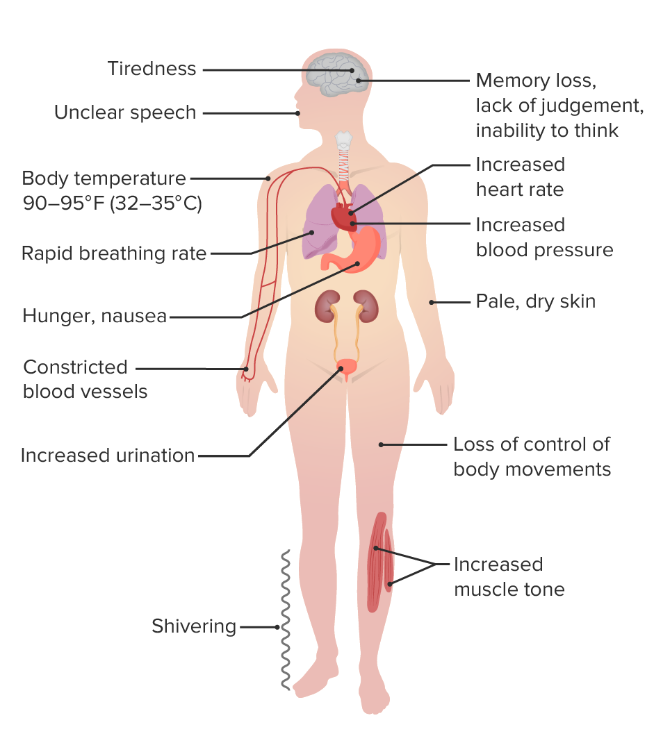
Clinical presentation of mild hypothermia
Image by Lecturio.
Stages of hypothermiaHypothermiaHypothermia can be defined as a drop in the core body temperature below 35°C (95°F) and is classified into mild, moderate, severe, and profound forms based on the degree of temperature decrease. Hypothermia (used when accurate core temperatures cannot be obtained)
There are several classification systems. The following are the 2 most widely used systems for classifying accidental hypothermiaAccidental HypothermiaHypothermia, which are used when an accurate core temperature measurementTemperature MeasurementMeasurement of the temperature of a material, or of the body or an organ by various temperature sensing devices which measure changes in properties of the material that vary with temperature, such as elasticity; magnetic fields; or luminescence.Body Temperature Regulation cannot be obtained (e.g., prehospital setting or if a low-reading thermometer is unavailable ):
Wilderness Medical Society (WMS):[5,6]
Cold-stressed, not hypothermic:
Normal mental status
ShiveringShiveringInvoluntary contraction or twitching of the muscles. It is a physiologic method of heat production in man and other mammals.Body Temperature Regulation, but functioning normally
Normal mental status, but not functioning normally and unable to care for self
ShiveringShiveringInvoluntary contraction or twitching of the muscles. It is a physiologic method of heat production in man and other mammals.Body Temperature Regulation
Abnormal mental status with or without shiveringShiveringInvoluntary contraction or twitching of the muscles. It is a physiologic method of heat production in man and other mammals.Body Temperature Regulation
UnconsciousUnconsciousThose forces and content of the mind which are not ordinarily available to conscious awareness or to immediate recall.Psychotherapy
Estimated core temperature: < 28℃ (82.4°F)
Revised Swiss system (based on level of consciousness; Alert, Verbal, PainPainAn unpleasant sensation induced by noxious stimuli which are detected by nerve endings of nociceptive neurons.Pain: Types and Pathways, Unresponsive (AVPU)):[6]
Note: reliable only if the patient does not have a coexisting condition that can affect the level of consciousness (e.g., intoxication, traumatic brain injuryTraumatic brain injuryA form of acquired brain injury which occurs when a sudden trauma causes damage to the brain.Le Fort Fractures, or sedating medications)
Alert (corresponds to a GCSGCSA scale that assesses the response to stimuli in patients with craniocerebral injuries. The parameters are eye opening, motor response, and verbal response.Coma score of 15)
Low risk of hypothermic cardiac arrestCardiac arrestCardiac arrest is the sudden, complete cessation of cardiac output with hemodynamic collapse. Patients present as pulseless, unresponsive, and apneic. Rhythms associated with cardiac arrest are ventricular fibrillation/tachycardia, asystole, or pulseless electrical activity. Cardiac Arrest
Stage 2:
Verbal (corresponds to a GCSGCSA scale that assesses the response to stimuli in patients with craniocerebral injuries. The parameters are eye opening, motor response, and verbal response.Coma score of 9–14; includes confused patientsPatientsIndividuals participating in the health care system for the purpose of receiving therapeutic, diagnostic, or preventive procedures.Clinician–Patient Relationship)
Moderate risk of hypothermic cardiac arrestCardiac arrestCardiac arrest is the sudden, complete cessation of cardiac output with hemodynamic collapse. Patients present as pulseless, unresponsive, and apneic. Rhythms associated with cardiac arrest are ventricular fibrillation/tachycardia, asystole, or pulseless electrical activity. Cardiac Arrest
Stage 3:
Painful or unconsciousUnconsciousThose forces and content of the mind which are not ordinarily available to conscious awareness or to immediate recall.Psychotherapy and vital signs present (corresponds to GCSGCSA scale that assesses the response to stimuli in patients with craniocerebral injuries. The parameters are eye opening, motor response, and verbal response.Coma score <9)
High risk of hypothermic cardiac arrestCardiac arrestCardiac arrest is the sudden, complete cessation of cardiac output with hemodynamic collapse. Patients present as pulseless, unresponsive, and apneic. Rhythms associated with cardiac arrest are ventricular fibrillation/tachycardia, asystole, or pulseless electrical activity. Cardiac Arrest
Stage 4:
UnconsciousUnconsciousThose forces and content of the mind which are not ordinarily available to conscious awareness or to immediate recall.Psychotherapy and vital signs not present
Hypothermic cardiac arrestCardiac arrestCardiac arrest is the sudden, complete cessation of cardiac output with hemodynamic collapse. Patients present as pulseless, unresponsive, and apneic. Rhythms associated with cardiac arrest are ventricular fibrillation/tachycardia, asystole, or pulseless electrical activity. Cardiac Arrest
Assume the possibility of spinal cordSpinal cordThe spinal cord is the major conduction pathway connecting the brain to the body; it is part of the CNS. In cross section, the spinal cord is divided into an H-shaped area of gray matter (consisting of synapsing neuronal cell bodies) and a surrounding area of white matter (consisting of ascending and descending tracts of myelinated axons). Spinal Cord: Anatomy injury until reliably ruled out.
Local temperature-related injuries (frostbiteFrostbiteInjuries due to cold weather are common among children and athletes who are involved in sports played in cold conditions. Frostbite is a direct freezing injury to the peripheral tissues and occurs when the skin temperature drops below 0°C (32°F). Common sites of frostbite include the nose, ears, fingers, and toes. Frostbite)
PatientsPatientsIndividuals participating in the health care system for the purpose of receiving therapeutic, diagnostic, or preventive procedures.Clinician–Patient Relationship need to be handled gently: Rough maneuvers may precipitate arrhythmias because the hypothermic heart becomes sensitive to movement..
Temperature measurements:
Need to have a low-reading thermometer (< 34°C [93.2°F])
Rectal or bladderBladderA musculomembranous sac along the urinary tract. Urine flows from the kidneys into the bladder via the ureters, and is held there until urination.Pyelonephritis and Perinephric AbscessprobeProbeA device placed on the patient’s body to visualize a targetUltrasound (Sonography): can be used in mild-to-moderate hypothermiaHypothermiaHypothermia can be defined as a drop in the core body temperature below 35°C (95°F) and is classified into mild, moderate, severe, and profound forms based on the degree of temperature decrease. Hypothermia
Laboratory studies[3,7,8,12]
Healthy patientsPatientsIndividuals participating in the health care system for the purpose of receiving therapeutic, diagnostic, or preventive procedures.Clinician–Patient Relationship with mild accidental hypothermiaAccidental HypothermiaHypothermia may not require laboratory investigation.
PatientsPatientsIndividuals participating in the health care system for the purpose of receiving therapeutic, diagnostic, or preventive procedures.Clinician–Patient Relationship with moderate or severe hypothermiaSevere HypothermiaHypothermia may need the following studies:
Blood chemistry:
GlucoseGlucoseA primary source of energy for living organisms. It is naturally occurring and is found in fruits and other parts of plants in its free state. It is used therapeutically in fluid and nutrient replacement.Lactose Intolerance level for hypoglycemiaHypoglycemiaHypoglycemia is an emergency condition defined as a serum glucose level ≤ 70 mg/dL (≤ 3.9 mmol/L) in diabetic patients. In nondiabetic patients, there is no specific or defined limit for normal serum glucose levels, and hypoglycemia is defined mainly by its clinical features. Hypoglycemia or hyperglycemiaHyperglycemiaAbnormally high blood glucose level.Diabetes Mellitus
HyperkalemiaHyperkalemiaHyperkalemia is defined as a serum potassium (K+) concentration >5.2 mEq/L. Homeostatic mechanisms maintain the serum K+ concentration between 3.5 and 5.2 mEq/L, despite marked variation in dietary intake. Hyperkalemia can be due to a variety of causes, which include transcellular shifts, tissue breakdown, inadequate renal excretion, and drugs. Hyperkalemia or hypokalemiaHypokalemiaHypokalemia is defined as plasma potassium (K+) concentration < 3.5 mEq/L. Homeostatic mechanisms maintain plasma concentration between 3.5-5.2 mEq/L despite marked variation in dietary intake. Hypokalemia can be due to renal losses, GI losses, transcellular shifts, or poor dietary intake.Hypokalemia (Severe hyperkalemiaHyperkalemiaHyperkalemia is defined as a serum potassium (K+) concentration >5.2 mEq/L. Homeostatic mechanisms maintain the serum K+ concentration between 3.5 and 5.2 mEq/L, despite marked variation in dietary intake. Hyperkalemia can be due to a variety of causes, which include transcellular shifts, tissue breakdown, inadequate renal excretion, and drugs. Hyperkalemia is a marker for a poor prognosisPrognosisA prediction of the probable outcome of a disease based on a individual’s condition and the usual course of the disease as seen in similar situations.Non-Hodgkin Lymphomas; if K+ > 12 mEq/L, CPRCPRThe artificial substitution of heart and lung action as indicated for heart arrest resulting from electric shock, drowning, respiratory arrest, or other causes. The two major components of cardiopulmonary resuscitation are artificial ventilation and closed-chest cardiac massage.Cardiac Arrest attempts may be futile; see management section for HOPE score)[3,8,12]
Blood ureaUreaA compound formed in the liver from ammonia produced by the deamination of amino acids. It is the principal end product of protein catabolism and constitutes about one half of the total urinary solids.Urea CyclenitrogenNitrogenAn element with the atomic symbol n, atomic number 7, and atomic weight [14. 00643; 14. 00728]. Nitrogen exists as a diatomic gas and makes up about 78% of the earth’s atmosphere by volume. It is a constituent of proteins and nucleic acids and found in all living cells.Urea Cycle (BUN) and creatinine may indicate renal injury.
CreatineCreatineAn amino acid that occurs in vertebrate tissues and in urine. In muscle tissue, creatine generally occurs as phosphocreatine. Creatine is excreted as creatinine in the urine.Acute Kidney Injury kinase (CK) (for possible rhabdomyolysisRhabdomyolysisRhabdomyolysis is characterized by muscle necrosis and the release of toxic intracellular contents, especially myoglobin, into the circulation.Rhabdomyolysis)
ElectrolytesElectrolytesElectrolytes are mineral salts that dissolve in water and dissociate into charged particles called ions, which can be either be positively (cations) or negatively (anions) charged. Electrolytes are distributed in the extracellular and intracellular compartments in different concentrations. Electrolytes are essential for various basic life-sustaining functions.Electrolytes need to be monitored during rewarming.
LipaseLipaseAn enzyme of the hydrolase class that catalyzes the reaction of triacylglycerol and water to yield diacylglycerol and a fatty acid anion. It is produced by glands on the tongue and by the pancreas and initiates the digestion of dietary fats.Malabsorption and Maldigestion for cold-induced ischemic pancreatitisPancreatitisInflammation of the pancreas. Pancreatitis is classified as acute unless there are computed tomographic or endoscopic retrograde cholangiopancreatographic findings of chronic pancreatitis. The two most common forms of acute pancreatitis are alcoholic pancreatitis and gallstone pancreatitis.Acute Pancreatitis
Complete blood count (CBC):
HematocritHematocritThe volume of packed red blood cells in a blood specimen. The volume is measured by centrifugation in a tube with graduated markings, or with automated blood cell counters. It is an indicator of erythrocyte status in disease. For example, anemia shows a low value; polycythemia, a high value.Neonatal Polycythemia can be elevated from hypovolemiaHypovolemiaSepsis in Children.
Low platelet count or white blood cells from splenic or liverLiverThe liver is the largest gland in the human body. The liver is found in the superior right quadrant of the abdomen and weighs approximately 1.5 kilograms. Its main functions are detoxification, metabolism, nutrient storage (e.g., iron and vitamins), synthesis of coagulation factors, formation of bile, filtration, and storage of blood. Liver: Anatomy sequestration
Coagulation profile for coagulopathy (clotting factor activity may be reduced)
EthanolEthanolA clear, colorless liquid rapidly absorbed from the gastrointestinal tract and distributed throughout the body. It has bactericidal activity and is used often as a topical disinfectant. It is widely used as a solvent and preservative in pharmaceutical preparations as well as serving as the primary ingredient in alcoholic beverages.Ethanol Metabolism level
Arterial blood gasArterial blood gasRespiratory Alkalosis (ABG) for respiratory or metabolic acidosisAcidosisA pathologic condition of acid accumulation or depletion of base in the body. The two main types are respiratory acidosis and metabolic acidosis, due to metabolic acid build up.Respiratory Acidosis
Other studies[3,7,8,12]
Chest X-rayX-rayPenetrating electromagnetic radiation emitted when the inner orbital electrons of an atom are excited and release radiant energy. X-ray wavelengths range from 1 pm to 10 nm. Hard x-rays are the higher energy, shorter wavelength x-rays. Soft x-rays or grenz rays are less energetic and longer in wavelength. The short wavelength end of the x-ray spectrum overlaps the gamma rays wavelength range. The distinction between gamma rays and x-rays is based on their radiation source.Pulmonary Function Tests to evaluate for pulmonary edemaPulmonary edemaPulmonary edema is a condition caused by excess fluid within the lung parenchyma and alveoli as a consequence of a disease process. Based on etiology, pulmonary edema is classified as cardiogenic or noncardiogenic. Patients may present with progressive dyspnea, orthopnea, cough, or respiratory failure.Pulmonary Edema
ElectrocardiogramElectrocardiogramAn electrocardiogram (ECG) is a graphic representation of the electrical activity of the heart plotted against time. Adhesive electrodes are affixed to the skin surface allowing measurement of cardiac impulses from many angles. The ECG provides 3-dimensional information about the conduction system of the heart, the myocardium, and other cardiac structures. Electrocardiogram (ECG) (ECGECGAn electrocardiogram (ECG) is a graphic representation of the electrical activity of the heart plotted against time. Adhesive electrodes are affixed to the skin surface allowing measurement of cardiac impulses from many angles. The ECG provides 3-dimensional information about the conduction system of the heart, the myocardium, and other cardiac structures. Electrocardiogram (ECG)):
BradycardiaBradycardiaBradyarrhythmia is a rhythm in which the heart rate is less than 60/min. Bradyarrhythmia can be physiologic, without symptoms or hemodynamic change. Pathologic bradyarrhythmia results in reduced cardiac output and hemodynamic instability causing syncope, dizziness, or dyspnea.Bradyarrhythmias
Ventricular arrhythmias
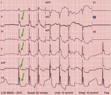
Osborn waves (J point elevation):
ECGECGAn electrocardiogram (ECG) is a graphic representation of the electrical activity of the heart plotted against time. Adhesive electrodes are affixed to the skin surface allowing measurement of cardiac impulses from many angles. The ECG provides 3-dimensional information about the conduction system of the heart, the myocardium, and other cardiac structures. Electrocardiogram (ECG) finding (in about 53% of cases, more frequent in severe hypothermiaSevere HypothermiaHypothermia)
Prominent convex deflections at the J point (junction of QRS and ST segments); best seen in the precordial leadsPrecordial leadsRecording obtained from the corresponding chest electrode and the central terminal.Electrocardiogram (ECG).
Often mistaken for ST-segment elevation myocardial infarctionMyocardial infarctionMI is ischemia and death of an area of myocardial tissue due to insufficient blood flow and oxygenation, usually from thrombus formation on a ruptured atherosclerotic plaque in the epicardial arteries. Clinical presentation is most commonly with chest pain, but women and patients with diabetes may have atypical symptoms.Myocardial Infarction (STEMI)
No prognostic significance
Resolves with rewarming
Osborn waves (J point elevation) associated with hypothermia
Image: “Osborn waves” by the First Department of Internal Medicine, Nippon Medical School, Tokyo, Japan. License: CC BY 2.5, edited by Lecturio.
Management may vary based on protocols in different locations. The following recommendations are based on US and international guidelines.
For all patientsPatientsIndividuals participating in the health care system for the purpose of receiving therapeutic, diagnostic, or preventive procedures.Clinician–Patient Relationship[7,8,10]
Perform a primary trauma survey.
Assess airway, breathing, and circulation (ABCs)
Follow basic and advanced life support treatment protocols.
Monitoring:
Cardiac telemetryTelemetryTransmission of the readings of instruments to a remote location by means of wires, radio waves, or other means.Crush Syndrome
Core temperature
If central line placement is needed:
Care should be taken to avoid inducing arrhythmia with the guidewire.
Consider femoral line placement.
If IV fluidsIV fluidsIntravenous fluids are one of the most common interventions administered in medicine to approximate physiologic bodily fluids. Intravenous fluids are divided into 2 categories: crystalloid and colloid solutions. Intravenous fluids have a wide variety of indications, including intravascular volume expansion, electrolyte manipulation, and maintenance fluids. Intravenous Fluids are given for hydration:[3]
Should be warmed (38–42°C (100–108°F))
Cold fluids can worsen hypothermiaHypothermiaHypothermia can be defined as a drop in the core body temperature below 35°C (95°F) and is classified into mild, moderate, severe, and profound forms based on the degree of temperature decrease. Hypothermia.
Rewarming techniques[3,5,7,12]
Rewarming is the cornerstone of management. The recommended rewarming technique(s) will vary depending on the severity of hypothermiaHypothermiaHypothermia can be defined as a drop in the core body temperature below 35°C (95°F) and is classified into mild, moderate, severe, and profound forms based on the degree of temperature decrease. Hypothermia, but may be classified as:
External rewarming:
Passive:
Remove wet clothes.
Cover the patient with an insulating material (blanket).
Warmed IV fluidsIV fluidsIntravenous fluids are one of the most common interventions administered in medicine to approximate physiologic bodily fluids. Intravenous fluids are divided into 2 categories: crystalloid and colloid solutions. Intravenous fluids have a wide variety of indications, including intravascular volume expansion, electrolyte manipulation, and maintenance fluids. Intravenous Fluids (38–42°C (100–108°F))[3]
Warmed, humidified oxygen
BladderBladderA musculomembranous sac along the urinary tract. Urine flows from the kidneys into the bladder via the ureters, and is held there until urination.Pyelonephritis and Perinephric Abscess irrigation with warmed fluid
Invasive:
Extracorporal blood rewarming (allows for the most rapid rewarming):
Venoarterial ECMO: preferred method, if available, in patientsPatientsIndividuals participating in the health care system for the purpose of receiving therapeutic, diagnostic, or preventive procedures.Clinician–Patient Relationship with severe hypothermiaSevere HypothermiaHypothermia and a nonperfusing cardiac rhythm
Venovenous rewarming
Cardiopulmonary bypass (CPB)
HemodialysisHemodialysisProcedures which temporarily or permanently remedy insufficient cleansing of body fluids by the kidneys.Crush Syndrome
Initiate passive external rewarmingPassive external rewarmingRemove wet clothes,cover the patient with an insulating material (blanket),bring to a warm environment.Hypothermia techniques.
The aim is to warm the torso > limbs to prevent the loss of the core temperature
More aggressive techniques may be considered if the rise in body temperatureBody TemperatureThe measure of the level of heat of a human or animal.Heatstroke < 0.5°C/hr.
Calorie replacement → provide warm, sweet drinks/food (avoid caffeineCaffeineA methylxanthine naturally occurring in some beverages and also used as a pharmacological agent. Caffeine’s most notable pharmacological effect is as a central nervous system stimulant, increasing alertness and producing agitation. Several cellular actions of caffeine have been observed, but it is not entirely clear how each contributes to its pharmacological profile. Among the most important are inhibition of cyclic nucleotide phosphodiesterases, antagonism of adenosine receptors, and modulation of intracellular calcium handling.Stimulants and alcohol)
Encourage active movement:
Avoid if patient has significant injuries
Note: WMS recommends that patientsPatientsIndividuals participating in the health care system for the purpose of receiving therapeutic, diagnostic, or preventive procedures.Clinician–Patient Relationship not stand or walk for 30 minutes once protected from heatHeatInflammation loss (may cause afterdrop in core temperature and ↓ blood pressure).[5]
Passive and active external rewarmingActive external rewarmingUse heated blankets/pads, warm baths, forced air systems, radiant heat lamps.Hypothermia
Consider minimally invasive internal rewarming
Full-body insulation, when possible
Keep patient in horizontal position
Immobilize patient, and use minimal, cautious movements to avoid arrhythmias.
Assess volume statusVolume StatusACES and RUSH: Resuscitation Ultrasound Protocols → provide IV fluidsIV fluidsIntravenous fluids are one of the most common interventions administered in medicine to approximate physiologic bodily fluids. Intravenous fluids are divided into 2 categories: crystalloid and colloid solutions. Intravenous fluids have a wide variety of indications, including intravascular volume expansion, electrolyte manipulation, and maintenance fluids. Intravenous Fluids, as indicated
Passive and active external rewarmingActive external rewarmingUse heated blankets/pads, warm baths, forced air systems, radiant heat lamps.Hypothermia
Minimally invasive internal rewarming
Consider the possible need for invasive internal rewarming (e.g., ECMO, CPB), particularly if the patient’s condition is unstable or rewarming attempts are failing.
High risk for cardiac arrestCardiac arrestCardiac arrest is the sudden, complete cessation of cardiac output with hemodynamic collapse. Patients present as pulseless, unresponsive, and apneic. Rhythms associated with cardiac arrest are ventricular fibrillation/tachycardia, asystole, or pulseless electrical activity. Cardiac Arrest:
Risk factors for imminent cardiac arrestCardiac arrestCardiac arrest is the sudden, complete cessation of cardiac output with hemodynamic collapse. Patients present as pulseless, unresponsive, and apneic. Rhythms associated with cardiac arrest are ventricular fibrillation/tachycardia, asystole, or pulseless electrical activity. Cardiac Arrest:
Systolic BP < 90 mm Hg
Ventricular arrhythmia
Manage cardiac arrestCardiac arrestCardiac arrest is the sudden, complete cessation of cardiac output with hemodynamic collapse. Patients present as pulseless, unresponsive, and apneic. Rhythms associated with cardiac arrest are ventricular fibrillation/tachycardia, asystole, or pulseless electrical activity. Cardiac Arrest, as detailed below.
Cardiac arrestCardiac arrestCardiac arrest is the sudden, complete cessation of cardiac output with hemodynamic collapse. Patients present as pulseless, unresponsive, and apneic. Rhythms associated with cardiac arrest are ventricular fibrillation/tachycardia, asystole, or pulseless electrical activity. Cardiac Arrest in hypothermic patientsPatientsIndividuals participating in the health care system for the purpose of receiving therapeutic, diagnostic, or preventive procedures.Clinician–Patient Relationship
General:[3,5,7,8]
“You’re not dead until you are warm and dead.”
CPRCPRThe artificial substitution of heart and lung action as indicated for heart arrest resulting from electric shock, drowning, respiratory arrest, or other causes. The two major components of cardiopulmonary resuscitation are artificial ventilation and closed-chest cardiac massage.Cardiac Arrest efforts should be continued, if practical, until body temperatureBody TemperatureThe measure of the level of heat of a human or animal.Heatstroke is 32–35°C (89.6–95°F).
Management does not differ much from the normal Advanced Cardiac Life Support (ACLS) algorithm.[7,8]
Estimate the survival probabilityProbabilityProbability is a mathematical tool used to study randomness and provide predictions about the likelihood of something happening. There are several basic rules of probability that can be used to help determine the probability of multiple events happening together, separately, or sequentially.Basics of Probability from hypothermic cardiac arrestCardiac arrestCardiac arrest is the sudden, complete cessation of cardiac output with hemodynamic collapse. Patients present as pulseless, unresponsive, and apneic. Rhythms associated with cardiac arrest are ventricular fibrillation/tachycardia, asystole, or pulseless electrical activity. Cardiac Arrest: HOPE (Hypothermia Outcome Prediction After ECLS) score[13]
Incorporates age, sexSexThe totality of characteristics of reproductive structure, functions, phenotype, and genotype, differentiating the male from the female organism.Gender Dysphoria, asphyxiaAsphyxiaA pathological condition caused by lack of oxygen, manifested in impending or actual cessation of life.Drowning, CPRCPRThe artificial substitution of heart and lung action as indicated for heart arrest resulting from electric shock, drowning, respiratory arrest, or other causes. The two major components of cardiopulmonary resuscitation are artificial ventilation and closed-chest cardiac massage.Cardiac Arrest duration, serum potassiumPotassiumAn element in the alkali group of metals with an atomic symbol k, atomic number 19, and atomic weight 39. 10. It is the chief cation in the intracellular fluid of muscle and other cells. Potassium ion is a strong electrolyte that plays a significant role in the regulation of fluid volume and maintenance of the water-electrolyte balance.Hyperkalemia, and temperature
Predicts survival better than serum potassiumPotassiumAn element in the alkali group of metals with an atomic symbol k, atomic number 19, and atomic weight 39. 10. It is the chief cation in the intracellular fluid of muscle and other cells. Potassium ion is a strong electrolyte that plays a significant role in the regulation of fluid volume and maintenance of the water-electrolyte balance.HyperkalemiathresholdThresholdMinimum voltage necessary to generate an action potential (an all-or-none response)Skeletal Muscle Contraction of ≥12 mmol/L alone
Provide warmed O2 via ventilator or bag-valve-mask device.
Oxygenation and ventilationVentilationThe total volume of gas inspired or expired per unit of time, usually measured in liters per minute.Ventilation: Mechanics of Breathing should otherwise follow standard guidelines.
Pulse oximeter may not be able to detect a waveform.
CirculationCirculationThe movement of the blood as it is pumped through the cardiovascular system.ABCDE Assessment:[2,7–10]
Continuous CPRCPRThe artificial substitution of heart and lung action as indicated for heart arrest resulting from electric shock, drowning, respiratory arrest, or other causes. The two major components of cardiopulmonary resuscitation are artificial ventilation and closed-chest cardiac massage.Cardiac Arrest should be performed per typical guidelines (perfusion not detected).
Note: If core temperature < 28℃, delayed or intermittent CPRCPRThe artificial substitution of heart and lung action as indicated for heart arrest resulting from electric shock, drowning, respiratory arrest, or other causes. The two major components of cardiopulmonary resuscitation are artificial ventilation and closed-chest cardiac massage.Cardiac Arrest may be used if on-site CPRCPRThe artificial substitution of heart and lung action as indicated for heart arrest resulting from electric shock, drowning, respiratory arrest, or other causes. The two major components of cardiopulmonary resuscitation are artificial ventilation and closed-chest cardiac massage.Cardiac Arrest is too dangerous or not feasible in prehospital setting (e.g., need to move to a safer location).[8,9]
May be difficult to assess if patient is truly in cardiac arrestCardiac arrestCardiac arrest is the sudden, complete cessation of cardiac output with hemodynamic collapse. Patients present as pulseless, unresponsive, and apneic. Rhythms associated with cardiac arrest are ventricular fibrillation/tachycardia, asystole, or pulseless electrical activity. Cardiac Arrest:[8]
Allow a full 60 seconds for a pulse check.
Handheld DopplerDopplerUltrasonography applying the doppler effect, with frequency-shifted ultrasound reflections produced by moving targets (usually red blood cells) in the bloodstream along the ultrasound axis in direct proportion to the velocity of movement of the targets, to determine both direction and velocity of blood flow.Ultrasound (Sonography) may be useful to verify the presence of a pulse.
Consider evaluating for cardiac motion on an ultrasound, if available.
VasopressorsVasopressorsSepsis in Children (e.g., epinephrineEpinephrineThe active sympathomimetic hormone from the adrenal medulla. It stimulates both the alpha- and beta- adrenergic systems, causes systemic vasoconstriction and gastrointestinal relaxation, stimulates the heart, and dilates bronchi and cerebral vessels.Sympathomimetic Drugs) in cases of no perfusion:
May not be effective (hypothermic heart is not as responsive) until core temperature > 30°C (86°F)
Once rewarmed to > 30°C (86°F), epinephrineEpinephrineThe active sympathomimetic hormone from the adrenal medulla. It stimulates both the alpha- and beta- adrenergic systems, causes systemic vasoconstriction and gastrointestinal relaxation, stimulates the heart, and dilates bronchi and cerebral vessels.Sympathomimetic Drugs 1 mg is administered but double the interval between doses until core temperature is >35°C.
May not work until core temperature > 30°C (86°F):
European guidelines recommend delaying repeated defibrillationDefibrillationVentricular Fibrillation (V-fib), after 3 unsuccessful attempts, until core temperature ≥ 30℃, then restarting.[7]
American Heart AssociationAmerican Heart AssociationA voluntary organization concerned with the prevention and treatment of heart and vascular diseases.Heart Failure does not recommend deviating from guidelines.[8]
Infection/sepsisSepsisSystemic inflammatory response syndrome with a proven or suspected infectious etiology. When sepsis is associated with organ dysfunction distant from the site of infection, it is called severe sepsis. When sepsis is accompanied by hypotension despite adequate fluid infusion, it is called septic shock.Sepsis and Septic Shock (empiric broad-spectrumBroad-SpectrumFluoroquinolones IV antibiotics should be started if the body temperatureBody TemperatureThe measure of the level of heat of a human or animal.Heatstroke rises < 0.7°C/hr)
Adrenal insufficiencyAdrenal InsufficiencyConditions in which the production of adrenal corticosteroids falls below the requirement of the body. Adrenal insufficiency can be caused by defects in the adrenal glands, the pituitary gland, or the hypothalamus.Adrenal Insufficiency and Addison Disease
HypoglycemiaHypoglycemiaHypoglycemia is an emergency condition defined as a serum glucose level ≤ 70 mg/dL (≤ 3.9 mmol/L) in diabetic patients. In nondiabetic patients, there is no specific or defined limit for normal serum glucose levels, and hypoglycemia is defined mainly by its clinical features. Hypoglycemia
HypothyroidismHypothyroidismHypothyroidism is a condition characterized by a deficiency of thyroid hormones. Iodine deficiency is the most common cause worldwide, but Hashimoto’s disease (autoimmune thyroiditis) is the leading cause in non-iodine-deficient regions. Hypothyroidism
Complications of rewarming[2,3,12]
Further decrease of body temperatureBody TemperatureThe measure of the level of heat of a human or animal.Heatstroke (afterdrop):
Known risk of external rewarming measures
Associated with ↑ risk for serious arrhythmias
HypotensionHypotensionHypotension is defined as low blood pressure, specifically < 90/60 mm Hg, and is most commonly a physiologic response. Hypotension may be mild, serious, or life threatening, depending on the cause. Hypotension/shockShockShock is a life-threatening condition associated with impaired circulation that results in tissue hypoxia. The different types of shock are based on the underlying cause: distributive (↑ cardiac output (CO), ↓ systemic vascular resistance (SVR)), cardiogenic (↓ CO, ↑ SVR), hypovolemic (↓ CO, ↑ SVR), obstructive (↓ CO), and mixed. Types of Shock (from peripheral vasodilationVasodilationThe physiological widening of blood vessels by relaxing the underlying vascular smooth muscle.Pulmonary Hypertension Drugs and hypovolemiaHypovolemiaSepsis in Children) → may necessitate:
Electrolyte abnormalities → monitor and correct, as needed
Arrhythmias → minimize movement; generally do not need treatment
RhabdomyolysisRhabdomyolysisRhabdomyolysis is characterized by muscle necrosis and the release of toxic intracellular contents, especially myoglobin, into the circulation.Rhabdomyolysis → IV fluid resuscitationResuscitationThe restoration to life or consciousness of one apparently dead. .Neonatal Respiratory Distress Syndrome
Delayed pulmonary, renal, or neurologic complications
PrognosisPrognosisA prediction of the probable outcome of a disease based on a individual’s condition and the usual course of the disease as seen in similar situations.Non-Hodgkin Lymphomas[5,7,8,12]
Factors associated with death within 24 hours:
Prehospital cardiac arrestCardiac arrestCardiac arrest is the sudden, complete cessation of cardiac output with hemodynamic collapse. Patients present as pulseless, unresponsive, and apneic. Rhythms associated with cardiac arrest are ventricular fibrillation/tachycardia, asystole, or pulseless electrical activity. Cardiac Arrest
HypothermiaHypothermiaHypothermia can be defined as a drop in the core body temperature below 35°C (95°F) and is classified into mild, moderate, severe, and profound forms based on the degree of temperature decrease. Hypothermia with asphyxiaAsphyxiaA pathological condition caused by lack of oxygen, manifested in impending or actual cessation of life.Drowning carries a worse prognosisPrognosisA prediction of the probable outcome of a disease based on a individual’s condition and the usual course of the disease as seen in similar situations.Non-Hodgkin Lymphomas:
DrowningDrowningDrowning occurs due to respiratory impairment from submersion or immersion in a liquid medium. Aspiration of water leads to hypoxemia, which affects all organ systems, resulting in respiratory insufficiency and acute respiratory distress syndrome (ARDS), cardiac arrhythmias, and neuronal damage. Drowning
Avalanche burial
HypothermiaHypothermiaHypothermia can be defined as a drop in the core body temperature below 35°C (95°F) and is classified into mild, moderate, severe, and profound forms based on the degree of temperature decrease. Hypothermia with cardiac arrestCardiac arrestCardiac arrest is the sudden, complete cessation of cardiac output with hemodynamic collapse. Patients present as pulseless, unresponsive, and apneic. Rhythms associated with cardiac arrest are ventricular fibrillation/tachycardia, asystole, or pulseless electrical activity. Cardiac Arrest:
50% neurologically intact survival if extracorporeal circulationCirculationThe movement of the blood as it is pumped through the cardiovascular system.ABCDE Assessment is used
< 37% neurologically intact survival with other methods
Severe hyperkalemiaHyperkalemiaHyperkalemia is defined as a serum potassium (K+) concentration >5.2 mEq/L. Homeostatic mechanisms maintain the serum K+ concentration between 3.5 and 5.2 mEq/L, despite marked variation in dietary intake. Hyperkalemia can be due to a variety of causes, which include transcellular shifts, tissue breakdown, inadequate renal excretion, and drugs. Hyperkalemia with low temperature may predict futility of resuscitationResuscitationThe restoration to life or consciousness of one apparently dead. .Neonatal Respiratory Distress Syndrome efforts.
Differential Diagnosis
HypothyroidismHypothyroidismHypothyroidism is a condition characterized by a deficiency of thyroid hormones. Iodine deficiency is the most common cause worldwide, but Hashimoto’s disease (autoimmune thyroiditis) is the leading cause in non-iodine-deficient regions. Hypothyroidism: deficiency of T3T3A T3 thyroid hormone normally synthesized and secreted by the thyroid gland in much smaller quantities than thyroxine (T4). Most T3 is derived from peripheral monodeiodination of T4 at the 5′ position of the outer ring of the iodothyronine nucleus. The hormone finally delivered and used by the tissues is mainly t3.Thyroid Hormones and T4T4The major hormone derived from the thyroid gland. Thyroxine is synthesized via the iodination of tyrosines (monoiodotyrosine) and the coupling of iodotyrosines (diiodotyrosine) in the thyroglobulin. Thyroxine is released from thyroglobulin by proteolysis and secreted into the blood. Thyroxine is peripherally deiodinated to form triiodothyronine which exerts a broad spectrum of stimulatory effects on cell metabolism.Thyroid HormonesthyroidThyroidThe thyroid gland is one of the largest endocrine glands in the human body. The thyroid gland is a highly vascular, brownish-red gland located in the visceral compartment of the anterior region of the neck.Thyroid Gland: AnatomyhormonesHormonesHormones are messenger molecules that are synthesized in one part of the body and move through the bloodstream to exert specific regulatory effects on another part of the body. Hormones play critical roles in coordinating cellular activities throughout the body in response to the constant changes in both the internal and external environments. Hormones: Overview and Types. Clinical features of hypothyroidismHypothyroidismHypothyroidism is a condition characterized by a deficiency of thyroid hormones. Iodine deficiency is the most common cause worldwide, but Hashimoto’s disease (autoimmune thyroiditis) is the leading cause in non-iodine-deficient regions. Hypothyroidism are primarily due to the accumulation of matrix substances and a decreased metabolic rate. Severe hypothyroidismHypothyroidismHypothyroidism is a condition characterized by a deficiency of thyroid hormones. Iodine deficiency is the most common cause worldwide, but Hashimoto’s disease (autoimmune thyroiditis) is the leading cause in non-iodine-deficient regions. Hypothyroidism is associated with hypothermiaHypothermiaHypothermia can be defined as a drop in the core body temperature below 35°C (95°F) and is classified into mild, moderate, severe, and profound forms based on the degree of temperature decrease. Hypothermia secondary to decreased metabolic heat productionHeat ProductionFever.
Adrenal insufficiencyAdrenal InsufficiencyConditions in which the production of adrenal corticosteroids falls below the requirement of the body. Adrenal insufficiency can be caused by defects in the adrenal glands, the pituitary gland, or the hypothalamus.Adrenal Insufficiency and Addison Disease: inadequate production of adrenocortical hormonesHormonesHormones are messenger molecules that are synthesized in one part of the body and move through the bloodstream to exert specific regulatory effects on another part of the body. Hormones play critical roles in coordinating cellular activities throughout the body in response to the constant changes in both the internal and external environments. Hormones: Overview and Types (glucocorticoidsGlucocorticoidsGlucocorticoids are a class within the corticosteroid family. Glucocorticoids are chemically and functionally similar to endogenous cortisol. There are a wide array of indications, which primarily benefit from the antiinflammatory and immunosuppressive effects of this class of drugs.Glucocorticoids, mineralocorticoidsMineralocorticoidsMineralocorticoids are a drug class within the corticosteroid family and fludrocortisone is the primary medication within this class. Fludrocortisone is a fluorinated analog of cortisone. The fluorine moiety protects the drug from isoenzyme inactivation in the kidney, allowing it to exert its mineralocorticoid effect.Mineralocorticoids, and adrenal androgensAndrogensAndrogens are naturally occurring steroid hormones responsible for development and maintenance of the male sex characteristics, including penile, scrotal, and clitoral growth, development of sexual hair, deepening of the voice, and musculoskeletal growth. Androgens and Antiandrogens). Primary adrenal insufficiencyPrimary adrenal insufficiencyAn adrenal disease characterized by the progressive destruction of the adrenal cortex, resulting in insufficient production of aldosterone and hydrocortisone. Clinical symptoms include anorexia; nausea; weight loss; muscle weakness; and hyperpigmentation of the skin due to increase in circulating levels of acth precursor hormone which stimulates melanocytes.Adrenal Insufficiency and Addison Disease (Addison disease) is caused by diseases in the gland itself. Secondary adrenal insufficiencySecondary adrenal insufficiencyDeficiency in pituitary adrenocorticotropic hormone (ACTH) secretion.Adrenal Insufficiency and Addison Disease occurs due to decreased production of ACTH either from prolonged glucocorticoid therapy or disease in the pituitaryPituitaryA small, unpaired gland situated in the sella turcica. It is connected to the hypothalamus by a short stalk which is called the infundibulum.Hormones: Overview and Types/hypothalamic glands. Both scenarios put the patient at risk of developing hypothermiaHypothermiaHypothermia can be defined as a drop in the core body temperature below 35°C (95°F) and is classified into mild, moderate, severe, and profound forms based on the degree of temperature decrease. Hypothermia.
SepsisSepsisSystemic inflammatory response syndrome with a proven or suspected infectious etiology. When sepsis is associated with organ dysfunction distant from the site of infection, it is called severe sepsis. When sepsis is accompanied by hypotension despite adequate fluid infusion, it is called septic shock.Sepsis and Septic Shock:bacteremiaBacteremiaThe presence of viable bacteria circulating in the blood. Fever, chills, tachycardia, and tachypnea are common acute manifestations of bacteremia. The majority of cases are seen in already hospitalized patients, most of whom have underlying diseases or procedures which render their bloodstreams susceptible to invasion.Glycopeptides associated with signs of systemic toxicityToxicityDosage Calculation and progression to multi-organ failure. Late sepsisSepsisSystemic inflammatory response syndrome with a proven or suspected infectious etiology. When sepsis is associated with organ dysfunction distant from the site of infection, it is called severe sepsis. When sepsis is accompanied by hypotension despite adequate fluid infusion, it is called septic shock.Sepsis and Septic Shock can be associated with hypothermiaHypothermiaHypothermia can be defined as a drop in the core body temperature below 35°C (95°F) and is classified into mild, moderate, severe, and profound forms based on the degree of temperature decrease. Hypothermia. Vital signs (e.g., tachycardiaTachycardiaAbnormally rapid heartbeat, usually with a heart rate above 100 beats per minute for adults. Tachycardia accompanied by disturbance in the cardiac depolarization (cardiac arrhythmia) is called tachyarrhythmia.Sepsis in Children) that are inconsistent with the degree of accidental hypothermiaAccidental HypothermiaHypothermia should raise the suspicion of an alternative diagnosis.
Billing and Coding
Diagnosis Codes:
This code is used to diagnose accidental hypothermiaAccidental HypothermiaHypothermia, a dangerous drop in core body temperatureBody TemperatureThe measure of the level of heat of a human or animal.Heatstroke, typically caused by prolonged exposure to cold. The code specifies the cause and encounter type.
Coding System
Code
Description
ICD-10-CM
T68.XX1A
HypothermiaHypothermiaHypothermia can be defined as a drop in the core body temperature below 35°C (95°F) and is classified into mild, moderate, severe, and profound forms based on the degree of temperature decrease. Hypothermia, accidental, initial encounter
An ECGECGAn electrocardiogram (ECG) is a graphic representation of the electrical activity of the heart plotted against time. Adhesive electrodes are affixed to the skin surface allowing measurement of cardiac impulses from many angles. The ECG provides 3-dimensional information about the conduction system of the heart, the myocardium, and other cardiac structures. Electrocardiogram (ECG) is a critical part of the evaluation, as hypothermiaHypothermiaHypothermia can be defined as a drop in the core body temperature below 35°C (95°F) and is classified into mild, moderate, severe, and profound forms based on the degree of temperature decrease. Hypothermia can cause characteristic changes (like the Osborn J wave) and lead to life-threatening arrhythmias.
Coding System
Code
Description
CPT
93000
ElectrocardiogramElectrocardiogramAn electrocardiogram (ECG) is a graphic representation of the electrical activity of the heart plotted against time. Adhesive electrodes are affixed to the skin surface allowing measurement of cardiac impulses from many angles. The ECG provides 3-dimensional information about the conduction system of the heart, the myocardium, and other cardiac structures. Electrocardiogram (ECG), routine ECGECGAn electrocardiogram (ECG) is a graphic representation of the electrical activity of the heart plotted against time. Adhesive electrodes are affixed to the skin surface allowing measurement of cardiac impulses from many angles. The ECG provides 3-dimensional information about the conduction system of the heart, the myocardium, and other cardiac structures. Electrocardiogram (ECG) with at least 12 leads; with interpretation and report
These codes represent methods of active internal rewarming for moderate to severe hypothermiaSevere HypothermiaHypothermia, including warm IV fluidsIV fluidsIntravenous fluids are one of the most common interventions administered in medicine to approximate physiologic bodily fluids. Intravenous fluids are divided into 2 categories: crystalloid and colloid solutions. Intravenous fluids have a wide variety of indications, including intravascular volume expansion, electrolyte manipulation, and maintenance fluids. Intravenous Fluids, gastric lavage, and, in the most severe cases, cardiopulmonary bypass.
Coding System
Code
Description
CPT
33946
Extracorporeal membrane oxygenation (ECMO)/extracorporeal life support (ECLS) provided by physician; initiation, veno-arterial
Complications:
These codes document the most severe complications of hypothermiaHypothermiaHypothermia can be defined as a drop in the core body temperature below 35°C (95°F) and is classified into mild, moderate, severe, and profound forms based on the degree of temperature decrease. Hypothermia, including ventricular fibrillationVentricular fibrillationVentricular fibrillation (VF or V-fib) is a type of ventricular tachyarrhythmia (> 300/min) often preceded by ventricular tachycardia. In this arrhythmia, the ventricle beats rapidly and sporadically. The ventricular contraction is uncoordinated, leading to a decrease in cardiac output and immediate hemodynamic collapse. Ventricular Fibrillation (V-fib) (a common cause of death) and rhabdomyolysisRhabdomyolysisRhabdomyolysis is characterized by muscle necrosis and the release of toxic intracellular contents, especially myoglobin, into the circulation.Rhabdomyolysis from prolonged immobility and muscle injury.
Coding System
Code
Description
ICD-10-CM
I49.01
Ventricular fibrillationVentricular fibrillationVentricular fibrillation (VF or V-fib) is a type of ventricular tachyarrhythmia (> 300/min) often preceded by ventricular tachycardia. In this arrhythmia, the ventricle beats rapidly and sporadically. The ventricular contraction is uncoordinated, leading to a decrease in cardiac output and immediate hemodynamic collapse. Ventricular Fibrillation (V-fib)
ICD-10-CM
M62.82
RhabdomyolysisRhabdomyolysisRhabdomyolysis is characterized by muscle necrosis and the release of toxic intracellular contents, especially myoglobin, into the circulation.Rhabdomyolysis
Brown, D. J. A., Brugger, H., Boyd, J., Paal, P. (2012). Accidental hypothermia. New England Journal of Medicine, 367(20), 1930–1938. https://doi.org/10.1056/NEJMra1114208
Dow, J., Giesbrecht, G. G., Danzl, D. F., Brugger, H., et al. (2019). Wilderness Medical Society clinical practice guidelines for the out-of-hospital evaluation and treatment of accidental hypothermia: 2019 update. Wilderness & Environmental Medicine, 30(4), S47–S69. https://doi.org/10.1016/j.wem.2019.10.002
Musi, M. E., Sheets, A., Zafren, K., Brugger, H., Paal, P., Hölzl, N., Pasquier, M. (2021). Clinical staging of accidental hypothermia: the Revised Swiss System. Resuscitation, 162, 182–187. https://doi.org/10.1016/j.resuscitation.2021.02.038
Lott, C., Truhlář, A., Alfonzo, A., Barelli, A., et al. (2021). European Resuscitation Council Guidelines 2021: cardiac arrest in special circumstances. Resuscitation, 161, 152–219. https://doi.org/10.1016/j.resuscitation.2021.02.011
Panchal, A. R., Bartos, J. A., Cabañas, J. G., Donnino, M. W., et al. (2020). Part 3: Adult Basic and Advanced Life Support: 2020 American Heart Association guidelines for cardiopulmonary resuscitation and emergency cardiovascular care. Circulation, 142(16_suppl_2). https://doi.org/10.1161/CIR.0000000000000916
Australian and New Zealand Committee on Resuscitation (ANZCOR). (2021). Guideline 9.3.3—hypothermia and cold related injury. Retrieved November 15, 2022, from https://resus.org.au/the-arc-guidelines/
Marino, P. L. (2014). Hypothermia. In Marino’s The ICU Book (4th ed., pp. 731–733). Wolters Kluwer Health.
Cao, D., Arens, A. M., Chow, S. L., Easter, S. R., Hoffman, R. S., Lagina, A. T., 3rd, Lavonas, E. J., Patil, K. D., Sutherland, L. D., Tijssen, J. A., Wang, G. S., Zelop, C. M., Rodriguez, A. J., Drennan, I. R., & McBride, M. E. (2025). Part 10: Adult and Pediatric Special Circumstances of Resuscitation: 2025 American Heart Association Guidelines for Cardiopulmonary Resuscitation and Emergency Cardiovascular Care. Circulation, 152(16_suppl_2), S578–S672. https://doi.org/10.1161/CIR.0000000000001380
Create your free account or log in to continue reading!