Atrial flutter is a regular narrow-complex tachycardia characterized by a rapid atrial rate (typically around 300/min), atrioventricular (AV) node conduction block with a ventricular response of approximately 150/minute, and a “sawtooth” pattern on an electrocardiogram (ECG). There are many cardiac and noncardiac causes, but patients usually have underlying structural heart disease. Symptoms include palpitations, shortness of breath, chest pain, dizziness, and nausea. Management is similar to that for atrial fibrillation, focusing on rate control and preventing systemic embolization.
Uncommon in patientsPatientsIndividuals participating in the health care system for the purpose of receiving therapeutic, diagnostic, or preventive procedures.Clinician–Patient Relationship without underlying structural heart disease (higher incidenceIncidenceThe number of new cases of a given disease during a given period in a specified population. It also is used for the rate at which new events occur in a defined population. It is differentiated from prevalence, which refers to all cases in the population at a given time.Measures of Disease Frequency with left atrial enlargement, or with left ventricular or biventricular heart failureHeart FailureA heterogeneous condition in which the heart is unable to pump out sufficient blood to meet the metabolic need of the body. Heart failure can be caused by structural defects, functional abnormalities (ventricular dysfunction), or a sudden overload beyond its capacity. Chronic heart failure is more common than acute heart failure which results from sudden insult to cardiac function, such as myocardial infarction.Total Anomalous Pulmonary Venous Return (TAPVR))
Less common than atrial fibrillationAtrial fibrillationAtrial fibrillation (AF or Afib) is a supraventricular tachyarrhythmia and the most common kind of arrhythmia. It is caused by rapid, uncontrolled atrial contractions and uncoordinated ventricular responses. Atrial Fibrillation
Approximately twice as common in men as in women
Etiology[1,4,6,9]
Cardiac causes:
Structural diseases:
Coronary arteryCoronary ArteryTruncus Arteriosus disease and myocardial infarctionMyocardial infarctionMI is ischemia and death of an area of myocardial tissue due to insufficient blood flow and oxygenation, usually from thrombus formation on a ruptured atherosclerotic plaque in the epicardial arteries. Clinical presentation is most commonly with chest pain, but women and patients with diabetes may have atypical symptoms.Myocardial Infarction
Heart failureHeart FailureA heterogeneous condition in which the heart is unable to pump out sufficient blood to meet the metabolic need of the body. Heart failure can be caused by structural defects, functional abnormalities (ventricular dysfunction), or a sudden overload beyond its capacity. Chronic heart failure is more common than acute heart failure which results from sudden insult to cardiac function, such as myocardial infarction.Total Anomalous Pulmonary Venous Return (TAPVR)
Valvular disease
Hypertensive heart disease
CardiomyopathyCardiomyopathyCardiomyopathy refers to a group of myocardial diseases associated with structural changes of the heart muscles (myocardium) and impaired systolic and/or diastolic function in the absence of other heart disorders (coronary artery disease, hypertension, valvular disease, and congenital heart disease). Cardiomyopathy: Overview and Types
Post-cardiac surgery (such as bypass surgery) or ablation
Congenital heart defects:
Ebstein anomaly
Atrial septal defectAtrial Septal DefectAtrial septal defects (ASDs) are benign acyanotic congenital heart defects characterized by an opening in the interatrial septum that causes blood to flow from the left atrium (LA) to the right atrium (RA) (left-to-right shunt). Atrial Septal Defect (ASD)
Tetralogy of FallotTetralogy of FallotTetralogy of Fallot is the most common cyanotic congenital heart disease. The disease is the confluence of 4 pathologic cardiac features: overriding aorta, ventricular septal defect, right ventricular outflow obstruction, and right ventricular hypertrophy. Tetralogy of Fallot
Inflammatory diseases:
PericarditisPericarditisPericarditis is an inflammation of the pericardium, often with fluid accumulation. It can be caused by infection (often viral), myocardial infarction, drugs, malignancies, metabolic disorders, autoimmune disorders, or trauma. Acute, subacute, and chronic forms exist. Pericarditis
MyocarditisMyocarditisMyocarditis is an inflammatory disease of the myocardium, which may occur alone or in association with a systemic process. There are numerous etiologies of myocarditis, but all lead to inflammation and myocyte injury, most often leading to signs and symptoms of heart failure. Myocarditis
Conduction system abnormalities:
Sinus node dysfunctionSinus node dysfunctionA condition caused by dysfunctions related to the sinoatrial node including impulse generation (cardiac sinus arrest) and impulse conduction (sinoatrial exit block). It is characterized by persistent bradycardia, chronic atrial fibrillation, and failure to resume sinus rhythm following cardioversion. This syndrome can be congenital or acquired, particularly after surgical correction for heart defects.Bradyarrhythmias
Pulmonary embolismPulmonary EmbolismPulmonary embolism (PE) is a potentially fatal condition that occurs as a result of intraluminal obstruction of the main pulmonary artery or its branches. The causative factors include thrombi, air, amniotic fluid, and fat. In PE, gas exchange is impaired due to the decreased return of deoxygenated blood to the lungs. Pulmonary Embolism
Obstructive sleep apneaSleep apneaRepeated cessation of breathing for > 10 seconds during sleep and results in sleep interruption, fatigue, and daytime sleepiness.Obstructive Sleep Apnea
Other causes:
Electrolyte imbalance
Acute illness (e.g., sepsisSepsisSystemic inflammatory response syndrome with a proven or suspected infectious etiology. When sepsis is associated with organ dysfunction distant from the site of infection, it is called severe sepsis. When sepsis is accompanied by hypotension despite adequate fluid infusion, it is called septic shock.Sepsis and Septic Shock, shockShockShock is a life-threatening condition associated with impaired circulation that results in tissue hypoxia. The different types of shock are based on the underlying cause: distributive (↑ cardiac output (CO), ↓ systemic vascular resistance (SVR)), cardiogenic (↓ CO, ↑ SVR), hypovolemic (↓ CO, ↑ SVR), obstructive (↓ CO), and mixed. Types of Shock)
ObesityObesityObesity is a condition associated with excess body weight, specifically with the deposition of excessive adipose tissue. Obesity is considered a global epidemic. Major influences come from the western diet and sedentary lifestyles, but the exact mechanisms likely include a mixture of genetic and environmental factors. Obesity
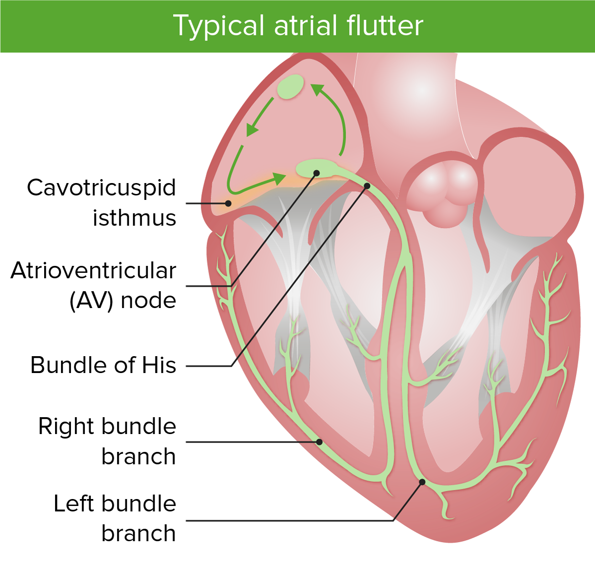
Atrial flutterAtrial flutterAtrial flutter is a regular supraventricular tachycardia characterized by an atrial heart rate between 240/min and 340/min (typically 300/min), atrioventricular (AV) node conduction block, and a “sawtooth” pattern on an electrocardiogram (ECG). Atrial Flutter is caused by a macroreentrant electrical loopMacroreentrant electrical loopReentrant circuit covers a large area of the atrium.Atrial Flutter (reentrant circuit covers a large area of the atrium):
Leads to an atrial heart rateHeart rateThe number of times the heart ventricles contract per unit of time, usually per minute.Cardiac Physiology between 240/min and 340/min
Atrioventricular nodeAtrioventricular nodeA small nodular mass of specialized muscle fibers located in the interatrial septum near the opening of the coronary sinus. It gives rise to the atrioventricular bundle of the conduction system of the heart.Heart: Anatomy conduction block results in a lower ratio of ventricular to atrial beats.
Atrioventricular nodeAtrioventricular nodeA small nodular mass of specialized muscle fibers located in the interatrial septum near the opening of the coronary sinus. It gives rise to the atrioventricular bundle of the conduction system of the heart.Heart: Anatomy cannot conduct at the same rate as the atrial activity.
Usually seen as a 2:1 ratio if the patient is not on AV nodal blocking medications
Leads to an average ventricular heart rateHeart rateThe number of times the heart ventricles contract per unit of time, usually per minute.Cardiac Physiology of approximately 150/min
Usually converts back into sinus rhythmSinus rhythmA heart rate and rhythm driven by the regular firing of the SA node (60–100 beats per minute)Cardiac Physiology or deteriorates into atrial fibrillationAtrial fibrillationAtrial fibrillation (AF or Afib) is a supraventricular tachyarrhythmia and the most common kind of arrhythmia. It is caused by rapid, uncontrolled atrial contractions and uncoordinated ventricular responses. Atrial Fibrillation
Originates in any region of the right or left atria, usually around scarScarDermatologic Examination tissue (scarScarDermatologic Examination tissue may be from intrinsic heart disease or iatrogenicIatrogenicAny adverse condition in a patient occurring as the result of treatment by a physician, surgeon, or other health professional, especially infections acquired by a patient during the course of treatment.Anterior Cord Syndrome from a cardiac procedure)
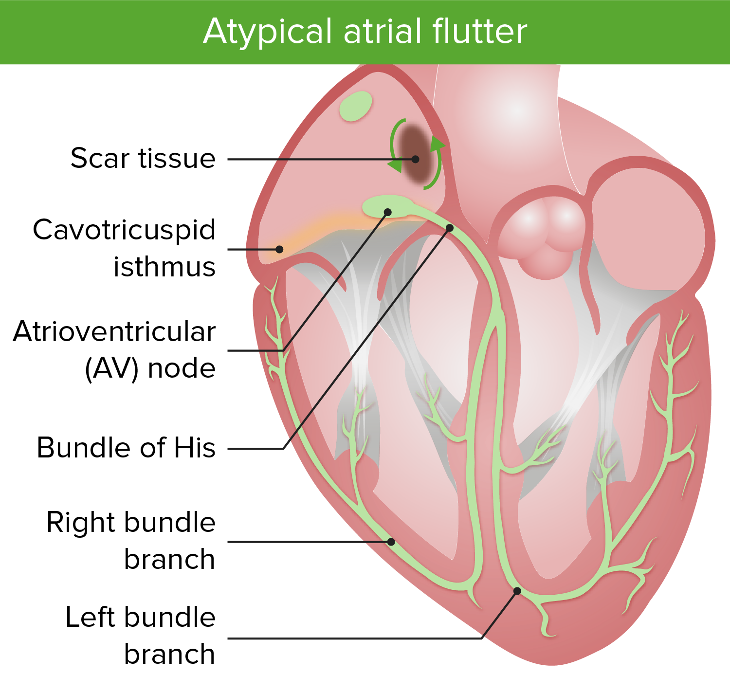
Image shows the macroreentrant circuits in atypical atrial flutter. Notice that the atypical flutter is focused around an area of scar tissue.
Image by Lecturio.
Clinical Presentation
Symptoms[4,6,10,12]
Atrial flutterAtrial flutterAtrial flutter is a regular supraventricular tachycardia characterized by an atrial heart rate between 240/min and 340/min (typically 300/min), atrioventricular (AV) node conduction block, and a “sawtooth” pattern on an electrocardiogram (ECG). Atrial Flutter may be asymptomatic or present with:
SyncopeSyncopeSyncope is a short-term loss of consciousness and loss of postural stability followed by spontaneous return of consciousness to the previous neurologic baseline without the need for resuscitation. The condition is caused by transient interruption of cerebral blood flow that may be benign or related to a underlying life-threatening condition. Syncope
Shortness of breathShortness of breathDyspnea is the subjective sensation of breathing discomfort. Dyspnea is a normal manifestation of heavy physical or psychological exertion, but also may be caused by underlying conditions (both pulmonary and extrapulmonary).Dyspnea
Chest painPainAn unpleasant sensation induced by noxious stimuli which are detected by nerve endings of nociceptive neurons.Pain: Types and Pathways
FatigueFatigueThe state of weariness following a period of exertion, mental or physical, characterized by a decreased capacity for work and reduced efficiency to respond to stimuli.Fibromyalgia
NauseaNauseaAn unpleasant sensation in the stomach usually accompanied by the urge to vomit. Common causes are early pregnancy, sea and motion sickness, emotional stress, intense pain, food poisoning, and various enteroviruses.Antiemetics
AnxietyAnxietyFeelings or emotions of dread, apprehension, and impending disaster but not disabling as with anxiety disorders.Generalized Anxiety Disorder
Patients may present with conditions resulting from atrial flutter (see table in “Complications”).
Physical exam[6,10]
TachycardiaTachycardiaAbnormally rapid heartbeat, usually with a heart rate above 100 beats per minute for adults. Tachycardia accompanied by disturbance in the cardiac depolarization (cardiac arrhythmia) is called tachyarrhythmia.Sepsis in Children, usually with a regularRegularInsulin pulse
HypotensionHypotensionHypotension is defined as low blood pressure, specifically < 90/60 mm Hg, and is most commonly a physiologic response. Hypotension may be mild, serious, or life threatening, depending on the cause. Hypotension
Diaphoresis
Indicators of an underlying cause:
Heart murmur → valvular disease
Pericardial rub → pericarditisPericarditisPericarditis is an inflammation of the pericardium, often with fluid accumulation. It can be caused by infection (often viral), myocardial infarction, drugs, malignancies, metabolic disorders, autoimmune disorders, or trauma. Acute, subacute, and chronic forms exist. Pericarditis
Exophthalmos → hyperthyroidismHyperthyroidismHypersecretion of thyroid hormones from the thyroid gland. Elevated levels of thyroid hormones increase basal metabolic rate.Thyrotoxicosis and Hyperthyroidism
Prolonged expirationExpirationVentilation: Mechanics of Breathing or wheeze → COPDCOPDChronic obstructive pulmonary disease (COPD) is a lung disease characterized by progressive, largely irreversible airflow obstruction. The condition usually presents in middle-aged or elderly persons with a history of cigarette smoking. Signs and symptoms include prolonged expiration, wheezing, diminished breath sounds, progressive dyspnea, and chronic cough. Chronic Obstructive Pulmonary Disease (COPD)
Complications[4,6,10]
Table: Complication
Possible symptoms
Exam findings
Cardiac:
Long-term tachycardiaTachycardiaAbnormally rapid heartbeat, usually with a heart rate above 100 beats per minute for adults. Tachycardia accompanied by disturbance in the cardiac depolarization (cardiac arrhythmia) is called tachyarrhythmia.Sepsis in Children can cause cardiac structural changes.
Increased oxygen demand of the myocardiumMyocardiumThe muscle tissue of the heart. It is composed of striated, involuntary muscle cells connected to form the contractile pump to generate blood flow.Heart: Anatomy
Heart failureHeart FailureA heterogeneous condition in which the heart is unable to pump out sufficient blood to meet the metabolic need of the body. Heart failure can be caused by structural defects, functional abnormalities (ventricular dysfunction), or a sudden overload beyond its capacity. Chronic heart failure is more common than acute heart failure which results from sudden insult to cardiac function, such as myocardial infarction.Total Anomalous Pulmonary Venous Return (TAPVR)
DyspneaDyspneaDyspnea is the subjective sensation of breathing discomfort. Dyspnea is a normal manifestation of heavy physical or psychological exertion, but also may be caused by underlying conditions (both pulmonary and extrapulmonary). Dyspnea
EdemaEdemaEdema is a condition in which excess serous fluid accumulates in the body cavity or interstitial space of connective tissues. Edema is a symptom observed in several medical conditions. It can be categorized into 2 types, namely, peripheral (in the extremities) and internal (in an organ or body cavity). Edema
Lower extremity pitting edemaPitting edemaEdema caused by excess fluid without excess colloid. Leaves “pits” due to fluid displacement when pressure is applied to the areaEdema
Myocardial ischemiaMyocardial ischemiaA disorder of cardiac function caused by insufficient blood flow to the muscle tissue of the heart. The decreased blood flow may be due to narrowing of the coronary arteries (coronary artery disease), to obstruction by a thrombus (coronary thrombosis), or less commonly, to diffuse narrowing of arterioles and other small vessels within the heart.Coronary Heart Disease
Chest pressure
DyspneaDyspneaDyspnea is the subjective sensation of breathing discomfort. Dyspnea is a normal manifestation of heavy physical or psychological exertion, but also may be caused by underlying conditions (both pulmonary and extrapulmonary). Dyspnea
May be related to intermittent periods of atrial fibrillationAtrial fibrillationAtrial fibrillation (AF or Afib) is a supraventricular tachyarrhythmia and the most common kind of arrhythmia. It is caused by rapid, uncontrolled atrial contractions and uncoordinated ventricular responses. Atrial Fibrillation, resulting in stasis of blood and thrombus formation
Stroke/transient ischemic attackTransient ischemic attackTransient ischemic attack (TIA) is a temporary episode of neurologic dysfunction caused by ischemia without infarction that resolves completely when blood supply is restored. Transient ischemic attack is a neurologic emergency that warrants urgent medical attention. Transient Ischemic Attack (TIA) (TIATIATransient ischemic attack (TIA) is a temporary episode of neurologic dysfunction caused by ischemia without infarction that resolves completely when blood supply is restored. Transient ischemic attack is a neurologic emergency that warrants urgent medical attention. Transient Ischemic Attack (TIA))
DysarthriaDysarthriaDisorders of speech articulation caused by imperfect coordination of pharynx, larynx, tongue, or face muscles. This may result from cranial nerve diseases; neuromuscular diseases; cerebellar diseases; basal ganglia diseases; brain stem diseases; or diseases of the corticobulbar tracts. The cortical language centers are intact in this condition.Wilson Disease
NauseaNauseaAn unpleasant sensation in the stomach usually accompanied by the urge to vomit. Common causes are early pregnancy, sea and motion sickness, emotional stress, intense pain, food poisoning, and various enteroviruses.Antiemetics and vomitingVomitingThe forcible expulsion of the contents of the stomach through the mouth.Hypokalemia
Left shoulder painPainAn unpleasant sensation induced by noxious stimuli which are detected by nerve endings of nociceptive neurons.Pain: Types and Pathways
FeverFeverFever is defined as a measured body temperature of at least 38°C (100.4°F). Fever is caused by circulating endogenous and/or exogenous pyrogens that increase levels of prostaglandin E2 in the hypothalamus. Fever is commonly associated with chills, rigors, sweating, and flushing of the skin. Fever
Bowel infarctInfarctArea of necrotic cells in an organ, arising mainly from hypoxia and ischemiaIschemic Cell Damage
Postprandial painPainAn unpleasant sensation induced by noxious stimuli which are detected by nerve endings of nociceptive neurons.Pain: Types and Pathways
NauseaNauseaAn unpleasant sensation in the stomach usually accompanied by the urge to vomit. Common causes are early pregnancy, sea and motion sickness, emotional stress, intense pain, food poisoning, and various enteroviruses.Antiemetics and vomitingVomitingThe forcible expulsion of the contents of the stomach through the mouth.Hypokalemia
Abdominal tenderness
Abdominal distension
Absent bowel sounds
Peritoneal signs
Renal infarctInfarctArea of necrotic cells in an organ, arising mainly from hypoxia and ischemiaIschemic Cell Damage
NauseaNauseaAn unpleasant sensation in the stomach usually accompanied by the urge to vomit. Common causes are early pregnancy, sea and motion sickness, emotional stress, intense pain, food poisoning, and various enteroviruses.Antiemetics and vomitingVomitingThe forcible expulsion of the contents of the stomach through the mouth.Hypokalemia
Flank tenderness
Elevated blood pressure
FeverFeverFever is defined as a measured body temperature of at least 38°C (100.4°F). Fever is caused by circulating endogenous and/or exogenous pyrogens that increase levels of prostaglandin E2 in the hypothalamus. Fever is commonly associated with chills, rigors, sweating, and flushing of the skin. Fever
Diagnosis
Diagnostic testsDiagnostic testsDiagnostic tests are important aspects in making a diagnosis. Some of the most important epidemiological values of diagnostic tests include sensitivity and specificity, false positives and false negatives, positive and negative predictive values, likelihood ratios, and pre-test and post-test probabilities. Epidemiological Values of Diagnostic Tests[1,4,10,12]
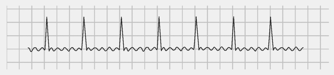
ElectrocardiographyElectrocardiographyRecording of the moment-to-moment electromotive forces of the heart as projected onto various sites on the body’s surface, delineated as a scalar function of time. The recording is monitored by a tracing on slow moving chart paper or by observing it on a cardioscope, which is a cathode ray tube display.Electrocardiogram (ECG):
Atrial rate of approximately 300/min (range, 240–340)
Rhythm usually regularRegularInsulin, but can rarely be irregular
Counterclockwise flutter: negative in leads II, III, aVF; positive in V1
Clockwise flutter: positive in leads II, III, aVF; negative in V1
Atypical atrial flutterAtypical atrial flutterAtrial Flutter: variableVariableVariables represent information about something that can change. The design of the measurement scales, or of the methods for obtaining information, will determine the data gathered and the characteristics of that data. As a result, a variable can be qualitative or quantitative, and may be further classified into subgroups.Types of Variables flutter wave morphology
Narrow QRS complexes
Consistent ratio between atrial and ventricular rates (usually a ventricular rate of approximately 150/min in 2:1 conduction)
May give clues to underlying cardiac disease or complications:
Evidence of LV hypertrophyHypertrophyGeneral increase in bulk of a part or organ due to cell enlargement and accumulation of fluids and secretions, not due to tumor formation, nor to an increase in the number of cells (hyperplasia).Cellular Adaptation
Rate-related myocardial ischemiaMyocardial ischemiaA disorder of cardiac function caused by insufficient blood flow to the muscle tissue of the heart. The decreased blood flow may be due to narrowing of the coronary arteries (coronary artery disease), to obstruction by a thrombus (coronary thrombosis), or less commonly, to diffuse narrowing of arterioles and other small vessels within the heart.Coronary Heart Disease (ST depressions that resolve with improvement in heart rateHeart rateThe number of times the heart ventricles contract per unit of time, usually per minute.Cardiac Physiology)
Myocardial infarctionMyocardial infarctionMI is ischemia and death of an area of myocardial tissue due to insufficient blood flow and oxygenation, usually from thrombus formation on a ruptured atherosclerotic plaque in the epicardial arteries. Clinical presentation is most commonly with chest pain, but women and patients with diabetes may have atypical symptoms.Myocardial Infarction
Holter monitoring:
Portable device that continuously records cardiac activity (usually worn for 24–48 hours)
Not required for diagnosis, but can catch transient events if symptoms are nonspecific or short-lived
Evaluate for structural disease, such as atrial dilation or valvular disease
Look for wall motion abnormalities
Assess ventricular systolic function
Transesophageal echocardiographyEchocardiographyUltrasonic recording of the size, motion, and composition of the heart and surrounding tissues. The standard approach is transthoracic.Tricuspid Valve Atresia (TVA):
Evaluate for associated ischemic heart diseaseIschemic heart diseaseCoronary heart disease (CHD), or ischemic heart disease, describes a situation in which an inadequate supply of blood to the myocardium exists due to a stenosis of the coronary arteries, typically from atherosclerosis. Coronary Heart Disease
Can reproduce exercise-induced atrial flutterAtrial flutterAtrial flutter is a regular supraventricular tachycardia characterized by an atrial heart rate between 240/min and 340/min (typically 300/min), atrioventricular (AV) node conduction block, and a “sawtooth” pattern on an electrocardiogram (ECG). Atrial Flutter
Other tests to search for an underlying cause:
Serum electrolytesElectrolytesElectrolytes are mineral salts that dissolve in water and dissociate into charged particles called ions, which can be either be positively (cations) or negatively (anions) charged. Electrolytes are distributed in the extracellular and intracellular compartments in different concentrations. Electrolytes are essential for various basic life-sustaining functions.Electrolytes → electrolyte imbalance
B-type natriuretic peptide (BNPBNPA peptide that is secreted by the brain and the heart atria, stored mainly in cardiac ventricular myocardium. It can cause natriuresis; diuresis; vasodilation; and inhibits secretion of renin and aldosterone. It improves heart function. It contains 32 amino acids.Renal Sodium and Water Regulation) → heart failureHeart FailureA heterogeneous condition in which the heart is unable to pump out sufficient blood to meet the metabolic need of the body. Heart failure can be caused by structural defects, functional abnormalities (ventricular dysfunction), or a sudden overload beyond its capacity. Chronic heart failure is more common than acute heart failure which results from sudden insult to cardiac function, such as myocardial infarction.Total Anomalous Pulmonary Venous Return (TAPVR)
Troponins → myocardial ischemiaMyocardial ischemiaA disorder of cardiac function caused by insufficient blood flow to the muscle tissue of the heart. The decreased blood flow may be due to narrowing of the coronary arteries (coronary artery disease), to obstruction by a thrombus (coronary thrombosis), or less commonly, to diffuse narrowing of arterioles and other small vessels within the heart.Coronary Heart Disease
Thyroid-stimulating hormoneThyroid-stimulating hormoneA glycoprotein hormone secreted by the adenohypophysis. Thyrotropin stimulates thyroid gland by increasing the iodide transport, synthesis and release of thyroid hormones (thyroxine and triiodothyronine).Thyroid Hormones (TSH) → screen for hyperthyroidismHyperthyroidismHypersecretion of thyroid hormones from the thyroid gland. Elevated levels of thyroid hormones increase basal metabolic rate.Thyrotoxicosis and Hyperthyroidism
WBCs → sepsisSepsisSystemic inflammatory response syndrome with a proven or suspected infectious etiology. When sepsis is associated with organ dysfunction distant from the site of infection, it is called severe sepsis. When sepsis is accompanied by hypotension despite adequate fluid infusion, it is called septic shock.Sepsis and Septic Shock
DigoxinDigoxinA cardiotonic glycoside obtained mainly from digitalis lanata; it consists of three sugars and the aglycone digoxigenin. Digoxin has positive inotropic and negative chronotropic activity. It is used to control ventricular rate in atrial fibrillation and in the management of congestive heart failure with atrial fibrillation. Its use in congestive heart failure and sinus rhythm is less certain. The margin between toxic and therapeutic doses is small.Cardiac Glycosides level → digitalis toxicityToxicityDosage Calculation
CT pulmonary angiographyAngiographyRadiography of blood vessels after injection of a contrast medium.Cardiac Surgery → pulmonary embolismPulmonary EmbolismPulmonary embolism (PE) is a potentially fatal condition that occurs as a result of intraluminal obstruction of the main pulmonary artery or its branches. The causative factors include thrombi, air, amniotic fluid, and fat. In PE, gas exchange is impaired due to the decreased return of deoxygenated blood to the lungs. Pulmonary Embolism
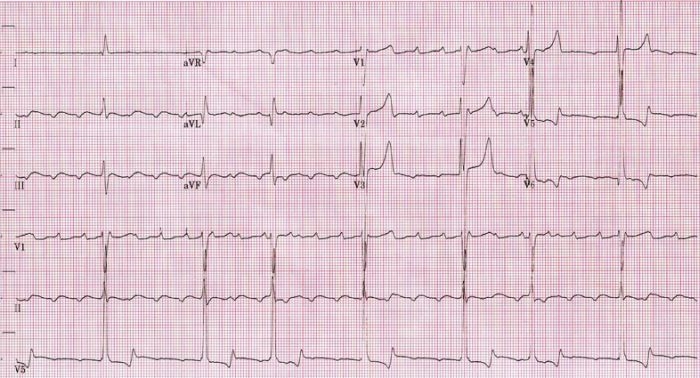
An ECG showing slow atrial flutter at an atrial cycle length of 400 ms (atrial rate of 150/min).
Image: “Atrial rate of 150/min” by Dept. of Cardiology, Beth Israel Medical Center. 16th Street and 1st Avenue. New York, New York 10003, USA. License: CC BY 2.5.
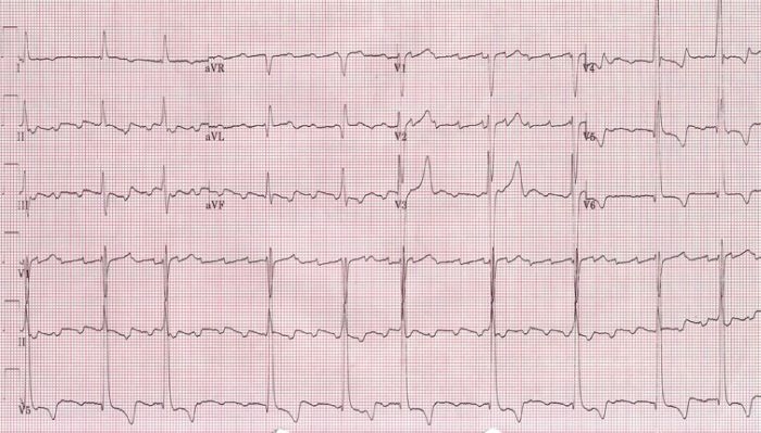
An ECG showing a faster atrial flutter at an atrial cycle length of 280 ms (atrial rate of 214/min).
Image: “Atrial rate of 214/min” by Dept. of Cardiology, Beth Israel Medical Center. 16th Street and 1st Avenue. New York, New York 10003, USA. License: CC BY 2.5.
An ECG showing the classic “sawtooth” appearance in atrial flutter.
Management may vary depending on practice location. The following information is based on US, Canadian, and European literature and guidelines.
The 3 important issues in managing atrial flutterAtrial flutterAtrial flutter is a regular supraventricular tachycardia characterized by an atrial heart rate between 240/min and 340/min (typically 300/min), atrioventricular (AV) node conduction block, and a “sawtooth” pattern on an electrocardiogram (ECG). Atrial Flutter are assessing the need for cardioversionCardioversionAtrial Fibrillation, slowing the ventricular rate, and preventing systemic embolizationEmbolizationA method of hemostasis utilizing various agents such as gelfoam, silastic, metal, glass, or plastic pellets, autologous clot, fat, and muscle as emboli. It has been used in the treatment of spinal cord and intracranial arteriovenous malformations, renal arteriovenous fistulas, gastrointestinal bleeding, epistaxis, hypersplenism, certain highly vascular tumors, traumatic rupture of blood vessels, and control of operative hemorrhage.Gastrointestinal Bleeding with antithrombotic therapy. Rhythm control is indicated to treat symptoms and prevent recurrence.
Indications for hospitalizationHospitalizationThe confinement of a patient in a hospital.Delirium[2,3]
Highly symptomatic atrial flutterAtrial flutterAtrial flutter is a regular supraventricular tachycardia characterized by an atrial heart rate between 240/min and 340/min (typically 300/min), atrioventricular (AV) node conduction block, and a “sawtooth” pattern on an electrocardiogram (ECG). Atrial Flutter
Acute coronary syndrome with chest painPainAn unpleasant sensation induced by noxious stimuli which are detected by nerve endings of nociceptive neurons.Pain: Types and Pathways and ECGECGAn electrocardiogram (ECG) is a graphic representation of the electrical activity of the heart plotted against time. Adhesive electrodes are affixed to the skin surface allowing measurement of cardiac impulses from many angles. The ECG provides 3-dimensional information about the conduction system of the heart, the myocardium, and other cardiac structures. Electrocardiogram (ECG) changes
Acute heart failureHeart FailureA heterogeneous condition in which the heart is unable to pump out sufficient blood to meet the metabolic need of the body. Heart failure can be caused by structural defects, functional abnormalities (ventricular dysfunction), or a sudden overload beyond its capacity. Chronic heart failure is more common than acute heart failure which results from sudden insult to cardiac function, such as myocardial infarction.Total Anomalous Pulmonary Venous Return (TAPVR) not improved with treatment in the emergency department
Acute management
The most important starting point in management is to assess the patient’s clinical stability. This will determine the best next steps. Additionally, identificationIdentificationDefense Mechanisms and treatment of any underlying causes (cardiac ischemiaIschemiaA hypoperfusion of the blood through an organ or tissue caused by a pathologic constriction or obstruction of its blood vessels, or an absence of blood circulation.Ischemic Cell Damage, sepsisSepsisSystemic inflammatory response syndrome with a proven or suspected infectious etiology. When sepsis is associated with organ dysfunction distant from the site of infection, it is called severe sepsis. When sepsis is accompanied by hypotension despite adequate fluid infusion, it is called septic shock.Sepsis and Septic Shock) should be pursued.
Systolic blood pressure (SBPSBPAscites) < 90 mm Hg or other signs of shockShockShock is a life-threatening condition associated with impaired circulation that results in tissue hypoxia. The different types of shock are based on the underlying cause: distributive (↑ cardiac output (CO), ↓ systemic vascular resistance (SVR)), cardiogenic (↓ CO, ↑ SVR), hypovolemic (↓ CO, ↑ SVR), obstructive (↓ CO), and mixed. Types of Shock
Evidence of cardiac ischemiaIschemiaA hypoperfusion of the blood through an organ or tissue caused by a pathologic constriction or obstruction of its blood vessels, or an absence of blood circulation.Ischemic Cell Damage: chest painPainAn unpleasant sensation induced by noxious stimuli which are detected by nerve endings of nociceptive neurons.Pain: Types and Pathways or marked ST depression on ECGECGAn electrocardiogram (ECG) is a graphic representation of the electrical activity of the heart plotted against time. Adhesive electrodes are affixed to the skin surface allowing measurement of cardiac impulses from many angles. The ECG provides 3-dimensional information about the conduction system of the heart, the myocardium, and other cardiac structures. Electrocardiogram (ECG)
Pulmonary edemaPulmonary edemaPulmonary edema is a condition caused by excess fluid within the lung parenchyma and alveoli as a consequence of a disease process. Based on etiology, pulmonary edema is classified as cardiogenic or noncardiogenic. Patients may present with progressive dyspnea, orthopnea, cough, or respiratory failure.Pulmonary Edema
More difficult to control rate with atrial flutterAtrial flutterAtrial flutter is a regular supraventricular tachycardia characterized by an atrial heart rate between 240/min and 340/min (typically 300/min), atrioventricular (AV) node conduction block, and a “sawtooth” pattern on an electrocardiogram (ECG). Atrial Flutter than with atrial fibrillationAtrial fibrillationAtrial fibrillation (AF or Afib) is a supraventricular tachyarrhythmia and the most common kind of arrhythmia. It is caused by rapid, uncontrolled atrial contractions and uncoordinated ventricular responses. Atrial Fibrillation
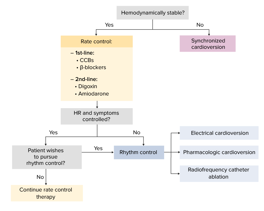
Simplified algorithm regarding rate versus rhythm control for atrial flutter:
Note that this does not include anticoagulation guidelines, which should be considered in the management of atrial flutter.
CCBs: calcium channel blockers HR: heart rate
Image by Lecturio.
Rate control
Common drug classes used for rate control:
CCBsCCBsCalcium channel blockers (CCBS) are a class of medications that inhibit voltage-dependent L-type calcium channels of cardiac and vascular smooth muscle cells. The inhibition of these channels produces vasodilation and myocardial depression. There are 2 major classes of CCBS: dihydropyridines and non-dihydropyridines.Class 4 Antiarrhythmic Drugs (Calcium Channel Blockers):
1st-line option
Avoid if severe heart failureHeart FailureA heterogeneous condition in which the heart is unable to pump out sufficient blood to meet the metabolic need of the body. Heart failure can be caused by structural defects, functional abnormalities (ventricular dysfunction), or a sudden overload beyond its capacity. Chronic heart failure is more common than acute heart failure which results from sudden insult to cardiac function, such as myocardial infarction.Total Anomalous Pulmonary Venous Return (TAPVR) or known LV dysfunction (LVEF ≤40%)
Avoid if with 2nd/3rd-degree AV blockAV blockAtrioventricular (AV) block is a bradyarrhythmia caused by delay, or interruption, in the electrical conduction between the atria and the ventricles. Atrioventricular block occurs due to either anatomic or functional impairment, and is classified into 3 types. Atrioventricular block (AV block) without a pacemakerPacemakerA device designed to stimulate, by electric impulses, contraction of the heart muscles. It may be temporary (external) or permanent (internal or internal-external).Bradyarrhythmias
Most common choice: IV diltiazemDiltiazemA benzothiazepine derivative with vasodilating action due to its antagonism of the actions of calcium ion on membrane functions.Class 4 Antiarrhythmic Drugs (Calcium Channel Blockers) (bolus dosing or continuous infusion)
Once rate is controlled, follow up with oral dose within 30 minutes
IV verapamilVerapamilA calcium channel blocker that is a class IV anti-arrhythmia agent.Pulmonary Hypertension Drugs is rarely used but still available
Beta blockers: an alternative to IV diltiazemDiltiazemA benzothiazepine derivative with vasodilating action due to its antagonism of the actions of calcium ion on membrane functions.Class 4 Antiarrhythmic Drugs (Calcium Channel Blockers), not used as an addition:
Another 1st-line option
Avoid in acute heart failureHeart FailureA heterogeneous condition in which the heart is unable to pump out sufficient blood to meet the metabolic need of the body. Heart failure can be caused by structural defects, functional abnormalities (ventricular dysfunction), or a sudden overload beyond its capacity. Chronic heart failure is more common than acute heart failure which results from sudden insult to cardiac function, such as myocardial infarction.Total Anomalous Pulmonary Venous Return (TAPVR) or chronic lung disease (prefer cardioselective agents, e.g., metoprololMetoprololA selective adrenergic beta-1 blocking agent that is commonly used to treat angina pectoris; hypertension; and cardiac arrhythmias.Antiadrenergic Drugs)
Most common choice: IV metoprololMetoprololA selective adrenergic beta-1 blocking agent that is commonly used to treat angina pectoris; hypertension; and cardiac arrhythmias.Antiadrenergic Drugs,esmololEsmololAntiadrenergic Drugs
Once rate is controlled, follow up with oral dose within 30 minutes
DigoxinDigoxinA cardiotonic glycoside obtained mainly from digitalis lanata; it consists of three sugars and the aglycone digoxigenin. Digoxin has positive inotropic and negative chronotropic activity. It is used to control ventricular rate in atrial fibrillation and in the management of congestive heart failure with atrial fibrillation. Its use in congestive heart failure and sinus rhythm is less certain. The margin between toxic and therapeutic doses is small.Cardiac Glycosides:
2nd-line option
Has positive inotropic effect, which may be helpful in heart failureHeart FailureA heterogeneous condition in which the heart is unable to pump out sufficient blood to meet the metabolic need of the body. Heart failure can be caused by structural defects, functional abnormalities (ventricular dysfunction), or a sudden overload beyond its capacity. Chronic heart failure is more common than acute heart failure which results from sudden insult to cardiac function, such as myocardial infarction.Total Anomalous Pulmonary Venous Return (TAPVR) with reduced ejection fractionEjection fractionCardiac Cycle
Less hypotensive effect than beta-blockersBeta-blockersDrugs that bind to but do not activate beta-adrenergic receptors thereby blocking the actions of beta-adrenergic agonists. Adrenergic beta-antagonists are used for treatment of hypertension, cardiac arrhythmias, angina pectoris, glaucoma, migraine headaches, and anxiety.Class 2 Antiarrhythmic Drugs (Beta Blockers) and CCBsCCBsCalcium channel blockers (CCBS) are a class of medications that inhibit voltage-dependent L-type calcium channels of cardiac and vascular smooth muscle cells. The inhibition of these channels produces vasodilation and myocardial depression. There are 2 major classes of CCBS: dihydropyridines and non-dihydropyridines.Class 4 Antiarrhythmic Drugs (Calcium Channel Blockers) → can be useful for acute management of critically ill patientsPatientsIndividuals participating in the health care system for the purpose of receiving therapeutic, diagnostic, or preventive procedures.Clinician–Patient Relationship
Caution in patientsPatientsIndividuals participating in the health care system for the purpose of receiving therapeutic, diagnostic, or preventive procedures.Clinician–Patient Relationship with chronic kidney diseaseChronic Kidney DiseaseChronic kidney disease (CKD) is kidney impairment that lasts for ≥ 3 months, implying that it is irreversible. Hypertension and diabetes are the most common causes; however, there are a multitude of other etiologies. In the early to moderate stages, CKD is usually asymptomatic and is primarily diagnosed by laboratory abnormalities.Chronic Kidney Disease
AmiodaroneAmiodaroneAn antianginal and class III antiarrhythmic drug. It increases the duration of ventricular and atrial muscle action by inhibiting potassium channels and voltage-gated sodium channels. There is a resulting decrease in heart rate and in vascular resistance.Pulmonary Fibrosis:
2nd-line option
Less hypotensive effect than 1st-line options → can be useful for acute management of critically ill patientsPatientsIndividuals participating in the health care system for the purpose of receiving therapeutic, diagnostic, or preventive procedures.Clinician–Patient Relationship
Table: Rate control agents* for atrial flutterAtrial flutterAtrial flutter is a regular supraventricular tachycardia characterized by an atrial heart rate between 240/min and 340/min (typically 300/min), atrioventricular (AV) node conduction block, and a “sawtooth” pattern on an electrocardiogram (ECG). Atrial Flutter (adults)[2,4]
Repeat 0.35 mg/kg every 15–20 minutes, as needed, for up to 3 doses
Short-acting: 30–60 mg every 6 hours
Extended-release: 120–240 mg daily
MetoprololMetoprololA selective adrenergic beta-1 blocking agent that is commonly used to treat angina pectoris; hypertension; and cardiac arrhythmias.Antiadrenergic Drugs
2.5–5 mg IV over 2 minutes
Repeat dose every 15–20 minutes, as needed, for up to 3 doses
Short-acting: 25–50 mg twice daily
Extended-release: 50–100 mg daily
2nd-line
DigoxinDigoxinA cardiotonic glycoside obtained mainly from digitalis lanata; it consists of three sugars and the aglycone digoxigenin. Digoxin has positive inotropic and negative chronotropic activity. It is used to control ventricular rate in atrial fibrillation and in the management of congestive heart failure with atrial fibrillation. Its use in congestive heart failure and sinus rhythm is less certain. The margin between toxic and therapeutic doses is small.Cardiac Glycosides
AmiodaroneAmiodaroneAn antianginal and class III antiarrhythmic drug. It increases the duration of ventricular and atrial muscle action by inhibiting potassium channels and voltage-gated sodium channels. There is a resulting decrease in heart rate and in vascular resistance.Pulmonary Fibrosis
300 mg over 1 hour
Followed by 10–50 mg/hr over 24 hours
100–200 mg daily
*List is not exhaustive.
Antithrombotic therapy for stroke prevention[3–5,10–12]
Principles similar to atrial fibrillationAtrial fibrillationAtrial fibrillation (AF or Afib) is a supraventricular tachyarrhythmia and the most common kind of arrhythmia. It is caused by rapid, uncontrolled atrial contractions and uncoordinated ventricular responses. Atrial Fibrillation
AnticoagulationAnticoagulationPulmonary Hypertension Drugs (long-term) should be considered in cases of atrial flutterAtrial flutterAtrial flutter is a regular supraventricular tachycardia characterized by an atrial heart rate between 240/min and 340/min (typically 300/min), atrioventricular (AV) node conduction block, and a “sawtooth” pattern on an electrocardiogram (ECG). Atrial Flutter with unknown onset or that persists > 48 hours or when there is a high risk of stroke.
Risk stratify using the CHA2DS2-VASc scoring system for nonvalvular atrial fibrillationAtrial fibrillationAtrial fibrillation (AF or Afib) is a supraventricular tachyarrhythmia and the most common kind of arrhythmia. It is caused by rapid, uncontrolled atrial contractions and uncoordinated ventricular responses. Atrial Fibrillation (see below):
Ensure that benefits of anticoagulationAnticoagulationPulmonary Hypertension Drugs outweigh risks → can assess risk of bleeding (and possibly reduce risk if patient has modifiable factors) using the following:
Check for contraindicationsContraindicationsA condition or factor associated with a recipient that makes the use of a drug, procedure, or physical agent improper or inadvisable. Contraindications may be absolute (life threatening) or relative (higher risk of complications in which benefits may outweigh risks).Noninvasive Ventilation to anticoagulationAnticoagulationPulmonary Hypertension Drugs.
Options
Direct oral anticoagulantsAnticoagulantsAnticoagulants are drugs that retard or interrupt the coagulation cascade. The primary classes of available anticoagulants include heparins, vitamin K-dependent antagonists (e.g., warfarin), direct thrombin inhibitors, and factor Xa inhibitors. Anticoagulants (DOACsDOACsAnticoagulants):
Preferred over warfarinWarfarinAn anticoagulant that acts by inhibiting the synthesis of vitamin K-dependent coagulation factors. Warfarin is indicated for the prophylaxis and/or treatment of venous thrombosis and its extension, pulmonary embolism, and atrial fibrillation with embolization. It is also used as an adjunct in the prophylaxis of systemic embolism after myocardial infarction. Warfarin is also used as a rodenticide.Anticoagulants
RivaroxabanRivaroxabanA morpholine and thiophene derivative that functions as a factor Xa inhibitor and is used in the treatment and prevention of deep-vein thrombosis and pulmonary embolism. It is also used for the prevention of stroke and systemic embolization in patients with non-valvular atrial fibrillation, and for the prevention of atherothrombotic events in patients after an acute coronary syndrome.Anticoagulants
DabigatranDabigatranA thrombin inhibitor which acts by binding and blocking thrombogenic activity and the prevention of thrombus formation. It is used to reduce the risk of stroke and systemic embolism in patients with nonvalvular atrial fibrillation.Anticoagulants
WarfarinWarfarinAn anticoagulant that acts by inhibiting the synthesis of vitamin K-dependent coagulation factors. Warfarin is indicated for the prophylaxis and/or treatment of venous thrombosis and its extension, pulmonary embolism, and atrial fibrillation with embolization. It is also used as an adjunct in the prophylaxis of systemic embolism after myocardial infarction. Warfarin is also used as a rodenticide.Anticoagulants:
Use if DOACsDOACsAnticoagulants are contraindicated, such as in patientsPatientsIndividuals participating in the health care system for the purpose of receiving therapeutic, diagnostic, or preventive procedures.Clinician–Patient Relationship with:
Severe renal impairment (GFRGFRThe volume of water filtered out of plasma through glomerular capillary walls into Bowman’s capsules per unit of time. It is considered to be equivalent to inulin clearance.Kidney Function Tests < 15 mL/min)
Goal INR 2–3 (unless other values are indicated by another condition)
Bridging therapy with heparin (or low-molecular-weight heparin) may be considered in patientsPatientsIndividuals participating in the health care system for the purpose of receiving therapeutic, diagnostic, or preventive procedures.Clinician–Patient Relationship at high risk for stroke (always weigh the risk of thrombosisThrombosisFormation and development of a thrombus or blood clot in the blood vessel.Epidemic Typhus against the risk of bleeding when making a decision).
Give IV unfractionated heparinUnfractionated heparinA highly acidic mucopolysaccharide formed of equal parts of sulfated d-glucosamine and d-glucuronic acid with sulfaminic bridges. The molecular weight ranges from six to twenty thousand. Heparin occurs in and is obtained from liver, lung, mast cells, etc. , of vertebrates. Its function is unknown, but it is used to prevent blood clotting in vivo and vitro, in the form of many different salts.Anticoagulants or subcutaneous low molecular weight heparin as soon as feasible.
Continue OAC for ≥ 4 weeks (due to potential atrial “stunning” or temporary reduction in atrial contractile function).
Long-term OAC thereafter is based on CHA₂DS₂-VASc.
For atrial flutterAtrial flutterAtrial flutter is a regular supraventricular tachycardia characterized by an atrial heart rate between 240/min and 340/min (typically 300/min), atrioventricular (AV) node conduction block, and a “sawtooth” pattern on an electrocardiogram (ECG). Atrial Flutter < 48 hours:
Definite history of onset is generally difficult.
If onset is confirmed as < 48 hours, give periprocedural anticoagulationAnticoagulationPulmonary Hypertension Drugs and continue for ≥ 4 weeks (unless CHA2DS2-VASc dictates otherwise).
If onset cannot be accurately defined, treat as atrial flutterAtrial flutterAtrial flutter is a regular supraventricular tachycardia characterized by an atrial heart rate between 240/min and 340/min (typically 300/min), atrioventricular (AV) node conduction block, and a “sawtooth” pattern on an electrocardiogram (ECG). Atrial Flutter of uncertain onset or ≥ 48 hours.
For atrial flutterAtrial flutterAtrial flutter is a regular supraventricular tachycardia characterized by an atrial heart rate between 240/min and 340/min (typically 300/min), atrioventricular (AV) node conduction block, and a “sawtooth” pattern on an electrocardiogram (ECG). Atrial Flutter ≥ 48 hours:
Long-term anticoagulationAnticoagulationPulmonary Hypertension Drugs may be required for recurrent or chronic atrial flutterAtrial flutterAtrial flutter is a regular supraventricular tachycardia characterized by an atrial heart rate between 240/min and 340/min (typically 300/min), atrioventricular (AV) node conduction block, and a “sawtooth” pattern on an electrocardiogram (ECG). Atrial Flutter.
Table: Cha2DS2-VASc score
C
Congestive heart failureHeart FailureA heterogeneous condition in which the heart is unable to pump out sufficient blood to meet the metabolic need of the body. Heart failure can be caused by structural defects, functional abnormalities (ventricular dysfunction), or a sudden overload beyond its capacity. Chronic heart failure is more common than acute heart failure which results from sudden insult to cardiac function, such as myocardial infarction.Total Anomalous Pulmonary Venous Return (TAPVR)
1
H
HypertensionHypertensionHypertension, or high blood pressure, is a common disease that manifests as elevated systemic arterial pressures. Hypertension is most often asymptomatic and is found incidentally as part of a routine physical examination or during triage for an unrelated medical encounter. Hypertension
1
A
Age (≥ 75 years)
2
D
DiabetesDiabetesDiabetes mellitus (DM) is a metabolic disease characterized by hyperglycemia and dysfunction of the regulation of glucose metabolism by insulin. Type 1 DM is diagnosed mostly in children and young adults as the result of autoimmune destruction of β cells in the pancreas and the resulting lack of insulin. Type 2 DM has a significant association with obesity and is characterized by insulin resistance.Diabetes Mellitus mellitus
1
S
Stroke, TIATIATransient ischemic attack (TIA) is a temporary episode of neurologic dysfunction caused by ischemia without infarction that resolves completely when blood supply is restored. Transient ischemic attack is a neurologic emergency that warrants urgent medical attention. Transient Ischemic Attack (TIA), or thromboembolismThromboembolismObstruction of a blood vessel (embolism) by a blood clot (thrombus) in the blood stream.Systemic Lupus Erythematosus
2
V
Vascular disease
1
A
Age 65–74 years
1
Sc
SexSexThe totality of characteristics of reproductive structure, functions, phenotype, and genotype, differentiating the male from the female organism.Gender Dysphoria category (female)
RivaroxabanRivaroxabanA morpholine and thiophene derivative that functions as a factor Xa inhibitor and is used in the treatment and prevention of deep-vein thrombosis and pulmonary embolism. It is also used for the prevention of stroke and systemic embolization in patients with non-valvular atrial fibrillation, and for the prevention of atherothrombotic events in patients after an acute coronary syndrome.Anticoagulants
20 mg daily
DabigatranDabigatranA thrombin inhibitor which acts by binding and blocking thrombogenic activity and the prevention of thrombus formation. It is used to reduce the risk of stroke and systemic embolism in patients with nonvalvular atrial fibrillation.Anticoagulants
Direct thrombinThrombinAn enzyme formed from prothrombin that converts fibrinogen to fibrin.Hemostasis inhibitors
150 mg twice daily
*Doses may need to be adjusted for older adults, for those with renal or hepatic disease, or based on weight.
Rhythm control[2,3]
Some patientsPatientsIndividuals participating in the health care system for the purpose of receiving therapeutic, diagnostic, or preventive procedures.Clinician–Patient Relationship may prefer to continue rate control management. However, many patientsPatientsIndividuals participating in the health care system for the purpose of receiving therapeutic, diagnostic, or preventive procedures.Clinician–Patient Relationship may wish to pursue cardioversionCardioversionAtrial Fibrillation to sinus rhythmSinus rhythmA heart rate and rhythm driven by the regular firing of the SA node (60–100 beats per minute)Cardiac Physiology. The decision on which treatment to pursue should be based on cardiologist expertise and patient preference.
Procedural sedation per local practice (e.g., fentanylFentanylA potent narcotic analgesic, abuse of which leads to habituation or addiction. It is primarily a mu-opioid agonist. Fentanyl is also used as an adjunct to general anesthetics, and as an anesthetic for induction and maintenance.Opioid Analgesics, propofolPropofolAn intravenous anesthetic agent which has the advantage of a very rapid onset after infusion or bolus injection plus a very short recovery period of a couple of minutes. Propofol has been used as anticonvulsants and antiemetics.Intravenous Anesthetics)
Primary options: both carry ↑ risk of torsades de pointesTorsades de pointesA malignant form of polymorphic ventricular tachycardia that is characterized by heart rate between 200 and 250 beats per minute, and QRS complexes with changing amplitude and twisting of the points. The term also describes the syndrome of tachycardia with prolonged ventricular repolarization, long qt intervals exceeding 500 milliseconds or bradycardia. Torsades de pointes may be self-limited or may progress to ventricular fibrillation.Ventricular Tachycardia, so continuous ECGECGAn electrocardiogram (ECG) is a graphic representation of the electrical activity of the heart plotted against time. Adhesive electrodes are affixed to the skin surface allowing measurement of cardiac impulses from many angles. The ECG provides 3-dimensional information about the conduction system of the heart, the myocardium, and other cardiac structures. Electrocardiogram (ECG) and QT intervalQT intervalElectrocardiogram (ECG) monitoring is important during administration.
AmiodaroneAmiodaroneAn antianginal and class III antiarrhythmic drug. It increases the duration of ventricular and atrial muscle action by inhibiting potassium channels and voltage-gated sodium channels. There is a resulting decrease in heart rate and in vascular resistance.Pulmonary Fibrosis is not recommended owing to low efficacy and delayed onset of action.
Preferred in most patientsPatientsIndividuals participating in the health care system for the purpose of receiving therapeutic, diagnostic, or preventive procedures.Clinician–Patient Relationship, except those with reversible causes of atrial flutterAtrial flutterAtrial flutter is a regular supraventricular tachycardia characterized by an atrial heart rate between 240/min and 340/min (typically 300/min), atrioventricular (AV) node conduction block, and a “sawtooth” pattern on an electrocardiogram (ECG). Atrial Flutter
Follow-up[2,3]
After discharge, patientsPatientsIndividuals participating in the health care system for the purpose of receiving therapeutic, diagnostic, or preventive procedures.Clinician–Patient Relationship should be seen in clinic within 7 days if starting:
Rate control therapy
WarfarinWarfarinAn anticoagulant that acts by inhibiting the synthesis of vitamin K-dependent coagulation factors. Warfarin is indicated for the prophylaxis and/or treatment of venous thrombosis and its extension, pulmonary embolism, and atrial fibrillation with embolization. It is also used as an adjunct in the prophylaxis of systemic embolism after myocardial infarction. Warfarin is also used as a rodenticide.Anticoagulants
Otherwise, should be seen in clinic within 4–6 weeks
Team should include:
Primary care physician
Cardiologist
Differential Diagnosis
Atrial fibrillationAtrial fibrillationAtrial fibrillation (AF or Afib) is a supraventricular tachyarrhythmia and the most common kind of arrhythmia. It is caused by rapid, uncontrolled atrial contractions and uncoordinated ventricular responses. Atrial Fibrillation: the most common form of supraventricular arrhythmia. Symptoms are highly variableVariableVariables represent information about something that can change. The design of the measurement scales, or of the methods for obtaining information, will determine the data gathered and the characteristics of that data. As a result, a variable can be qualitative or quantitative, and may be further classified into subgroups.Types of Variables, but include palpitationsPalpitationsEbstein’s Anomaly, syncopeSyncopeSyncope is a short-term loss of consciousness and loss of postural stability followed by spontaneous return of consciousness to the previous neurologic baseline without the need for resuscitation. The condition is caused by transient interruption of cerebral blood flow that may be benign or related to a underlying life-threatening condition. Syncope, shortness of breathShortness of breathDyspnea is the subjective sensation of breathing discomfort. Dyspnea is a normal manifestation of heavy physical or psychological exertion, but also may be caused by underlying conditions (both pulmonary and extrapulmonary).Dyspnea, or chest painPainAn unpleasant sensation induced by noxious stimuli which are detected by nerve endings of nociceptive neurons.Pain: Types and Pathways. Atrial fibrillationAtrial fibrillationAtrial fibrillation (AF or Afib) is a supraventricular tachyarrhythmia and the most common kind of arrhythmia. It is caused by rapid, uncontrolled atrial contractions and uncoordinated ventricular responses. Atrial Fibrillation is diagnosed with an irregularly irregular rhythm without visible P waves on an ECGECGAn electrocardiogram (ECG) is a graphic representation of the electrical activity of the heart plotted against time. Adhesive electrodes are affixed to the skin surface allowing measurement of cardiac impulses from many angles. The ECG provides 3-dimensional information about the conduction system of the heart, the myocardium, and other cardiac structures. Electrocardiogram (ECG), which differentiates it from atrial flutterAtrial flutterAtrial flutter is a regular supraventricular tachycardia characterized by an atrial heart rate between 240/min and 340/min (typically 300/min), atrioventricular (AV) node conduction block, and a “sawtooth” pattern on an electrocardiogram (ECG). Atrial Flutter. Treatment focuses on rate or rhythm control, as well as anticoagulationAnticoagulationPulmonary Hypertension Drugs to reduce thromboembolic risk.
Multifocal atrial tachycardiaMultifocal atrial tachycardiaTachyarrhythmias (MATMATLeptospira/Leptospirosis): an atrial tachyarrhythmiaTachyarrhythmiaA tachyarrhythmia is a rapid heart rhythm, regular or irregular, with a rate > 100 beats/min. Tachyarrhythmia may or may not be accompanied by symptoms of hemodynamic change.Tachyarrhythmias due to pacemakerPacemakerA device designed to stimulate, by electric impulses, contraction of the heart muscles. It may be temporary (external) or permanent (internal or internal-external).Bradyarrhythmias activity from multiple atrial locations. MATMATLeptospira/Leptospirosis is seen in older adults with chronic pulmonary conditions. Diagnosis is made by ECGECGAn electrocardiogram (ECG) is a graphic representation of the electrical activity of the heart plotted against time. Adhesive electrodes are affixed to the skin surface allowing measurement of cardiac impulses from many angles. The ECG provides 3-dimensional information about the conduction system of the heart, the myocardium, and other cardiac structures. Electrocardiogram (ECG) showing ≥ 3 P-wave morphologies, distinguishing it from the uniform, “sawtooth” pattern of atrial flutterAtrial flutterAtrial flutter is a regular supraventricular tachycardia characterized by an atrial heart rate between 240/min and 340/min (typically 300/min), atrioventricular (AV) node conduction block, and a “sawtooth” pattern on an electrocardiogram (ECG). Atrial Flutter. Treatment includes rate or rhythm control.
Paroxysmal supraventricular tachycardiaTachycardiaAbnormally rapid heartbeat, usually with a heart rate above 100 beats per minute for adults. Tachycardia accompanied by disturbance in the cardiac depolarization (cardiac arrhythmia) is called tachyarrhythmia.Sepsis in Children: often due to a reentry pathway. PatientsPatientsIndividuals participating in the health care system for the purpose of receiving therapeutic, diagnostic, or preventive procedures.Clinician–Patient Relationship may have abrupt onset of chest painPainAn unpleasant sensation induced by noxious stimuli which are detected by nerve endings of nociceptive neurons.Pain: Types and Pathways, shortness of breathShortness of breathDyspnea is the subjective sensation of breathing discomfort. Dyspnea is a normal manifestation of heavy physical or psychological exertion, but also may be caused by underlying conditions (both pulmonary and extrapulmonary).Dyspnea, or dizzinessDizzinessAn imprecise term which may refer to a sense of spatial disorientation, motion of the environment, or lightheadedness.Lateral Medullary Syndrome (Wallenberg Syndrome). An ECGECGAn electrocardiogram (ECG) is a graphic representation of the electrical activity of the heart plotted against time. Adhesive electrodes are affixed to the skin surface allowing measurement of cardiac impulses from many angles. The ECG provides 3-dimensional information about the conduction system of the heart, the myocardium, and other cardiac structures. Electrocardiogram (ECG) shows narrow QRS complexes with a rate between 150/min and 240/min. The fast rate and lack of flutter waves differentiates paroxysmal supraventricular tachycardiaTachycardiaAbnormally rapid heartbeat, usually with a heart rate above 100 beats per minute for adults. Tachycardia accompanied by disturbance in the cardiac depolarization (cardiac arrhythmia) is called tachyarrhythmia.Sepsis in Children from atrial flutterAtrial flutterAtrial flutter is a regular supraventricular tachycardia characterized by an atrial heart rate between 240/min and 340/min (typically 300/min), atrioventricular (AV) node conduction block, and a “sawtooth” pattern on an electrocardiogram (ECG). Atrial Flutter. Treatment depends on hemodynamicsHemodynamicsThe movement and the forces involved in the movement of the blood through the cardiovascular system.Vascular Resistance, Flow, and Mean Arterial Pressure and the type of SVTSVTSupraventricular tachycardias are related disorders in which the elevation in heart rate is driven by pathophysiology in the atria. This group falls under the larger umbrella of tachyarrhythmias and includes paroxysmal supraventricular tachycardias (PSVTs), ventricular pre-excitation syndromes (i.e. Wolff-Parkinson-White syndrome), atrial flutter, multifocal atrial tachycardia, and atrial fibrillation. Supraventricular Tachycardias, but may include vagal maneuvers, adenosineAdenosineA nucleoside that is composed of adenine and d-ribose. Adenosine or adenosine derivatives play many important biological roles in addition to being components of DNA and RNA. Adenosine itself is a neurotransmitter.Class 5 Antiarrhythmic Drugs, and cardioversionCardioversionAtrial Fibrillation.
Sinus tachycardiaTachycardiaAbnormally rapid heartbeat, usually with a heart rate above 100 beats per minute for adults. Tachycardia accompanied by disturbance in the cardiac depolarization (cardiac arrhythmia) is called tachyarrhythmia.Sepsis in Children: due to increased SA node activity as a physiologic response to a stressor. Sinus tachycardiaTachycardiaAbnormally rapid heartbeat, usually with a heart rate above 100 beats per minute for adults. Tachycardia accompanied by disturbance in the cardiac depolarization (cardiac arrhythmia) is called tachyarrhythmia.Sepsis in Children may be normal and asymptomatic, or due to an active disease process. Symptoms are usually related to the underlying cause. An ECGECGAn electrocardiogram (ECG) is a graphic representation of the electrical activity of the heart plotted against time. Adhesive electrodes are affixed to the skin surface allowing measurement of cardiac impulses from many angles. The ECG provides 3-dimensional information about the conduction system of the heart, the myocardium, and other cardiac structures. Electrocardiogram (ECG) will show a heart rateHeart rateThe number of times the heart ventricles contract per unit of time, usually per minute.Cardiac Physiology > 100/min, narrow QRS complexes, and regularRegularInsulin P waves (which differentiates sinus tachycardiaTachycardiaAbnormally rapid heartbeat, usually with a heart rate above 100 beats per minute for adults. Tachycardia accompanied by disturbance in the cardiac depolarization (cardiac arrhythmia) is called tachyarrhythmia.Sepsis in Children from atrial flutterAtrial flutterAtrial flutter is a regular supraventricular tachycardia characterized by an atrial heart rate between 240/min and 340/min (typically 300/min), atrioventricular (AV) node conduction block, and a “sawtooth” pattern on an electrocardiogram (ECG). Atrial Flutter). Treatment focuses on the precipitating disease.
Billing and Coding
Diagnosis Codes:
Atrial flutterAtrial flutterAtrial flutter is a regular supraventricular tachycardia characterized by an atrial heart rate between 240/min and 340/min (typically 300/min), atrioventricular (AV) node conduction block, and a “sawtooth” pattern on an electrocardiogram (ECG). Atrial Flutter is coded based on whether it is typical (counterclockwise circuit in the right atrium) or atypical.
IntracardiacIntracardiacTotal Anomalous Pulmonary Venous Return (TAPVR) catheter ablation of a discrete mechanism of atrial flutterAtrial flutterAtrial flutter is a regular supraventricular tachycardia characterized by an atrial heart rate between 240/min and 340/min (typically 300/min), atrioventricular (AV) node conduction block, and a “sawtooth” pattern on an electrocardiogram (ECG). Atrial Flutter (ieIEInfective endocarditis (IE) is caused by infection or inflammation of the inner lining of the heart (endocardium), most commonly affecting the heart valves.Endocarditis, cavotricuspid isthmusIsthmusUterus, Cervix, and Fallopian Tubes: Anatomy)
Medications:
Similar to atrial fibrillationAtrial fibrillationAtrial fibrillation (AF or Afib) is a supraventricular tachyarrhythmia and the most common kind of arrhythmia. It is caused by rapid, uncontrolled atrial contractions and uncoordinated ventricular responses. Atrial Fibrillation, these codes are for medications used for rate control (e.g., diltiazemDiltiazemA benzothiazepine derivative with vasodilating action due to its antagonism of the actions of calcium ion on membrane functions.Class 4 Antiarrhythmic Drugs (Calcium Channel Blockers)) and stroke prevention with anticoagulantsAnticoagulantsAnticoagulants are drugs that retard or interrupt the coagulation cascade. The primary classes of available anticoagulants include heparins, vitamin K-dependent antagonists (e.g., warfarin), direct thrombin inhibitors, and factor Xa inhibitors. Anticoagulants (e.g., rivaroxabanRivaroxabanA morpholine and thiophene derivative that functions as a factor Xa inhibitor and is used in the treatment and prevention of deep-vein thrombosis and pulmonary embolism. It is also used for the prevention of stroke and systemic embolization in patients with non-valvular atrial fibrillation, and for the prevention of atherothrombotic events in patients after an acute coronary syndrome.Anticoagulants), based on the patient’s stroke risk score.
RivaroxabanRivaroxabanA morpholine and thiophene derivative that functions as a factor Xa inhibitor and is used in the treatment and prevention of deep-vein thrombosis and pulmonary embolism. It is also used for the prevention of stroke and systemic embolization in patients with non-valvular atrial fibrillation, and for the prevention of atherothrombotic events in patients after an acute coronary syndrome.Anticoagulants (ingredient)
References
Olgin, J. E. (2019). Approach to the patient with suspected arrhythmia. In Crow, M. K., et al. (Eds.), Goldman-Cecil Medicine. (26th ed., vol 1, pp. 318–326).
Brandes, A., et al. (2020). Cardioversion of atrial fibrillation and atrial flutter revisited: current evidence and practical guidance for a common procedure. Europace, 22(8), 1149-61. https://www.ncbi.nlm.nih.gov/pmc/articles/PMC7399700/
Page, R. L., Joglar, J. A., et al. (2016). 2015 ACC/AHA/HRS guideline for the management of adult patients with supraventricular tachycardia: a report of the American College of Cardiology/American Heart Association task force on clinical practice guidelines and the Heart Rhythm Society. Journal of the American College of Cardiology, 67(13), e27–e115. https://www.sciencedirect.com/science/article/pii/S0735109715058404?via%3Dihub
Nunes-Ferreira, A., Alves, M., et al. (2021). Anticoagulation after typical atrial flutter ablation: systematic review and meta-analysis. PACE, 44(10), 1701–1710. https://onlinelibrary.wiley.com/doi/10.1111/pace.14342
Heidenreich, P. A., et al. (2020). 2020 update to the 2016 ACC/AHA clinical performance and quality measures for adults with atrial fibrillation or atrial flutter: a report of the American College of Cardiology, American Heart Association task force on performance measures. Circulation: Quality and Outcomes, 14:e000100. https://www.ahajournals.org/doi/10.1161/HCQ.0000000000000100
Katritsis, D. G., et al. (2017). European Heart Rhythm Association (EHRA) consensus document on the management of supraventricular arrhythmias, endorsed by the Heart Rhythm Society (HRS), Asia-Pacific Heart Rhythm Society (APHRS), and Sociedad Latinoamericana de Estimulacion Cardiaca y Electrofisiologia (SOLAECE). EP Europace, 19(3), 465–511. https://academic.oup.com/europace/article/19/3/465/2631183?login=false
Create your free account or log in to continue reading!