Ventricular tachycardia is any heart rhythm faster than 100/min, with 3 or more irregular beats in a row, arising distal to the bundle of His. Ventricular tachycardia is the most common form of wide-complex tachycardia, and it is associated with a high mortality rate. Ventricular tachycardia is often caused by myocardial ischemia, structural disease, congenital conditions, or electrolyte derangement. Individuals may present with chest pain, dyspnea, palpitations, syncope, and hemodynamic instability. Diagnosis is based on characteristic ECG findings of wide-complex QRS, fusion, and capture beats. Management may require antiarrhythmic medications or electrical cardioversion to avoid complications such as heart failure, multiorgan failure, and cardiac arrest.
Ventricular tachycardiaTachycardiaAbnormally rapid heartbeat, usually with a heart rate above 100 beats per minute for adults. Tachycardia accompanied by disturbance in the cardiac depolarization (cardiac arrhythmia) is called tachyarrhythmia.Sepsis in Children (particularly sustained VTVTVentricular tachycardia is any heart rhythm faster than 100 beats/min, with 3 or more irregular beats in a row, arising distal to the bundle of his. Ventricular tachycardia is the most common form of wide-complex tachycardia, and it is associated with a high mortality rate.Ventricular Tachycardia) and ventricular fibrillationVentricular fibrillationVentricular fibrillation (VF or V-fib) is a type of ventricular tachyarrhythmia (> 300/min) often preceded by ventricular tachycardia. In this arrhythmia, the ventricle beats rapidly and sporadically. The ventricular contraction is uncoordinated, leading to a decrease in cardiac output and immediate hemodynamic collapse. Ventricular Fibrillation (V-fib), is a leading cause of sudden cardiac deathSudden cardiac deathCardiac arrest is the sudden, complete cessation of cardiac output with hemodynamic collapse. Patients present as pulseless, unresponsive, and apneic. Rhythms associated with cardiac arrest are ventricular fibrillation/tachycardia, asystole, or pulseless electrical activity.Cardiac Arrest.
IncidenceIncidenceThe number of new cases of a given disease during a given period in a specified population. It also is used for the rate at which new events occur in a defined population. It is differentiated from prevalence, which refers to all cases in the population at a given time.Measures of Disease Frequency ↑ with age
More common in men
Torsades de pointesTorsades de pointesA malignant form of polymorphic ventricular tachycardia that is characterized by heart rate between 200 and 250 beats per minute, and QRS complexes with changing amplitude and twisting of the points. The term also describes the syndrome of tachycardia with prolonged ventricular repolarization, long qt intervals exceeding 500 milliseconds or bradycardia. Torsades de pointes may be self-limited or may progress to ventricular fibrillation.Ventricular Tachycardia more common in women
Etiology[5]
Heart disease:
Coronary arteryCoronary ArteryTruncus Arteriosus disease (most common), especially with prior MIMIMI is ischemia and death of an area of myocardial tissue due to insufficient blood flow and oxygenation, usually from thrombus formation on a ruptured atherosclerotic plaque in the epicardial arteries. Clinical presentation is most commonly with chest pain, but women and patients with diabetes may have atypical symptoms.Myocardial Infarction
AtherosclerosisAtherosclerosisAtherosclerosis is a common form of arterial disease in which lipid deposition forms a plaque in the blood vessel walls. Atherosclerosis is an incurable disease, for which there are clearly defined risk factors that often can be reduced through a change in lifestyle and behavior of the patient. Atherosclerosis
SmokingSmokingWillful or deliberate act of inhaling and exhaling smoke from burning substances or agents held by hand.Interstitial Lung Diseases
DiabetesDiabetesDiabetes mellitus (DM) is a metabolic disease characterized by hyperglycemia and dysfunction of the regulation of glucose metabolism by insulin. Type 1 DM is diagnosed mostly in children and young adults as the result of autoimmune destruction of β cells in the pancreas and the resulting lack of insulin. Type 2 DM has a significant association with obesity and is characterized by insulin resistance.Diabetes Mellitus mellitus
Structural disease:
Heart failureHeart FailureA heterogeneous condition in which the heart is unable to pump out sufficient blood to meet the metabolic need of the body. Heart failure can be caused by structural defects, functional abnormalities (ventricular dysfunction), or a sudden overload beyond its capacity. Chronic heart failure is more common than acute heart failure which results from sudden insult to cardiac function, such as myocardial infarction.Total Anomalous Pulmonary Venous Return (TAPVR)
CardiomyopathyCardiomyopathyCardiomyopathy refers to a group of myocardial diseases associated with structural changes of the heart muscles (myocardium) and impaired systolic and/or diastolic function in the absence of other heart disorders (coronary artery disease, hypertension, valvular disease, and congenital heart disease). Cardiomyopathy: Overview and Types
MyocarditisMyocarditisMyocarditis is an inflammatory disease of the myocardium, which may occur alone or in association with a systemic process. There are numerous etiologies of myocarditis, but all lead to inflammation and myocyte injury, most often leading to signs and symptoms of heart failure. Myocarditis
Congenital disorders:
Long-QT syndrome
Wolff-Parkinson-White syndromeWolff-Parkinson-White SyndromeA form of ventricular pre-excitation characterized by a short PR interval and a long QRS interval with a delta wave. In this syndrome, atrial impulses are abnormally conducted to the heart ventricles via an accessory conducting pathway that is located between the wall of the right or left atria and the ventricles, also known as a bundle of kent. The inherited form can be caused by mutation of prkag2 gene encoding a gamma-2 regulatory subunit of amp-activated protein kinase.Supraventricular Tachycardias
Electrolyte derangement:
HypokalemiaHypokalemiaHypokalemia is defined as plasma potassium (K+) concentration < 3.5 mEq/L. Homeostatic mechanisms maintain plasma concentration between 3.5-5.2 mEq/L despite marked variation in dietary intake. Hypokalemia can be due to renal losses, GI losses, transcellular shifts, or poor dietary intake.Hypokalemia
HypomagnesemiaHypomagnesemiaA nutritional condition produced by a deficiency of magnesium in the diet, characterized by anorexia, nausea, vomiting, lethargy, and weakness. Symptoms are paresthesias, muscle cramps, irritability, decreased attention span, and mental confusion, possibly requiring months to appear. Deficiency of body magnesium can exist even when serum values are normal. In addition, magnesium deficiency may be organ-selective, since certain tissues become deficient before others. Electrolytes
HypocalcemiaHypocalcemiaHypocalcemia, a serum calcium < 8.5 mg/dL, can result from various conditions. The causes may include hypoparathyroidism, drugs, disorders leading to vitamin D deficiency, and more. Calcium levels are regulated and affected by different elements such as dietary intake, parathyroid hormone (PTH), vitamin D, pH, and albumin. Presentation can range from an asymptomatic (mild deficiency) to a life-threatening condition (acute, significant deficiency). Hypocalcemia
QT-prolonging agents:
Antibiotics:
MacrolidesMacrolidesMacrolides and ketolides are antibiotics that inhibit bacterial protein synthesis by binding to the 50S ribosomal subunit and blocking transpeptidation. These antibiotics have a broad spectrum of antimicrobial activity but are best known for their coverage of atypical microorganisms. Macrolides and Ketolides
FluoroquinolonesFluoroquinolonesFluoroquinolones are a group of broad-spectrum, bactericidal antibiotics inhibiting bacterial DNA replication. Fluoroquinolones cover gram-negative, anaerobic, and atypical organisms, as well as some gram-positive and multidrug-resistant (MDR) organisms. Fluoroquinolones
Antiarrhythmics:
AmiodaroneAmiodaroneAn antianginal and class III antiarrhythmic drug. It increases the duration of ventricular and atrial muscle action by inhibiting potassium channels and voltage-gated sodium channels. There is a resulting decrease in heart rate and in vascular resistance.Pulmonary Fibrosis
QuinidineQuinidineAn optical isomer of quinine, extracted from the bark of the cinchona tree and similar plant species. This alkaloid dampens the excitability of cardiac and skeletal muscles by blocking sodium and potassium currents across cellular membranes. It prolongs cellular action potentials, and decreases automaticity. Quinidine also blocks muscarinic and alpha-adrenergic neurotransmission.Class 1 Antiarrhythmic Drugs (Sodium Channel Blockers)
1st-generation antipsychoticAntipsychoticAntipsychotics, also called neuroleptics, are used to treat psychotic disorders and alleviate agitation, mania, and aggression. Antipsychotics are notable for their use in treating schizophrenia and bipolar disorder and are divided into 1st-generation antipsychotics (FGAs) and atypical or 2nd-generation antipsychotics. First-Generation Antipsychotics agent such as haloperidolHaloperidolA phenyl-piperidinyl-butyrophenone that is used primarily to treat schizophrenia and other psychoses. It is also used in schizoaffective disorder, delusional disorders, ballism, and tourette syndrome (a drug of choice) and occasionally as adjunctive therapy in intellectual disability and the chorea of huntington disease. It is a potent antiemetic and is used in the treatment of intractable hiccups.First-Generation Antipsychotics
Tricyclic antidepressantsTricyclic antidepressantsTricyclic antidepressants (TCAs) are a class of medications used in the management of mood disorders, primarily depression. These agents, named after their 3-ring chemical structure, act via reuptake inhibition of neurotransmitters (particularly norepinephrine and serotonin) in the brain.Tricyclic Antidepressants
IdiopathicIdiopathicDermatomyositis: right ventricular outflow tract (RVOT) tachycardiaTachycardiaAbnormally rapid heartbeat, usually with a heart rate above 100 beats per minute for adults. Tachycardia accompanied by disturbance in the cardiac depolarization (cardiac arrhythmia) is called tachyarrhythmia.Sepsis in Children
Pathophysiology
Ventricular tachycardiaTachycardiaAbnormally rapid heartbeat, usually with a heart rate above 100 beats per minute for adults. Tachycardia accompanied by disturbance in the cardiac depolarization (cardiac arrhythmia) is called tachyarrhythmia.Sepsis in Children is due to abnormal ectopic contractions in the ventricle[5,8]:
Cardiac outputCardiac outputThe volume of blood passing through the heart per unit of time. It is usually expressed as liters (volume) per minute so as not to be confused with stroke volume (volume per beat).Cardiac Mechanics is maintained.
Life-threatening ventricular tachycardiaTachycardiaAbnormally rapid heartbeat, usually with a heart rate above 100 beats per minute for adults. Tachycardia accompanied by disturbance in the cardiac depolarization (cardiac arrhythmia) is called tachyarrhythmia.Sepsis in Children: if ectopic signal has a variableVariableVariables represent information about something that can change. The design of the measurement scales, or of the methods for obtaining information, will determine the data gathered and the characteristics of that data. As a result, a variable can be qualitative or quantitative, and may be further classified into subgroups.Types of Variables location or rate
Remaining ventricular tissue depolarizes again as the impulse exits the scarScarDermatologic Examination (known as reentry).
Ventricular contractions are rapid and out of sync with the atria:
If tolerated, the arrhythmia may cause cardiomyopathyCardiomyopathyCardiomyopathy refers to a group of myocardial diseases associated with structural changes of the heart muscles (myocardium) and impaired systolic and/or diastolic function in the absence of other heart disorders (coronary artery disease, hypertension, valvular disease, and congenital heart disease). Cardiomyopathy: Overview and Types over time.
If not tolerated, acute decompensation, including cardiac arrestCardiac arrestCardiac arrest is the sudden, complete cessation of cardiac output with hemodynamic collapse. Patients present as pulseless, unresponsive, and apneic. Rhythms associated with cardiac arrest are ventricular fibrillation/tachycardia, asystole, or pulseless electrical activity. Cardiac Arrest, may occur.
Medications and genetic disorders that result in a prolonged or delayed repolarizationRepolarizationMembrane Potential may result in early afterdepolarizations and torsades de pointesTorsades de pointesA malignant form of polymorphic ventricular tachycardia that is characterized by heart rate between 200 and 250 beats per minute, and QRS complexes with changing amplitude and twisting of the points. The term also describes the syndrome of tachycardia with prolonged ventricular repolarization, long qt intervals exceeding 500 milliseconds or bradycardia. Torsades de pointes may be self-limited or may progress to ventricular fibrillation.Ventricular Tachycardia.[5,8]
Often due to catecholamine excess, ischemiaIschemiaA hypoperfusion of the blood through an organ or tissue caused by a pathologic constriction or obstruction of its blood vessels, or an absence of blood circulation.Ischemic Cell Damage, or electrolyte imbalance
Seen in idiopathicIdiopathicDermatomyositisVTVTVentricular tachycardia is any heart rhythm faster than 100 beats/min, with 3 or more irregular beats in a row, arising distal to the bundle of his. Ventricular tachycardia is the most common form of wide-complex tachycardia, and it is associated with a high mortality rate.Ventricular Tachycardia, such as RVOT VTVTVentricular tachycardia is any heart rhythm faster than 100 beats/min, with 3 or more irregular beats in a row, arising distal to the bundle of his. Ventricular tachycardia is the most common form of wide-complex tachycardia, and it is associated with a high mortality rate.Ventricular Tachycardia
PrematurePrematureChildbirth before 37 weeks of pregnancy (259 days from the first day of the mother’s last menstrual period, or 245 days after fertilization).Necrotizing Enterocolitis sudden death (< 40 years old)
Brugada syndromeBrugada syndromeAn autosomal dominant defect of cardiac conduction that is characterized by an abnormal st-segment in leads v1-v3 on the electrocardiogram resembling a right bundle-branch block; high risk of ventricular tachycardia; or ventricular fibrillation; syncopal episode; and possible sudden death. This syndrome is linked to mutations of gene encoding the cardiac sodium channel alpha subunit.Ventricular Tachycardia
Hypertrophic cardiomyopathyHypertrophic CardiomyopathyHypertrophic cardiomyopathy (HCM) is the most commonly inherited cardiomyopathy, which is characterized by an asymmetric increase in thickness (hypertrophy) of the left ventricular wall, diastolic dysfunction, and often left ventricular outflow tract obstruction. Hypertrophic Cardiomyopathy
Signs and Symptoms[3,5]
May be asymptomatic
Chest painPainAn unpleasant sensation induced by noxious stimuli which are detected by nerve endings of nociceptive neurons.Pain: Types and Pathways
HypotensionHypotensionHypotension is defined as low blood pressure, specifically < 90/60 mm Hg, and is most commonly a physiologic response. Hypotension may be mild, serious, or life threatening, depending on the cause. Hypotension
SyncopeSyncopeSyncope is a short-term loss of consciousness and loss of postural stability followed by spontaneous return of consciousness to the previous neurologic baseline without the need for resuscitation. The condition is caused by transient interruption of cerebral blood flow that may be benign or related to a underlying life-threatening condition. Syncope
FatigueFatigueThe state of weariness following a period of exertion, mental or physical, characterized by a decreased capacity for work and reduced efficiency to respond to stimuli.Fibromyalgia
DyspneaDyspneaDyspnea is the subjective sensation of breathing discomfort. Dyspnea is a normal manifestation of heavy physical or psychological exertion, but also may be caused by underlying conditions (both pulmonary and extrapulmonary). Dyspnea
Sudden death
Exam[3,5]
HypotensionHypotensionHypotension is defined as low blood pressure, specifically < 90/60 mm Hg, and is most commonly a physiologic response. Hypotension may be mild, serious, or life threatening, depending on the cause. Hypotension
Diagnosis is by ECGECGAn electrocardiogram (ECG) is a graphic representation of the electrical activity of the heart plotted against time. Adhesive electrodes are affixed to the skin surface allowing measurement of cardiac impulses from many angles. The ECG provides 3-dimensional information about the conduction system of the heart, the myocardium, and other cardiac structures. Electrocardiogram (ECG) or cardiac monitoring.[6–8]
There are 2 primary types of ventricular tachycardiaTachycardiaAbnormally rapid heartbeat, usually with a heart rate above 100 beats per minute for adults. Tachycardia accompanied by disturbance in the cardiac depolarization (cardiac arrhythmia) is called tachyarrhythmia.Sepsis in Children:[6–8]
Sustained: lasts ≥ 30 seconds or causes hemodynamic instability within 30 seconds
Polymorphic:
Wide QRS complexes (> 120 msec)
Torsades de pointesTorsades de pointesA malignant form of polymorphic ventricular tachycardia that is characterized by heart rate between 200 and 250 beats per minute, and QRS complexes with changing amplitude and twisting of the points. The term also describes the syndrome of tachycardia with prolonged ventricular repolarization, long qt intervals exceeding 500 milliseconds or bradycardia. Torsades de pointes may be self-limited or may progress to ventricular fibrillation.Ventricular Tachycardia: beat-to-beat axis deviation of the QRS complexes around the baseline
Results from long-QT syndrome (LQTSLQTSLong qt syndrome (LQTS) is a disorder of ventricular myocardial repolarization that produces qt prolongation on electrocardiogram (ECG). Long qt syndrome is associated with an increased risk of developing life-threatening cardiac arrhythmias, specifically torsades de pointes.Long QT Syndrome) which can be inborn or acquired
Most patientsPatientsIndividuals participating in the health care system for the purpose of receiving therapeutic, diagnostic, or preventive procedures.Clinician–Patient Relationship have QTc of ≥ 500 msec (when arrhythmia is detected).
Polymorphic VTVTVentricular tachycardia is any heart rhythm faster than 100 beats/min, with 3 or more irregular beats in a row, arising distal to the bundle of his. Ventricular tachycardia is the most common form of wide-complex tachycardia, and it is associated with a high mortality rate.Ventricular Tachycardia without prolonged QTProlonged QTClass 3 Antiarrhythmic Drugs (Potassium Channel Blockers) (normal QTc):
Often due to myocardial ischemiaMyocardial ischemiaA disorder of cardiac function caused by insufficient blood flow to the muscle tissue of the heart. The decreased blood flow may be due to narrowing of the coronary arteries (coronary artery disease), to obstruction by a thrombus (coronary thrombosis), or less commonly, to diffuse narrowing of arterioles and other small vessels within the heart.Coronary Heart Disease (can present as arrhythmia on stress test, or on presentation for ST-elevation MIMIMI is ischemia and death of an area of myocardial tissue due to insufficient blood flow and oxygenation, usually from thrombus formation on a ruptured atherosclerotic plaque in the epicardial arteries. Clinical presentation is most commonly with chest pain, but women and patients with diabetes may have atypical symptoms.Myocardial Infarction, or as sudden death)
Other causes include genetic diseases, short-QT and Brugada syndromes.
Brugada syndromeBrugada syndromeAn autosomal dominant defect of cardiac conduction that is characterized by an abnormal st-segment in leads v1-v3 on the electrocardiogram resembling a right bundle-branch block; high risk of ventricular tachycardia; or ventricular fibrillation; syncopal episode; and possible sudden death. This syndrome is linked to mutations of gene encoding the cardiac sodium channel alpha subunit.Ventricular Tachycardia may lead to polymorphic VTVTVentricular tachycardia is any heart rhythm faster than 100 beats/min, with 3 or more irregular beats in a row, arising distal to the bundle of his. Ventricular tachycardia is the most common form of wide-complex tachycardia, and it is associated with a high mortality rate.Ventricular Tachycardia or VFVFVentricular fibrillation (VF or V-fib) is a type of ventricular tachyarrhythmia (> 300/min) often preceded by ventricular tachycardia. In this arrhythmia, the ventricle beats rapidly and sporadically. The ventricular contraction is uncoordinated, leading to a decrease in cardiac output and immediate hemodynamic collapse.Ventricular Fibrillation (V-fib), particularly during rest or sleepSleepA readily reversible suspension of sensorimotor interaction with the environment, usually associated with recumbency and immobility.Physiology of Sleep, and is characterized by coved-type ST elevation in V1–V3.
Catecholaminergic polymorphic VTVTVentricular tachycardia is any heart rhythm faster than 100 beats/min, with 3 or more irregular beats in a row, arising distal to the bundle of his. Ventricular tachycardia is the most common form of wide-complex tachycardia, and it is associated with a high mortality rate.Ventricular Tachycardia (CPVT), typically triggered by stress or exercise, is another inherited cause.
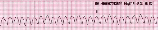
Rhythm strip of ventricular tachycardia showing a regular, wide complex rhythm with a rate over 100/min This is a shockable rhythm.
Image: “Lead II rhythm ventricular tachycardia Vtach VT” by Glenlarson. License: CC0 1.0
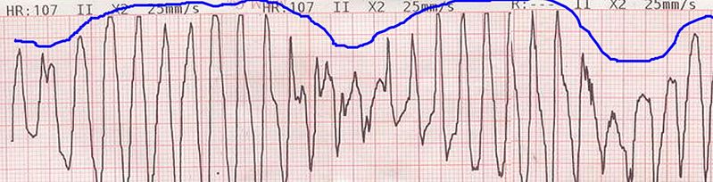
Example of an ECG tracing showing beat-to-beat axis deviation of the QRS complexes around the baseline in torsades de pointes
Image: “Torsade de pointes” by Panthro. License: CC0 1.0
The following recommendations are based on the most recent US and UK advanced life support guidelines.
Management of ventricular tachycardiaTachycardiaAbnormally rapid heartbeat, usually with a heart rate above 100 beats per minute for adults. Tachycardia accompanied by disturbance in the cardiac depolarization (cardiac arrhythmia) is called tachyarrhythmia.Sepsis in Children is based on whether a pulse is present and, if it is, whether the individual is hemodynamically stable.
For pulseless ventricular tachycardiaTachycardiaAbnormally rapid heartbeat, usually with a heart rate above 100 beats per minute for adults. Tachycardia accompanied by disturbance in the cardiac depolarization (cardiac arrhythmia) is called tachyarrhythmia.Sepsis in Children
Follow the adult cardiac arrestCardiac arrestCardiac arrest is the sudden, complete cessation of cardiac output with hemodynamic collapse. Patients present as pulseless, unresponsive, and apneic. Rhythms associated with cardiac arrest are ventricular fibrillation/tachycardia, asystole, or pulseless electrical activity. Cardiac Arrest algorithm.[6–8]
Confirm cardiac arrestCardiac arrestCardiac arrest is the sudden, complete cessation of cardiac output with hemodynamic collapse. Patients present as pulseless, unresponsive, and apneic. Rhythms associated with cardiac arrest are ventricular fibrillation/tachycardia, asystole, or pulseless electrical activity. Cardiac Arrest.
Patient unresponsive
Check pulse (checking should take < 10 seconds)
Not breathing
Start CPRCPRThe artificial substitution of heart and lung action as indicated for heart arrest resulting from electric shock, drowning, respiratory arrest, or other causes. The two major components of cardiopulmonary resuscitation are artificial ventilation and closed-chest cardiac massage.Cardiac Arrest to help maintain blood flowBlood flowBlood flow refers to the movement of a certain volume of blood through the vasculature over a given unit of time (e.g., mL per minute).Vascular Resistance, Flow, and Mean Arterial Pressure through the body.
Continue in 2-minute intervals (stopping only for pulse/rhythm check and to administer shockShockShock is a life-threatening condition associated with impaired circulation that results in tissue hypoxia. The different types of shock are based on the underlying cause: distributive (↑ cardiac output (CO), ↓ systemic vascular resistance (SVR)), cardiogenic (↓ CO, ↑ SVR), hypovolemic (↓ CO, ↑ SVR), obstructive (↓ CO), and mixed. Types of Shock energy).
Have someone get an automated external defibrillatorDefibrillatorCardiac electrical stimulators that apply brief high-voltage electroshocks to the heart. These stimulators are used to restore normal rhythm and contractile function in hearts of patients who are experiencing ventricular fibrillation or ventricular tachycardia that is not accompanied by a palpable pulse. Some defibrillators may also be used to correct certain noncritical dysrhythmias (called synchronized defibrillation or cardioversion), using relatively low-level discharges synchronized to the patient’s ECG waveform.Cardiac Arrest (AEDAEDCardiac electrical stimulators that apply brief high-voltage electroshocks to the heart. These stimulators are used to restore normal rhythm and contractile function in hearts of patients who are experiencing ventricular fibrillation or ventricular tachycardia that is not accompanied by a palpable pulse. Some defibrillators may also be used to correct certain noncritical dysrhythmias (called synchronized defibrillation or cardioversion), using relatively low-level discharges synchronized to the patient’s ECG waveform.Cardiopulmonary Resuscitation (CPR))/defibrillatorDefibrillatorCardiac electrical stimulators that apply brief high-voltage electroshocks to the heart. These stimulators are used to restore normal rhythm and contractile function in hearts of patients who are experiencing ventricular fibrillation or ventricular tachycardia that is not accompanied by a palpable pulse. Some defibrillators may also be used to correct certain noncritical dysrhythmias (called synchronized defibrillation or cardioversion), using relatively low-level discharges synchronized to the patient’s ECG waveform.Cardiac Arrest.
Senior airwayAirwayABCDE Assessment expert should attempt to secure the airwayAirwayABCDE Assessment concurrently with CPRCPRThe artificial substitution of heart and lung action as indicated for heart arrest resulting from electric shock, drowning, respiratory arrest, or other causes. The two major components of cardiopulmonary resuscitation are artificial ventilation and closed-chest cardiac massage.Cardiac Arrest.
IV or intraosseous (IO) access should be obtained concurrently with CPRCPRThe artificial substitution of heart and lung action as indicated for heart arrest resulting from electric shock, drowning, respiratory arrest, or other causes. The two major components of cardiopulmonary resuscitation are artificial ventilation and closed-chest cardiac massage.Cardiac Arrest.
If return of spontaneous circulationCirculationThe movement of the blood as it is pumped through the cardiovascular system.ABCDE Assessment (ROSC), begin post-resuscitation care (see Table).
Change compressions-provider during pulse check if possible.
Delivery of an electrical shockShockShock is a life-threatening condition associated with impaired circulation that results in tissue hypoxia. The different types of shock are based on the underlying cause: distributive (↑ cardiac output (CO), ↓ systemic vascular resistance (SVR)), cardiogenic (↓ CO, ↑ SVR), hypovolemic (↓ CO, ↑ SVR), obstructive (↓ CO), and mixed. Types of Shock to the heart
Can use manual defibrillatorDefibrillatorCardiac electrical stimulators that apply brief high-voltage electroshocks to the heart. These stimulators are used to restore normal rhythm and contractile function in hearts of patients who are experiencing ventricular fibrillation or ventricular tachycardia that is not accompanied by a palpable pulse. Some defibrillators may also be used to correct certain noncritical dysrhythmias (called synchronized defibrillation or cardioversion), using relatively low-level discharges synchronized to the patient’s ECG waveform.Cardiac Arrest or AEDAEDCardiac electrical stimulators that apply brief high-voltage electroshocks to the heart. These stimulators are used to restore normal rhythm and contractile function in hearts of patients who are experiencing ventricular fibrillation or ventricular tachycardia that is not accompanied by a palpable pulse. Some defibrillators may also be used to correct certain noncritical dysrhythmias (called synchronized defibrillation or cardioversion), using relatively low-level discharges synchronized to the patient’s ECG waveform.Cardiopulmonary Resuscitation (CPR)
Select shockShockShock is a life-threatening condition associated with impaired circulation that results in tissue hypoxia. The different types of shock are based on the underlying cause: distributive (↑ cardiac output (CO), ↓ systemic vascular resistance (SVR)), cardiogenic (↓ CO, ↑ SVR), hypovolemic (↓ CO, ↑ SVR), obstructive (↓ CO), and mixed. Types of Shock energy and charge defibrillatorDefibrillatorCardiac electrical stimulators that apply brief high-voltage electroshocks to the heart. These stimulators are used to restore normal rhythm and contractile function in hearts of patients who are experiencing ventricular fibrillation or ventricular tachycardia that is not accompanied by a palpable pulse. Some defibrillators may also be used to correct certain noncritical dysrhythmias (called synchronized defibrillation or cardioversion), using relatively low-level discharges synchronized to the patient’s ECG waveform.Cardiac Arrest:
Biphasic: use manufacturer’s recommendations (initially, 120–200 J); if unknown, use maximum available.
Monophasic: 360 J
Continue CPRCPRThe artificial substitution of heart and lung action as indicated for heart arrest resulting from electric shock, drowning, respiratory arrest, or other causes. The two major components of cardiopulmonary resuscitation are artificial ventilation and closed-chest cardiac massage.Cardiac Arrest while charging.
Move away from the patient to deliver shockShockShock is a life-threatening condition associated with impaired circulation that results in tissue hypoxia. The different types of shock are based on the underlying cause: distributive (↑ cardiac output (CO), ↓ systemic vascular resistance (SVR)), cardiogenic (↓ CO, ↑ SVR), hypovolemic (↓ CO, ↑ SVR), obstructive (↓ CO), and mixed. Types of Shock.
Resume compressions immediately after shockShockShock is a life-threatening condition associated with impaired circulation that results in tissue hypoxia. The different types of shock are based on the underlying cause: distributive (↑ cardiac output (CO), ↓ systemic vascular resistance (SVR)), cardiogenic (↓ CO, ↑ SVR), hypovolemic (↓ CO, ↑ SVR), obstructive (↓ CO), and mixed. Types of Shock (checking rhythm only when prompted by AEDAEDCardiac electrical stimulators that apply brief high-voltage electroshocks to the heart. These stimulators are used to restore normal rhythm and contractile function in hearts of patients who are experiencing ventricular fibrillation or ventricular tachycardia that is not accompanied by a palpable pulse. Some defibrillators may also be used to correct certain noncritical dysrhythmias (called synchronized defibrillation or cardioversion), using relatively low-level discharges synchronized to the patient’s ECG waveform.Cardiopulmonary Resuscitation (CPR)).
Drug treatment (IV/IO):
EpinephrineEpinephrineThe active sympathomimetic hormone from the adrenal medulla. It stimulates both the alpha- and beta- adrenergic systems, causes systemic vasoconstriction and gastrointestinal relaxation, stimulates the heart, and dilates bronchi and cerebral vessels.Sympathomimetic Drugs (adrenaline): 1 mg every 3–5 minutes (after 2nd shockShockShock is a life-threatening condition associated with impaired circulation that results in tissue hypoxia. The different types of shock are based on the underlying cause: distributive (↑ cardiac output (CO), ↓ systemic vascular resistance (SVR)), cardiogenic (↓ CO, ↑ SVR), hypovolemic (↓ CO, ↑ SVR), obstructive (↓ CO), and mixed. Types of Shock)
Consider the addition of antiarrhythmics, if needed:
AmiodaroneAmiodaroneAn antianginal and class III antiarrhythmic drug. It increases the duration of ventricular and atrial muscle action by inhibiting potassium channels and voltage-gated sodium channels. There is a resulting decrease in heart rate and in vascular resistance.Pulmonary Fibrosis (after 3rd shockShockShock is a life-threatening condition associated with impaired circulation that results in tissue hypoxia. The different types of shock are based on the underlying cause: distributive (↑ cardiac output (CO), ↓ systemic vascular resistance (SVR)), cardiogenic (↓ CO, ↑ SVR), hypovolemic (↓ CO, ↑ SVR), obstructive (↓ CO), and mixed. Types of Shock):
1st dose: 300 mg bolus
2nd dose: 150 mg, or
LidocaineLidocaineA local anesthetic and cardiac depressant used as an antiarrhythmic agent. Its actions are more intense and its effects more prolonged than those of procaine but its duration of action is shorter than that of bupivacaine or prilocaine.Local Anesthetics (after 3rd shockShockShock is a life-threatening condition associated with impaired circulation that results in tissue hypoxia. The different types of shock are based on the underlying cause: distributive (↑ cardiac output (CO), ↓ systemic vascular resistance (SVR)), cardiogenic (↓ CO, ↑ SVR), hypovolemic (↓ CO, ↑ SVR), obstructive (↓ CO), and mixed. Types of Shock (US only)):
1st dose: 1‒1.5 mg/kg IV/IO bolus
2nd dose: 0.5‒0.75 mg/kg (every 5 to 10 minutes)
Check for and treat reversible causes (see Table 1).
Return to step 5 every 2 minutes.
Identify reversible causes:
Table: Reversible causes
5 HsHSHypertrophic scars and keloids are raised, red, and rigid (3 rs) scars that develop during cutaneous wound healing and are characterized by a local abnormal proliferation of fibroblasts with over-production of collagen. Over-expression of growth factors and decreased production of molecules that promote matrix breakdown appear to be involved in the etiology.Hypertrophic and Keloid Scars
5 Ts
Hypovolemia
Tension pneumothoraxPneumothoraxA pneumothorax is a life-threatening condition in which air collects in the pleural space, causing partial or full collapse of the lung. A pneumothorax can be traumatic or spontaneous. Patients present with a sudden onset of sharp chest pain, dyspnea, and diminished breath sounds on exam.Pneumothorax
Hypoxia
TamponadeTamponadePericardial effusion, usually of rapid onset, exceeding ventricular filling pressures and causing collapse of the heart with a markedly reduced cardiac output.Pericarditis(cardiac)
Hydrogen ions (acidosisAcidosisA pathologic condition of acid accumulation or depletion of base in the body. The two main types are respiratory acidosis and metabolic acidosis, due to metabolic acid build up.Respiratory Acidosis)
Toxins
Hypokalemia or hyperkalemiaHyperkalemiaHyperkalemia is defined as a serum potassium (K+) concentration >5.2 mEq/L. Homeostatic mechanisms maintain the serum K+ concentration between 3.5 and 5.2 mEq/L, despite marked variation in dietary intake. Hyperkalemia can be due to a variety of causes, which include transcellular shifts, tissue breakdown, inadequate renal excretion, and drugs. Hyperkalemia
Obtain ECGECGAn electrocardiogram (ECG) is a graphic representation of the electrical activity of the heart plotted against time. Adhesive electrodes are affixed to the skin surface allowing measurement of cardiac impulses from many angles. The ECG provides 3-dimensional information about the conduction system of the heart, the myocardium, and other cardiac structures. Electrocardiogram (ECG), chest X-rayX-rayPenetrating electromagnetic radiation emitted when the inner orbital electrons of an atom are excited and release radiant energy. X-ray wavelengths range from 1 pm to 10 nm. Hard x-rays are the higher energy, shorter wavelength x-rays. Soft x-rays or grenz rays are less energetic and longer in wavelength. The short wavelength end of the x-ray spectrum overlaps the gamma rays wavelength range. The distinction between gamma rays and x-rays is based on their radiation source.Pulmonary Function Tests, and arterial blood gasArterial blood gasRespiratory Alkalosis (ABG) if not already completed.
Goals: systolic blood pressure (SBPSBPAscites) > 90 mm Hg, mean arterial pressureMean Arterial PressureMean arterial pressure (MAP) is the average systemic arterial pressure and is directly related to cardiac output (CO) and systemic vascular resistance (SVR). The SVR and MAP are affected by the vascular anatomy as well as a number of local and neurohumoral factors.Vascular Resistance, Flow, and Mean Arterial Pressure (MAP) > 65 mm Hg, SpO2 92-98%, PaCO2 35‒45 mm Hg
Temperature management
Treat underlying cause.
For ventricular tachycardiaTachycardiaAbnormally rapid heartbeat, usually with a heart rate above 100 beats per minute for adults. Tachycardia accompanied by disturbance in the cardiac depolarization (cardiac arrhythmia) is called tachyarrhythmia.Sepsis in Children with a pulse
Wide complex tachycardiaTachycardiaAbnormally rapid heartbeat, usually with a heart rate above 100 beats per minute for adults. Tachycardia accompanied by disturbance in the cardiac depolarization (cardiac arrhythmia) is called tachyarrhythmia.Sepsis in Children (regularRegularInsulin and monomorphic) of unknown etiology:
Close observation with ECGECGAn electrocardiogram (ECG) is a graphic representation of the electrical activity of the heart plotted against time. Adhesive electrodes are affixed to the skin surface allowing measurement of cardiac impulses from many angles. The ECG provides 3-dimensional information about the conduction system of the heart, the myocardium, and other cardiac structures. Electrocardiogram (ECG) monitoring as rapid deterioration can occur
Cardiology consultation
Vagal maneuvers if appropriate
IV adenosineAdenosineA nucleoside that is composed of adenine and d-ribose. Adenosine or adenosine derivatives play many important biological roles in addition to being components of DNA and RNA. Adenosine itself is a neurotransmitter.Class 5 Antiarrhythmic Drugs (6 mg initially; if no effect, give 12 mg); Use carefully, as it may not terminate VTVTVentricular tachycardia is any heart rhythm faster than 100 beats/min, with 3 or more irregular beats in a row, arising distal to the bundle of his. Ventricular tachycardia is the most common form of wide-complex tachycardia, and it is associated with a high mortality rate.Ventricular Tachycardia and is mainly useful when diagnosis is uncertain.
If no response, proceed with treatment for VTVTVentricular tachycardia is any heart rhythm faster than 100 beats/min, with 3 or more irregular beats in a row, arising distal to the bundle of his. Ventricular tachycardia is the most common form of wide-complex tachycardia, and it is associated with a high mortality rate.Ventricular Tachycardia.
AmiodaroneAmiodaroneAn antianginal and class III antiarrhythmic drug. It increases the duration of ventricular and atrial muscle action by inhibiting potassium channels and voltage-gated sodium channels. There is a resulting decrease in heart rate and in vascular resistance.Pulmonary Fibrosis
LidocaineLidocaineA local anesthetic and cardiac depressant used as an antiarrhythmic agent. Its actions are more intense and its effects more prolonged than those of procaine but its duration of action is shorter than that of bupivacaine or prilocaine.Local Anesthetics
IVmagnesium sulfateMagnesium SulfateA small colorless crystal used as an anticonvulsant, a cathartic, and an electrolyte replenisher in the treatment of pre-eclampsia and eclampsia. It causes direct inhibition of action potentials in myometrial muscle cells. Excitation and contraction are uncoupled, which decreases the frequency and force of contractions.Laxatives (emergency treatment)
IsoproterenolIsoproterenolIsopropyl analog of epinephrine; beta-sympathomimetic that acts on the heart, bronchi, skeletal muscle, alimentary tract, etc. It is used mainly as bronchodilator and heart stimulant.Sympathomimetic Drugs for polymorphic VTVTVentricular tachycardia is any heart rhythm faster than 100 beats/min, with 3 or more irregular beats in a row, arising distal to the bundle of his. Ventricular tachycardia is the most common form of wide-complex tachycardia, and it is associated with a high mortality rate.Ventricular Tachycardia triggered by bradycardiaBradycardiaBradyarrhythmia is a rhythm in which the heart rate is less than 60/min. Bradyarrhythmia can be physiologic, without symptoms or hemodynamic change. Pathologic bradyarrhythmia results in reduced cardiac output and hemodynamic instability causing syncope, dizziness, or dyspnea.Bradyarrhythmias (pause-dependent torsade de pointes)
Temporary overdrive pacing (if no response to initial management)
Antiarrhythmic agent that will not prolong the QT intervalQT intervalElectrocardiogram (ECG) (ProcainamideProcainamideA class ia antiarrhythmic drug that is structurally-related to procaine.Class 1 Antiarrhythmic Drugs (Sodium Channel Blockers) and amiodaroneAmiodaroneAn antianginal and class III antiarrhythmic drug. It increases the duration of ventricular and atrial muscle action by inhibiting potassium channels and voltage-gated sodium channels. There is a resulting decrease in heart rate and in vascular resistance.Pulmonary Fibrosis are contraindicated.)
Synchronized cardioversionSynchronized cardioversionAtrial Flutter (if medical therapy is unsuccessful or patient becomes unstable but has a pulse) at 50–200 J monophasic
Correction of electrolyte abnormalities
Discontinue any drugs that induce QT prolongation (e.g., azithromycinAzithromycinA semi-synthetic macrolide antibiotic structurally related to erythromycin. It has been used in the treatment of Mycobacterium avium intracellulare infections, toxoplasmosis, and cryptosporidiosis.Macrolides and Ketolides)
Polymorphic ventricular tachycardiaPolymorphic ventricular tachycardiaVentricular Tachycardia (in patientsPatientsIndividuals participating in the health care system for the purpose of receiving therapeutic, diagnostic, or preventive procedures.Clinician–Patient Relationship with organic heart disease, most often myocardial ischemiaMyocardial ischemiaA disorder of cardiac function caused by insufficient blood flow to the muscle tissue of the heart. The decreased blood flow may be due to narrowing of the coronary arteries (coronary artery disease), to obstruction by a thrombus (coronary thrombosis), or less commonly, to diffuse narrowing of arterioles and other small vessels within the heart.Coronary Heart Disease):
IV beta-blockersBeta-blockersDrugs that bind to but do not activate beta-adrenergic receptors thereby blocking the actions of beta-adrenergic agonists. Adrenergic beta-antagonists are used for treatment of hypertension, cardiac arrhythmias, angina pectoris, glaucoma, migraine headaches, and anxiety.Class 2 Antiarrhythmic Drugs (Beta Blockers) (metoprololMetoprololA selective adrenergic beta-1 blocking agent that is commonly used to treat angina pectoris; hypertension; and cardiac arrhythmias.Antiadrenergic Drugs 5 mg (up to 15 mg) IV as BP allows)
Consider IV amiodaroneAmiodaroneAn antianginal and class III antiarrhythmic drug. It increases the duration of ventricular and atrial muscle action by inhibiting potassium channels and voltage-gated sodium channels. There is a resulting decrease in heart rate and in vascular resistance.Pulmonary Fibrosis or lidocaineLidocaineA local anesthetic and cardiac depressant used as an antiarrhythmic agent. Its actions are more intense and its effects more prolonged than those of procaine but its duration of action is shorter than that of bupivacaine or prilocaine.Local Anesthetics
If left ventricular dysfunction is present, cardioversionCardioversionAtrial Fibrillation should be undertaken with amiodaroneAmiodaroneAn antianginal and class III antiarrhythmic drug. It increases the duration of ventricular and atrial muscle action by inhibiting potassium channels and voltage-gated sodium channels. There is a resulting decrease in heart rate and in vascular resistance.Pulmonary Fibrosis or lidocaineLidocaineA local anesthetic and cardiac depressant used as an antiarrhythmic agent. Its actions are more intense and its effects more prolonged than those of procaine but its duration of action is shorter than that of bupivacaine or prilocaine.Local Anesthetics.[6–8]
If ventricular tachycardiaTachycardiaAbnormally rapid heartbeat, usually with a heart rate above 100 beats per minute for adults. Tachycardia accompanied by disturbance in the cardiac depolarization (cardiac arrhythmia) is called tachyarrhythmia.Sepsis in Children is the result of digitalis toxicityToxicityDosage Calculation, manage with antidigitalis antibody and electrocardiovert to achieve normal sinus rhythmNormal sinus rhythmElectrocardiogram (ECG).[6–8]
Table: Acute antiarrhythmic medications for stable wide complex tachycardiaTachycardiaAbnormally rapid heartbeat, usually with a heart rate above 100 beats per minute for adults. Tachycardia accompanied by disturbance in the cardiac depolarization (cardiac arrhythmia) is called tachyarrhythmia.Sepsis in Children[6–10] Wide complex tachycardiaTachycardiaAbnormally rapid heartbeat, usually with a heart rate above 100 beats per minute for adults. Tachycardia accompanied by disturbance in the cardiac depolarization (cardiac arrhythmia) is called tachyarrhythmia.Sepsis in Children (unknown etiology) in a stable patient
Drug
Dose
Notes
AdenosineAdenosineA nucleoside that is composed of adenine and d-ribose. Adenosine or adenosine derivatives play many important biological roles in addition to being components of DNA and RNA. Adenosine itself is a neurotransmitter.Class 5 Antiarrhythmic Drugs
Initial dose: 6 mg IV over 1–2 seconds followed by normal salineNormal salineA crystalloid solution that contains 9. 0g of sodium chloride per liter of water. It has a variety of uses, including: as a contact lens solution, in ophthalmic solutions and nasal lavage, in wound irrigation, and for fluid therapy.Intravenous Fluids flush 2nd dose 12 mg IV over 1–2 seconds followed by normal salineNormal salineA crystalloid solution that contains 9. 0g of sodium chloride per liter of water. It has a variety of uses, including: as a contact lens solution, in ophthalmic solutions and nasal lavage, in wound irrigation, and for fluid therapy.Intravenous Fluids flush
May be used to distinguish monomorphic wide complex tachycardiaTachycardiaAbnormally rapid heartbeat, usually with a heart rate above 100 beats per minute for adults. Tachycardia accompanied by disturbance in the cardiac depolarization (cardiac arrhythmia) is called tachyarrhythmia.Sepsis in Children from supraventricular tachycardiaTachycardiaAbnormally rapid heartbeat, usually with a heart rate above 100 beats per minute for adults. Tachycardia accompanied by disturbance in the cardiac depolarization (cardiac arrhythmia) is called tachyarrhythmia.Sepsis in Children
The 2nd dose can be given 1–2 minutes after the 1st
Table: Acute antiarrhythmic medications for stable wide complex tachycardiaTachycardiaAbnormally rapid heartbeat, usually with a heart rate above 100 beats per minute for adults. Tachycardia accompanied by disturbance in the cardiac depolarization (cardiac arrhythmia) is called tachyarrhythmia.Sepsis in Children[6–10] Monomorphic VTVTVentricular tachycardia is any heart rhythm faster than 100 beats/min, with 3 or more irregular beats in a row, arising distal to the bundle of his. Ventricular tachycardia is the most common form of wide-complex tachycardia, and it is associated with a high mortality rate.Ventricular Tachycardia
Initial infusion:20–50 mg/min IV Maintenance infusion1–4 mg/min IV
Continue initial infusion until arrhythmia is terminated, QRS is increased by > 50%, or maximum dose of 17 mg/kg is reached. Avoid in prolonged–QT syndrome or congestive heart failureHeart FailureA heterogeneous condition in which the heart is unable to pump out sufficient blood to meet the metabolic need of the body. Heart failure can be caused by structural defects, functional abnormalities (ventricular dysfunction), or a sudden overload beyond its capacity. Chronic heart failure is more common than acute heart failure which results from sudden insult to cardiac function, such as myocardial infarction.Total Anomalous Pulmonary Venous Return (TAPVR)
AmiodaroneAmiodaroneAn antianginal and class III antiarrhythmic drug. It increases the duration of ventricular and atrial muscle action by inhibiting potassium channels and voltage-gated sodium channels. There is a resulting decrease in heart rate and in vascular resistance.Pulmonary Fibrosis
Initial:150 mg IV over 10 minutes Maintenance infusion 1 mg/min IV for next 6 hours
Initial dose may be repeated if VTVTVentricular tachycardia is any heart rhythm faster than 100 beats/min, with 3 or more irregular beats in a row, arising distal to the bundle of his. Ventricular tachycardia is the most common form of wide-complex tachycardia, and it is associated with a high mortality rate.Ventricular Tachycardia recurs.
Preferred if left ventricular (LV) dysfunction present
LidocaineLidocaineA local anesthetic and cardiac depressant used as an antiarrhythmic agent. Its actions are more intense and its effects more prolonged than those of procaine but its duration of action is shorter than that of bupivacaine or prilocaine.Local Anesthetics
Initial infusion: 1–1.5 mg/kg IV, may repeat with 0.5–0.75 mg/kg every 5–10 minutes as necessary Maintenance infusion: 1–4 mg/min IV
Maximum dose: 3 mg/kg
Consider when VTVTVentricular tachycardia is any heart rhythm faster than 100 beats/min, with 3 or more irregular beats in a row, arising distal to the bundle of his. Ventricular tachycardia is the most common form of wide-complex tachycardia, and it is associated with a high mortality rate.Ventricular Tachycardia is associated with myocardial ischemiaMyocardial ischemiaA disorder of cardiac function caused by insufficient blood flow to the muscle tissue of the heart. The decreased blood flow may be due to narrowing of the coronary arteries (coronary artery disease), to obstruction by a thrombus (coronary thrombosis), or less commonly, to diffuse narrowing of arterioles and other small vessels within the heart.Coronary Heart Disease or infarction.
Table: Acute antiarrhythmic medications for stable wide complex tachycardiaTachycardiaAbnormally rapid heartbeat, usually with a heart rate above 100 beats per minute for adults. Tachycardia accompanied by disturbance in the cardiac depolarization (cardiac arrhythmia) is called tachyarrhythmia.Sepsis in Children[6–10] Polymorphic VTVTVentricular tachycardia is any heart rhythm faster than 100 beats/min, with 3 or more irregular beats in a row, arising distal to the bundle of his. Ventricular tachycardia is the most common form of wide-complex tachycardia, and it is associated with a high mortality rate.Ventricular Tachycardia
Drug
Dose
Notes
MagnesiumMagnesiumA metallic element that has the atomic symbol mg, atomic number 12, and atomic weight 24. 31. It is important for the activity of many enzymes, especially those involved in oxidative phosphorylation.Electrolytes
IsoproterenolIsoproterenolIsopropyl analog of epinephrine; beta-sympathomimetic that acts on the heart, bronchi, skeletal muscle, alimentary tract, etc. It is used mainly as bronchodilator and heart stimulant.Sympathomimetic Drugs
2–10 µg/min IV titrated to clinical response (adults)
0.05–0.1 µg/kg/min (children)
For polymorphic VTVTVentricular tachycardia is any heart rhythm faster than 100 beats/min, with 3 or more irregular beats in a row, arising distal to the bundle of his. Ventricular tachycardia is the most common form of wide-complex tachycardia, and it is associated with a high mortality rate.Ventricular Tachycardia triggered by bradycardiaBradycardiaBradyarrhythmia is a rhythm in which the heart rate is less than 60/min. Bradyarrhythmia can be physiologic, without symptoms or hemodynamic change. Pathologic bradyarrhythmia results in reduced cardiac output and hemodynamic instability causing syncope, dizziness, or dyspnea.Bradyarrhythmias
Nonpharmacologic methods[6–9]
The most effective long-term therapy for ↓ mortalityMortalityAll deaths reported in a given population.Measures of Health Status is an implantable cardioverter-defibrillator (ICD).
PatientsPatientsIndividuals participating in the health care system for the purpose of receiving therapeutic, diagnostic, or preventive procedures.Clinician–Patient Relationship who survive after sustained ventricular tachycardiaTachycardiaAbnormally rapid heartbeat, usually with a heart rate above 100 beats per minute for adults. Tachycardia accompanied by disturbance in the cardiac depolarization (cardiac arrhythmia) is called tachyarrhythmia.Sepsis in Children (from structural heart disease or cardiomyopathyCardiomyopathyCardiomyopathy refers to a group of myocardial diseases associated with structural changes of the heart muscles (myocardium) and impaired systolic and/or diastolic function in the absence of other heart disorders (coronary artery disease, hypertension, valvular disease, and congenital heart disease). Cardiomyopathy: Overview and Types) are candidates for ICD placement.
In patientsPatientsIndividuals participating in the health care system for the purpose of receiving therapeutic, diagnostic, or preventive procedures.Clinician–Patient Relationship who experience frequent ICD shocks, antiarrhythmic agents are added.
Catheter ablation:
Treatment option if antiarrhythmic agents are not effective or not tolerated by the patient
Also considered for those who have ICD and are taking antiarrhythmic drugs but have recurrent sustained VTs
Cardiac surgeryCardiac surgeryCardiac surgery is the surgical management of cardiac abnormalities and of the great vessels of the thorax. In general terms, surgical intervention of the heart is performed to directly restore adequate pump function, correct inherent structural issues, and reestablish proper blood supply via the coronary circulation.Cardiac Surgery:
Rarely performed
Has a role in cases of failed catheter ablation and antiarrhythmic drugs
Additional investigation
Evaluating for underlying causes[6–9]
Cardiac enzymesEnzymesEnzymes are complex protein biocatalysts that accelerate chemical reactions without being consumed by them. Due to the body’s constant metabolic needs, the absence of enzymes would make life unsustainable, as reactions would occur too slowly without these molecules. Basics of Enzymes: ↑ after MIMIMI is ischemia and death of an area of myocardial tissue due to insufficient blood flow and oxygenation, usually from thrombus formation on a ruptured atherosclerotic plaque in the epicardial arteries. Clinical presentation is most commonly with chest pain, but women and patients with diabetes may have atypical symptoms.Myocardial Infarction
Coronary angiographyAngiographyRadiography of blood vessels after injection of a contrast medium.Cardiac Surgery to evaluate myocardial ischemiaMyocardial ischemiaA disorder of cardiac function caused by insufficient blood flow to the muscle tissue of the heart. The decreased blood flow may be due to narrowing of the coronary arteries (coronary artery disease), to obstruction by a thrombus (coronary thrombosis), or less commonly, to diffuse narrowing of arterioles and other small vessels within the heart.Coronary Heart Disease
ElectrolytesElectrolytesElectrolytes are mineral salts that dissolve in water and dissociate into charged particles called ions, which can be either be positively (cations) or negatively (anions) charged. Electrolytes are distributed in the extracellular and intracellular compartments in different concentrations. Electrolytes are essential for various basic life-sustaining functions.Electrolytes:
HypokalemiaHypokalemiaHypokalemia is defined as plasma potassium (K+) concentration < 3.5 mEq/L. Homeostatic mechanisms maintain plasma concentration between 3.5-5.2 mEq/L despite marked variation in dietary intake. Hypokalemia can be due to renal losses, GI losses, transcellular shifts, or poor dietary intake.Hypokalemia
HypomagnesemiaHypomagnesemiaA nutritional condition produced by a deficiency of magnesium in the diet, characterized by anorexia, nausea, vomiting, lethargy, and weakness. Symptoms are paresthesias, muscle cramps, irritability, decreased attention span, and mental confusion, possibly requiring months to appear. Deficiency of body magnesium can exist even when serum values are normal. In addition, magnesium deficiency may be organ-selective, since certain tissues become deficient before others. Electrolytes
HypocalcemiaHypocalcemiaHypocalcemia, a serum calcium < 8.5 mg/dL, can result from various conditions. The causes may include hypoparathyroidism, drugs, disorders leading to vitamin D deficiency, and more. Calcium levels are regulated and affected by different elements such as dietary intake, parathyroid hormone (PTH), vitamin D, pH, and albumin. Presentation can range from an asymptomatic (mild deficiency) to a life-threatening condition (acute, significant deficiency). Hypocalcemia
Urine drug screen for medications or stimulantsStimulantsStimulants are used by the general public to increase alertness and energy, decrease fatigue, and promote mental focus. Stimulants have medical uses for individuals with ADHD and sleep disorders, and are also used in combination with analgesics in pain management. Stimulants that may affect heart rateHeart rateThe number of times the heart ventricles contract per unit of time, usually per minute.Cardiac Physiology
EchocardiographyEchocardiographyUltrasonic recording of the size, motion, and composition of the heart and surrounding tissues. The standard approach is transthoracic.Tricuspid Valve Atresia (TVA) if structural cause is suspected
Cardiac CT or MRI also evaluates for structural heart disease.
Just as with echocardiographyEchocardiographyUltrasonic recording of the size, motion, and composition of the heart and surrounding tissues. The standard approach is transthoracic.Tricuspid Valve Atresia (TVA), left ventricular ejection fractionEjection fractionCardiac Cycle (LVEF), left and right ventricular function, valves, and coronary anatomy can be assessed.
Diagnosing suspected arrhythmias[9]
The following tests can be performed in patientsPatientsIndividuals participating in the health care system for the purpose of receiving therapeutic, diagnostic, or preventive procedures.Clinician–Patient Relationship presenting with suspicious symptoms (e.g., syncopeSyncopeSyncope is a short-term loss of consciousness and loss of postural stability followed by spontaneous return of consciousness to the previous neurologic baseline without the need for resuscitation. The condition is caused by transient interruption of cerebral blood flow that may be benign or related to a underlying life-threatening condition. Syncope) and/or nonsustained VTVTVentricular tachycardia is any heart rhythm faster than 100 beats/min, with 3 or more irregular beats in a row, arising distal to the bundle of his. Ventricular tachycardia is the most common form of wide-complex tachycardia, and it is associated with a high mortality rate.Ventricular Tachycardia. These can detect arrhythmias, their frequency, and their relationship with symptoms.
Holter monitor can be used to detect intermittent ventricular tachycardiaTachycardiaAbnormally rapid heartbeat, usually with a heart rate above 100 beats per minute for adults. Tachycardia accompanied by disturbance in the cardiac depolarization (cardiac arrhythmia) is called tachyarrhythmia.Sepsis in Children not detected by regularRegularInsulinECGECGAn electrocardiogram (ECG) is a graphic representation of the electrical activity of the heart plotted against time. Adhesive electrodes are affixed to the skin surface allowing measurement of cardiac impulses from many angles. The ECG provides 3-dimensional information about the conduction system of the heart, the myocardium, and other cardiac structures. Electrocardiogram (ECG).
Can be worn for weeks and are activated by patientsPatientsIndividuals participating in the health care system for the purpose of receiving therapeutic, diagnostic, or preventive procedures.Clinician–Patient Relationship when symptoms occur
Differential Diagnosis
HypokalemiaHypokalemiaHypokalemia is defined as plasma potassium (K+) concentration < 3.5 mEq/L. Homeostatic mechanisms maintain plasma concentration between 3.5-5.2 mEq/L despite marked variation in dietary intake. Hypokalemia can be due to renal losses, GI losses, transcellular shifts, or poor dietary intake.Hypokalemia:plasmaPlasmaThe residual portion of blood that is left after removal of blood cells by centrifugation without prior blood coagulation.Transfusion ProductspotassiumPotassiumAn element in the alkali group of metals with an atomic symbol k, atomic number 19, and atomic weight 39. 10. It is the chief cation in the intracellular fluid of muscle and other cells. Potassium ion is a strong electrolyte that plays a significant role in the regulation of fluid volume and maintenance of the water-electrolyte balance.Hyperkalemia concentration < 3.5 mEq/L. HypokalemiaHypokalemiaHypokalemia is defined as plasma potassium (K+) concentration < 3.5 mEq/L. Homeostatic mechanisms maintain plasma concentration between 3.5-5.2 mEq/L despite marked variation in dietary intake. Hypokalemia can be due to renal losses, GI losses, transcellular shifts, or poor dietary intake.Hypokalemia can be due to renal losses, GI losses, transcellularTranscellularThe movement of one cell into, through, and out of another cell.Tubular System shifts, or poor dietary intake. HypokalemiaHypokalemiaHypokalemia is defined as plasma potassium (K+) concentration < 3.5 mEq/L. Homeostatic mechanisms maintain plasma concentration between 3.5-5.2 mEq/L despite marked variation in dietary intake. Hypokalemia can be due to renal losses, GI losses, transcellular shifts, or poor dietary intake.Hypokalemia is usually asymptomatic if minor; however, it can lead to cardiac arrhythmias, muscle weakness, rhabdomyolysisRhabdomyolysisRhabdomyolysis is characterized by muscle necrosis and the release of toxic intracellular contents, especially myoglobin, into the circulation.Rhabdomyolysis, paralysis, and respiratory failureRespiratory failureRespiratory failure is a syndrome that develops when the respiratory system is unable to maintain oxygenation and/or ventilation. Respiratory failure may be acute or chronic and is classified as hypoxemic, hypercapnic, or a combination of the two. Respiratory Failure. Diagnosis is by history and lab testing. Management is by correcting the potassiumPotassiumAn element in the alkali group of metals with an atomic symbol k, atomic number 19, and atomic weight 39. 10. It is the chief cation in the intracellular fluid of muscle and other cells. Potassium ion is a strong electrolyte that plays a significant role in the regulation of fluid volume and maintenance of the water-electrolyte balance.Hyperkalemia deficit and treating the underlying cause.
Coronary arteryCoronary ArteryTruncus Arteriosus disease: inadequate supply of blood to the myocardiumMyocardiumThe muscle tissue of the heart. It is composed of striated, involuntary muscle cells connected to form the contractile pump to generate blood flow.Heart: Anatomy, typically caused by atherosclerosisAtherosclerosisAtherosclerosis is a common form of arterial disease in which lipid deposition forms a plaque in the blood vessel walls. Atherosclerosis is an incurable disease, for which there are clearly defined risk factors that often can be reduced through a change in lifestyle and behavior of the patient. Atherosclerosis. The myocardiumMyocardiumThe muscle tissue of the heart. It is composed of striated, involuntary muscle cells connected to form the contractile pump to generate blood flow.Heart: Anatomy becomes ischemic when oxygen supply does not meet oxygen demand. Diagnosis is based on history and ECGECGAn electrocardiogram (ECG) is a graphic representation of the electrical activity of the heart plotted against time. Adhesive electrodes are affixed to the skin surface allowing measurement of cardiac impulses from many angles. The ECG provides 3-dimensional information about the conduction system of the heart, the myocardium, and other cardiac structures. Electrocardiogram (ECG) findings, cardiac stress tests, or heart catheterization. Management is primarily based on reducing oxygen demand of the heart and increasing the delivery of oxygen.
Myocardial infarctionMyocardial infarctionMI is ischemia and death of an area of myocardial tissue due to insufficient blood flow and oxygenation, usually from thrombus formation on a ruptured atherosclerotic plaque in the epicardial arteries. Clinical presentation is most commonly with chest pain, but women and patients with diabetes may have atypical symptoms.Myocardial Infarction:ischemiaIschemiaA hypoperfusion of the blood through an organ or tissue caused by a pathologic constriction or obstruction of its blood vessels, or an absence of blood circulation.Ischemic Cell Damage of an area of myocardial tissue due to insufficient blood flowBlood flowBlood flow refers to the movement of a certain volume of blood through the vasculature over a given unit of time (e.g., mL per minute).Vascular Resistance, Flow, and Mean Arterial Pressure and oxygenation. The typical clinical presentation of MIMIMI is ischemia and death of an area of myocardial tissue due to insufficient blood flow and oxygenation, usually from thrombus formation on a ruptured atherosclerotic plaque in the epicardial arteries. Clinical presentation is most commonly with chest pain, but women and patients with diabetes may have atypical symptoms.Myocardial Infarction is crushingCrushingBlunt Chest Trauma substernal chest painPainAn unpleasant sensation induced by noxious stimuli which are detected by nerve endings of nociceptive neurons.Pain: Types and Pathways radiating to the left armArmThe arm, or “upper arm” in common usage, is the region of the upper limb that extends from the shoulder to the elbow joint and connects inferiorly to the forearm through the cubital fossa. It is divided into 2 fascial compartments (anterior and posterior).Arm: Anatomy or neckNeckThe part of a human or animal body connecting the head to the rest of the body.Peritonsillar Abscess. Diagnosis is by history, ECGECGAn electrocardiogram (ECG) is a graphic representation of the electrical activity of the heart plotted against time. Adhesive electrodes are affixed to the skin surface allowing measurement of cardiac impulses from many angles. The ECG provides 3-dimensional information about the conduction system of the heart, the myocardium, and other cardiac structures. Electrocardiogram (ECG) changes, increases in cardiac enzymesEnzymesEnzymes are complex protein biocatalysts that accelerate chemical reactions without being consumed by them. Due to the body’s constant metabolic needs, the absence of enzymes would make life unsustainable, as reactions would occur too slowly without these molecules. Basics of Enzymes, and evidence of wall-motion abnormalities. Management depends on the type of MIMIMI is ischemia and death of an area of myocardial tissue due to insufficient blood flow and oxygenation, usually from thrombus formation on a ruptured atherosclerotic plaque in the epicardial arteries. Clinical presentation is most commonly with chest pain, but women and patients with diabetes may have atypical symptoms.Myocardial Infarction; can include thrombolytic therapy and percutaneous intervention. All individuals receive nitratesNitratesNitrates are a class of medications that cause systemic vasodilation (veins > arteries) by smooth muscle relaxation. Nitrates are primarily indicated for the treatment of angina, where preferential venodilation causes pooling of blood, decreased preload, and ultimately decreased myocardial O2 demand.Nitrates, painPainAn unpleasant sensation induced by noxious stimuli which are detected by nerve endings of nociceptive neurons.Pain: Types and Pathways control, aspirinAspirinThe prototypical analgesic used in the treatment of mild to moderate pain. It has anti-inflammatory and antipyretic properties and acts as an inhibitor of cyclooxygenase which results in the inhibition of the biosynthesis of prostaglandins. Aspirin also inhibits platelet aggregation and is used in the prevention of arterial and venous thrombosis.Nonsteroidal Antiinflammatory Drugs (NSAIDs), anticoagulationAnticoagulationPulmonary Hypertension Drugs, and beta-blockersBeta-blockersDrugs that bind to but do not activate beta-adrenergic receptors thereby blocking the actions of beta-adrenergic agonists. Adrenergic beta-antagonists are used for treatment of hypertension, cardiac arrhythmias, angina pectoris, glaucoma, migraine headaches, and anxiety.Class 2 Antiarrhythmic Drugs (Beta Blockers).
Long-QT syndrome: disorder of the heart’s electrical activity. Long-QT syndrome affects the repolarizationRepolarizationMembrane Potential of the heart after a heartbeat and may be congenital or acquired. The syndrome is typically characterized by a prolongation of the QT intervalQT intervalElectrocardiogram (ECG) on the ECGECGAn electrocardiogram (ECG) is a graphic representation of the electrical activity of the heart plotted against time. Adhesive electrodes are affixed to the skin surface allowing measurement of cardiac impulses from many angles. The ECG provides 3-dimensional information about the conduction system of the heart, the myocardium, and other cardiac structures. Electrocardiogram (ECG). Presentation may be with syncopeSyncopeSyncope is a short-term loss of consciousness and loss of postural stability followed by spontaneous return of consciousness to the previous neurologic baseline without the need for resuscitation. The condition is caused by transient interruption of cerebral blood flow that may be benign or related to a underlying life-threatening condition. Syncope or cardiac arrestCardiac arrestCardiac arrest is the sudden, complete cessation of cardiac output with hemodynamic collapse. Patients present as pulseless, unresponsive, and apneic. Rhythms associated with cardiac arrest are ventricular fibrillation/tachycardia, asystole, or pulseless electrical activity. Cardiac Arrest..
Ventricular fibrillationVentricular fibrillationVentricular fibrillation (VF or V-fib) is a type of ventricular tachyarrhythmia (> 300/min) often preceded by ventricular tachycardia. In this arrhythmia, the ventricle beats rapidly and sporadically. The ventricular contraction is uncoordinated, leading to a decrease in cardiac output and immediate hemodynamic collapse. Ventricular Fibrillation (V-fib): type of ventricular tachyarrhythmiaTachyarrhythmiaA tachyarrhythmia is a rapid heart rhythm, regular or irregular, with a rate > 100 beats/min. Tachyarrhythmia may or may not be accompanied by symptoms of hemodynamic change.Tachyarrhythmias (> 300/min) often preceded by ventricular tachycardiaTachycardiaAbnormally rapid heartbeat, usually with a heart rate above 100 beats per minute for adults. Tachycardia accompanied by disturbance in the cardiac depolarization (cardiac arrhythmia) is called tachyarrhythmia.Sepsis in Children. In this arrhythmia, the ventricle beats rapidly and sporadically. Ventricular contraction is absent. Instead, the ventricle quivers, leading to an absence of cardiac outputCardiac outputThe volume of blood passing through the heart per unit of time. It is usually expressed as liters (volume) per minute so as not to be confused with stroke volume (volume per beat).Cardiac Mechanics and immediate hemodynamic collapse. Ventricular fibrillationVentricular fibrillationVentricular fibrillation (VF or V-fib) is a type of ventricular tachyarrhythmia (> 300/min) often preceded by ventricular tachycardia. In this arrhythmia, the ventricle beats rapidly and sporadically. The ventricular contraction is uncoordinated, leading to a decrease in cardiac output and immediate hemodynamic collapse. Ventricular Fibrillation (V-fib) is most commonly caused by underlying ischemic heart diseaseIschemic heart diseaseCoronary heart disease (CHD), or ischemic heart disease, describes a situation in which an inadequate supply of blood to the myocardium exists due to a stenosis of the coronary arteries, typically from atherosclerosis. Coronary Heart Disease.
Cardiac arrestCardiac arrestCardiac arrest is the sudden, complete cessation of cardiac output with hemodynamic collapse. Patients present as pulseless, unresponsive, and apneic. Rhythms associated with cardiac arrest are ventricular fibrillation/tachycardia, asystole, or pulseless electrical activity. Cardiac Arrest: sudden complete cessation of cardiac outputCardiac outputThe volume of blood passing through the heart per unit of time. It is usually expressed as liters (volume) per minute so as not to be confused with stroke volume (volume per beat).Cardiac Mechanics with hemodynamic collapse. Individuals with cardiac arrestCardiac arrestCardiac arrest is the sudden, complete cessation of cardiac output with hemodynamic collapse. Patients present as pulseless, unresponsive, and apneic. Rhythms associated with cardiac arrest are ventricular fibrillation/tachycardia, asystole, or pulseless electrical activity. Cardiac Arrest present in a pulseless, unresponsive, and apneic state. Rhythms associated with cardiac arrestCardiac arrestCardiac arrest is the sudden, complete cessation of cardiac output with hemodynamic collapse. Patients present as pulseless, unresponsive, and apneic. Rhythms associated with cardiac arrest are ventricular fibrillation/tachycardia, asystole, or pulseless electrical activity. Cardiac Arrest are V-fibV-fibVentricular fibrillation (VF or V-fib) is a type of ventricular tachyarrhythmia (> 300/min) often preceded by ventricular tachycardia. In this arrhythmia, the ventricle beats rapidly and sporadically. The ventricular contraction is uncoordinated, leading to a decrease in cardiac output and immediate hemodynamic collapse. Ventricular Fibrillation (V-fib)/tachycardiaTachycardiaAbnormally rapid heartbeat, usually with a heart rate above 100 beats per minute for adults. Tachycardia accompanied by disturbance in the cardiac depolarization (cardiac arrhythmia) is called tachyarrhythmia.Sepsis in Children, asystoleAsystoleNo discernible electrical activity, flatline on electrocardiogram (P waves and QRS complexes are not present).Cardiac Arrest, or pulseless electrical activityPulseless electrical activityElectrocardiogram (ECG) shows a cardiac rhythm without a palpable pulse. May be organized (with normal-appearing ECG complexes) or unorganized (no discernible complexes on ecg). From electromechanical dissociation, or no cardiac filling (“empty heart”).Cardiac Arrest. Management of cardiac arrestCardiac arrestCardiac arrest is the sudden, complete cessation of cardiac output with hemodynamic collapse. Patients present as pulseless, unresponsive, and apneic. Rhythms associated with cardiac arrest are ventricular fibrillation/tachycardia, asystole, or pulseless electrical activity. Cardiac Arrest starts with BLSBLSAirway Management outside the hospital and advanced cardiac life support (ACLS) when in the hospital.
Billing and Coding
Diagnosis Codes:
This code is used to document an episode of Ventricular TachycardiaTachycardiaAbnormally rapid heartbeat, usually with a heart rate above 100 beats per minute for adults. Tachycardia accompanied by disturbance in the cardiac depolarization (cardiac arrhythmia) is called tachyarrhythmia.Sepsis in Children (V-tach), a rapid heart rhythm originating in the ventricles that can be stable or unstable and may lead to ventricular fibrillationVentricular fibrillationVentricular fibrillation (VF or V-fib) is a type of ventricular tachyarrhythmia (> 300/min) often preceded by ventricular tachycardia. In this arrhythmia, the ventricle beats rapidly and sporadically. The ventricular contraction is uncoordinated, leading to a decrease in cardiac output and immediate hemodynamic collapse. Ventricular Fibrillation (V-fib).
Domain
Code
Description
ICD-10-CM
I47.2
Ventricular tachycardiaTachycardiaAbnormally rapid heartbeat, usually with a heart rate above 100 beats per minute for adults. Tachycardia accompanied by disturbance in the cardiac depolarization (cardiac arrhythmia) is called tachyarrhythmia.Sepsis in Children
ICD-11
BC81.0
Sustained ventricular tachycardiaTachycardiaAbnormally rapid heartbeat, usually with a heart rate above 100 beats per minute for adults. Tachycardia accompanied by disturbance in the cardiac depolarization (cardiac arrhythmia) is called tachyarrhythmia.Sepsis in Children
SNOMED CT
17354005
Ventricular tachycardiaTachycardiaAbnormally rapid heartbeat, usually with a heart rate above 100 beats per minute for adults. Tachycardia accompanied by disturbance in the cardiac depolarization (cardiac arrhythmia) is called tachyarrhythmia.Sepsis in Children (disorder)
Procedures/Interventions:
These codes are used for treatments for V-tach, including immediate electrical cardioversionCardioversionAtrial Fibrillation for unstable patientsUnstable PatientsBlunt Chest Trauma, catheter ablation to destroy the abnormal heart tissue causing the rhythm, and ICD placement for long-term prevention.
Comprehensive electrophysiologic evaluation including catheter ablation
CPT
33249
Insertion or replacement of permanent implantable defibrillatorDefibrillatorCardiac electrical stimulators that apply brief high-voltage electroshocks to the heart. These stimulators are used to restore normal rhythm and contractile function in hearts of patients who are experiencing ventricular fibrillation or ventricular tachycardia that is not accompanied by a palpable pulse. Some defibrillators may also be used to correct certain noncritical dysrhythmias (called synchronized defibrillation or cardioversion), using relatively low-level discharges synchronized to the patient’s ECG waveform.Cardiac Arrest system
Medications:
These codes are used to prescribe antiarrhythmic drugs like amiodaroneAmiodaroneAn antianginal and class III antiarrhythmic drug. It increases the duration of ventricular and atrial muscle action by inhibiting potassium channels and voltage-gated sodium channels. There is a resulting decrease in heart rate and in vascular resistance.Pulmonary Fibrosis or sotalolSotalolAn adrenergic beta-antagonist that is used in the treatment of life-threatening arrhythmias.Class 3 Antiarrhythmic Drugs (Potassium Channel Blockers), which are used to terminate an episode of V-tach or for long-term suppressionSuppressionDefense Mechanisms to prevent recurrence.
Domain
Code
Description
RxNorm
703
AmiodaroneAmiodaroneAn antianginal and class III antiarrhythmic drug. It increases the duration of ventricular and atrial muscle action by inhibiting potassium channels and voltage-gated sodium channels. There is a resulting decrease in heart rate and in vascular resistance.Pulmonary Fibrosis (ingredient)
AmiodaroneAmiodaroneAn antianginal and class III antiarrhythmic drug. It increases the duration of ventricular and atrial muscle action by inhibiting potassium channels and voltage-gated sodium channels. There is a resulting decrease in heart rate and in vascular resistance.Pulmonary Fibrosis
Complications & Supportive Procedures:
This ICD-10 code is used to document the most dangerous potential outcome of V-tach, which is its degeneration into the non-perfusing, chaotic rhythm of ventricular fibrillationVentricular fibrillationVentricular fibrillation (VF or V-fib) is a type of ventricular tachyarrhythmia (> 300/min) often preceded by ventricular tachycardia. In this arrhythmia, the ventricle beats rapidly and sporadically. The ventricular contraction is uncoordinated, leading to a decrease in cardiac output and immediate hemodynamic collapse. Ventricular Fibrillation (V-fib), leading to cardiac arrestCardiac arrestCardiac arrest is the sudden, complete cessation of cardiac output with hemodynamic collapse. Patients present as pulseless, unresponsive, and apneic. Rhythms associated with cardiac arrest are ventricular fibrillation/tachycardia, asystole, or pulseless electrical activity. Cardiac Arrest.
Domain
Code
Description
ICD-10-CM
I49.01
Ventricular fibrillationVentricular fibrillationVentricular fibrillation (VF or V-fib) is a type of ventricular tachyarrhythmia (> 300/min) often preceded by ventricular tachycardia. In this arrhythmia, the ventricle beats rapidly and sporadically. The ventricular contraction is uncoordinated, leading to a decrease in cardiac output and immediate hemodynamic collapse. Ventricular Fibrillation (V-fib)
References
Kashou, A. H., Noseworthy, P. A., DeSimone, C. V., Deshmukh, A. J., Asirvatham, S. J., May, A. M. (2020). Wide complex tachycardia differentiation: A reappraisal of the state‐of‐the‐art. Journal of the American Heart Association, 9(11).
Al-Khatib et. Al., (2018). 2017 AHA/ACC/HRS guideline for management of patients with ventricular arrhythmias and the prevention of sudden cardiac death. Journal of the American College of Cardiology 72(14).
Link et. Al., (2010). Part 6: Electrical Therapies: Automated External DEFIBRILLATORS, Defibrillation, CARDIOVERSION, and Pacing * 2010 American Heart Association guidelines for cardiopulmonary resuscitation and Emergency CARDIOVASCULAR CARE. Circulation 122(18 Suppl 3).
(1997). A comparison of antiarrhythmic-drug therapy with implantable defibrillators in patients resuscitated from near-fatal ventricular arrhythmias. New England Journal of Medicine 337:1576–1584.
Al-Khatib, S. M., Stevenson, W. G., Ackerman, M. J., Bryant, W. J., Callans, D. J., Curtis, A. B., Deal, B. J., Dickfeld, T., Field, M. E., Fonarow, G. C., Gillis, A. M., Granger, C. B., Hammill, S. C., Hlatky, M. A., Joglar, J. A., Kay, G. N., Matlock, D. D., Myerburg, R. J., Page, R. L. (2018). 2017 AHA/ACC/HRS guideline for management of patients with ventricular arrhythmias and the prevention of sudden cardiac death: a report of the American College of Cardiology/American Heart Association Task Force on Clinical Practice Guidelines and the Heart Rhythm Society. Journal of the American College of Cardiology, 72(14), e91–e220. https://doi.org/10.1016/j.jacc.2017.10.054
Viskin, S., Chorin, E., Viskin, D., Hochstadt, A., Schwartz, A. L., Rosso, R. (2021). Polymorphic ventricular tachycardia: terminology, mechanism, diagnosis, and emergency therapy. Circulation, 144(10), 823–839. https://doi.org/10.1161/CIRCULATIONAHA.121.055783
Create your free account or log in to continue reading!