Long QT syndrome (LQTS) is a disorder of ventricular myocardial repolarization that produces QT prolongation on electrocardiogram (ECG). Long QT syndrome is associated with an increased risk of developing life-threatening cardiac arrhythmias, specifically torsades de pointes. The condition may be congenital or acquired. Congenital LQTS is attributed to genetic mutations affecting cardiac ion channels. Acquired LQTS usually results from drug therapy or electrolyte abnormalities. Patients can be asymptomatic or present with palpitations, syncope, seizures, and even sudden cardiac death. Diagnosis is established with ECG along with medical and family history, laboratory workup, and other cardiac tests. Treatment is determined by etiology. Acquired LQTS requires removal of the offending drug or drug combinations and correction of electrolyte abnormalities. Congenital LQTS management involves beta blockers, aggressive treatment of electrolyte imbalances, avoidance of medications that prolong the QT interval, and placement of an implantable cardioverter–defibrillator (ICD).
Long QT syndromeLong QT syndromeLong QT syndrome (LQTS) is a disorder of ventricular myocardial repolarization that produces QT prolongation on electrocardiogram (ECG). Long QT syndrome is associated with an increased risk of developing life-threatening cardiac arrhythmias, specifically torsades de pointes.Long QT Syndrome (LQTSLQTSLong qt syndrome (LQTS) is a disorder of ventricular myocardial repolarization that produces qt prolongation on electrocardiogram (ECG). Long qt syndrome is associated with an increased risk of developing life-threatening cardiac arrhythmias, specifically torsades de pointes.Long QT Syndrome): a ventricular electrical disorder (congenital or acquired) characterized by delayed myocardial repolarizationRepolarizationMembrane Potential and demonstrated as prolonged QT intervalQT intervalElectrocardiogram (ECG) on electrocardiogramElectrocardiogramAn electrocardiogram (ECG) is a graphic representation of the electrical activity of the heart plotted against time. Adhesive electrodes are affixed to the skin surface allowing measurement of cardiac impulses from many angles. The ECG provides 3-dimensional information about the conduction system of the heart, the myocardium, and other cardiac structures. Electrocardiogram (ECG) (ECGECGAn electrocardiogram (ECG) is a graphic representation of the electrical activity of the heart plotted against time. Adhesive electrodes are affixed to the skin surface allowing measurement of cardiac impulses from many angles. The ECG provides 3-dimensional information about the conduction system of the heart, the myocardium, and other cardiac structures. Electrocardiogram (ECG))[4,6]
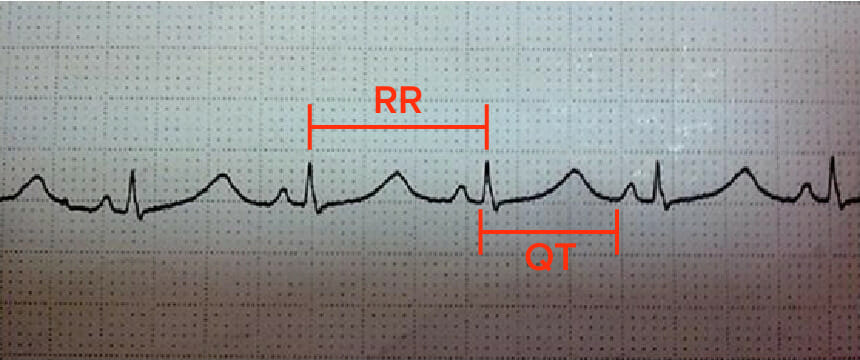
The measurement from the start of the Q wave to the end of the T waveT waveElectrocardiogram (ECG) on an ECGECGAn electrocardiogram (ECG) is a graphic representation of the electrical activity of the heart plotted against time. Adhesive electrodes are affixed to the skin surface allowing measurement of cardiac impulses from many angles. The ECG provides 3-dimensional information about the conduction system of the heart, the myocardium, and other cardiac structures. Electrocardiogram (ECG)
After pubertyPubertyPuberty is a complex series of physical, psychosocial, and cognitive transitions usually experienced by adolescents (11-19 years of age). Puberty is marked by a growth in stature and the development of secondary sexual characteristics, achievement of fertility, and changes in most body systems.Puberty, women tend to have a longer QT intervalQT intervalElectrocardiogram (ECG) than men.
Torsades de pointesTorsades de pointesA malignant form of polymorphic ventricular tachycardia that is characterized by heart rate between 200 and 250 beats per minute, and QRS complexes with changing amplitude and twisting of the points. The term also describes the syndrome of tachycardia with prolonged ventricular repolarization, long qt intervals exceeding 500 milliseconds or bradycardia. Torsades de pointes may be self-limited or may progress to ventricular fibrillation.Ventricular Tachycardia:
A life-threatening arrhythmia associated with LQTSLQTSLong qt syndrome (LQTS) is a disorder of ventricular myocardial repolarization that produces qt prolongation on electrocardiogram (ECG). Long qt syndrome is associated with an increased risk of developing life-threatening cardiac arrhythmias, specifically torsades de pointes.Long QT Syndrome
Irregular QRS complexes “twisting” around the isoelectric line (torsades de pointesTorsades de pointesA malignant form of polymorphic ventricular tachycardia that is characterized by heart rate between 200 and 250 beats per minute, and QRS complexes with changing amplitude and twisting of the points. The term also describes the syndrome of tachycardia with prolonged ventricular repolarization, long qt intervals exceeding 500 milliseconds or bradycardia. Torsades de pointes may be self-limited or may progress to ventricular fibrillation.Ventricular Tachycardia = “twisting of points”)
QTc ≥ 500 msMSMultiple sclerosis (MS) is a chronic inflammatory autoimmune disease that leads to demyelination of the nerves in the CNS. Young women are more predominantly affected by this most common demyelinating condition.Multiple Sclerosis is associated with a markedly increased risk of torsades de pointesTorsades de pointesA malignant form of polymorphic ventricular tachycardia that is characterized by heart rate between 200 and 250 beats per minute, and QRS complexes with changing amplitude and twisting of the points. The term also describes the syndrome of tachycardia with prolonged ventricular repolarization, long qt intervals exceeding 500 milliseconds or bradycardia. Torsades de pointes may be self-limited or may progress to ventricular fibrillation.Ventricular Tachycardia.
Rate of 160–250/min
Often terminates spontaneously but can lead to sudden cardiac deathSudden cardiac deathCardiac arrest is the sudden, complete cessation of cardiac output with hemodynamic collapse. Patients present as pulseless, unresponsive, and apneic. Rhythms associated with cardiac arrest are ventricular fibrillation/tachycardia, asystole, or pulseless electrical activity.Cardiac Arrest
ECG in a patient with long QT syndrome:
The corrected QT interval (QTc) is calculated by dividing the QT interval (0.48 seconds) by the square root of the preceding RR interval (0.825 seconds). In this case, the QTc is 0.582 seconds (582 milliseconds).
Defects in genesGenesA category of nucleic acid sequences that function as units of heredity and which code for the basic instructions for the development, reproduction, and maintenance of organisms.DNA Types and Structure coding for ion channelsChannelsThe Cell: Cell Membrane (i.e., cardiac channelopathies)
Change in flowFlowBlood flows through the heart, arteries, capillaries, and veins in a closed, continuous circuit. Flow is the movement of volume per unit of time. Flow is affected by the pressure gradient and the resistance fluid encounters between 2 points. Vascular resistance is the opposition to flow, which is caused primarily by blood friction against vessel walls.Vascular Resistance, Flow, and Mean Arterial Pressure of positive ions affects repolarizing current in cardiomyocytes → prolongs the action potentialAction PotentialAbrupt changes in the membrane potential that sweep along the cell membrane of excitable cells in response to excitation stimuli.Membrane Potential (QT prolongation) → leads to early afterdepolarizations → increased risk of torsades de pointesTorsades de pointesA malignant form of polymorphic ventricular tachycardia that is characterized by heart rate between 200 and 250 beats per minute, and QRS complexes with changing amplitude and twisting of the points. The term also describes the syndrome of tachycardia with prolonged ventricular repolarization, long qt intervals exceeding 500 milliseconds or bradycardia. Torsades de pointes may be self-limited or may progress to ventricular fibrillation.Ventricular Tachycardia
These genetic mutationsGenetic MutationsCarcinogenesis account for 80% of LQTSLQTSLong qt syndrome (LQTS) is a disorder of ventricular myocardial repolarization that produces qt prolongation on electrocardiogram (ECG). Long qt syndrome is associated with an increased risk of developing life-threatening cardiac arrhythmias, specifically torsades de pointes.Long QT Syndrome cases:
Loss-of-function mutations in KCNQ1-encoded and KCNH2-encoded potassiumPotassiumAn element in the alkali group of metals with an atomic symbol k, atomic number 19, and atomic weight 39. 10. It is the chief cation in the intracellular fluid of muscle and other cells. Potassium ion is a strong electrolyte that plays a significant role in the regulation of fluid volume and maintenance of the water-electrolyte balance.Hyperkalemia (K) channelsChannelsThe Cell: Cell Membrane: prolong action potentialAction PotentialAbrupt changes in the membrane potential that sweep along the cell membrane of excitable cells in response to excitation stimuli.Membrane Potential duration by reducing K efflux
Gain-of-function mutations in the SCN5A-encoded sodiumSodiumA member of the alkali group of metals. It has the atomic symbol na, atomic number 11, and atomic weight 23.Hyponatremia (Na) channelsChannelsThe Cell: Cell Membrane: prolong action potentialAction PotentialAbrupt changes in the membrane potential that sweep along the cell membrane of excitable cells in response to excitation stimuli.Membrane Potential duration by the contribution of increased late sodiumSodiumA member of the alkali group of metals. It has the atomic symbol na, atomic number 11, and atomic weight 23.Hyponatremia currents (which raises Na influx)
Sympathetic innervation of the heart:
A triggerTriggerThe type of signal that initiates the inspiratory phase by the ventilatorInvasive Mechanical Ventilation for torsades de pointesTorsades de pointesA malignant form of polymorphic ventricular tachycardia that is characterized by heart rate between 200 and 250 beats per minute, and QRS complexes with changing amplitude and twisting of the points. The term also describes the syndrome of tachycardia with prolonged ventricular repolarization, long qt intervals exceeding 500 milliseconds or bradycardia. Torsades de pointes may be self-limited or may progress to ventricular fibrillation.Ventricular Tachycardia is a surge in sympathetic tone (such as an extreme emotional event).
Congenital LQTSLQTSLong qt syndrome (LQTS) is a disorder of ventricular myocardial repolarization that produces qt prolongation on electrocardiogram (ECG). Long qt syndrome is associated with an increased risk of developing life-threatening cardiac arrhythmias, specifically torsades de pointes.Long QT Syndrome is more strongly genotype-specific in its triggers.
Sympathetic triggers are particularly characteristic of congenital LQTSLQTSLong qt syndrome (LQTS) is a disorder of ventricular myocardial repolarization that produces qt prolongation on electrocardiogram (ECG). Long qt syndrome is associated with an increased risk of developing life-threatening cardiac arrhythmias, specifically torsades de pointes.Long QT Syndrome (especially LQT1/LQT2), but can also precipitate torsades in acquired LQTSLQTSLong qt syndrome (LQTS) is a disorder of ventricular myocardial repolarization that produces qt prolongation on electrocardiogram (ECG). Long qt syndrome is associated with an increased risk of developing life-threatening cardiac arrhythmias, specifically torsades de pointes.Long QT Syndrome.
LQT1: Triggers are typically exercise, especially swimming or diving (vagal/sympathetic surge).
LQT2: Triggers are often auditory stimuli (alarm clocks, telephones) or sudden emotional stress.
LQT3: Events often occur during sleepSleepA readily reversible suspension of sensorimotor interaction with the environment, usually associated with recumbency and immobility.Physiology of Sleep or rest (bradycardia-dependent), as the slower heart rateHeart rateThe number of times the heart ventricles contract per unit of time, usually per minute.Cardiac Physiology allows more time for the late sodiumSodiumA member of the alkali group of metals. It has the atomic symbol na, atomic number 11, and atomic weight 23.Hyponatremia current to manifest.
Types of congenital LQTSLQTSLong qt syndrome (LQTS) is a disorder of ventricular myocardial repolarization that produces qt prolongation on electrocardiogram (ECG). Long qt syndrome is associated with an increased risk of developing life-threatening cardiac arrhythmias, specifically torsades de pointes.Long QT Syndrome:
Up to 45% of LQTSLQTSLong qt syndrome (LQTS) is a disorder of ventricular myocardial repolarization that produces qt prolongation on electrocardiogram (ECG). Long qt syndrome is associated with an increased risk of developing life-threatening cardiac arrhythmias, specifically torsades de pointes.Long QT Syndrome cases
Defect in KCNQ1geneGeneA category of nucleic acid sequences that function as units of heredity and which code for the basic instructions for the development, reproduction, and maintenance of organisms.Basic Terms of Genetics
Type 2:
Up to 40% of LQTSLQTSLong qt syndrome (LQTS) is a disorder of ventricular myocardial repolarization that produces qt prolongation on electrocardiogram (ECG). Long qt syndrome is associated with an increased risk of developing life-threatening cardiac arrhythmias, specifically torsades de pointes.Long QT Syndrome cases
Defect in KCNH2geneGeneA category of nucleic acid sequences that function as units of heredity and which code for the basic instructions for the development, reproduction, and maintenance of organisms.Basic Terms of Genetics
Up to 10% of LQTSLQTSLong qt syndrome (LQTS) is a disorder of ventricular myocardial repolarization that produces qt prolongation on electrocardiogram (ECG). Long qt syndrome is associated with an increased risk of developing life-threatening cardiac arrhythmias, specifically torsades de pointes.Long QT Syndrome cases
Defect in SCN5AgeneGeneA category of nucleic acid sequences that function as units of heredity and which code for the basic instructions for the development, reproduction, and maintenance of organisms.Basic Terms of Genetics
Variety of associated conditions:
Romano-Ward syndromeRomano-Ward syndromeA form of long qt syndrome that is without congenital deafness. It is caused by mutation of the kcnq1 gene which encodes a protein in the voltage-gated potassium channel.Long QT Syndrome: majority of cases; congenital LQTSLQTSLong qt syndrome (LQTS) is a disorder of ventricular myocardial repolarization that produces qt prolongation on electrocardiogram (ECG). Long qt syndrome is associated with an increased risk of developing life-threatening cardiac arrhythmias, specifically torsades de pointes.Long QT Syndrome without extracardiac manifestations (autosomal dominantAutosomal dominantAutosomal inheritance, both dominant and recessive, refers to the transmission of genes from the 22 autosomal chromosomes. Autosomal dominant diseases are expressed when only 1 copy of the dominant allele is inherited. Autosomal Recessive and Autosomal Dominant Inheritance)
Jervell and Lange-Nielsen syndrome: congenital LQTSLQTSLong qt syndrome (LQTS) is a disorder of ventricular myocardial repolarization that produces qt prolongation on electrocardiogram (ECG). Long qt syndrome is associated with an increased risk of developing life-threatening cardiac arrhythmias, specifically torsades de pointes.Long QT Syndrome + sensorineural hearing lossSensorineural hearing lossHearing loss resulting from damage to the cochlea and the sensorineural elements which lie internally beyond the oval and round windows. These elements include the auditory nerve and its connections in the brainstem.Hearing Loss (autosomal recessiveAutosomal recessiveAutosomal inheritance, both dominant and recessive, refers to the transmission of genes from the 22 autosomal chromosomes. Autosomal recessive diseases are only expressed when 2 copies of the recessive allele are inherited.Autosomal Recessive and Autosomal Dominant Inheritance)
Anderson-Tawil syndrome: congenital LQTSLQTSLong qt syndrome (LQTS) is a disorder of ventricular myocardial repolarization that produces qt prolongation on electrocardiogram (ECG). Long qt syndrome is associated with an increased risk of developing life-threatening cardiac arrhythmias, specifically torsades de pointes.Long QT Syndrome + periodic paralysis
Timothy syndromeTimothy syndromeLong QT Syndrome: congenital LQTSLQTSLong qt syndrome (LQTS) is a disorder of ventricular myocardial repolarization that produces qt prolongation on electrocardiogram (ECG). Long qt syndrome is associated with an increased risk of developing life-threatening cardiac arrhythmias, specifically torsades de pointes.Long QT Syndrome + cutaneous syndactylySyndactylyA congenital anomaly of the hand or foot, marked by the webbing between adjacent fingers or toes. Syndactylies are classified as complete or incomplete by the degree of joining. Syndactylies can also be simple or complex. Simple syndactyly indicates joining of only skin or soft tissue; complex syndactyly marks joining of bony elements.Development of the Limbs + autism
IatrogenicIatrogenicAny adverse condition in a patient occurring as the result of treatment by a physician, surgeon, or other health professional, especially infections acquired by a patient during the course of treatment.Anterior Cord Syndrome/pharmacologic:
Most common cause
LQTSLQTSLong qt syndrome (LQTS) is a disorder of ventricular myocardial repolarization that produces qt prolongation on electrocardiogram (ECG). Long qt syndrome is associated with an increased risk of developing life-threatening cardiac arrhythmias, specifically torsades de pointes.Long QT Syndrome caused by blocking potassiumPotassiumAn element in the alkali group of metals with an atomic symbol k, atomic number 19, and atomic weight 39. 10. It is the chief cation in the intracellular fluid of muscle and other cells. Potassium ion is a strong electrolyte that plays a significant role in the regulation of fluid volume and maintenance of the water-electrolyte balance.Hyperkalemia outflow during the rapid repolarizationRepolarizationMembrane Potential phase:
Antibiotics: macrolidesMacrolidesMacrolides and ketolides are antibiotics that inhibit bacterial protein synthesis by binding to the 50S ribosomal subunit and blocking transpeptidation. These antibiotics have a broad spectrum of antimicrobial activity but are best known for their coverage of atypical microorganisms. Macrolides and Ketolides, fluoroquinolonesFluoroquinolonesFluoroquinolones are a group of broad-spectrum, bactericidal antibiotics inhibiting bacterial DNA replication. Fluoroquinolones cover gram-negative, anaerobic, and atypical organisms, as well as some gram-positive and multidrug-resistant (MDR) organisms. Fluoroquinolones
Antifungals: ketoconazoleKetoconazoleBroad spectrum antifungal agent used for long periods at high doses, especially in immunosuppressed patients.Azoles, itraconazoleItraconazoleA triazole antifungal agent that inhibits cytochrome p-450-dependent enzymes required for ergosterol synthesis.Azoles
HIVHIVAnti-HIV Drugs antiretroviral drugs (e.g., saquinavirSaquinavirAn HIV protease inhibitor which acts as an analog of an HIV protease cleavage site. It is a highly specific inhibitor of HIV-1 and HIV-2 proteases, and also inhibits cytochrome p-450 cyp3a.Anti-HIV Drugs particularly via drug–drug interactions)
Antimalarial drugsAntimalarial drugsMalaria, a vector-borne parasitic disease caused by Plasmodium spp., is transmitted via injection of sporozoites or immature forms of the parasite into a person’s bloodstream. Sporozoites then infect the hepatocytes and differentiate into schizonts, which subsequently rupture, and merozoites invade red blood cells. Antimalarial Drugs: chloroquineChloroquineThe prototypical antimalarial agent with a mechanism that is not well understood. It has also been used to treat rheumatoid arthritis, systemic lupus erythematosus, and in the systemic therapy of amebic liver abscesses.Antimalarial Drugs and hydroxychloroquineHydroxychloroquineA chemotherapeutic agent that acts against erythrocytic forms of malarial parasites. Hydroxychloroquine appears to concentrate in food vacuoles of affected protozoa. It inhibits plasmodial heme polymerase.Immunosuppressants
OpioidsOpioidsOpiates are drugs that are derived from the sap of the opium poppy. Opiates have been used since antiquity for the relief of acute severe pain. Opioids are synthetic opiates with properties that are substantially similar to those of opiates. Opioid Analgesics (methadoneMethadoneA synthetic opioid that is used as the hydrochloride. It is an opioid analgesic that is primarily a mu-opioid agonist.Opioid Analgesics)
Gastric motilityGastric motilityGastrointestinal Motility drugs (cisapride), antiemeticsAntiemeticsAntiemetics are medications used to treat and/or prevent nausea and vomiting. These drugs act on different target receptors. The main classes include benzodiazepines, corticosteroids, atypical antipsychotics, cannabinoids, and antagonists of the following receptors: serotonin, dopamine, and muscarinic and neurokinin receptors.Antiemetics (ondansetronOndansetronA competitive serotonin type 3 receptor antagonist. It is effective in the treatment of nausea and vomiting caused by cytotoxic chemotherapy drugs, including cisplatin, and has reported anxiolytic and neuroleptic properties.Antiemetics)
Antineoplastics (e.g., anthracyclinesAnthracyclinesOrganic compounds that have a tetrahydronaphthacenedione ring structure attached by a glycosidic linkage to the amino sugar daunosamine.Antitumor Antibiotics, ivosidenib, lenvatinib, mobocertinib, selpercatinib, vandetanib)
Beta-2 agonists
Metabolic disorders:
Electrolyte disturbances: hypokalemiaHypokalemiaHypokalemia is defined as plasma potassium (K+) concentration < 3.5 mEq/L. Homeostatic mechanisms maintain plasma concentration between 3.5-5.2 mEq/L despite marked variation in dietary intake. Hypokalemia can be due to renal losses, GI losses, transcellular shifts, or poor dietary intake.Hypokalemia, hypocalcemiaHypocalcemiaHypocalcemia, a serum calcium < 8.5 mg/dL, can result from various conditions. The causes may include hypoparathyroidism, drugs, disorders leading to vitamin D deficiency, and more. Calcium levels are regulated and affected by different elements such as dietary intake, parathyroid hormone (PTH), vitamin D, pH, and albumin. Presentation can range from an asymptomatic (mild deficiency) to a life-threatening condition (acute, significant deficiency). Hypocalcemia, hypomagnesemiaHypomagnesemiaA nutritional condition produced by a deficiency of magnesium in the diet, characterized by anorexia, nausea, vomiting, lethargy, and weakness. Symptoms are paresthesias, muscle cramps, irritability, decreased attention span, and mental confusion, possibly requiring months to appear. Deficiency of body magnesium can exist even when serum values are normal. In addition, magnesium deficiency may be organ-selective, since certain tissues become deficient before others. Electrolytes
HypothyroidismHypothyroidismHypothyroidism is a condition characterized by a deficiency of thyroid hormones. Iodine deficiency is the most common cause worldwide, but Hashimoto’s disease (autoimmune thyroiditis) is the leading cause in non-iodine-deficient regions. Hypothyroidism
AnorexiaAnorexiaThe lack or loss of appetite accompanied by an aversion to food and the inability to eat. It is the defining characteristic of the disorder anorexia nervosa.Anorexia Nervosa nervosa, starvation (due to resultant electrolyte abnormalities)
BradyarrhythmiasBradyarrhythmiasBradyarrhythmia is a rhythm in which the heart rate is less than 60/min. Bradyarrhythmia can be physiologic, without symptoms or hemodynamic change. Pathologic bradyarrhythmia results in reduced cardiac output and hemodynamic instability causing syncope, dizziness, or dyspnea. Bradyarrhythmias:
Sinus node dysfunctionSinus node dysfunctionA condition caused by dysfunctions related to the sinoatrial node including impulse generation (cardiac sinus arrest) and impulse conduction (sinoatrial exit block). It is characterized by persistent bradycardia, chronic atrial fibrillation, and failure to resume sinus rhythm following cardioversion. This syndrome can be congenital or acquired, particularly after surgical correction for heart defects.Bradyarrhythmias
Atrioventricular (AV) block (2nd or 3rd degree)
Other:
Myocardial infarctionMyocardial infarctionMI is ischemia and death of an area of myocardial tissue due to insufficient blood flow and oxygenation, usually from thrombus formation on a ruptured atherosclerotic plaque in the epicardial arteries. Clinical presentation is most commonly with chest pain, but women and patients with diabetes may have atypical symptoms.Myocardial Infarction (MIMIMI is ischemia and death of an area of myocardial tissue due to insufficient blood flow and oxygenation, usually from thrombus formation on a ruptured atherosclerotic plaque in the epicardial arteries. Clinical presentation is most commonly with chest pain, but women and patients with diabetes may have atypical symptoms.Myocardial Infarction)
HypothermiaHypothermiaHypothermia can be defined as a drop in the core body temperature below 35°C (95°F) and is classified into mild, moderate, severe, and profound forms based on the degree of temperature decrease. Hypothermia
When a patient seeks medical attentionAttentionFocusing on certain aspects of current experience to the exclusion of others. It is the act of heeding or taking notice or concentrating.Psychiatric Assessment after a cardiac event in a family member
As an incidental finding in an ECGECGAn electrocardiogram (ECG) is a graphic representation of the electrical activity of the heart plotted against time. Adhesive electrodes are affixed to the skin surface allowing measurement of cardiac impulses from many angles. The ECG provides 3-dimensional information about the conduction system of the heart, the myocardium, and other cardiac structures. Electrocardiogram (ECG) done for another indication
Those with symptoms present with:
SyncopeSyncopeSyncope is a short-term loss of consciousness and loss of postural stability followed by spontaneous return of consciousness to the previous neurologic baseline without the need for resuscitation. The condition is caused by transient interruption of cerebral blood flow that may be benign or related to a underlying life-threatening condition. Syncope
SyncopeSyncopeSyncope is a short-term loss of consciousness and loss of postural stability followed by spontaneous return of consciousness to the previous neurologic baseline without the need for resuscitation. The condition is caused by transient interruption of cerebral blood flow that may be benign or related to a underlying life-threatening condition. Syncope followed by seizure (may be misdiagnosed as primary seizure disorder)
Sudden cardiac arrestCardiac arrestCardiac arrest is the sudden, complete cessation of cardiac output with hemodynamic collapse. Patients present as pulseless, unresponsive, and apneic. Rhythms associated with cardiac arrest are ventricular fibrillation/tachycardia, asystole, or pulseless electrical activity. Cardiac Arrest (SCA)
In rare cases, the sentinel event is sudden cardiac deathSudden cardiac deathCardiac arrest is the sudden, complete cessation of cardiac output with hemodynamic collapse. Patients present as pulseless, unresponsive, and apneic. Rhythms associated with cardiac arrest are ventricular fibrillation/tachycardia, asystole, or pulseless electrical activity.Cardiac Arrest (SCDSCDSickle cell disease (SCD) is a group of genetic disorders in which an abnormal Hb molecule (HbS) transforms RBCs into sickle-shaped cells, resulting in chronic anemia, vasoocclusive episodes, pain, and organ damage.Sickle Cell Disease).
LQTSLQTSLong qt syndrome (LQTS) is a disorder of ventricular myocardial repolarization that produces qt prolongation on electrocardiogram (ECG). Long qt syndrome is associated with an increased risk of developing life-threatening cardiac arrhythmias, specifically torsades de pointes.Long QT Syndrometype 1Type 1Spinal Muscular Atrophy:
Cardiac events preceded by exercise (62% of events) or stress
Risk during sleepSleepA readily reversible suspension of sensorimotor interaction with the environment, usually associated with recumbency and immobility.Physiology of Sleep is low.
LQTSLQTSLong qt syndrome (LQTS) is a disorder of ventricular myocardial repolarization that produces qt prolongation on electrocardiogram (ECG). Long qt syndrome is associated with an increased risk of developing life-threatening cardiac arrhythmias, specifically torsades de pointes.Long QT Syndrome type 2:
Arrhythmia after an extreme emotional event, exercise, or auditory stimuli (sudden noise or alarm/telephone ringing)
Postpartum cardiac event: almost exclusively in LQTSLQTSLong qt syndrome (LQTS) is a disorder of ventricular myocardial repolarization that produces qt prolongation on electrocardiogram (ECG). Long qt syndrome is associated with an increased risk of developing life-threatening cardiac arrhythmias, specifically torsades de pointes.Long QT Syndrome 2
Cardiac events can also occur at rest or during sleepSleepA readily reversible suspension of sensorimotor interaction with the environment, usually associated with recumbency and immobility.Physiology of Sleep.
LQTSLQTSLong qt syndrome (LQTS) is a disorder of ventricular myocardial repolarization that produces qt prolongation on electrocardiogram (ECG). Long qt syndrome is associated with an increased risk of developing life-threatening cardiac arrhythmias, specifically torsades de pointes.Long QT Syndrometype 3Type 3Spinal Muscular Atrophy
Highest risk of cardiac events while at rest or asleep
Fewer events with exercise or stress because the QTc shortens with tachycardiaTachycardiaAbnormally rapid heartbeat, usually with a heart rate above 100 beats per minute for adults. Tachycardia accompanied by disturbance in the cardiac depolarization (cardiac arrhythmia) is called tachyarrhythmia.Sepsis in Children
Symptoms are only present with an episode of arrhythmia.
Symptoms vary with the rate and duration of torsades de pointesTorsades de pointesA malignant form of polymorphic ventricular tachycardia that is characterized by heart rate between 200 and 250 beats per minute, and QRS complexes with changing amplitude and twisting of the points. The term also describes the syndrome of tachycardia with prolonged ventricular repolarization, long qt intervals exceeding 500 milliseconds or bradycardia. Torsades de pointes may be self-limited or may progress to ventricular fibrillation.Ventricular Tachycardia as well as with comorbiditiesComorbiditiesThe presence of co-existing or additional diseases with reference to an initial diagnosis or with reference to the index condition that is the subject of study. Comorbidity may affect the ability of affected individuals to function and also their survival; it may be used as a prognostic indicator for length of hospital stay, cost factors, and outcome or survival.St. Louis Encephalitis Virus:
SyncopeSyncopeSyncope is a short-term loss of consciousness and loss of postural stability followed by spontaneous return of consciousness to the previous neurologic baseline without the need for resuscitation. The condition is caused by transient interruption of cerebral blood flow that may be benign or related to a underlying life-threatening condition. Syncope
Sudden cardiac arrestCardiac arrestCardiac arrest is the sudden, complete cessation of cardiac output with hemodynamic collapse. Patients present as pulseless, unresponsive, and apneic. Rhythms associated with cardiac arrest are ventricular fibrillation/tachycardia, asystole, or pulseless electrical activity. Cardiac Arrest
Diagnosis
The diagnosis of long QT syndromeLong QT syndromeLong QT syndrome (LQTS) is a disorder of ventricular myocardial repolarization that produces QT prolongation on electrocardiogram (ECG). Long QT syndrome is associated with an increased risk of developing life-threatening cardiac arrhythmias, specifically torsades de pointes.Long QT Syndrome can be made via an ECGECGAn electrocardiogram (ECG) is a graphic representation of the electrical activity of the heart plotted against time. Adhesive electrodes are affixed to the skin surface allowing measurement of cardiac impulses from many angles. The ECG provides 3-dimensional information about the conduction system of the heart, the myocardium, and other cardiac structures. Electrocardiogram (ECG) of the patient and/or 1st-degree relatives. A careful medication review is indicated for all patientsPatientsIndividuals participating in the health care system for the purpose of receiving therapeutic, diagnostic, or preventive procedures.Clinician–Patient Relationship.
ECGECGAn electrocardiogram (ECG) is a graphic representation of the electrical activity of the heart plotted against time. Adhesive electrodes are affixed to the skin surface allowing measurement of cardiac impulses from many angles. The ECG provides 3-dimensional information about the conduction system of the heart, the myocardium, and other cardiac structures. Electrocardiogram (ECG) findings[4,6,13,14,17]
The QT intervalQT intervalElectrocardiogram (ECG) needs to be “corrected” to account for varying heart rates and is often automatically reported as the QTc.
QTc interval > 450 msMSMultiple sclerosis (MS) is a chronic inflammatory autoimmune disease that leads to demyelination of the nerves in the CNS. Young women are more predominantly affected by this most common demyelinating condition.Multiple Sclerosis in males
QTc interval > 470 msMSMultiple sclerosis (MS) is a chronic inflammatory autoimmune disease that leads to demyelination of the nerves in the CNS. Young women are more predominantly affected by this most common demyelinating condition.Multiple Sclerosis in females
LQTSLQTSLong qt syndrome (LQTS) is a disorder of ventricular myocardial repolarization that produces qt prolongation on electrocardiogram (ECG). Long qt syndrome is associated with an increased risk of developing life-threatening cardiac arrhythmias, specifically torsades de pointes.Long QT Syndrome can degenerate into fatal arrhythmias, especially torsades de pointesTorsades de pointesA malignant form of polymorphic ventricular tachycardia that is characterized by heart rate between 200 and 250 beats per minute, and QRS complexes with changing amplitude and twisting of the points. The term also describes the syndrome of tachycardia with prolonged ventricular repolarization, long qt intervals exceeding 500 milliseconds or bradycardia. Torsades de pointes may be self-limited or may progress to ventricular fibrillation.Ventricular Tachycardia:
Short-lived but can have multiple successive episodes
Can progress to ventricular fibrillationVentricular fibrillationVentricular fibrillation (VF or V-fib) is a type of ventricular tachyarrhythmia (> 300/min) often preceded by ventricular tachycardia. In this arrhythmia, the ventricle beats rapidly and sporadically. The ventricular contraction is uncoordinated, leading to a decrease in cardiac output and immediate hemodynamic collapse. Ventricular Fibrillation (V-fib)
ECGECGAn electrocardiogram (ECG) is a graphic representation of the electrical activity of the heart plotted against time. Adhesive electrodes are affixed to the skin surface allowing measurement of cardiac impulses from many angles. The ECG provides 3-dimensional information about the conduction system of the heart, the myocardium, and other cardiac structures. Electrocardiogram (ECG) findings include:
Prolonged QTc in last sinus beat preceding onset of arrhythmia
TachycardiaTachycardiaAbnormally rapid heartbeat, usually with a heart rate above 100 beats per minute for adults. Tachycardia accompanied by disturbance in the cardiac depolarization (cardiac arrhythmia) is called tachyarrhythmia.Sepsis in Children of 160–250/min
Wide QRS complexes (> 120 msMSMultiple sclerosis (MS) is a chronic inflammatory autoimmune disease that leads to demyelination of the nerves in the CNS. Young women are more predominantly affected by this most common demyelinating condition.Multiple Sclerosis) with QRS axisQRS axisElectrocardiogram (ECG) rotating over a sequence of 5–20 beats
Ambulatory ECGECGAn electrocardiogram (ECG) is a graphic representation of the electrical activity of the heart plotted against time. Adhesive electrodes are affixed to the skin surface allowing measurement of cardiac impulses from many angles. The ECG provides 3-dimensional information about the conduction system of the heart, the myocardium, and other cardiac structures. Electrocardiogram (ECG) monitoring: may provide corroborative information for suspected cases of congenital LQTSLQTSLong qt syndrome (LQTS) is a disorder of ventricular myocardial repolarization that produces qt prolongation on electrocardiogram (ECG). Long qt syndrome is associated with an increased risk of developing life-threatening cardiac arrhythmias, specifically torsades de pointes.Long QT Syndrome
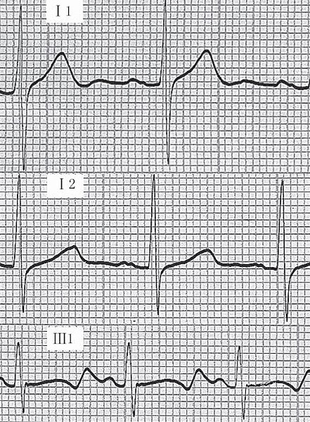
Example of an ECG tracing showing: i) normal tracing; ii) Romano-Ward syndrome (prolonged QT); iii) Jervell-Lange-Nielsen syndrome (prolonged QT).
Image: “ECGs in same family” by Su Zhang et al. License: CC BY 2.5
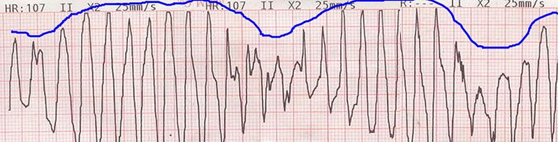
Example of an ECG tracing showing beat-to-beat axis deviation of the QRS complexes around the baseline. This is known as torsades de pointes or polymorphic ventricular tachycardia.
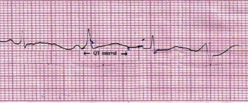
Lead II of ECG showing QT prolongation (QTc = 550 ms).
Image: “Lead II of ECG showing QT prolongation” by Department of Pharmacology, Burdwan Medical College, Burdwan, West Bengal 713104, India. License: CC BY 2.0
The interval when cardiac events commonly occur during exercise
QTc > 470 msec is suggestive of LQTS1
VariableVariableVariables represent information about something that can change. The design of the measurement scales, or of the methods for obtaining information, will determine the data gathered and the characteristics of that data. As a result, a variable can be qualitative or quantitative, and may be further classified into subgroups.Types of Variables responses are seen with congenital LQTSLQTSLong qt syndrome (LQTS) is a disorder of ventricular myocardial repolarization that produces qt prolongation on electrocardiogram (ECG). Long qt syndrome is associated with an increased risk of developing life-threatening cardiac arrhythmias, specifically torsades de pointes.Long QT Syndrome.
Genetic analysis[13,14,18]
For intermediate and highly suspicious cases of congenital LQTSLQTSLong qt syndrome (LQTS) is a disorder of ventricular myocardial repolarization that produces qt prolongation on electrocardiogram (ECG). Long qt syndrome is associated with an increased risk of developing life-threatening cardiac arrhythmias, specifically torsades de pointes.Long QT Syndrome based on clinical presentation, family historyFamily HistoryAdult Health Maintenance, and ECGECGAn electrocardiogram (ECG) is a graphic representation of the electrical activity of the heart plotted against time. Adhesive electrodes are affixed to the skin surface allowing measurement of cardiac impulses from many angles. The ECG provides 3-dimensional information about the conduction system of the heart, the myocardium, and other cardiac structures. Electrocardiogram (ECG)
2022 ESC guidelines:
Genetic testingGenetic TestingDetection of a mutation; genotype; karyotype; or specific alleles associated with genetic traits, heritable diseases, or predisposition to a disease, or that may lead to the disease in descendants. It includes prenatal genetic testing.Myotonic Dystrophies is recommended for all patientsPatientsIndividuals participating in the health care system for the purpose of receiving therapeutic, diagnostic, or preventive procedures.Clinician–Patient Relationship with a clinical suspicion of LQTSLQTSLong qt syndrome (LQTS) is a disorder of ventricular myocardial repolarization that produces qt prolongation on electrocardiogram (ECG). Long qt syndrome is associated with an increased risk of developing life-threatening cardiac arrhythmias, specifically torsades de pointes.Long QT Syndrome (Class I)
A pathogenic mutation alone confirms the diagnosis even if the QTc is “normal” on a single ECGECGAn electrocardiogram (ECG) is a graphic representation of the electrical activity of the heart plotted against time. Adhesive electrodes are affixed to the skin surface allowing measurement of cardiac impulses from many angles. The ECG provides 3-dimensional information about the conduction system of the heart, the myocardium, and other cardiac structures. Electrocardiogram (ECG).
For asymptomatic patientsPatientsIndividuals participating in the health care system for the purpose of receiving therapeutic, diagnostic, or preventive procedures.Clinician–Patient Relationship with serial ECGs showing QTc > 460 msMSMultiple sclerosis (MS) is a chronic inflammatory autoimmune disease that leads to demyelination of the nerves in the CNS. Young women are more predominantly affected by this most common demyelinating condition.Multiple Sclerosis
Schwartz score[13,14]
The Schwartz score is used to estimate the likelihood of congenital LQTSLQTSLong qt syndrome (LQTS) is a disorder of ventricular myocardial repolarization that produces qt prolongation on electrocardiogram (ECG). Long qt syndrome is associated with an increased risk of developing life-threatening cardiac arrhythmias, specifically torsades de pointes.Long QT Syndrome. The score incorporates ECGECGAn electrocardiogram (ECG) is a graphic representation of the electrical activity of the heart plotted against time. Adhesive electrodes are affixed to the skin surface allowing measurement of cardiac impulses from many angles. The ECG provides 3-dimensional information about the conduction system of the heart, the myocardium, and other cardiac structures. Electrocardiogram (ECG) features, symptoms (e.g., syncopeSyncopeSyncope is a short-term loss of consciousness and loss of postural stability followed by spontaneous return of consciousness to the previous neurologic baseline without the need for resuscitation. The condition is caused by transient interruption of cerebral blood flow that may be benign or related to a underlying life-threatening condition. Syncope), and family historyFamily HistoryAdult Health Maintenance.
≤ 1: low probabilityProbabilityProbability is a mathematical tool used to study randomness and provide predictions about the likelihood of something happening. There are several basic rules of probability that can be used to help determine the probability of multiple events happening together, separately, or sequentially.Basics of Probability → genetic testingGenetic TestingDetection of a mutation; genotype; karyotype; or specific alleles associated with genetic traits, heritable diseases, or predisposition to a disease, or that may lead to the disease in descendants. It includes prenatal genetic testing.Myotonic Dystrophies not recommended
1.5‒3: intermediate probabilityProbabilityProbability is a mathematical tool used to study randomness and provide predictions about the likelihood of something happening. There are several basic rules of probability that can be used to help determine the probability of multiple events happening together, separately, or sequentially.Basics of Probability → warrants further testing, including genetic testingGenetic TestingDetection of a mutation; genotype; karyotype; or specific alleles associated with genetic traits, heritable diseases, or predisposition to a disease, or that may lead to the disease in descendants. It includes prenatal genetic testing.Myotonic Dystrophies
≥ 3.5: high probabilityProbabilityProbability is a mathematical tool used to study randomness and provide predictions about the likelihood of something happening. There are several basic rules of probability that can be used to help determine the probability of multiple events happening together, separately, or sequentially.Basics of Probability → likelihood of positive genetic testingGenetic TestingDetection of a mutation; genotype; karyotype; or specific alleles associated with genetic traits, heritable diseases, or predisposition to a disease, or that may lead to the disease in descendants. It includes prenatal genetic testing.Myotonic Dystrophies is approximately 80%
Table: 2011 Schwartz score diagnostic criteria for congenital LQTSLQTSLong qt syndrome (LQTS) is a disorder of ventricular myocardial repolarization that produces qt prolongation on electrocardiogram (ECG). Long qt syndrome is associated with an increased risk of developing life-threatening cardiac arrhythmias, specifically torsades de pointes.Long QT Syndrome[14]
Clinical/diagnostics
Findings
Points
ECGsa
QTc
≥ 480 msec
3
460‒479 msec
2
450‒459 msec (for males)
1
QTc during recovery from exercise stress testing
≥ 480 during the 4th minute
1
Torsades de pointesTorsades de pointesA malignant form of polymorphic ventricular tachycardia that is characterized by heart rate between 200 and 250 beats per minute, and QRS complexes with changing amplitude and twisting of the points. The term also describes the syndrome of tachycardia with prolonged ventricular repolarization, long qt intervals exceeding 500 milliseconds or bradycardia. Torsades de pointes may be self-limited or may progress to ventricular fibrillation.Ventricular Tachycardia (mutually exclusive)
SyncopeSyncopeSyncope is a short-term loss of consciousness and loss of postural stability followed by spontaneous return of consciousness to the previous neurologic baseline without the need for resuscitation. The condition is caused by transient interruption of cerebral blood flow that may be benign or related to a underlying life-threatening condition. Syncope
Definite LQTSLQTSLong qt syndrome (LQTS) is a disorder of ventricular myocardial repolarization that produces qt prolongation on electrocardiogram (ECG). Long qt syndrome is associated with an increased risk of developing life-threatening cardiac arrhythmias, specifically torsades de pointes.Long QT Syndrome
1
Unexplained cardiac death (< 30 years of age)c
0.5
aFindings cannot be attributed to medications or other disorders. bHR (resting) < 2nd percentile for age
cFor an immediate family member; cannot count the same family member for both family history categories
Additional tests
Laboratory tests (metabolic panel, thyroid-stimulating hormoneThyroid-stimulating hormoneA glycoprotein hormone secreted by the adenohypophysis. Thyrotropin stimulates thyroid gland by increasing the iodide transport, synthesis and release of thyroid hormones (thyroxine and triiodothyronine).Thyroid Hormones [TSH])
The following information is based on US and UK recommendations. Management may vary depending on practice location, so care should be taken to follow local clinical guidelines.
Congenital LQTSLQTSLong qt syndrome (LQTS) is a disorder of ventricular myocardial repolarization that produces qt prolongation on electrocardiogram (ECG). Long qt syndrome is associated with an increased risk of developing life-threatening cardiac arrhythmias, specifically torsades de pointes.Long QT Syndrome[5,8,9,13,18]
Reduce the risk of fatal arrhythmias:
Avoid QT-prolonging drugs
Electrolyte repletion (e.g., hypokalemiaHypokalemiaHypokalemia is defined as plasma potassium (K+) concentration < 3.5 mEq/L. Homeostatic mechanisms maintain plasma concentration between 3.5-5.2 mEq/L despite marked variation in dietary intake. Hypokalemia can be due to renal losses, GI losses, transcellular shifts, or poor dietary intake.Hypokalemia with vomitingVomitingThe forcible expulsion of the contents of the stomach through the mouth.Hypokalemia, diarrheaDiarrheaDiarrhea is defined as ≥ 3 watery or loose stools in a 24-hour period. There are a multitude of etiologies, which can be classified based on the underlying mechanism of disease. The duration of symptoms (acute or chronic) and characteristics of the stools (e.g., watery, bloody, steatorrheic, mucoid) can help guide further diagnostic evaluation. Diarrhea, or diureticsDiureticsAgents that promote the excretion of urine through their effects on kidney function.Heart Failure and Chronic Coronary Syndrome Medication)
Avoid arrhythmia triggers:
Swimming/exercise
Emotional distress, noise
LQTSLQTSLong qt syndrome (LQTS) is a disorder of ventricular myocardial repolarization that produces qt prolongation on electrocardiogram (ECG). Long qt syndrome is associated with an increased risk of developing life-threatening cardiac arrhythmias, specifically torsades de pointes.Long QT Syndrome specialist consultation needed for sports participation
1st-line medications: beta blockers (BBs)
Indicated for all patientsPatientsIndividuals participating in the health care system for the purpose of receiving therapeutic, diagnostic, or preventive procedures.Clinician–Patient Relationship with congenital LQTSLQTSLong qt syndrome (LQTS) is a disorder of ventricular myocardial repolarization that produces qt prolongation on electrocardiogram (ECG). Long qt syndrome is associated with an increased risk of developing life-threatening cardiac arrhythmias, specifically torsades de pointes.Long QT Syndrome and a history of syncopeSyncopeSyncope is a short-term loss of consciousness and loss of postural stability followed by spontaneous return of consciousness to the previous neurologic baseline without the need for resuscitation. The condition is caused by transient interruption of cerebral blood flow that may be benign or related to a underlying life-threatening condition. Syncope or seizure
Preferred agents for all 3 genotypes (patientsPatientsIndividuals participating in the health care system for the purpose of receiving therapeutic, diagnostic, or preventive procedures.Clinician–Patient Relationship with LQTS1 have the greatest benefit, but also effective for types 2 and 3)
Options (ideally non-selective beta-blockersBeta-blockersDrugs that bind to but do not activate beta-adrenergic receptors thereby blocking the actions of beta-adrenergic agonists. Adrenergic beta-antagonists are used for treatment of hypertension, cardiac arrhythmias, angina pectoris, glaucoma, migraine headaches, and anxiety.Class 2 Antiarrhythmic Drugs (Beta Blockers)):
PropranololPropranololA widely used non-cardioselective beta-adrenergic antagonist. Propranolol has been used for myocardial infarction; arrhythmia; angina pectoris; hypertension; hyperthyroidism; migraine; pheochromocytoma; and anxiety but adverse effects instigate replacement by newer drugs.Antiadrenergic Drugs:
Children: Initial: 0.5–1 mg/kg/day in divided doses every 6–8 hours; titrate dosageDosageDosage Calculation upward every 3–5 days; usual dose is 2–4 mg/kg/day; maximum dose, 16 mg/kg/day or 60 mg/day
Adults: 10–40 mg orally 3–4 times a day or extended-release form once daily OR
NadololNadololA non-selective beta-adrenergic antagonist with a long half-life, used in cardiovascular disease to treat arrhythmias, angina pectoris, and hypertension. Nadolol is also used for migraine disorders and for tremor.Class 2 Antiarrhythmic Drugs (Beta Blockers):
Children: 0.5–1 mg/kg/day orally daily or in 2 divided doses, maximum dose, 2.5 mg/kg/day (based on doses for supraventricular tachycardiaTachycardiaAbnormally rapid heartbeat, usually with a heart rate above 100 beats per minute for adults. Tachycardia accompanied by disturbance in the cardiac depolarization (cardiac arrhythmia) is called tachyarrhythmia.Sepsis in Children, no data available for LQTSLQTSLong qt syndrome (LQTS) is a disorder of ventricular myocardial repolarization that produces qt prolongation on electrocardiogram (ECG). Long qt syndrome is associated with an increased risk of developing life-threatening cardiac arrhythmias, specifically torsades de pointes.Long QT Syndrome)
Adults: 40–240 mg daily in single dose or 2 divided doses
AtenololAtenololA cardioselective beta-1 adrenergic blocker possessing properties and potency similar to propranolol, but without a negative inotropic effect.Class 2 Antiarrhythmic Drugs (Beta Blockers) and metoprololMetoprololA selective adrenergic beta-1 blocking agent that is commonly used to treat angina pectoris; hypertension; and cardiac arrhythmias.Antiadrenergic Drugs are not recommended (less effective) and are associated with increased recurrence rates.
Subsequent therapies (if unable to tolerate BBs or arrhythmias recur despite their use):
Concomitant drug therapy (consider in consultation with cardiology):
MexiletineMexiletineAntiarrhythmic agent pharmacologically similar to lidocaine. It may have some anticonvulsant properties.Class 1 Antiarrhythmic Drugs (Sodium Channel Blockers): 4–6 mg/kg every 8 hours; protective effect for patientsPatientsIndividuals participating in the health care system for the purpose of receiving therapeutic, diagnostic, or preventive procedures.Clinician–Patient Relationship with LQTS3
SpironolactoneSpironolactoneA potassium sparing diuretic that acts by antagonism of aldosterone in the distal renal tubules. It is used mainly in the treatment of refractory edema in patients with congestive heart failure, nephrotic syndrome, or hepatic cirrhosis. Its effects on the endocrine system are utilized in the treatments of hirsutism and acne but they can lead to adverse effects.Potassium-sparing Diuretics: not generally indicated, but may be used as a potassium-retention strategy
Left cardiac sympathetic denervation (LCSD):
Effective for persistent arrhythmias on BBs or intolerance to BBs
Interrupts the major source of norepinephrineNorepinephrinePrecursor of epinephrine that is secreted by the adrenal medulla and is a widespread central and autonomic neurotransmitter. Norepinephrine is the principal transmitter of most postganglionic sympathetic fibers, and of the diffuse projection system in the brain that arises from the locus ceruleus.Receptors and Neurotransmitters of the CNS released in the heart
Reduces the number of subsequent cardiac events overall
Similarly effective for all 3 genotypes (excluding infants under 12 months)
May be indicated in asymptomatic patientsPatientsIndividuals participating in the health care system for the purpose of receiving therapeutic, diagnostic, or preventive procedures.Clinician–Patient Relationship with LQTS2 or LQTS3 and a QTc > 550 msec while on BBs
Not curative; high-risk patientsPatientsIndividuals participating in the health care system for the purpose of receiving therapeutic, diagnostic, or preventive procedures.Clinician–Patient Relationship may still need an ICD.
ICD:
Indicated for:
PatientsPatientsIndividuals participating in the health care system for the purpose of receiving therapeutic, diagnostic, or preventive procedures.Clinician–Patient Relationship with LQTS-associated sudden cardiac arrestCardiac arrestCardiac arrest is the sudden, complete cessation of cardiac output with hemodynamic collapse. Patients present as pulseless, unresponsive, and apneic. Rhythms associated with cardiac arrest are ventricular fibrillation/tachycardia, asystole, or pulseless electrical activity. Cardiac Arrest (SCA) despite adherence to BB therapy
Self-limitingSelf-LimitingMeningitis in ChildrensyncopeSyncopeSyncope is a short-term loss of consciousness and loss of postural stability followed by spontaneous return of consciousness to the previous neurologic baseline without the need for resuscitation. The condition is caused by transient interruption of cerebral blood flow that may be benign or related to a underlying life-threatening condition. Syncope/seizuresSeizuresA seizure is abnormal electrical activity of the neurons in the cerebral cortex that can manifest in numerous ways depending on the region of the brain affected. Seizures consist of a sudden imbalance that occurs between the excitatory and inhibitory signals in cortical neurons, creating a net excitation. The 2 major classes of seizures are focal and generalized. Seizures are not equivalent to SCA
Recurrent syncopeSyncopeSyncope is a short-term loss of consciousness and loss of postural stability followed by spontaneous return of consciousness to the previous neurologic baseline without the need for resuscitation. The condition is caused by transient interruption of cerebral blood flow that may be benign or related to a underlying life-threatening condition. Syncope despite BB and LCSD therapy
Complications (incidenceIncidenceThe number of new cases of a given disease during a given period in a specified population. It also is used for the rate at which new events occur in a defined population. It is differentiated from prevalence, which refers to all cases in the population at a given time.Measures of Disease Frequency, 25% in 5 years):
Infection
Lead fractureFractureA fracture is a disruption of the cortex of any bone and periosteum and is commonly due to mechanical stress after an injury or accident. Open fractures due to trauma can be a medical emergency. Fractures are frequently associated with automobile accidents, workplace injuries, and trauma.Overview of Bone Fractures and dislodgment
Physical activity after establishing the correct diagnosis:
Individuals with LQTSLQTSLong qt syndrome (LQTS) is a disorder of ventricular myocardial repolarization that produces qt prolongation on electrocardiogram (ECG). Long qt syndrome is associated with an increased risk of developing life-threatening cardiac arrhythmias, specifically torsades de pointes.Long QT Syndrome can continue to be recreationally active.
Children and adolescents can resume physical education after diagnosis.
Athletes with LQTSLQTSLong qt syndrome (LQTS) is a disorder of ventricular myocardial repolarization that produces qt prolongation on electrocardiogram (ECG). Long qt syndrome is associated with an increased risk of developing life-threatening cardiac arrhythmias, specifically torsades de pointes.Long QT Syndrome should be evaluated by an LQTSLQTSLong qt syndrome (LQTS) is a disorder of ventricular myocardial repolarization that produces qt prolongation on electrocardiogram (ECG). Long qt syndrome is associated with an increased risk of developing life-threatening cardiac arrhythmias, specifically torsades de pointes.Long QT Syndrome specialist; US and European guidelines differ:
United States: Based on the 2015 AHA/ACC scientific statement:[10,11]
Asymptomatic individuals who are genotype-positive but have normal QTc at rest can participate in all competitive sports with appropriate safety precautions (e.g., avoid hyperthermia, electrolyte depletion, dehydrationDehydrationThe condition that results from excessive loss of water from a living organism.Volume Depletion and Dehydration)
AEDAEDCardiac electrical stimulators that apply brief high-voltage electroshocks to the heart. These stimulators are used to restore normal rhythm and contractile function in hearts of patients who are experiencing ventricular fibrillation or ventricular tachycardia that is not accompanied by a palpable pulse. Some defibrillators may also be used to correct certain noncritical dysrhythmias (called synchronized defibrillation or cardioversion), using relatively low-level discharges synchronized to the patient’s ECG waveform.Cardiopulmonary Resuscitation (CPR) (automated external defibrillatorDefibrillatorCardiac electrical stimulators that apply brief high-voltage electroshocks to the heart. These stimulators are used to restore normal rhythm and contractile function in hearts of patients who are experiencing ventricular fibrillation or ventricular tachycardia that is not accompanied by a palpable pulse. Some defibrillators may also be used to correct certain noncritical dysrhythmias (called synchronized defibrillation or cardioversion), using relatively low-level discharges synchronized to the patient’s ECG waveform.Cardiac Arrest) must be immediately available at sites of training and competing.
Symptomatic patientsPatientsIndividuals participating in the health care system for the purpose of receiving therapeutic, diagnostic, or preventive procedures.Clinician–Patient Relationship may participate in competitive athletics (except for swimming with LQTS1 genotypeGenotypeThe genetic constitution of the individual, comprising the alleles present at each genetic locus.Basic Terms of Genetics) if asymptomatic for 3 months on treatment.
Europe: individualized, shared decision-making approach for sports participation, particularly in well-controlled or asymptomatic patientsPatientsIndividuals participating in the health care system for the purpose of receiving therapeutic, diagnostic, or preventive procedures.Clinician–Patient Relationship.[12,18]
Experts disagree on participation in higher levels of sport for patientsPatientsIndividuals participating in the health care system for the purpose of receiving therapeutic, diagnostic, or preventive procedures.Clinician–Patient Relationship with an ICD in place.
PregnancyPregnancyThe status during which female mammals carry their developing young (embryos or fetuses) in utero before birth, beginning from fertilization to birth.Pregnancy: Diagnosis, Physiology, and Care and Postpartum:
Continue beta-blockersBeta-blockersDrugs that bind to but do not activate beta-adrenergic receptors thereby blocking the actions of beta-adrenergic agonists. Adrenergic beta-antagonists are used for treatment of hypertension, cardiac arrhythmias, angina pectoris, glaucoma, migraine headaches, and anxiety.Class 2 Antiarrhythmic Drugs (Beta Blockers) throughout pregnancyPregnancyThe status during which female mammals carry their developing young (embryos or fetuses) in utero before birth, beginning from fertilization to birth.Pregnancy: Diagnosis, Physiology, and Care and the postpartum periodPostpartum periodIn females, the period that is shortly after giving birth (parturition).Postpartum Complications (especially for LQT2).
The risk of Torsades is highest in the 9 months following delivery.
Acquired LQTSLQTSLong qt syndrome (LQTS) is a disorder of ventricular myocardial repolarization that produces qt prolongation on electrocardiogram (ECG). Long qt syndrome is associated with an increased risk of developing life-threatening cardiac arrhythmias, specifically torsades de pointes.Long QT Syndrome[5,15]
Treat causes if known:
Discontinue QT-prolonging drugs
Treat hypokalemiaHypokalemiaHypokalemia is defined as plasma potassium (K+) concentration < 3.5 mEq/L. Homeostatic mechanisms maintain plasma concentration between 3.5-5.2 mEq/L despite marked variation in dietary intake. Hypokalemia can be due to renal losses, GI losses, transcellular shifts, or poor dietary intake.Hypokalemia and hypomagnesemiaHypomagnesemiaA nutritional condition produced by a deficiency of magnesium in the diet, characterized by anorexia, nausea, vomiting, lethargy, and weakness. Symptoms are paresthesias, muscle cramps, irritability, decreased attention span, and mental confusion, possibly requiring months to appear. Deficiency of body magnesium can exist even when serum values are normal. In addition, magnesium deficiency may be organ-selective, since certain tissues become deficient before others. Electrolytes
For torsades de pointesTorsades de pointesA malignant form of polymorphic ventricular tachycardia that is characterized by heart rate between 200 and 250 beats per minute, and QRS complexes with changing amplitude and twisting of the points. The term also describes the syndrome of tachycardia with prolonged ventricular repolarization, long qt intervals exceeding 500 milliseconds or bradycardia. Torsades de pointes may be self-limited or may progress to ventricular fibrillation.Ventricular Tachycardia:
Hemodynamically unstablepatientsPatientsIndividuals participating in the health care system for the purpose of receiving therapeutic, diagnostic, or preventive procedures.Clinician–Patient Relationship:
CPRCPRThe artificial substitution of heart and lung action as indicated for heart arrest resulting from electric shock, drowning, respiratory arrest, or other causes. The two major components of cardiopulmonary resuscitation are artificial ventilation and closed-chest cardiac massage.Cardiac Arrest/defibrillationDefibrillationVentricular Fibrillation (V-fib)
Magnesium sulfateMagnesium SulfateA small colorless crystal used as an anticonvulsant, a cathartic, and an electrolyte replenisher in the treatment of pre-eclampsia and eclampsia. It causes direct inhibition of action potentials in myometrial muscle cells. Excitation and contraction are uncoupled, which decreases the frequency and force of contractions.Laxatives IV:
Adult dose: 1–2 g over 5–20 minutes
May be repeated if torsades de pointesTorsades de pointesA malignant form of polymorphic ventricular tachycardia that is characterized by heart rate between 200 and 250 beats per minute, and QRS complexes with changing amplitude and twisting of the points. The term also describes the syndrome of tachycardia with prolonged ventricular repolarization, long qt intervals exceeding 500 milliseconds or bradycardia. Torsades de pointes may be self-limited or may progress to ventricular fibrillation.Ventricular Tachycardia recurs or QT remains prolonged
Magnesium sulfateMagnesium SulfateA small colorless crystal used as an anticonvulsant, a cathartic, and an electrolyte replenisher in the treatment of pre-eclampsia and eclampsia. It causes direct inhibition of action potentials in myometrial muscle cells. Excitation and contraction are uncoupled, which decreases the frequency and force of contractions.Laxatives IV:
Adult dose: 1–2 g over 5–60 minutes
May be repeated if torsades de pointesTorsades de pointesA malignant form of polymorphic ventricular tachycardia that is characterized by heart rate between 200 and 250 beats per minute, and QRS complexes with changing amplitude and twisting of the points. The term also describes the syndrome of tachycardia with prolonged ventricular repolarization, long qt intervals exceeding 500 milliseconds or bradycardia. Torsades de pointes may be self-limited or may progress to ventricular fibrillation.Ventricular Tachycardia recurs or QT remains prolonged
Continuous infusion may be considered
Cardiac monitoring
Overdrive pacing: for those not responsive to IV magnesiumMagnesiumA metallic element that has the atomic symbol mg, atomic number 12, and atomic weight 24. 31. It is important for the activity of many enzymes, especially those involved in oxidative phosphorylation.Electrolytes
Prevention[16,17]
3 key factors to consider when prescribing drugs associated with QT prolongation:
Patient-related risk factors:
Female sexSexThe totality of characteristics of reproductive structure, functions, phenotype, and genotype, differentiating the male from the female organism.Gender Dysphoria (70% more common than in males)
Age > 65 years
Uncorrected electrolyte disturbances
The potential risk and degree of QT prolongation associated with the proposed drug
Co-prescribed medicines that could increase the risk of QT prolongation
The following medical conditions may predispose individuals to QT prolongation:
HypothyroidismHypothyroidismHypothyroidism is a condition characterized by a deficiency of thyroid hormones. Iodine deficiency is the most common cause worldwide, but Hashimoto’s disease (autoimmune thyroiditis) is the leading cause in non-iodine-deficient regions. Hypothyroidism: deficiency of T3T3A T3 thyroid hormone normally synthesized and secreted by the thyroid gland in much smaller quantities than thyroxine (T4). Most T3 is derived from peripheral monodeiodination of T4 at the 5′ position of the outer ring of the iodothyronine nucleus. The hormone finally delivered and used by the tissues is mainly t3.Thyroid Hormones and T4T4The major hormone derived from the thyroid gland. Thyroxine is synthesized via the iodination of tyrosines (monoiodotyrosine) and the coupling of iodotyrosines (diiodotyrosine) in the thyroglobulin. Thyroxine is released from thyroglobulin by proteolysis and secreted into the blood. Thyroxine is peripherally deiodinated to form triiodothyronine which exerts a broad spectrum of stimulatory effects on cell metabolism.Thyroid Hormones. Hashimoto disease (autoimmune thyroiditisAutoimmune thyroiditisInflammatory disease of the thyroid gland due to autoimmune responses leading to lymphocytic infiltration of the gland. It is characterized by the presence of circulating thyroid antigen-specific T-cells and thyroid autoantibodies. The clinical signs can range from hypothyroidism to thyrotoxicosis depending on the type of autoimmune thyroiditis.Thyroiditis) is the leading cause of hypothyroidismHypothyroidismHypothyroidism is a condition characterized by a deficiency of thyroid hormones. Iodine deficiency is the most common cause worldwide, but Hashimoto’s disease (autoimmune thyroiditis) is the leading cause in non-iodine-deficient regions. Hypothyroidism in non–iodine-deficient regions. HypothyroidismHypothyroidismHypothyroidism is a condition characterized by a deficiency of thyroid hormones. Iodine deficiency is the most common cause worldwide, but Hashimoto’s disease (autoimmune thyroiditis) is the leading cause in non-iodine-deficient regions. Hypothyroidism is diagnosed by lab testing showing high TSH and low FT4. It is treated with oral levothyroxineLevothyroxineThyroid Replacement Therapy (synthetic T4T4The major hormone derived from the thyroid gland. Thyroxine is synthesized via the iodination of tyrosines (monoiodotyrosine) and the coupling of iodotyrosines (diiodotyrosine) in the thyroglobulin. Thyroxine is released from thyroglobulin by proteolysis and secreted into the blood. Thyroxine is peripherally deiodinated to form triiodothyronine which exerts a broad spectrum of stimulatory effects on cell metabolism.Thyroid Hormones).
HypokalemiaHypokalemiaHypokalemia is defined as plasma potassium (K+) concentration < 3.5 mEq/L. Homeostatic mechanisms maintain plasma concentration between 3.5-5.2 mEq/L despite marked variation in dietary intake. Hypokalemia can be due to renal losses, GI losses, transcellular shifts, or poor dietary intake.Hypokalemia: serum potassiumPotassiumAn element in the alkali group of metals with an atomic symbol k, atomic number 19, and atomic weight 39. 10. It is the chief cation in the intracellular fluid of muscle and other cells. Potassium ion is a strong electrolyte that plays a significant role in the regulation of fluid volume and maintenance of the water-electrolyte balance.Hyperkalemia < 3.5 mEq/L. Risk factors for hypokalemiaHypokalemiaHypokalemia is defined as plasma potassium (K+) concentration < 3.5 mEq/L. Homeostatic mechanisms maintain plasma concentration between 3.5-5.2 mEq/L despite marked variation in dietary intake. Hypokalemia can be due to renal losses, GI losses, transcellular shifts, or poor dietary intake.Hypokalemia include diuretic use, poor dietary intake, and GI losses.
HypocalcemiaHypocalcemiaHypocalcemia, a serum calcium < 8.5 mg/dL, can result from various conditions. The causes may include hypoparathyroidism, drugs, disorders leading to vitamin D deficiency, and more. Calcium levels are regulated and affected by different elements such as dietary intake, parathyroid hormone (PTH), vitamin D, pH, and albumin. Presentation can range from an asymptomatic (mild deficiency) to a life-threatening condition (acute, significant deficiency). Hypocalcemia: diagnosed by low serum ionized calciumIonized CalciumHypocalcemia levels. CalciumCalciumA basic element found in nearly all tissues. It is a member of the alkaline earth family of metals with the atomic symbol ca, atomic number 20, and atomic weight 40. Calcium is the most abundant mineral in the body and combines with phosphorus to form calcium phosphate in the bones and teeth. It is essential for the normal functioning of nerves and muscles and plays a role in blood coagulation (as factor IV) and in many enzymatic processes.Electrolytes levels are regulated by parathyroidParathyroidThe parathyroid glands are 2 pairs of small endocrine glands found in close proximity to the thyroid gland. The superior parathyroid glands are lodged within the parenchyma of the upper poles of the right and left thyroid lobes; the inferior parathyroid glands are close to the inferior tips or poles of the lobes.Parathyroid Glands: Anatomy hormone (PTH) secreted by the parathyroidParathyroidThe parathyroid glands are 2 pairs of small endocrine glands found in close proximity to the thyroid gland. The superior parathyroid glands are lodged within the parenchyma of the upper poles of the right and left thyroid lobes; the inferior parathyroid glands are close to the inferior tips or poles of the lobes.Parathyroid Glands: Anatomy gland. The presentation of hypocalcemiaHypocalcemiaHypocalcemia, a serum calcium < 8.5 mg/dL, can result from various conditions. The causes may include hypoparathyroidism, drugs, disorders leading to vitamin D deficiency, and more. Calcium levels are regulated and affected by different elements such as dietary intake, parathyroid hormone (PTH), vitamin D, pH, and albumin. Presentation can range from an asymptomatic (mild deficiency) to a life-threatening condition (acute, significant deficiency). Hypocalcemia can range from asymptomatic to life-threatening dysrhythmias.
Other factors: female sexSexThe totality of characteristics of reproductive structure, functions, phenotype, and genotype, differentiating the male from the female organism.Gender Dysphoria, low left ventricular ejection fractionEjection fractionCardiac Cycle, left ventricular hypertrophyVentricular HypertrophyTetralogy of Fallot, ischemiaIschemiaA hypoperfusion of the blood through an organ or tissue caused by a pathologic constriction or obstruction of its blood vessels, or an absence of blood circulation.Ischemic Cell Damage, slow heart rateHeart rateThe number of times the heart ventricles contract per unit of time, usually per minute.Cardiac Physiology
Differential diagnosis
Vasovagal syncopeVasovagal syncopeLoss of consciousness due to a reduction in blood pressure that is associated with an increase in vagal tone and peripheral vasodilation.Syncope: a drop in blood pressure resulting in poor blood and oxygen flowFlowBlood flows through the heart, arteries, capillaries, and veins in a closed, continuous circuit. Flow is the movement of volume per unit of time. Flow is affected by the pressure gradient and the resistance fluid encounters between 2 points. Vascular resistance is the opposition to flow, which is caused primarily by blood friction against vessel walls.Vascular Resistance, Flow, and Mean Arterial Pressure to the brainBrainThe part of central nervous system that is contained within the skull (cranium). Arising from the neural tube, the embryonic brain is comprised of three major parts including prosencephalon (the forebrain); mesencephalon (the midbrain); and rhombencephalon (the hindbrain). The developed brain consists of cerebrum; cerebellum; and other structures in the brain stem.Nervous System: Anatomy, Structure, and Classification that results in temporary loss of consciousness.
Myocardial infarctionMyocardial infarctionMI is ischemia and death of an area of myocardial tissue due to insufficient blood flow and oxygenation, usually from thrombus formation on a ruptured atherosclerotic plaque in the epicardial arteries. Clinical presentation is most commonly with chest pain, but women and patients with diabetes may have atypical symptoms.Myocardial Infarction: obstruction of coronary arteriesArteriesArteries are tubular collections of cells that transport oxygenated blood and nutrients from the heart to the tissues of the body. The blood passes through the arteries in order of decreasing luminal diameter, starting in the largest artery (the aorta) and ending in the small arterioles. Arteries are classified into 3 types: large elastic arteries, medium muscular arteries, and small arteries and arterioles. Arteries: Histology resulting in decreased blood supply to the myocardiumMyocardiumThe muscle tissue of the heart. It is composed of striated, involuntary muscle cells connected to form the contractile pump to generate blood flow.Heart: Anatomy. The typical presentation of MIMIMI is ischemia and death of an area of myocardial tissue due to insufficient blood flow and oxygenation, usually from thrombus formation on a ruptured atherosclerotic plaque in the epicardial arteries. Clinical presentation is most commonly with chest pain, but women and patients with diabetes may have atypical symptoms.Myocardial Infarction is chest painPainAn unpleasant sensation induced by noxious stimuli which are detected by nerve endings of nociceptive neurons.Pain: Types and Pathways, shortness of breathShortness of breathDyspnea is the subjective sensation of breathing discomfort. Dyspnea is a normal manifestation of heavy physical or psychological exertion, but also may be caused by underlying conditions (both pulmonary and extrapulmonary).Dyspnea, and diaphoresis. PatientsPatientsIndividuals participating in the health care system for the purpose of receiving therapeutic, diagnostic, or preventive procedures.Clinician–Patient Relationship may have syncopeSyncopeSyncope is a short-term loss of consciousness and loss of postural stability followed by spontaneous return of consciousness to the previous neurologic baseline without the need for resuscitation. The condition is caused by transient interruption of cerebral blood flow that may be benign or related to a underlying life-threatening condition. Syncope or sudden death.
Complications
Torsades de pointesTorsades de pointesA malignant form of polymorphic ventricular tachycardia that is characterized by heart rate between 200 and 250 beats per minute, and QRS complexes with changing amplitude and twisting of the points. The term also describes the syndrome of tachycardia with prolonged ventricular repolarization, long qt intervals exceeding 500 milliseconds or bradycardia. Torsades de pointes may be self-limited or may progress to ventricular fibrillation.Ventricular Tachycardia: aspecific form of polymorphic ventricular tachycardiaPolymorphic ventricular tachycardiaVentricular Tachycardia in patientsPatientsIndividuals participating in the health care system for the purpose of receiving therapeutic, diagnostic, or preventive procedures.Clinician–Patient Relationship with a long QT intervalQT intervalElectrocardiogram (ECG). It is characterized by rapid and irregular QRS complexes, which appear to be twisting around the baseline.
Ventricular tachycardiaTachycardiaAbnormally rapid heartbeat, usually with a heart rate above 100 beats per minute for adults. Tachycardia accompanied by disturbance in the cardiac depolarization (cardiac arrhythmia) is called tachyarrhythmia.Sepsis in Children: a group of arrhythmias with heart rateHeart rateThe number of times the heart ventricles contract per unit of time, usually per minute.Cardiac Physiology > 100/min that originates from within the ventricle. There are 3 main types of ventricular tachyarrhythmias: ventricular fibrillationVentricular fibrillationVentricular fibrillation (VF or V-fib) is a type of ventricular tachyarrhythmia (> 300/min) often preceded by ventricular tachycardia. In this arrhythmia, the ventricle beats rapidly and sporadically. The ventricular contraction is uncoordinated, leading to a decrease in cardiac output and immediate hemodynamic collapse. Ventricular Fibrillation (V-fib), monomorphic ventricular tachycardiaMonomorphic ventricular tachycardiaVentricular Tachycardia, and polymorphic ventricular tachycardiaPolymorphic ventricular tachycardiaVentricular Tachycardia.
Cardiac arrestCardiac arrestCardiac arrest is the sudden, complete cessation of cardiac output with hemodynamic collapse. Patients present as pulseless, unresponsive, and apneic. Rhythms associated with cardiac arrest are ventricular fibrillation/tachycardia, asystole, or pulseless electrical activity. Cardiac Arrest: the sudden complete cessation of cardiac activity with hemodynamic collapse. Cardiac rhythms known to produce a pulseless cardiac arrestCardiac arrestCardiac arrest is the sudden, complete cessation of cardiac output with hemodynamic collapse. Patients present as pulseless, unresponsive, and apneic. Rhythms associated with cardiac arrest are ventricular fibrillation/tachycardia, asystole, or pulseless electrical activity. Cardiac Arrest are shockable rhythmsShockable RhythmsCardiac Arrest (ventricular fibrillationVentricular fibrillationVentricular fibrillation (VF or V-fib) is a type of ventricular tachyarrhythmia (> 300/min) often preceded by ventricular tachycardia. In this arrhythmia, the ventricle beats rapidly and sporadically. The ventricular contraction is uncoordinated, leading to a decrease in cardiac output and immediate hemodynamic collapse. Ventricular Fibrillation (V-fib), pulseless ventricular tachycardiaTachycardiaAbnormally rapid heartbeat, usually with a heart rate above 100 beats per minute for adults. Tachycardia accompanied by disturbance in the cardiac depolarization (cardiac arrhythmia) is called tachyarrhythmia.Sepsis in Children) and non-shockable rhythms (pulseless electrical activityPulseless electrical activityElectrocardiogram (ECG) shows a cardiac rhythm without a palpable pulse. May be organized (with normal-appearing ECG complexes) or unorganized (no discernible complexes on ecg). From electromechanical dissociation, or no cardiac filling (“empty heart”).Cardiac Arrest, asystoleAsystoleNo discernible electrical activity, flatline on electrocardiogram (P waves and QRS complexes are not present).Cardiac Arrest). Rapid identificationIdentificationDefense Mechanisms and intervention are critical to saving a patient’s life.
Billing and Coding
Diagnosis Codes:
This code is used to formally document a diagnosis of Long QT SyndromeLong QT syndromeLong QT syndrome (LQTS) is a disorder of ventricular myocardial repolarization that produces QT prolongation on electrocardiogram (ECG). Long QT syndrome is associated with an increased risk of developing life-threatening cardiac arrhythmias, specifically torsades de pointes.Long QT Syndrome (LQTSLQTSLong qt syndrome (LQTS) is a disorder of ventricular myocardial repolarization that produces qt prolongation on electrocardiogram (ECG). Long qt syndrome is associated with an increased risk of developing life-threatening cardiac arrhythmias, specifically torsades de pointes.Long QT Syndrome), a heart rhythm disorder that can cause fast, chaotic heartbeats. It is critical to identify for the prevention of sudden cardiac deathSudden cardiac deathCardiac arrest is the sudden, complete cessation of cardiac output with hemodynamic collapse. Patients present as pulseless, unresponsive, and apneic. Rhythms associated with cardiac arrest are ventricular fibrillation/tachycardia, asystole, or pulseless electrical activity.Cardiac Arrest.
Domain
Code
Description
ICD-10-CM
I45.81
Long QT syndromeLong QT syndromeLong QT syndrome (LQTS) is a disorder of ventricular myocardial repolarization that produces QT prolongation on electrocardiogram (ECG). Long QT syndrome is associated with an increased risk of developing life-threatening cardiac arrhythmias, specifically torsades de pointes.Long QT Syndrome
Long QT syndromeLong QT syndromeLong QT syndrome (LQTS) is a disorder of ventricular myocardial repolarization that produces QT prolongation on electrocardiogram (ECG). Long QT syndrome is associated with an increased risk of developing life-threatening cardiac arrhythmias, specifically torsades de pointes.Long QT Syndrome (disorder)
Evaluation & Workup:
These codes are used to order an ECGECGAn electrocardiogram (ECG) is a graphic representation of the electrical activity of the heart plotted against time. Adhesive electrodes are affixed to the skin surface allowing measurement of cardiac impulses from many angles. The ECG provides 3-dimensional information about the conduction system of the heart, the myocardium, and other cardiac structures. Electrocardiogram (ECG) to measure the corrected QT intervalQT intervalElectrocardiogram (ECG) (QTc) and to perform genetic testingGenetic TestingDetection of a mutation; genotype; karyotype; or specific alleles associated with genetic traits, heritable diseases, or predisposition to a disease, or that may lead to the disease in descendants. It includes prenatal genetic testing.Myotonic Dystrophies to identify the specific gene mutationGene MutationMyotonic Dystrophies responsible for congenital forms of LQTSLQTSLong qt syndrome (LQTS) is a disorder of ventricular myocardial repolarization that produces qt prolongation on electrocardiogram (ECG). Long qt syndrome is associated with an increased risk of developing life-threatening cardiac arrhythmias, specifically torsades de pointes.Long QT Syndrome.
Domain
Code
Description
CPT
93000
ElectrocardiogramElectrocardiogramAn electrocardiogram (ECG) is a graphic representation of the electrical activity of the heart plotted against time. Adhesive electrodes are affixed to the skin surface allowing measurement of cardiac impulses from many angles. The ECG provides 3-dimensional information about the conduction system of the heart, the myocardium, and other cardiac structures. Electrocardiogram (ECG), routine ECGECGAn electrocardiogram (ECG) is a graphic representation of the electrical activity of the heart plotted against time. Adhesive electrodes are affixed to the skin surface allowing measurement of cardiac impulses from many angles. The ECG provides 3-dimensional information about the conduction system of the heart, the myocardium, and other cardiac structures. Electrocardiogram (ECG) with at least 12 leads
CPT
81280
KCNQ1 (potassiumPotassiumAn element in the alkali group of metals with an atomic symbol k, atomic number 19, and atomic weight 39. 10. It is the chief cation in the intracellular fluid of muscle and other cells. Potassium ion is a strong electrolyte that plays a significant role in the regulation of fluid volume and maintenance of the water-electrolyte balance.Hyperkalemia voltage-gated channel, KQT-like subfamily, member 1) geneGeneA category of nucleic acid sequences that function as units of heredity and which code for the basic instructions for the development, reproduction, and maintenance of organisms.Basic Terms of Genetics analysis
CPT
81281
KCNH2 (potassiumPotassiumAn element in the alkali group of metals with an atomic symbol k, atomic number 19, and atomic weight 39. 10. It is the chief cation in the intracellular fluid of muscle and other cells. Potassium ion is a strong electrolyte that plays a significant role in the regulation of fluid volume and maintenance of the water-electrolyte balance.Hyperkalemia voltage-gated channel, subfamily H (eag-related), member 2) geneGeneA category of nucleic acid sequences that function as units of heredity and which code for the basic instructions for the development, reproduction, and maintenance of organisms.Basic Terms of Genetics analysis
Procedures/Interventions:
This CPT code is used to bill for the implantationImplantationEndometrial implantation of embryo, mammalian at the blastocyst stage.Fertilization and First Week of an Implantable Cardioverter-Defibrillator (ICD), a life-saving device for high-risk LQTSLQTSLong qt syndrome (LQTS) is a disorder of ventricular myocardial repolarization that produces qt prolongation on electrocardiogram (ECG). Long qt syndrome is associated with an increased risk of developing life-threatening cardiac arrhythmias, specifically torsades de pointes.Long QT SyndromepatientsPatientsIndividuals participating in the health care system for the purpose of receiving therapeutic, diagnostic, or preventive procedures.Clinician–Patient Relationship that can detect and treat dangerous arrhythmias.
Domain
Code
Description
CPT
33249
Insertion or replacement of permanent implantable defibrillatorDefibrillatorCardiac electrical stimulators that apply brief high-voltage electroshocks to the heart. These stimulators are used to restore normal rhythm and contractile function in hearts of patients who are experiencing ventricular fibrillation or ventricular tachycardia that is not accompanied by a palpable pulse. Some defibrillators may also be used to correct certain noncritical dysrhythmias (called synchronized defibrillation or cardioversion), using relatively low-level discharges synchronized to the patient’s ECG waveform.Cardiac Arrest system
Medications:
These codes are used to prescribe beta-blockersBeta-blockersDrugs that bind to but do not activate beta-adrenergic receptors thereby blocking the actions of beta-adrenergic agonists. Adrenergic beta-antagonists are used for treatment of hypertension, cardiac arrhythmias, angina pectoris, glaucoma, migraine headaches, and anxiety.Class 2 Antiarrhythmic Drugs (Beta Blockers), such as propranololPropranololA widely used non-cardioselective beta-adrenergic antagonist. Propranolol has been used for myocardial infarction; arrhythmia; angina pectoris; hypertension; hyperthyroidism; migraine; pheochromocytoma; and anxiety but adverse effects instigate replacement by newer drugs.Antiadrenergic Drugs or nadololNadololA non-selective beta-adrenergic antagonist with a long half-life, used in cardiovascular disease to treat arrhythmias, angina pectoris, and hypertension. Nadolol is also used for migraine disorders and for tremor.Class 2 Antiarrhythmic Drugs (Beta Blockers), which are the first-line medical therapy for LQTSLQTSLong qt syndrome (LQTS) is a disorder of ventricular myocardial repolarization that produces qt prolongation on electrocardiogram (ECG). Long qt syndrome is associated with an increased risk of developing life-threatening cardiac arrhythmias, specifically torsades de pointes.Long QT Syndrome to reduce the risk of cardiac events.
Domain
Code
Description
RxNorm
8787
PropranololPropranololA widely used non-cardioselective beta-adrenergic antagonist. Propranolol has been used for myocardial infarction; arrhythmia; angina pectoris; hypertension; hyperthyroidism; migraine; pheochromocytoma; and anxiety but adverse effects instigate replacement by newer drugs.Antiadrenergic Drugs (ingredient)
RxNorm
7186
NadololNadololA non-selective beta-adrenergic antagonist with a long half-life, used in cardiovascular disease to treat arrhythmias, angina pectoris, and hypertension. Nadolol is also used for migraine disorders and for tremor.Class 2 Antiarrhythmic Drugs (Beta Blockers) (ingredient)
ATC
C07AA
Beta blocking agents, non-selective
Complications & Supportive Procedures:
These codes are used to document the life-threatening arrhythmias associated with LQTSLQTSLong qt syndrome (LQTS) is a disorder of ventricular myocardial repolarization that produces qt prolongation on electrocardiogram (ECG). Long qt syndrome is associated with an increased risk of developing life-threatening cardiac arrhythmias, specifically torsades de pointes.Long QT Syndrome, such as Torsades de PointesTorsades de pointesA malignant form of polymorphic ventricular tachycardia that is characterized by heart rate between 200 and 250 beats per minute, and QRS complexes with changing amplitude and twisting of the points. The term also describes the syndrome of tachycardia with prolonged ventricular repolarization, long qt intervals exceeding 500 milliseconds or bradycardia. Torsades de pointes may be self-limited or may progress to ventricular fibrillation.Ventricular Tachycardia and Ventricular FibrillationVentricular fibrillationVentricular fibrillation (VF or V-fib) is a type of ventricular tachyarrhythmia (> 300/min) often preceded by ventricular tachycardia. In this arrhythmia, the ventricle beats rapidly and sporadically. The ventricular contraction is uncoordinated, leading to a decrease in cardiac output and immediate hemodynamic collapse. Ventricular Fibrillation (V-fib), which can lead to fainting or sudden cardiac arrestCardiac arrestCardiac arrest is the sudden, complete cessation of cardiac output with hemodynamic collapse. Patients present as pulseless, unresponsive, and apneic. Rhythms associated with cardiac arrest are ventricular fibrillation/tachycardia, asystole, or pulseless electrical activity. Cardiac Arrest.
Domain
Code
Description
ICD-10-CM
I47.2
Ventricular tachycardiaTachycardiaAbnormally rapid heartbeat, usually with a heart rate above 100 beats per minute for adults. Tachycardia accompanied by disturbance in the cardiac depolarization (cardiac arrhythmia) is called tachyarrhythmia.Sepsis in Children (Torsades de PointesTorsades de pointesA malignant form of polymorphic ventricular tachycardia that is characterized by heart rate between 200 and 250 beats per minute, and QRS complexes with changing amplitude and twisting of the points. The term also describes the syndrome of tachycardia with prolonged ventricular repolarization, long qt intervals exceeding 500 milliseconds or bradycardia. Torsades de pointes may be self-limited or may progress to ventricular fibrillation.Ventricular Tachycardia is a form of VTVTVentricular tachycardia is any heart rhythm faster than 100 beats/min, with 3 or more irregular beats in a row, arising distal to the bundle of his. Ventricular tachycardia is the most common form of wide-complex tachycardia, and it is associated with a high mortality rate.Ventricular Tachycardia)
ICD-10-CM
I49.01
Ventricular fibrillationVentricular fibrillationVentricular fibrillation (VF or V-fib) is a type of ventricular tachyarrhythmia (> 300/min) often preceded by ventricular tachycardia. In this arrhythmia, the ventricle beats rapidly and sporadically. The ventricular contraction is uncoordinated, leading to a decrease in cardiac output and immediate hemodynamic collapse. Ventricular Fibrillation (V-fib)
ICD-10-CM
R55
SyncopeSyncopeSyncope is a short-term loss of consciousness and loss of postural stability followed by spontaneous return of consciousness to the previous neurologic baseline without the need for resuscitation. The condition is caused by transient interruption of cerebral blood flow that may be benign or related to a underlying life-threatening condition. Syncope and collapse
Schiavone, M., Gasperetti, A., et al. (2022). Arrhythmogenic risk and mechanisms of QT-prolonging drugs to treat COVID-19. Cardiac Electrophysiology Clinics 14(1):95–104. doi: 10.1016/j.ccep.2021.10.009
Tisdale, J.E., Chung, M.K., et al. (2020). Drug-induced arrhythmias: a scientific statement from the American Heart Association. Circulation 142(15):e214–e233. doi: 10.1161/CIR.0000000000000905
Vandenberk, B., Vandael, E., et al. (2018). QT correction across the heart rate spectrum, in atrial fibrillation and ventricular conduction defects. Pacing and Clinical Electrophysiology 41(9):1101–1108. doi: 10.1111/pace.13423
Al-Khatib, S.M., Stevenson, W.G., et al. (2018). 2017 AHA/ACC/HRS guideline for management of patients with ventricular arrhythmias and the prevention of sudden cardiac death: a report of the American College of Cardiology/American Heart Association Task Force on Clinical Practice Guidelines and the Heart Rhythm Society. Journal of the American College of Cardiology 72(14):e91–e220. doi: 10.1016/j.jacc.2017.10.054
Garan, H. (2019). Ventricular arrhythmias. Chapter 59 of Crow, M.K., et al. (Eds.), Goldman-Cecil Medicine, 26th ed., vol. 1, pp. 343–350.
Schwartz, P.J. (2021). 1970-2020: 50 years of research on the long QT syndrome-from almost zero knowledge to precision medicine. European Heart Journal 42(11):1063–1072. doi: 10.1093/eurheartj/ehaa769
Schwartz, P.J., Ackerman, M.J. (2022). Cardiac sympathetic denervation in the prevention of genetically mediated life-threatening ventricular arrhythmias. European Heart Journal 43(22):2096–2102. doi: 10.1093/eurheartj/ehac134
Dusi, V., Pugliese, L., et al. (2022). Left cardiac sympathetic denervation for long QT syndrome: 50 years’ experience provides guidance for management. JACC: Clinical Electrophysiology 8(3):281–294. doi: 10.1016/j.jacep.2021.09.002
Tobert, K.E., Bos, J.M., Garmany, R., Ackerman, M.J. (2021). Return-to-play for athletes with long QT syndrome or genetic heart diseases predisposing to sudden death. Journal of the American College of Cardiology 78(6):594–604. doi: 10.1016/j.jacc.2021.04.026
Zipes, D.P., Link, M.S., Ackerman, M.J., Kovacs, R.J., Myerburg, R.J., Estes, N.A.M. (2015). Eligibility and disqualification recommendations for competitive athletes With cardiovascular abnormalities: task force 9: arrhythmias and conduction defects: a scientific statement from the American Heart Association and American College of Cardiology. Journal of the American College of Cardiology 66(21):2412–2423. doi: 10.1016/j.jacc.2015.09.041
Pelliccia, A., Fagard, R., et al. (2005). Recommendations for competitive sports participation in athletes with cardiovascular disease: a consensus document from the Study Group of Sports Cardiology of the Working Group of Cardiac Rehabilitation and Exercise Physiology and the Working Group of Myocardial and Pericardial Diseases of the European Society of Cardiology. European Heart Journal 26(14):1422–1445. doi: 10.1093/eurheartj/ehi325
Wilde, A.A.M., Amin, A.S., Postema, P.G. (2022). Diagnosis, management and therapeutic strategies for congenital long QT syndrome. Heart 108:332–338. https://heart.bmj.com/content/108/5/332
Zeppenfeld, K., Tfelt-Hansen, J., de Riva, M., Winkel, B. G., Behr, E. R., Blom, N. A., Charron, P., Corrado, D., Dagres, N., de Chillou, C., Eckardt, L., Friede, T., Haugaa, K. H., Hocini, M., Lambiase, P. D., Marijon, E., Merino, J. L., Peichl, P., Priori, S. G., Reichlin, T., … ESC Scientific Document Group (2022). 2022 ESC Guidelines for the management of patients with ventricular arrhythmias and the prevention of sudden cardiac death. European heart journal, 43(40), 3997–4126. https://doi.org/10.1093/eurheartj/ehac262
Create your free account or log in to continue reading!