Polycythemia vera (PV) is a chronic myeloproliferative neoplasm characterized by the overproduction of RBCs. In addition, the WBC and platelet counts are also increased, which differentiates PV from erythrocytosis seen with chronic hypoxia and other chronic conditions. Polycythemia vera is presumed to have a genetic basis due to mutations in the Janus kinase-2 gene. The clinical presentation may consist of symptoms related to increased blood volume and viscosity, such as headache, visual changes, and venous or arterial thrombosis;however, many cases are found incidentally with asymptomatic elevated hemoglobin levels on a CBC. Diagnosis is based on peripheral blood analysis and bone marrow biopsy findings. Management is with phlebotomy or drug therapy. The prognosis is generally good, and patient survival is anticipated to improve further with the wide use of new therapies.
PolycythemiaPolycythemiaAn increase in the total red cell mass of the blood.Renal Cell Carcinoma vera (PVPVPolycythemia vera (PV) is a chronic myeloproliferative neoplasm characterized by the overproduction of rbcs. In addition, the wbc and platelet counts are also increased, which differentiate pv from erythrocytosis seen with chronic hypoxia and other chronic conditions.Polycythemia Vera) is a chronic myeloproliferative neoplasm characterized by the overproduction of RBCsRBCsErythrocytes, or red blood cells (RBCs), are the most abundant cells in the blood. While erythrocytes in the fetus are initially produced in the yolk sac then the liver, the bone marrow eventually becomes the main site of production.Erythrocytes: Histology (erythrocytosis), WBCs, and plateletsPlateletsPlatelets are small cell fragments involved in hemostasis. Thrombopoiesis takes place primarily in the bone marrow through a series of cell differentiation and is influenced by several cytokines. Platelets are formed after fragmentation of the megakaryocyte cytoplasm. Platelets: Histology. This triad differentiates PVPVPolycythemia vera (PV) is a chronic myeloproliferative neoplasm characterized by the overproduction of rbcs. In addition, the wbc and platelet counts are also increased, which differentiate pv from erythrocytosis seen with chronic hypoxia and other chronic conditions.Polycythemia Vera from erythrocytosis seen with chronic hypoxiaHypoxiaSub-optimal oxygen levels in the ambient air of living organisms.Ischemic Cell Damage and other conditions.
Secondary polycythemiaSecondary PolycythemiaPolycythemia Vera is due to an increase in erythropoietinErythropoietinGlycoprotein hormone, secreted chiefly by the kidney in the adult and the liver in the fetus, that acts on erythroid stem cells of the bone marrow to stimulate proliferation and differentiation.Erythrocytes: Histology (EPOEPOGlycoprotein hormone, secreted chiefly by the kidney in the adult and the liver in the fetus, that acts on erythroid stem cells of the bone marrow to stimulate proliferation and differentiation.Erythrocytes: Histology) caused by environmental circumstances or other conditions:
Lung disease/chronic hypoxiaHypoxiaSub-optimal oxygen levels in the ambient air of living organisms.Ischemic Cell Damage
Sleep apneaSleep apneaRepeated cessation of breathing for > 10 seconds during sleep and results in sleep interruption, fatigue, and daytime sleepiness.Obstructive Sleep Apnea
SmokingSmokingWillful or deliberate act of inhaling and exhaling smoke from burning substances or agents held by hand.Interstitial Lung Diseases
High altitude
EPO-secreting tumorsEPO-secreting tumorsPolycythemia Vera, such as renal cell carcinomaRenal cell carcinomaRenal cell carcinoma (RCC) is a tumor that arises from the lining of the renal tubular system within the renal cortex. Renal cell carcinoma is responsible for 80%-85% of all primary renal neoplasms. Most RCCs arise sporadically, but smoking, hypertension, and obesity are linked to its development. Renal Cell Carcinoma
Epidemiology[1]
Affects an estimated 44–57 of 100,000 individuals in the US
IncidenceIncidenceThe number of new cases of a given disease during a given period in a specified population. It also is used for the rate at which new events occur in a defined population. It is differentiated from prevalence, which refers to all cases in the population at a given time.Measures of Disease Frequency is slightly higher in men than women for unclear reasons
Seen in all ages, but the incidenceIncidenceThe number of new cases of a given disease during a given period in a specified population. It also is used for the rate at which new events occur in a defined population. It is differentiated from prevalence, which refers to all cases in the population at a given time.Measures of Disease Frequency increases with age and peaks between 50 and 70 years; median age at diagnosis is 64.
Most patientsPatientsIndividuals participating in the health care system for the purpose of receiving therapeutic, diagnostic, or preventive procedures.Clinician–Patient Relationship have a mutationMutationGenetic mutations are errors in DNA that can cause protein misfolding and dysfunction. There are various types of mutations, including chromosomal, point, frameshift, and expansion mutations. Types of Mutations of the Janus kinase-2 geneGeneA category of nucleic acid sequences that function as units of heredity and which code for the basic instructions for the development, reproduction, and maintenance of organisms.Basic Terms of Genetics (JAK2) on chromosome 9Chromosome 9Friedreich Ataxia, which encodes for a protein essential for RBC production.
Modifiable risk factors:
SmokingSmokingWillful or deliberate act of inhaling and exhaling smoke from burning substances or agents held by hand.Interstitial Lung Diseases
ObesityObesityObesity is a condition associated with excess body weight, specifically with the deposition of excessive adipose tissue. Obesity is considered a global epidemic. Major influences come from the western diet and sedentary lifestyles, but the exact mechanisms likely include a mixture of genetic and environmental factors. Obesity
HypertensionHypertensionHypertension, or high blood pressure, is a common disease that manifests as elevated systemic arterial pressures. Hypertension is most often asymptomatic and is found incidentally as part of a routine physical examination or during triage for an unrelated medical encounter. Hypertension
DiabetesDiabetesDiabetes mellitus (DM) is a metabolic disease characterized by hyperglycemia and dysfunction of the regulation of glucose metabolism by insulin. Type 1 DM is diagnosed mostly in children and young adults as the result of autoimmune destruction of β cells in the pancreas and the resulting lack of insulin. Type 2 DM has a significant association with obesity and is characterized by insulin resistance.Diabetes Mellitus
Hyperlipidemia
Nonmodifiable risk factors
Older age
Male sexSexThe totality of characteristics of reproductive structure, functions, phenotype, and genotype, differentiating the male from the female organism.Gender Dysphoria
Pathophysiology[7,9,12]
The JAK2 geneGeneA category of nucleic acid sequences that function as units of heredity and which code for the basic instructions for the development, reproduction, and maintenance of organisms.Basic Terms of Genetics is involved in signal transductionTransductionThe transfer of bacterial DNA by phages from an infected bacterium to another bacterium. This also refers to the transfer of genes into eukaryotic cells by viruses. This naturally occurring process is routinely employed as a gene transfer technique.Bacteriology for EPOEPOGlycoprotein hormone, secreted chiefly by the kidney in the adult and the liver in the fetus, that acts on erythroid stem cells of the bone marrow to stimulate proliferation and differentiation.Erythrocytes: Histology, thrombopoietinThrombopoietinA humoral factor that stimulates the production of thrombocytes (blood platelets). Thrombopoietin stimulates the proliferation of bone marrow megakaryocytes and their release of blood platelets. The process is called thrombopoiesis.Platelets: Histology, and granulocyte colony-stimulating factorGranulocyte colony-stimulating factorA glycoprotein of mw 25 kda containing internal disulfide bonds. It induces the survival, proliferation, and differentiation of neutrophilic granulocyte precursor cells and functionally activates mature blood neutrophils. Among the family of colony-stimulating factors, G-CSF is the most potent inducer of terminal differentiation to granulocytes and macrophages of leukemic myeloid cell lines.White Myeloid Cells: Histology (G-CSF).
A JAK2 alteration with the V617F mutationMutationGenetic mutations are errors in DNA that can cause protein misfolding and dysfunction. There are various types of mutations, including chromosomal, point, frameshift, and expansion mutations. Types of Mutations, found in 98% of patientsPatientsIndividuals participating in the health care system for the purpose of receiving therapeutic, diagnostic, or preventive procedures.Clinician–Patient Relationship with PVPVPolycythemia vera (PV) is a chronic myeloproliferative neoplasm characterized by the overproduction of rbcs. In addition, the wbc and platelet counts are also increased, which differentiate pv from erythrocytosis seen with chronic hypoxia and other chronic conditions.Polycythemia Vera, results in enhanced cytokine signaling that results in the increased production of all hematopoietic stem cellsHematopoietic stem cellsProgenitor cells from which all blood cells derived. They are found primarily in the bone marrow and also in small numbers in the peripheral blood.Bone Marrow: Composition and Hematopoiesis.
A small minority of patientsPatientsIndividuals participating in the health care system for the purpose of receiving therapeutic, diagnostic, or preventive procedures.Clinician–Patient Relationship do not exhibit JAK2 V617F mutationMutationGenetic mutations are errors in DNA that can cause protein misfolding and dysfunction. There are various types of mutations, including chromosomal, point, frameshift, and expansion mutations. Types of Mutations but have insertions and deletions in JAK2 exon 12.
Other mutations lead to sustained activation of JAK2 kinase, which causes excess blood cell production independent of EPOEPOGlycoprotein hormone, secreted chiefly by the kidney in the adult and the liver in the fetus, that acts on erythroid stem cells of the bone marrow to stimulate proliferation and differentiation.Erythrocytes: Histology:
Calreticulin (CALR) mutations have been found in patientsPatientsIndividuals participating in the health care system for the purpose of receiving therapeutic, diagnostic, or preventive procedures.Clinician–Patient Relationship with PVPVPolycythemia vera (PV) is a chronic myeloproliferative neoplasm characterized by the overproduction of rbcs. In addition, the wbc and platelet counts are also increased, which differentiate pv from erythrocytosis seen with chronic hypoxia and other chronic conditions.Polycythemia Vera without a JAK2mutationMutationGenetic mutations are errors in DNA that can cause protein misfolding and dysfunction. There are various types of mutations, including chromosomal, point, frameshift, and expansion mutations. Types of Mutations.
Lymphocytic adaptor protein (LNK) mutations have been found in patientsPatientsIndividuals participating in the health care system for the purpose of receiving therapeutic, diagnostic, or preventive procedures.Clinician–Patient Relationship with isolated erythrocytosis.
Clinical Presentation
PolycythemiaPolycythemiaAn increase in the total red cell mass of the blood.Renal Cell Carcinoma vera is often diagnosed incidentally when a CBC obtained for other reasons reveals increased hemoglobin and hematocritHematocritThe volume of packed red blood cells in a blood specimen. The volume is measured by centrifugation in a tube with graduated markings, or with automated blood cell counters. It is an indicator of erythrocyte status in disease. For example, anemia shows a low value; polycythemia, a high value.Neonatal Polycythemia. PatientsPatientsIndividuals participating in the health care system for the purpose of receiving therapeutic, diagnostic, or preventive procedures.Clinician–Patient Relationship also present with disease-related symptoms or complications.[1,6,12]
Constitutional:
FatigueFatigueThe state of weariness following a period of exertion, mental or physical, characterized by a decreased capacity for work and reduced efficiency to respond to stimuli.Fibromyalgia (91%)
InsomniaInsomniaInsomnia is a sleep disorder characterized by difficulty in the initiation, maintenance, and consolidation of sleep, leading to impairment of function. Patients may exhibit symptoms such as difficulty falling asleep, disrupted sleep, trouble going back to sleep, early awakenings, and feeling tired upon waking.Insomnia (68%)
Neurologic:
HeadacheHeadacheThe symptom of pain in the cranial region. It may be an isolated benign occurrence or manifestation of a wide variety of headache disorders.Brain Abscess
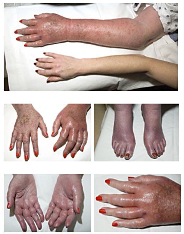
ErythromelalgiaErythromelalgiaA peripheral arterial disease that is characterized by the triad of erythema, burning pain, and increased skin temperature of the extremities (or red, painful extremities). Erythromelalgia may be classified as primary or idiopathic, familial or non-familial. Secondary erythromelalgia is associated with other diseases, the most common being myeloproliferative disorders.Polycythemia Vera, a burning painPainAn unpleasant sensation induced by noxious stimuli which are detected by nerve endings of nociceptive neurons.Pain: Types and Pathways in the feet or hands accompanied by erythemaErythemaRedness of the skin produced by congestion of the capillaries. This condition may result from a variety of disease processes.Chalazion, pallor, or cyanosisCyanosisA bluish or purplish discoloration of the skin and mucous membranes due to an increase in the amount of deoxygenated hemoglobin in the blood or a structural defect in the hemoglobin molecule.Pulmonary Examination, in the presence of palpable pulses (29% of cases)
PruritusPruritusAn intense itching sensation that produces the urge to rub or scratch the skin to obtain relief.Atopic Dermatitis (Eczema), especially after a hot bath or shower (36%)
Gouty tophiTophiMsu deposit in the soft tissue and synovium.Gout
SplenomegalySplenomegalySplenomegaly is pathologic enlargement of the spleen that is attributable to numerous causes, including infections, hemoglobinopathies, infiltrative processes, and outflow obstruction of the portal vein. Splenomegaly (36%)
Peptic ulcerPeptic ulcerPeptic ulcer disease (PUD) refers to the full-thickness ulcerations of duodenal or gastric mucosa. The ulcerations form when exposure to acid and digestive enzymes overcomes mucosal defense mechanisms. The most common etiologies include Helicobacter pylori (H. pylori) infection and prolonged use of non-steroidal anti-inflammatory drugs (NSAIDs). Peptic Ulcer Disease disease
Cardiovascular:
HypertensionHypertensionHypertension, or high blood pressure, is a common disease that manifests as elevated systemic arterial pressures. Hypertension is most often asymptomatic and is found incidentally as part of a routine physical examination or during triage for an unrelated medical encounter. Hypertension (46%)
Angina pectorisAngina pectorisThe symptom of paroxysmal pain consequent to myocardial ischemia usually of distinctive character, location and radiation. It is thought to be provoked by a transient stressful situation during which the oxygen requirements of the myocardium exceed that supplied by the coronary circulation.Stable and Unstable Angina
Stroke
Intermittent claudicationIntermittent claudicationA symptom complex characterized by pain and weakness in skeletal muscle group associated with exercise, such as leg pain and weakness brought on by walking. Such muscle limpness disappears after a brief rest and is often relates to arterial stenosis; muscle ischemia; and accumulation of lactate.Thromboangiitis Obliterans (Buerger Disease)
EcchymosisEcchymosisExtravasation of blood into the skin, resulting in a nonelevated, rounded or irregular, blue or purplish patch, larger than a petechia.Orbital Fractures
Gingival bleeding
Major hemorrhage (4%)
Erythromelalgia: A sensation of burning pain in the feet or hands accompanied by erythema, pallor, or cyanosis, in the presence of palpable pulses
Image: “Erythromelalgia” by Herbert L. Fred, MD and Hendrik A. van Dijk. License: CC BY 2.0
Diagnosis
PolycythemiaPolycythemiaAn increase in the total red cell mass of the blood.Renal Cell Carcinoma vera is suspected in patientsPatientsIndividuals participating in the health care system for the purpose of receiving therapeutic, diagnostic, or preventive procedures.Clinician–Patient Relationship with characteristic physical findings and/or increased levels of hemoglobin and hematocritHematocritThe volume of packed red blood cells in a blood specimen. The volume is measured by centrifugation in a tube with graduated markings, or with automated blood cell counters. It is an indicator of erythrocyte status in disease. For example, anemia shows a low value; polycythemia, a high value.Neonatal Polycythemia on a CBC. Testing for EPOEPOGlycoprotein hormone, secreted chiefly by the kidney in the adult and the liver in the fetus, that acts on erythroid stem cells of the bone marrow to stimulate proliferation and differentiation.Erythrocytes: Histology is indicated next (unless evaluation points to volume depletionVolume depletionVolume status is a balance between water and solutes, the majority of which is Na. Volume depletion refers to a loss of both water and Na, whereas dehydration refers only to a loss of water. Volume depletion can be caused by GI losses, renal losses, bleeding, poor oral Na intake, or third spacing of fluids.Volume Depletion and Dehydration alone).[8]
Initial evaluation[14]
Repeat CBC (especially if volume contraction is evident)
ElectrolytesElectrolytesElectrolytes are mineral salts that dissolve in water and dissociate into charged particles called ions, which can be either be positively (cations) or negatively (anions) charged. Electrolytes are distributed in the extracellular and intracellular compartments in different concentrations. Electrolytes are essential for various basic life-sustaining functions.Electrolytes, renal and hepatic function, urinalysisUrinalysisExamination of urine by chemical, physical, or microscopic means. Routine urinalysis usually includes performing chemical screening tests, determining specific gravity, observing any unusual color or odor, screening for bacteriuria, and examining the sediment microscopically.Urinary Tract Infections (UTIs) in Children:
Detect underlying liverLiverThe liver is the largest gland in the human body. The liver is found in the superior right quadrant of the abdomen and weighs approximately 1.5 kilograms. Its main functions are detoxification, metabolism, nutrient storage (e.g., iron and vitamins), synthesis of coagulation factors, formation of bile, filtration, and storage of blood. Liver: Anatomy and/or kidney disease, which can lead to erythrocytosis
EPOEPOGlycoprotein hormone, secreted chiefly by the kidney in the adult and the liver in the fetus, that acts on erythroid stem cells of the bone marrow to stimulate proliferation and differentiation.Erythrocytes: Histology
Rule out secondary causes of polycythemiaPolycythemiaAn increase in the total red cell mass of the blood.Renal Cell Carcinoma (high EPOEPOGlycoprotein hormone, secreted chiefly by the kidney in the adult and the liver in the fetus, that acts on erythroid stem cells of the bone marrow to stimulate proliferation and differentiation.Erythrocytes: Histology levels)[14]
High altitude
Chronic hypoxiaHypoxiaSub-optimal oxygen levels in the ambient air of living organisms.Ischemic Cell Damage:
Paraneoplastic syndrome (renal cell carcinomaRenal cell carcinomaRenal cell carcinoma (RCC) is a tumor that arises from the lining of the renal tubular system within the renal cortex. Renal cell carcinoma is responsible for 80%-85% of all primary renal neoplasms. Most RCCs arise sporadically, but smoking, hypertension, and obesity are linked to its development. Renal Cell Carcinoma)
Other underlying conditions leading to ↑ hemoglobin and hematocritHematocritThe volume of packed red blood cells in a blood specimen. The volume is measured by centrifugation in a tube with graduated markings, or with automated blood cell counters. It is an indicator of erythrocyte status in disease. For example, anemia shows a low value; polycythemia, a high value.Neonatal Polycythemia (with raised EPOEPOGlycoprotein hormone, secreted chiefly by the kidney in the adult and the liver in the fetus, that acts on erythroid stem cells of the bone marrow to stimulate proliferation and differentiation.Erythrocytes: Histology), such as parathyroidParathyroidThe parathyroid glands are 2 pairs of small endocrine glands found in close proximity to the thyroid gland. The superior parathyroid glands are lodged within the parenchyma of the upper poles of the right and left thyroid lobes; the inferior parathyroid glands are close to the inferior tips or poles of the lobes.Parathyroid Glands: AnatomytumorTumorInflammation, cerebellar hemangiomaHemangiomaA vascular anomaly due to proliferation of blood vessels that forms a tumor-like mass. The common types involve capillaries and veins. It can occur anywhere in the body but is most frequently noticed in the skin and subcutaneous tissue.Imaging of the Liver and Biliary Tract, pheochromocytomaPheochromocytomaPheochromocytoma is a catecholamine-secreting tumor derived from chromaffin cells. The majority of tumors originate in the adrenal medulla, but they may also arise from sympathetic ganglia (also referred to as paraganglioma). Symptoms are associated with excessive catecholamine production and commonly include hypertension, tachycardia, headache, and sweating. Pheochromocytoma
Hemoglobin and hematocritHematocritThe volume of packed red blood cells in a blood specimen. The volume is measured by centrifugation in a tube with graduated markings, or with automated blood cell counters. It is an indicator of erythrocyte status in disease. For example, anemia shows a low value; polycythemia, a high value.Neonatal Polycythemia
LeukocytesLeukocytesWhite blood cells. These include granular leukocytes (basophils; eosinophils; and neutrophils) as well as non-granular leukocytes (lymphocytes and monocytes).White Myeloid Cells: Histology
PlateletsPlateletsPlatelets are small cell fragments involved in hemostasis. Thrombopoiesis takes place primarily in the bone marrow through a series of cell differentiation and is influenced by several cytokines. Platelets are formed after fragmentation of the megakaryocyte cytoplasm. Platelets: Histology
Uric acidUric acidAn oxidation product, via xanthine oxidase, of oxypurines such as xanthine and hypoxanthine. It is the final oxidation product of purine catabolism in humans and primates, whereas in most other mammals urate oxidase further oxidizes it to allantoin.Nephrolithiasis and B12 (not needed for diagnosis)
IronIronA metallic element with atomic symbol fe, atomic number 26, and atomic weight 55. 85. It is an essential constituent of hemoglobins; cytochromes; and iron-binding proteins. It plays a role in cellular redox reactions and in the transport of oxygen.Trace Elements
EPOEPOGlycoprotein hormone, secreted chiefly by the kidney in the adult and the liver in the fetus, that acts on erythroid stem cells of the bone marrow to stimulate proliferation and differentiation.Erythrocytes: Histology
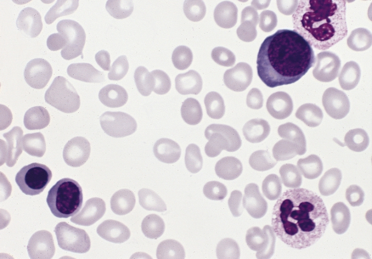
Peripheral smear: Findings depend on the stage at the time of diagnosis.
Erythrocytosis (excess RBCsRBCsErythrocytes, or red blood cells (RBCs), are the most abundant cells in the blood. While erythrocytes in the fetus are initially produced in the yolk sac then the liver, the bone marrow eventually becomes the main site of production.Erythrocytes: Histology)
Dacryocytes (teardrop-shaped RBCsRBCsErythrocytes, or red blood cells (RBCs), are the most abundant cells in the blood. While erythrocytes in the fetus are initially produced in the yolk sac then the liver, the bone marrow eventually becomes the main site of production.Erythrocytes: Histology)
Poikilocytosis (abnormally shaped RBCsRBCsErythrocytes, or red blood cells (RBCs), are the most abundant cells in the blood. While erythrocytes in the fetus are initially produced in the yolk sac then the liver, the bone marrow eventually becomes the main site of production.Erythrocytes: Histology)
Circulating nucleated RBCsRBCsErythrocytes, or red blood cells (RBCs), are the most abundant cells in the blood. While erythrocytes in the fetus are initially produced in the yolk sac then the liver, the bone marrow eventually becomes the main site of production.Erythrocytes: Histology
Hypochromia and microcytosis in the case of ironIronA metallic element with atomic symbol fe, atomic number 26, and atomic weight 55. 85. It is an essential constituent of hemoglobins; cytochromes; and iron-binding proteins. It plays a role in cellular redox reactions and in the transport of oxygen.Trace Elements deficiency
Thrombocytosis
LeukocytosisLeukocytosisA transient increase in the number of leukocytes in a body fluid.West Nile Virus in the absence of feverFeverFever is defined as a measured body temperature of at least 38°C (100.4°F). Fever is caused by circulating endogenous and/or exogenous pyrogens that increase levels of prostaglandin E2 in the hypothalamus. Fever is commonly associated with chills, rigors, sweating, and flushing of the skin. Fever or infection
Testing for JAK2, CALR, and LNK mutations (done sequentially)
Testing for JAK2 V617F:
Stage 1Stage 1Trypanosoma brucei/African trypanosomiasis investigation in patientsPatientsIndividuals participating in the health care system for the purpose of receiving therapeutic, diagnostic, or preventive procedures.Clinician–Patient Relationship with clinical features of PVPVPolycythemia vera (PV) is a chronic myeloproliferative neoplasm characterized by the overproduction of rbcs. In addition, the wbc and platelet counts are also increased, which differentiate pv from erythrocytosis seen with chronic hypoxia and other chronic conditions.Polycythemia Vera
Confirms diagnosis in majority of patientsPatientsIndividuals participating in the health care system for the purpose of receiving therapeutic, diagnostic, or preventive procedures.Clinician–Patient Relationship with PVPVPolycythemia vera (PV) is a chronic myeloproliferative neoplasm characterized by the overproduction of rbcs. In addition, the wbc and platelet counts are also increased, which differentiate pv from erythrocytosis seen with chronic hypoxia and other chronic conditions.Polycythemia Vera
Testing for JAK2 exon 12 mutationMutationGenetic mutations are errors in DNA that can cause protein misfolding and dysfunction. There are various types of mutations, including chromosomal, point, frameshift, and expansion mutations. Types of Mutations:
Obtained if with sustained elevated hemoglobin and hematocritHematocritThe volume of packed red blood cells in a blood specimen. The volume is measured by centrifugation in a tube with graduated markings, or with automated blood cell counters. It is an indicator of erythrocyte status in disease. For example, anemia shows a low value; polycythemia, a high value.Neonatal Polycythemia, subnormal EPOEPOGlycoprotein hormone, secreted chiefly by the kidney in the adult and the liver in the fetus, that acts on erythroid stem cells of the bone marrow to stimulate proliferation and differentiation.Erythrocytes: Histology, but JAK2 V617F–negative
PatientsPatientsIndividuals participating in the health care system for the purpose of receiving therapeutic, diagnostic, or preventive procedures.Clinician–Patient Relationship are often younger and have higher hemoglobin and lower white blood cell (WBC) and platelet countsPlatelet countsThe number of platelets per unit volume in a sample of venous blood.Coagulation Studies.
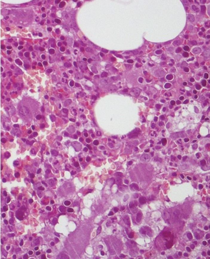
Bone marrowBone marrowThe soft tissue filling the cavities of bones. Bone marrow exists in two types, yellow and red. Yellow marrow is found in the large cavities of large bones and consists mostly of fat cells and a few primitive blood cells. Red marrow is a hematopoietic tissue and is the site of production of erythrocytes and granular leukocytes. Bone marrow is made up of a framework of connective tissue containing branching fibers with the frame being filled with marrow cells.Bone Marrow: Composition and HematopoiesisbiopsyBiopsyRemoval and pathologic examination of specimens from the living body.Ewing Sarcoma:
No bone marrowBone marrowThe soft tissue filling the cavities of bones. Bone marrow exists in two types, yellow and red. Yellow marrow is found in the large cavities of large bones and consists mostly of fat cells and a few primitive blood cells. Red marrow is a hematopoietic tissue and is the site of production of erythrocytes and granular leukocytes. Bone marrow is made up of a framework of connective tissue containing branching fibers with the frame being filled with marrow cells.Bone Marrow: Composition and Hematopoiesis findings absolutely differentiate PVPVPolycythemia vera (PV) is a chronic myeloproliferative neoplasm characterized by the overproduction of rbcs. In addition, the wbc and platelet counts are also increased, which differentiate pv from erythrocytosis seen with chronic hypoxia and other chronic conditions.Polycythemia Vera from other disorders of erythrocytosis.
Increased cellularity (versus fibrosisFibrosisAny pathological condition where fibrous connective tissue invades any organ, usually as a consequence of inflammation or other injury.Bronchiolitis Obliterans)
Typically shows panmyelosis (prominent erythroid, granulocytic, and megakaryocytic proliferation)
Large and clumped megakaryocytes
Occasional increase in reticulin fibers
Morphological bone marrow analysis for an individual with polycythemia vera confirms diagnosis of PV, with no evident change in bone marrow fibrosis or blast percentage, and no significant lymphoid infiltrate.
Image: “Erdheim-Chester Disease With Multiorgan Involvement, Following Polycythemia Vera: A Case Report” by Iurlo, A., et al. License: CC BY 4.0, cropped by Lecturio.
Blood smear of different RBC morphology in an individual with polycythemia vera:
3 red blood cell precursors are present and there is slight to moderate anisopoikilocytosis (Wright-Giemsa stain).
Image: “Polycythemia vera, blood smear” by The Armed Forces Institute of Pathology (AFIP). License: Public Domain
World Health Organization diagnostic criteria[10]
A diagnosis of PVPVPolycythemia vera (PV) is a chronic myeloproliferative neoplasm characterized by the overproduction of rbcs. In addition, the wbc and platelet counts are also increased, which differentiate pv from erythrocytosis seen with chronic hypoxia and other chronic conditions.Polycythemia Vera is made with the presence of either:
3 major criteria
1st 2 major criteria and the minor criterion
Major criteria:
Laboratory testing:
Hemoglobin > 16.5 g/dL in men, > 16.0 g/dL in women, OR
HematocritHematocritThe volume of packed red blood cells in a blood specimen. The volume is measured by centrifugation in a tube with graduated markings, or with automated blood cell counters. It is an indicator of erythrocyte status in disease. For example, anemia shows a low value; polycythemia, a high value.Neonatal Polycythemia > 49 % in men, > 48 % in women, OR
Increased red cell massMassThree-dimensional lesion that occupies a space within the breastImaging of the Breast (> 25 % above mean normal predicted value)
Bone marrowBone marrowThe soft tissue filling the cavities of bones. Bone marrow exists in two types, yellow and red. Yellow marrow is found in the large cavities of large bones and consists mostly of fat cells and a few primitive blood cells. Red marrow is a hematopoietic tissue and is the site of production of erythrocytes and granular leukocytes. Bone marrow is made up of a framework of connective tissue containing branching fibers with the frame being filled with marrow cells.Bone Marrow: Composition and HematopoiesisbiopsyBiopsyRemoval and pathologic examination of specimens from the living body.Ewing Sarcoma: prominent erythroid, granulocytic, and megakaryocytic proliferation (panmyelosis) with pleomorphicPleomorphicBacteroides, mature megakaryocytes
JAK2 V617F or JAK2 exon 12 mutationMutationGenetic mutations are errors in DNA that can cause protein misfolding and dysfunction. There are various types of mutations, including chromosomal, point, frameshift, and expansion mutations. Types of Mutations
Minor criterion:
EPOEPOGlycoprotein hormone, secreted chiefly by the kidney in the adult and the liver in the fetus, that acts on erythroid stem cells of the bone marrow to stimulate proliferation and differentiation.Erythrocytes: Histology level < normal reference range (helps differentiate from causes of secondary erythrocytosis, such as smokingSmokingWillful or deliberate act of inhaling and exhaling smoke from burning substances or agents held by hand.Interstitial Lung Diseases, sleep apneaSleep apneaRepeated cessation of breathing for > 10 seconds during sleep and results in sleep interruption, fatigue, and daytime sleepiness.Obstructive Sleep Apnea, exogenous testosteroneTestosteroneA potent androgenic steroid and major product secreted by the leydig cells of the testis. Its production is stimulated by luteinizing hormone from the pituitary gland. In turn, testosterone exerts feedback control of the pituitary LH and FSH secretion. Depending on the tissues, testosterone can be further converted to dihydrotestosterone or estradiol.Androgens and Antiandrogens)
Management and Prognosis
The goals of treatment are to reduce symptoms, prevent thrombosisThrombosisFormation and development of a thrombus or blood clot in the blood vessel.Epidemic Typhus, and improve qualityQualityActivities and programs intended to assure or improve the quality of care in either a defined medical setting or a program. The concept includes the assessment or evaluation of the quality of care; identification of problems or shortcomings in the delivery of care; designing activities to overcome these deficiencies; and follow-up monitoring to ensure effectiveness of corrective steps.Quality Measurement and Improvement of life and survival by decreasing leukemic transformationTransformationChange brought about to an organism’s genetic composition by unidirectional transfer (transfection; transduction, genetic; conjugation, genetic, etc.) and incorporation of foreign DNA into prokaryotic or eukaryotic cells by recombination of part or all of that DNA into the cell’s genome.Bacteriology and progression to myelofibrosis. The following is based on US, UK, and European guidelines.
General management[5,9,11,13]
All patientsPatientsIndividuals participating in the health care system for the purpose of receiving therapeutic, diagnostic, or preventive procedures.Clinician–Patient Relationship:
PhlebotomyPhlebotomyThe techniques used to draw blood from a vein for diagnostic purposes or for treatment of certain blood disorders such as erythrocytosis, hemochromatosis, polycythemia vera, and porphyria cutanea tarda.Hereditary Hemochromatosis to reduce hematocritHematocritThe volume of packed red blood cells in a blood specimen. The volume is measured by centrifugation in a tube with graduated markings, or with automated blood cell counters. It is an indicator of erythrocyte status in disease. For example, anemia shows a low value; polycythemia, a high value.Neonatal Polycythemia to < 45%
Low-dose aspirinAspirinThe prototypical analgesic used in the treatment of mild to moderate pain. It has anti-inflammatory and antipyretic properties and acts as an inhibitor of cyclooxygenase which results in the inhibition of the biosynthesis of prostaglandins. Aspirin also inhibits platelet aggregation and is used in the prevention of arterial and venous thrombosis.Nonsteroidal Antiinflammatory Drugs (NSAIDs) 81 mg daily (unless contraindicated) to reduce symptoms of microvascular events
In patientsPatientsIndividuals participating in the health care system for the purpose of receiving therapeutic, diagnostic, or preventive procedures.Clinician–Patient Relationship with plateletsPlateletsPlatelets are small cell fragments involved in hemostasis. Thrombopoiesis takes place primarily in the bone marrow through a series of cell differentiation and is influenced by several cytokines. Platelets are formed after fragmentation of the megakaryocyte cytoplasm. Platelets: Histology > 1 million/µL, test for acquired von Willebrand syndrome (aVWS).
Those with aVWS should not receive aspirinAspirinThe prototypical analgesic used in the treatment of mild to moderate pain. It has anti-inflammatory and antipyretic properties and acts as an inhibitor of cyclooxygenase which results in the inhibition of the biosynthesis of prostaglandins. Aspirin also inhibits platelet aggregation and is used in the prevention of arterial and venous thrombosis.Nonsteroidal Antiinflammatory Drugs (NSAIDs), as they are at risk for aspirin-associated bleeding.
SmokingSmokingWillful or deliberate act of inhaling and exhaling smoke from burning substances or agents held by hand.Interstitial Lung Diseases cessation (if applicable)
Address other modifiable atherosclerotic cardiovascular disease risk factors (e.g., diabetesDiabetesDiabetes mellitus (DM) is a metabolic disease characterized by hyperglycemia and dysfunction of the regulation of glucose metabolism by insulin. Type 1 DM is diagnosed mostly in children and young adults as the result of autoimmune destruction of β cells in the pancreas and the resulting lack of insulin. Type 2 DM has a significant association with obesity and is characterized by insulin resistance.Diabetes Mellitus, hyperlipidemia, hypertensionHypertensionHypertension, or high blood pressure, is a common disease that manifests as elevated systemic arterial pressures. Hypertension is most often asymptomatic and is found incidentally as part of a routine physical examination or during triage for an unrelated medical encounter. Hypertension, obesityObesityObesity is a condition associated with excess body weight, specifically with the deposition of excessive adipose tissue. Obesity is considered a global epidemic. Major influences come from the western diet and sedentary lifestyles, but the exact mechanisms likely include a mixture of genetic and environmental factors. Obesity)
Approach:
Low-risk patientsPatientsIndividuals participating in the health care system for the purpose of receiving therapeutic, diagnostic, or preventive procedures.Clinician–Patient Relationship (patientsPatientsIndividuals participating in the health care system for the purpose of receiving therapeutic, diagnostic, or preventive procedures.Clinician–Patient Relationship ≤ 60 years old and with no history of thrombosisThrombosisFormation and development of a thrombus or blood clot in the blood vessel.Epidemic Typhus) are treated with phlebotomyPhlebotomyThe techniques used to draw blood from a vein for diagnostic purposes or for treatment of certain blood disorders such as erythrocytosis, hemochromatosis, polycythemia vera, and porphyria cutanea tarda.Hereditary Hemochromatosis and aspirinAspirinThe prototypical analgesic used in the treatment of mild to moderate pain. It has anti-inflammatory and antipyretic properties and acts as an inhibitor of cyclooxygenase which results in the inhibition of the biosynthesis of prostaglandins. Aspirin also inhibits platelet aggregation and is used in the prevention of arterial and venous thrombosis.Nonsteroidal Antiinflammatory Drugs (NSAIDs).
High-risk patientsPatientsIndividuals participating in the health care system for the purpose of receiving therapeutic, diagnostic, or preventive procedures.Clinician–Patient Relationship (patientsPatientsIndividuals participating in the health care system for the purpose of receiving therapeutic, diagnostic, or preventive procedures.Clinician–Patient Relationship > 60 years old and with history of thrombosisThrombosisFormation and development of a thrombus or blood clot in the blood vessel.Epidemic Typhus) are recommended to also have cytoreductive therapy in addition to phlebotomyPhlebotomyThe techniques used to draw blood from a vein for diagnostic purposes or for treatment of certain blood disorders such as erythrocytosis, hemochromatosis, polycythemia vera, and porphyria cutanea tarda.Hereditary Hemochromatosis and aspirinAspirinThe prototypical analgesic used in the treatment of mild to moderate pain. It has anti-inflammatory and antipyretic properties and acts as an inhibitor of cyclooxygenase which results in the inhibition of the biosynthesis of prostaglandins. Aspirin also inhibits platelet aggregation and is used in the prevention of arterial and venous thrombosis.Nonsteroidal Antiinflammatory Drugs (NSAIDs).
Cytoreductive agents[5,9,11,14]
HydroxyureaHydroxyureaAn antineoplastic agent that inhibits DNA synthesis through the inhibition of ribonucleoside diphosphate reductase.Antimetabolite Chemotherapy (hydroxycarbamideHydroxycarbamideAn antineoplastic agent that inhibits DNA synthesis through the inhibition of ribonucleoside diphosphate reductase.Antimetabolite Chemotherapy in UK):
1st-line cytoreductive agent
Dosing:
Initial: 500 mg twice daily
Titrate to hematocritHematocritThe volume of packed red blood cells in a blood specimen. The volume is measured by centrifugation in a tube with graduated markings, or with automated blood cell counters. It is an indicator of erythrocyte status in disease. For example, anemia shows a low value; polycythemia, a high value.Neonatal Polycythemia target
InterferonsInterferonsInterferon (IFN) is a cytokine with antiviral properties (it interferes with viral infections) and various roles in immunoregulation. The different types are type I IFN (IFN-ɑ and IFN-β), type II IFN (IFN-ɣ), and type III IFN (IFN-ƛ). Interferons:[8]
Restore normal RBC counts; effect may persist after discontinuing.
Decreases JAK2 V617F alleleAlleleVariant forms of the same gene, occupying the same locus on homologous chromosomes, and governing the variants in production of the same gene product.Basic Terms of Genetics burden → improved myelofibrosis-free survival rate compared to hydroxyureaHydroxyureaAn antineoplastic agent that inhibits DNA synthesis through the inhibition of ribonucleoside diphosphate reductase.Antimetabolite Chemotherapy or phlebotomyPhlebotomyThe techniques used to draw blood from a vein for diagnostic purposes or for treatment of certain blood disorders such as erythrocytosis, hemochromatosis, polycythemia vera, and porphyria cutanea tarda.Hereditary Hemochromatosis
May lower the rate of disease progression to myelofibrosis or leukemia
Used in younger patientsPatientsIndividuals participating in the health care system for the purpose of receiving therapeutic, diagnostic, or preventive procedures.Clinician–Patient Relationship (< 40 years) or those who may become pregnant, who require further therapy beyond phlebotomyPhlebotomyThe techniques used to draw blood from a vein for diagnostic purposes or for treatment of certain blood disorders such as erythrocytosis, hemochromatosis, polycythemia vera, and porphyria cutanea tarda.Hereditary Hemochromatosis
Options:
Ropeginterferon alfa-2b (brand name Besremi):
Initially 100 mcg SC every 2 weeks
Increase dose every 2 weeks until Hct < 45%, plateletsPlateletsPlatelets are small cell fragments involved in hemostasis. Thrombopoiesis takes place primarily in the bone marrow through a series of cell differentiation and is influenced by several cytokines. Platelets are formed after fragmentation of the megakaryocyte cytoplasm. Platelets: Histology < 400,000/µL, and leukocytesLeukocytesWhite blood cells. These include granular leukocytes (basophils; eosinophils; and neutrophils) as well as non-granular leukocytes (lymphocytes and monocytes).White Myeloid Cells: Histology < 10,000/µL (maximum dose, 500 µg/dose)
Only for patientsPatientsIndividuals participating in the health care system for the purpose of receiving therapeutic, diagnostic, or preventive procedures.Clinician–Patient Relationship with severe pruritusPruritusAn intense itching sensation that produces the urge to rub or scratch the skin to obtain relief.Atopic Dermatitis (Eczema), symptomatic splenomegalySplenomegalySplenomegaly is pathologic enlargement of the spleen that is attributable to numerous causes, including infections, hemoglobinopathies, infiltrative processes, and outflow obstruction of the portal vein. Splenomegaly, or myelofibrosis that fails to respond to hydroxyureaHydroxyureaAn antineoplastic agent that inhibits DNA synthesis through the inhibition of ribonucleoside diphosphate reductase.Antimetabolite Chemotherapy
Dosing:
10 mg orally twice daily for 4 weeks
If needed, titrate by increasing dose in increments of 5 mg twice daily (no more frequently than every 2 weeks) to a maximum of 25 mg twice daily.
BusulfanBusulfanAn alkylating agent having a selective immunosuppressive effect on bone marrow. It has been used in the palliative treatment of chronic myeloid leukemia, but although symptomatic relief is provided, no permanent remission is brought about. According to the fourth annual report on carcinogens, busulfan is listed as a known carcinogen.Bronchiolitis Obliterans:
Alkylating agentAlkylating AgentAlkylating agents are cell cycle-independent antineoplastic drugs that work primarily by binding alkyl groups to various parts of DNA. The overall action produces cross-linking of DNA, leading to inhibition of DNA replication and DNA damage. Alkylating Agents and Platinum associated with cytopenia, pulmonary fibrosisFibrosisAny pathological condition where fibrous connective tissue invades any organ, usually as a consequence of inflammation or other injury.Bronchiolitis Obliterans, marrow aplasiaAplasiaCranial Nerve Palsies
Reserved for older patientsPatientsIndividuals participating in the health care system for the purpose of receiving therapeutic, diagnostic, or preventive procedures.Clinician–Patient Relationship (> 60 years old) who have PVPVPolycythemia vera (PV) is a chronic myeloproliferative neoplasm characterized by the overproduction of rbcs. In addition, the wbc and platelet counts are also increased, which differentiate pv from erythrocytosis seen with chronic hypoxia and other chronic conditions.Polycythemia Vera that has not responded to hydroxyureaHydroxyureaAn antineoplastic agent that inhibits DNA synthesis through the inhibition of ribonucleoside diphosphate reductase.Antimetabolite Chemotherapy and/or interferon
Initial dose is 2–4 mg a day.
PrognosisPrognosisA prediction of the probable outcome of a disease based on a individual’s condition and the usual course of the disease as seen in similar situations.Non-Hodgkin Lymphomas and complications[11]
ThrombosisThrombosisFormation and development of a thrombus or blood clot in the blood vessel.Epidemic Typhus is the most common cause of morbidityMorbidityThe proportion of patients with a particular disease during a given year per given unit of population.Measures of Health Status and death, followed by the complications of myelofibrosis and leukemic transformationTransformationChange brought about to an organism’s genetic composition by unidirectional transfer (transfection; transduction, genetic; conjugation, genetic, etc.) and incorporation of foreign DNA into prokaryotic or eukaryotic cells by recombination of part or all of that DNA into the cell’s genome.Bacteriology.
About 10%–30% of patientsPatientsIndividuals participating in the health care system for the purpose of receiving therapeutic, diagnostic, or preventive procedures.Clinician–Patient Relationship progress to a syndrome compatible with primary myelofibrosisPrimary myelofibrosisPrimary myelofibrosis (PMF) is a myeloproliferative neoplasm characterized by chronic myeloproliferation with nonclonal fibroblastic deposition, resulting in bone marrow fibrosis. The abnormality stems from genetic mutations of the hematopoietic stem cells (typically, JAK2 mutation). Primary symptoms are anemia and extramedullary hematopoiesis,.Primary Myelofibrosis but with better survival.
Leukemic transformationTransformationChange brought about to an organism’s genetic composition by unidirectional transfer (transfection; transduction, genetic; conjugation, genetic, etc.) and incorporation of foreign DNA into prokaryotic or eukaryotic cells by recombination of part or all of that DNA into the cell’s genome.Bacteriology from PVPVPolycythemia vera (PV) is a chronic myeloproliferative neoplasm characterized by the overproduction of rbcs. In addition, the wbc and platelet counts are also increased, which differentiate pv from erythrocytosis seen with chronic hypoxia and other chronic conditions.Polycythemia Vera occurs in about 2% of patientsPatientsIndividuals participating in the health care system for the purpose of receiving therapeutic, diagnostic, or preventive procedures.Clinician–Patient Relationship at 10 years to about 8% at 20 years.[12,13]
Differential Diagnosis
Secondary polycythemiaSecondary PolycythemiaPolycythemia Vera: an increase in RBCsRBCsErythrocytes, or red blood cells (RBCs), are the most abundant cells in the blood. While erythrocytes in the fetus are initially produced in the yolk sac then the liver, the bone marrow eventually becomes the main site of production.Erythrocytes: Histology due to chronic hypoxemiaHypoxemiaNeonatal Respiratory Distress Syndrome, familial erythrocythemia, paraneoplastic syndromesParaneoplastic syndromesParaneoplastic syndromes are a heterogeneous group of disorders caused by an abnormal immune response to a neoplasm. The substances produced are not due to the direct effect of the tumor, such as metastasis, mass effect, or invasion. Antibodies, hormones, cytokines, and other substances are generated and affect multiple organ systems. Paraneoplastic Syndromes, or relative polycythemiaPolycythemiaAn increase in the total red cell mass of the blood.Renal Cell Carcinoma due to contraction of plasmaPlasmaThe residual portion of blood that is left after removal of blood cells by centrifugation without prior blood coagulation.Transfusion Products volume as seen with dehydrationDehydrationThe condition that results from excessive loss of water from a living organism.Volume Depletion and Dehydration.
Essential thrombocytosis (primary thrombocythemia): a nonreactive, chronic myeloproliferative disorder in which excessive proliferation of megakaryocytes and plateletsPlateletsPlatelets are small cell fragments involved in hemostasis. Thrombopoiesis takes place primarily in the bone marrow through a series of cell differentiation and is influenced by several cytokines. Platelets are formed after fragmentation of the megakaryocyte cytoplasm. Platelets: Histology is seen. PatientsPatientsIndividuals participating in the health care system for the purpose of receiving therapeutic, diagnostic, or preventive procedures.Clinician–Patient Relationship are largely asymptomatic but may develop thrombosisThrombosisFormation and development of a thrombus or blood clot in the blood vessel.Epidemic Typhus and bleeding.
Chronic myeloid leukemiaChronic myeloid leukemiaChronic myeloid leukemia is a malignant proliferation of the granulocytic cell line characterized by a fairly normal differentiation. The underlying genetic abnormality is the Philadelphia chromosome, an abbreviated chromosome 22, resulting from reciprocal (9;22)(q34;q11) translocation. Chronic Myeloid Leukemia: a myeloproliferative disorder characterized by an increase in WBCs, including an increased number of granulocytesGranulocytesLeukocytes with abundant granules in the cytoplasm. They are divided into three groups according to the staining properties of the granules: neutrophilic, eosinophilic, and basophilic. Mature granulocytes are the neutrophils; eosinophils; and basophils.White Myeloid Cells: Histology and their immature precursors. Blast cells are also seen occasionally.
Primary myelofibrosisPrimary myelofibrosisPrimary myelofibrosis (PMF) is a myeloproliferative neoplasm characterized by chronic myeloproliferation with nonclonal fibroblastic deposition, resulting in bone marrow fibrosis. The abnormality stems from genetic mutations of the hematopoietic stem cells (typically, JAK2 mutation). Primary symptoms are anemia and extramedullary hematopoiesis,.Primary Myelofibrosis: a clonal disorder arising from the neoplastic transformationTransformationChange brought about to an organism’s genetic composition by unidirectional transfer (transfection; transduction, genetic; conjugation, genetic, etc.) and incorporation of foreign DNA into prokaryotic or eukaryotic cells by recombination of part or all of that DNA into the cell’s genome.Bacteriology of early hematopoietic stem cellsHematopoietic stem cellsProgenitor cells from which all blood cells derived. They are found primarily in the bone marrow and also in small numbers in the peripheral blood.Bone Marrow: Composition and Hematopoiesis. Primary myelofibrosisPrimary myelofibrosisPrimary myelofibrosis (PMF) is a myeloproliferative neoplasm characterized by chronic myeloproliferation with nonclonal fibroblastic deposition, resulting in bone marrow fibrosis. The abnormality stems from genetic mutations of the hematopoietic stem cells (typically, JAK2 mutation). Primary symptoms are anemia and extramedullary hematopoiesis,.Primary Myelofibrosis is characterized by anemiaAnemiaAnemia is a condition in which individuals have low Hb levels, which can arise from various causes. Anemia is accompanied by a reduced number of RBCs and may manifest with fatigue, shortness of breath, pallor, and weakness. Subtypes are classified by the size of RBCs, chronicity, and etiology. Anemia: Overview and Types, bone marrow fibrosisBone Marrow FibrosisPrimary Myelofibrosis, extramedullary hematopoiesisExtramedullary HematopoiesisPrimary Myelofibrosis, leukoerythroblastosis, teardrop-shaped RBCsRBCsErythrocytes, or red blood cells (RBCs), are the most abundant cells in the blood. While erythrocytes in the fetus are initially produced in the yolk sac then the liver, the bone marrow eventually becomes the main site of production.Erythrocytes: Histology, and hepatosplenomegalyHepatosplenomegalyCytomegalovirus.
EPOEPOGlycoprotein hormone, secreted chiefly by the kidney in the adult and the liver in the fetus, that acts on erythroid stem cells of the bone marrow to stimulate proliferation and differentiation.Erythrocytes: HistologyreceptorReceptorReceptors are proteins located either on the surface of or within a cell that can bind to signaling molecules known as ligands (e.g., hormones) and cause some type of response within the cell.Receptors mutations: a rare familial disorder that can mimic the basic findings of PVPVPolycythemia vera (PV) is a chronic myeloproliferative neoplasm characterized by the overproduction of rbcs. In addition, the wbc and platelet counts are also increased, which differentiate pv from erythrocytosis seen with chronic hypoxia and other chronic conditions.Polycythemia Vera (elevated hemoglobin and hematocritHematocritThe volume of packed red blood cells in a blood specimen. The volume is measured by centrifugation in a tube with graduated markings, or with automated blood cell counters. It is an indicator of erythrocyte status in disease. For example, anemia shows a low value; polycythemia, a high value.Neonatal Polycythemia with low serum EPOEPOGlycoprotein hormone, secreted chiefly by the kidney in the adult and the liver in the fetus, that acts on erythroid stem cells of the bone marrow to stimulate proliferation and differentiation.Erythrocytes: Histology). A positive family historyFamily HistoryAdult Health Maintenance, early age at disease onset, and the lack of PV-associated clinical findings can help distinguish EPOEPOGlycoprotein hormone, secreted chiefly by the kidney in the adult and the liver in the fetus, that acts on erythroid stem cells of the bone marrow to stimulate proliferation and differentiation.Erythrocytes: HistologyreceptorReceptorReceptors are proteins located either on the surface of or within a cell that can bind to signaling molecules known as ligands (e.g., hormones) and cause some type of response within the cell.Receptors mutations from primary PVPrimary PvPolycythemia Vera.
Billing and Coding
Diagnosis Codes:
This code is used to diagnose PolycythemiaPolycythemiaAn increase in the total red cell mass of the blood.Renal Cell Carcinoma Vera (PVPVPolycythemia vera (PV) is a chronic myeloproliferative neoplasm characterized by the overproduction of rbcs. In addition, the wbc and platelet counts are also increased, which differentiate pv from erythrocytosis seen with chronic hypoxia and other chronic conditions.Polycythemia Vera), a chronic myeloproliferative neoplasm characterized by the overproduction of red blood cellsRed blood cellsErythrocytes, or red blood cells (RBCs), are the most abundant cells in the blood. While erythrocytes in the fetus are initially produced in the yolk sac then the liver, the bone marrow eventually becomes the main site of production.Erythrocytes: Histology, which thickens the blood and increases the risk of clots.
This code is for the JAK2 V617F mutationMutationGenetic mutations are errors in DNA that can cause protein misfolding and dysfunction. There are various types of mutations, including chromosomal, point, frameshift, and expansion mutations. Types of Mutations analysis, a genetic test that is a major diagnostic criterion for PVPVPolycythemia vera (PV) is a chronic myeloproliferative neoplasm characterized by the overproduction of rbcs. In addition, the wbc and platelet counts are also increased, which differentiate pv from erythrocytosis seen with chronic hypoxia and other chronic conditions.Polycythemia Vera, as this mutationMutationGenetic mutations are errors in DNA that can cause protein misfolding and dysfunction. There are various types of mutations, including chromosomal, point, frameshift, and expansion mutations. Types of Mutations is present in over 95% of cases.
Coding System
Code
Description
CPT
81270
JAK2 (Janus kinase 2) geneGeneA category of nucleic acid sequences that function as units of heredity and which code for the basic instructions for the development, reproduction, and maintenance of organisms.Basic Terms of Genetics analysis
Procedures/Interventions:
This code is for therapeutic phlebotomyPhlebotomyThe techniques used to draw blood from a vein for diagnostic purposes or for treatment of certain blood disorders such as erythrocytosis, hemochromatosis, polycythemia vera, and porphyria cutanea tarda.Hereditary Hemochromatosis, the primary treatment for PVPVPolycythemia vera (PV) is a chronic myeloproliferative neoplasm characterized by the overproduction of rbcs. In addition, the wbc and platelet counts are also increased, which differentiate pv from erythrocytosis seen with chronic hypoxia and other chronic conditions.Polycythemia Vera, which involves regularly removing blood to reduce the red blood cell massMassThree-dimensional lesion that occupies a space within the breastImaging of the Breast and maintain a target hematocritHematocritThe volume of packed red blood cells in a blood specimen. The volume is measured by centrifugation in a tube with graduated markings, or with automated blood cell counters. It is an indicator of erythrocyte status in disease. For example, anemia shows a low value; polycythemia, a high value.Neonatal Polycythemia level.
Coding System
Code
Description
CPT
99195
PhlebotomyPhlebotomyThe techniques used to draw blood from a vein for diagnostic purposes or for treatment of certain blood disorders such as erythrocytosis, hemochromatosis, polycythemia vera, and porphyria cutanea tarda.Hereditary Hemochromatosis, therapeutic (separate procedure)
Medications:
This code is for hydroxyureaHydroxyureaAn antineoplastic agent that inhibits DNA synthesis through the inhibition of ribonucleoside diphosphate reductase.Antimetabolite Chemotherapy, a cytoreductive agent used in high-risk PVPVPolycythemia vera (PV) is a chronic myeloproliferative neoplasm characterized by the overproduction of rbcs. In addition, the wbc and platelet counts are also increased, which differentiate pv from erythrocytosis seen with chronic hypoxia and other chronic conditions.Polycythemia VerapatientsPatientsIndividuals participating in the health care system for the purpose of receiving therapeutic, diagnostic, or preventive procedures.Clinician–Patient Relationship to lower blood cell counts and reduce the risk of thrombosisThrombosisFormation and development of a thrombus or blood clot in the blood vessel.Epidemic Typhus.
Coding System
Code
Description
RxNorm
5526
HydroxyureaHydroxyureaAn antineoplastic agent that inhibits DNA synthesis through the inhibition of ribonucleoside diphosphate reductase.Antimetabolite Chemotherapy (ingredient)
Tefferi, A., et al. (2013). Survival and prognosis among 1545 patients with contemporary polycythemia vera: an international study. Leukemia, 27(9),1874–1881. https://pubmed.ncbi.nlm.nih.gov/23739289/
Tefferi, A., Barbui, T. (2020). Polycythemia vera and essential thrombocythemia: 2021 update on diagnosis, risk-stratification and management. American Journal of Hematology, 95(12), 1599–1613. https://doi.org/10.1002/ajh.26008
Regimbeau, M., Mary, R., Hermetet, F., Girodon, F. (2022). Genetic background of polycythemia vera. Genes, 13(4), 637. https://doi.org/10.3390/genes13040637
Kiladjian, J.-J., Klade, C., et al. (2022). Long-term outcomes of polycythemia vera patients treated with ropeginterferon alfa-2b. Leukemia, 36(5), 1408–1411. https://doi.org/10.1038/s41375-022-01528-x
Benevolo, G., Vassallo, F., Urbino, I., Giai, V. (2021). Polycythemia vera (PV): update on emerging treatment options. Therapeutics and Clinical Risk Management, 17, 209–221. https://doi.org/10.2147/TCRM.S213020
Arber, D. A., Orazi, A., Hasserjian, R., Thiele, J., Borowitz, M. J., Le Beau, M. M., Bloomfield, C. D., Cazzola, M., Vardiman, J. W. (2016). The 2016 revision to the World Health Organization classification of myeloid neoplasms and acute leukemia. Blood, 127(20), 2391–2405. https://doi.org/10.1182/blood-2016-03-643544
Gotlib, J. (2019). Polycythemia vera, essential thrombocythemia, and primary myelofibrosis. In Crow, M.K., et al. (Eds.), Goldman-Cecil Medicine (26th ed., vol. 1, pp. 1085–1093).
Luque Paz, D., Jouanneau-Courville, R., et al. (2020). Leukemic evolution of polycythemia vera and essential thrombocythemia: genomic profiles predict time to transformation. Blood Advances, 4(19), 4887–4897. https://doi.org/10.1182/bloodadvances.2020002271
McMullin, M. F., Harrison, C. N., et al. (2019). A guideline for the diagnosis and management of polycythaemia vera: a British Society for Haematology Guideline. British Journal of Haematology, 184(2), 176–191. https://doi.org/10.1111/bjh.15648
Create your free account or log in to continue reading!