Diagnostic procedures in gynecology are useful in identifying the presence of disease, determining the progression of disease, and monitoring the response of the organs to treatment. The major diagnostic procedures include speculum examinations, sonography (ultrasound), colposcopy, cervical biopsy and endocervical curettage, loop electrosurgical excision procedures, vulvar biopsy, endometrial biopsy, hysteroscopy, and hysterosalpingography (HSG). All of these procedures can be performed in the office setting or in a radiology suite, though in certain situations they are performed in the OR if more sedation or increased monitoring is required.
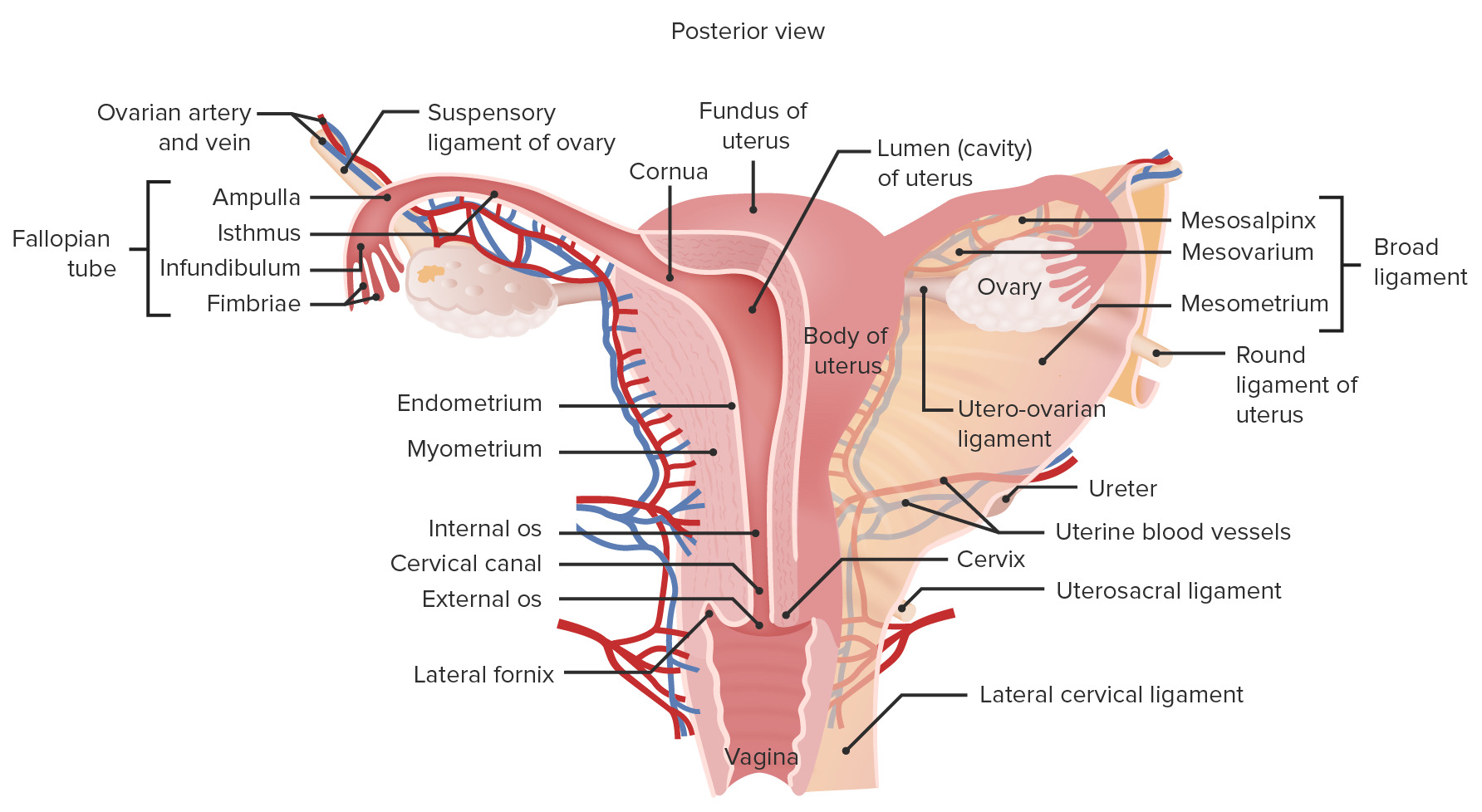
The female reproductive organs are split into the lower and upper genital tracts.
Lower genital tract
Includes:
CervixCervixThe uterus, cervix, and fallopian tubes are part of the internal female reproductive system. The most inferior portion of the uterus is the cervix, which connects the uterine cavity to the vagina. Externally, the cervix is lined by stratified squamous cells; however, the cervical canal is lined by columnar epithelium.Uterus, Cervix, and Fallopian Tubes: Anatomy
VaginaVaginaThe vagina is the female genital canal, extending from the vulva externally to the cervix uteri internally. The structures have sexual, reproductive, and urinary functions and a rich blood supply, mainly arising from the internal iliac artery.Vagina, Vulva, and Pelvic Floor: Anatomy
VulvaVulvaThe vulva is the external genitalia of the female and includes the mons pubis, labia majora, labia minora, clitoris, vestibule, vestibular bulb, and greater vestibular glands. Vagina, Vulva, and Pelvic Floor: Anatomy
Types of procedures used to assess the lower tract:
Speculum exam
ColposcopyColposcopyThe examination, therapy or surgery of the cervix and vagina by means of a specially designed endoscope introduced vaginally.Cervical Cancer Screening
Cervical biopsyBiopsyRemoval and pathologic examination of specimens from the living body.Ewing Sarcoma
Endocervical curettageCurettageA scraping, usually of the interior of a cavity or tract, for removal of new growth or other abnormal tissue, or to obtain material for tissue diagnosis. It is performed with a curet (curette), a spoon-shaped instrument designed for that purpose.Benign Bone Tumors
Loop electrosurgical excision procedure
Vulvar biopsyBiopsyRemoval and pathologic examination of specimens from the living body.Ewing Sarcoma
Upper genital tract
Includes:
UterusUterusThe uterus, cervix, and fallopian tubes are part of the internal female reproductive system. The uterus has a thick wall made of smooth muscle (the myometrium) and an inner mucosal layer (the endometrium). The most inferior portion of the uterus is the cervix, which connects the uterine cavity to the vagina.Uterus, Cervix, and Fallopian Tubes: Anatomy
Fallopian tubesFallopian tubesThe uterus, cervix, and fallopian tubes are part of the internal female reproductive system. The fallopian tubes receive an ovum after ovulation and help move it and/or a fertilized embryo toward the uterus via ciliated cells lining the tubes and peristaltic movements of its smooth muscle. Uterus, Cervix, and Fallopian Tubes: Anatomy
OvariesOvariesOvaries are the paired gonads of the female reproductive system that contain haploid gametes known as oocytes. The ovaries are located intraperitoneally in the pelvis, just posterior to the broad ligament, and are connected to the pelvic sidewall and to the uterus by ligaments. These organs function to secrete hormones (estrogen and progesterone) and to produce the female germ cells (oocytes).Ovaries: Anatomy
Types of procedures used to assess the upper tract:
Sonography
Endometrial biopsyBiopsyRemoval and pathologic examination of specimens from the living body.Ewing Sarcoma
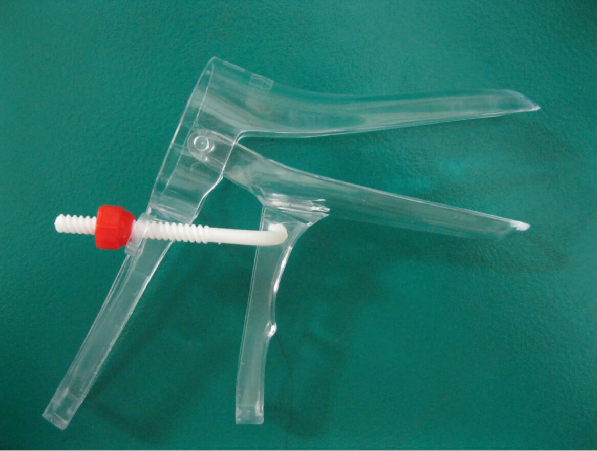
A speculumis a plastic or metal device used to mechanically open the vaginaVaginaThe vagina is the female genital canal, extending from the vulva externally to the cervix uteri internally. The structures have sexual, reproductive, and urinary functions and a rich blood supply, mainly arising from the internal iliac artery.Vagina, Vulva, and Pelvic Floor: Anatomy, allowing visualization and examination of the vaginal wall and ectocervix.
Pelvic painPainAn unpleasant sensation induced by noxious stimuli which are detected by nerve endings of nociceptive neurons.Pain: Types and Pathways
Abnormal discharge
Vaginal itching
Pelvic organ prolapsePelvic Organ ProlapsePelvic organ prolapse (POP) is a general term that refers to herniation of 1 or more pelvic organs (e.g., bladder, uterus, rectum) into the vaginal canal, and potentially all the way through the introitus. Weakness and insufficiency of the pelvic floor may result in POP.Pelvic Organ Prolapse
PregnancyPregnancyThe status during which female mammals carry their developing young (embryos or fetuses) in utero before birth, beginning from fertilization to birth.Pregnancy: Diagnosis, Physiology, and Care symptoms:
Loss of fluid
Concerns for preterm laborPreterm laborPreterm labor refers to regular uterine contractions leading to cervical change prior to 37 weeks of gestation; preterm birth refers to birth prior to 37 weeks of gestation. Preterm birth may be spontaneous due to preterm labor, preterm prelabor rupture of membranes (PPROM), or cervical insufficiency. Preterm Labor and Birth
Bleeding
As part of a routine wellness examination:
To obtain samples for cervical cancerCervical cancerCervical cancer, or invasive cervical carcinoma (ICC), is the 3rd most common cancer in women in the world, with > 50% of the cases being fatal. In the United States, ICC is the 13th most common cancer and the cause of < 3% of all cancer deaths due to the slow progression of precursor lesions and, more importantly, effective cancer screening. Cervical CancerscreeningScreeningPreoperative Care (e.g., a Pap smearPap smearCytological preparation of cells collected from a mucosal surface and stained with Papanicolaou stain.Cervical Cancer Screening)
Clinical utility when woman is asymptomatic and does notneed cervical cancerCervical cancerCervical cancer, or invasive cervical carcinoma (ICC), is the 3rd most common cancer in women in the world, with > 50% of the cases being fatal. In the United States, ICC is the 13th most common cancer and the cause of < 3% of all cancer deaths due to the slow progression of precursor lesions and, more importantly, effective cancer screening. Cervical CancerscreeningScreeningPreoperative Care is controversial → joint decision-making between woman and clinicianClinicianA physician, nurse practitioner, physician assistant, or another health professional who is directly involved in patient care and has a professional relationship with patients.Clinician–Patient Relationship is recommended
To gain access to the cervixCervixThe uterus, cervix, and fallopian tubes are part of the internal female reproductive system. The most inferior portion of the uterus is the cervix, which connects the uterine cavity to the vagina. Externally, the cervix is lined by stratified squamous cells; however, the cervical canal is lined by columnar epithelium.Uterus, Cervix, and Fallopian Tubes: Anatomy for other procedures
Note: A desire to initiate contraception without any other concerns does not require a speculum exam.
ContraindicationsContraindicationsA condition or factor associated with a recipient that makes the use of a drug, procedure, or physical agent improper or inadvisable. Contraindications may be absolute (life threatening) or relative (higher risk of complications in which benefits may outweigh risks).Noninvasive Ventilation
Preadolescent girls (if needed to evaluate for abuse or for other procedures → general anesthesiaGeneral anesthesiaProcedure in which patients are induced into an unconscious state through use of various medications so that they do not feel pain during surgery.Anesthesiology: History and Basic Concepts)
Severe immunosuppression: ↑ risk of bacterial translocation with speculum exams
Procedure
Placing the speculum:
Make sure you have all swabs, collection containers, and tools within reach prior to starting.
Wash your hands and wear gloves:
Tip: Avoid touching other items that are not part of the procedure once your hands are gloved (e.g., footFootThe foot is the terminal portion of the lower limb, whose primary function is to bear weight and facilitate locomotion. The foot comprises 26 bones, including the tarsal bones, metatarsal bones, and phalanges. The bones of the foot form longitudinal and transverse arches and are supported by various muscles, ligaments, and tendons.Foot: Anatomy stirrups, supply drawer, etcETCThe electron transport chain (ETC) sends electrons through a series of proteins, which generate an electrochemical proton gradient that produces energy in the form of adenosine triphosphate (ATP).Electron Transport Chain (ETC).)
Unlikely to cause true harm, but your hands will touch the vaginaVaginaThe vagina is the female genital canal, extending from the vulva externally to the cervix uteri internally. The structures have sexual, reproductive, and urinary functions and a rich blood supply, mainly arising from the internal iliac artery.Vagina, Vulva, and Pelvic Floor: Anatomy → don’t touch something “unclean” right before you do so
Select a speculum of appropriate size and shape.
Familiarize yourself with the speculum prior to the exam.
Lubricate the speculum with warm water or a water-soluble lubricant (some lubricants may interfere with sampling for cervical cytologyCervical cytologyA procedure in which ectocervical and endocervical cells are collected to evaluate the transformation zone (area at risk for cervical cancer).Cervical Cancer Screening and should be avoided).
Let the woman know that you are about to insert the speculum.
Tip: Gently touch the back of your handHandThe hand constitutes the distal part of the upper limb and provides the fine, precise movements needed in activities of daily living. It consists of 5 metacarpal bones and 14 phalanges, as well as numerous muscles innervated by the median and ulnar nerves. Hand: Anatomy to the woman’s inner thighThighThe thigh is the region of the lower limb found between the hip and the knee joint. There is a single bone in the thigh called the femur, which is surrounded by large muscles grouped into 3 fascial compartments. Thigh: Anatomy to let her adjust to the temperature/touch of your handHandThe hand constitutes the distal part of the upper limb and provides the fine, precise movements needed in activities of daily living. It consists of 5 metacarpal bones and 14 phalanges, as well as numerous muscles innervated by the median and ulnar nerves. Hand: Anatomy before touching the labia.
Manually spread the labia.
Introduce the speculum holding it at a downward angle, then slide it inward while applying gentle downward pressure along the posterior vaginal wall
Once the speculum is fully inserted, open it slowly and carefully.
Tip: Be careful not to open the blades prematurely.
This is very uncomfortable.
Typically, the fornixFornixVagina, Vulva, and Pelvic Floor: Anatomy of the vaginaVaginaThe vagina is the female genital canal, extending from the vulva externally to the cervix uteri internally. The structures have sexual, reproductive, and urinary functions and a rich blood supply, mainly arising from the internal iliac artery.Vagina, Vulva, and Pelvic Floor: Anatomy (upper portion around the cervixCervixThe uterus, cervix, and fallopian tubes are part of the internal female reproductive system. The most inferior portion of the uterus is the cervix, which connects the uterine cavity to the vagina. Externally, the cervix is lined by stratified squamous cells; however, the cervical canal is lined by columnar epithelium.Uterus, Cervix, and Fallopian Tubes: Anatomy) is able to comfortably tolerate much larger diameters than the introitus.
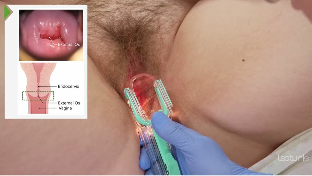
Rotate and adjust the speculum until it cups the cervixCervixThe uterus, cervix, and fallopian tubes are part of the internal female reproductive system. The most inferior portion of the uterus is the cervix, which connects the uterine cavity to the vagina. Externally, the cervix is lined by stratified squamous cells; however, the cervical canal is lined by columnar epithelium.Uterus, Cervix, and Fallopian Tubes: Anatomy and brings it into full view.
If there’s difficulty finding the cervixCervixThe uterus, cervix, and fallopian tubes are part of the internal female reproductive system. The most inferior portion of the uterus is the cervix, which connects the uterine cavity to the vagina. Externally, the cervix is lined by stratified squamous cells; however, the cervical canal is lined by columnar epithelium.Uterus, Cervix, and Fallopian Tubes: Anatomy, partially withdraw and try again.
Tip: Try to find the “smooth” surface of the cervixCervixThe uterus, cervix, and fallopian tubes are part of the internal female reproductive system. The most inferior portion of the uterus is the cervix, which connects the uterine cavity to the vagina. Externally, the cervix is lined by stratified squamous cells; however, the cervical canal is lined by columnar epithelium.Uterus, Cervix, and Fallopian Tubes: Anatomy hidden within the vaginal rugal folds → once found, use the speculum to “catch” the cervixCervixThe uterus, cervix, and fallopian tubes are part of the internal female reproductive system. The most inferior portion of the uterus is the cervix, which connects the uterine cavity to the vagina. Externally, the cervix is lined by stratified squamous cells; however, the cervical canal is lined by columnar epithelium.Uterus, Cervix, and Fallopian Tubes: Anatomy between the speculum blades
Note: The cervixCervixThe uterus, cervix, and fallopian tubes are part of the internal female reproductive system. The most inferior portion of the uterus is the cervix, which connects the uterine cavity to the vagina. Externally, the cervix is lined by stratified squamous cells; however, the cervical canal is lined by columnar epithelium.Uterus, Cervix, and Fallopian Tubes: Anatomy is often pointed directly downward.
May need to direct the tip of the speculum more posteriorly, then sweep upward while opening to catch the cervixCervixThe uterus, cervix, and fallopian tubes are part of the internal female reproductive system. The most inferior portion of the uterus is the cervix, which connects the uterine cavity to the vagina. Externally, the cervix is lined by stratified squamous cells; however, the cervical canal is lined by columnar epithelium.Uterus, Cervix, and Fallopian Tubes: Anatomy.
The bottom blade is longer than the top blade for this reason.
Less commonly, the cervixCervixThe uterus, cervix, and fallopian tubes are part of the internal female reproductive system. The most inferior portion of the uterus is the cervix, which connects the uterine cavity to the vagina. Externally, the cervix is lined by stratified squamous cells; however, the cervical canal is lined by columnar epithelium.Uterus, Cervix, and Fallopian Tubes: Anatomy points more upward (can be seen with retroverted/retroflexed uteri).
Position the light until you can visualize the cervixCervixThe uterus, cervix, and fallopian tubes are part of the internal female reproductive system. The most inferior portion of the uterus is the cervix, which connects the uterine cavity to the vagina. Externally, the cervix is lined by stratified squamous cells; however, the cervical canal is lined by columnar epithelium.Uterus, Cervix, and Fallopian Tubes: Anatomy well.
Maintain the open position of the speculum by tightening the thumbscrew on a metal speculum, or “clicking” it into place with a plastic speculum.
InspectionInspectionDermatologic Examination of the cervixCervixThe uterus, cervix, and fallopian tubes are part of the internal female reproductive system. The most inferior portion of the uterus is the cervix, which connects the uterine cavity to the vagina. Externally, the cervix is lined by stratified squamous cells; however, the cervical canal is lined by columnar epithelium.Uterus, Cervix, and Fallopian Tubes: Anatomy:
Envision the cervixCervixThe uterus, cervix, and fallopian tubes are part of the internal female reproductive system. The most inferior portion of the uterus is the cervix, which connects the uterine cavity to the vagina. Externally, the cervix is lined by stratified squamous cells; however, the cervical canal is lined by columnar epithelium.Uterus, Cervix, and Fallopian Tubes: Anatomy as a clock face, and describe the location of the lesion as a time.
E.g., a lesion in the middle of the upper cervical lip would be described as being at “12:00”
Speculum examination of the cervix
Image by Lecturio.
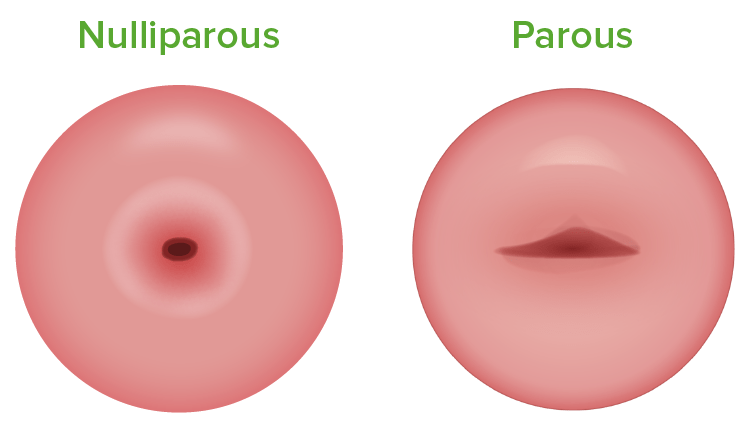
Differences between a nulliparous cervix (left) and a parous cervix (right)
Image by Lecturio.
Obtain specimens for cervical cytologyCervical cytologyA procedure in which ectocervical and endocervical cells are collected to evaluate the transformation zone (area at risk for cervical cancer).Cervical Cancer Screening:
Rotating it 90 to 180 degrees (do not over-rotate, as bleeding will obscure the sample)
Obtain 1 specimen from the ectocervixby:
Rotating a spatula 360 degrees across the entire surface of the cervixCervixThe uterus, cervix, and fallopian tubes are part of the internal female reproductive system. The most inferior portion of the uterus is the cervix, which connects the uterine cavity to the vagina. Externally, the cervix is lined by stratified squamous cells; however, the cervical canal is lined by columnar epithelium.Uterus, Cervix, and Fallopian Tubes: Anatomy
Placing 1 side of the spatula in the os, then rotating the spatula around like the hands on a clock
You can also get a combination specimen with the cervical brush (“broom”) instead:
Place the central, taller portion in the os.
Spin the brush around several times.
For best results:
The woman should not be menstruating. Note that this is not a contraindication; however, too much blood may make the sample uninterpretable.
Avoid intercourse, douches, or vaginal suppositoriesSuppositoriesMedicated dosage forms that are designed to be inserted into the rectal, vaginal, or urethral orifice of the body for absorption. Generally, the active ingredients are packaged in dosage forms containing fatty bases such as cocoa butter, hydrogenated oil, or glycerogelatin that are solid at room temperature but melt or dissolve at body temperature.Large Bowel Obstruction for 24–48 hours before the examination.
Inspect the vaginaVaginaThe vagina is the female genital canal, extending from the vulva externally to the cervix uteri internally. The structures have sexual, reproductive, and urinary functions and a rich blood supply, mainly arising from the internal iliac artery.Vagina, Vulva, and Pelvic Floor: Anatomy:
Unscrew the thumbscrew and open the speculum slightly further to release the cervixCervixThe uterus, cervix, and fallopian tubes are part of the internal female reproductive system. The most inferior portion of the uterus is the cervix, which connects the uterine cavity to the vagina. Externally, the cervix is lined by stratified squamous cells; however, the cervical canal is lined by columnar epithelium.Uterus, Cervix, and Fallopian Tubes: Anatomy.
Withdraw the speculum slowly while observing the vaginaVaginaThe vagina is the female genital canal, extending from the vulva externally to the cervix uteri internally. The structures have sexual, reproductive, and urinary functions and a rich blood supply, mainly arising from the internal iliac artery.Vagina, Vulva, and Pelvic Floor: Anatomy.
Maintain a slightly open position of the speculum as you withdraw it, carefully observing the mucosa as you do so, noting:
Color
Any inflammationInflammationInflammation is a complex set of responses to infection and injury involving leukocytes as the principal cellular mediators in the body’s defense against pathogenic organisms. Inflammation is also seen as a response to tissue injury in the process of wound healing. The 5 cardinal signs of inflammation are pain, heat, redness, swelling, and loss of function. Inflammation
Colposcopy, Cervical Biopsy, and Endocervical Curettage (ECC)
Description
ColposcopyColposcopyThe examination, therapy or surgery of the cervix and vagina by means of a specially designed endoscope introduced vaginally.Cervical Cancer Screening:
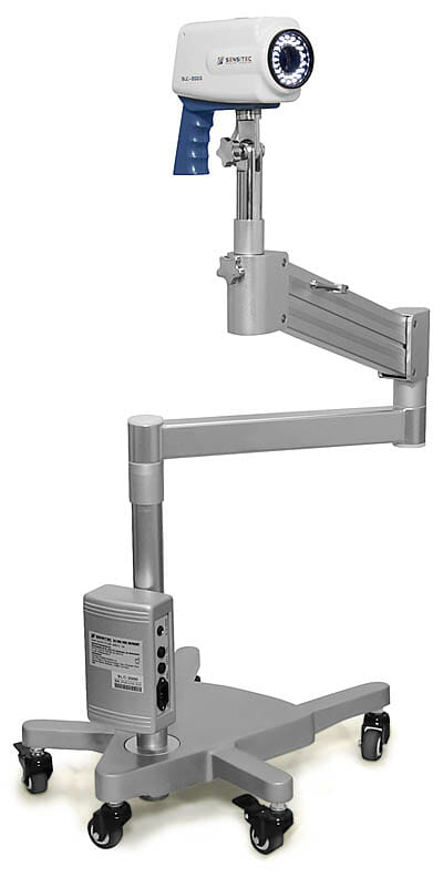
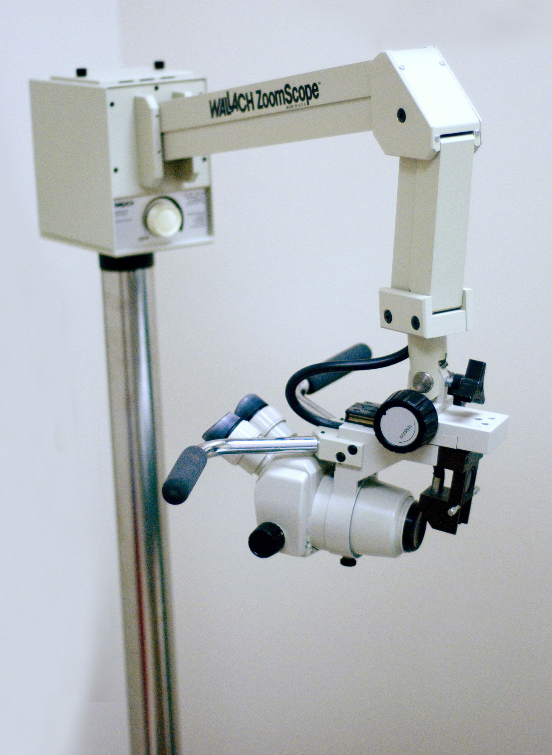
A colposcope (magnifying device) is used to provide an illuminated, magnified view of the ectocervix, vaginal wall, and vulvaVulvaThe vulva is the external genitalia of the female and includes the mons pubis, labia majora, labia minora, clitoris, vestibule, vestibular bulb, and greater vestibular glands. Vagina, Vulva, and Pelvic Floor: Anatomy.
Can be used for both diagnosis and treatment of identified lesions
Cervical biopsyBiopsyRemoval and pathologic examination of specimens from the living body.Ewing Sarcoma: A tissue sample from the ectocervix and/or transformationTransformationChange brought about to an organism’s genetic composition by unidirectional transfer (transfection; transduction, genetic; conjugation, genetic, etc.) and incorporation of foreign DNA into prokaryotic or eukaryotic cells by recombination of part or all of that DNA into the cell’s genome.Bacteriology zone is taken of any abnormal areas identified on colposcopyColposcopyThe examination, therapy or surgery of the cervix and vagina by means of a specially designed endoscope introduced vaginally.Cervical Cancer Screening.
Endocervical curettageCurettageA scraping, usually of the interior of a cavity or tract, for removal of new growth or other abnormal tissue, or to obtain material for tissue diagnosis. It is performed with a curet (curette), a spoon-shaped instrument designed for that purpose.Benign Bone Tumors (ECC):
A tissue sample is obtained from the endocervical canal.
Done at the time of colposcopyColposcopyThe examination, therapy or surgery of the cervix and vagina by means of a specially designed endoscope introduced vaginally.Cervical Cancer Screening and cervical biopsies if indicated based on history, HPVHPVHuman papillomavirus (HPV) is a nonenveloped, circular, double-stranded DNA virus belonging to the Papillomaviridae family. Humans are the only reservoir, and transmission occurs through close skin-to-skin or sexual contact. Human papillomaviruses infect basal epithelial cells and can affect cell-regulatory proteins to result in cell proliferation. Papillomavirus (HPV)screeningScreeningPreoperative Care, and cytology results
Abnormal cervical cytologyCervical cytologyA procedure in which ectocervical and endocervical cells are collected to evaluate the transformation zone (area at risk for cervical cancer).Cervical Cancer Screening (abnormal Pap smearPap smearCytological preparation of cells collected from a mucosal surface and stained with Papanicolaou stain.Cervical Cancer Screening)
High-risk types of HPVHPVHuman papillomavirus (HPV) is a nonenveloped, circular, double-stranded DNA virus belonging to the Papillomaviridae family. Humans are the only reservoir, and transmission occurs through close skin-to-skin or sexual contact. Human papillomaviruses infect basal epithelial cells and can affect cell-regulatory proteins to result in cell proliferation. Papillomavirus (HPV) detected on cervical HPV testingHPV testingCervical Cancer Screening
Evaluation of a palpably or visually abnormal cervixCervixThe uterus, cervix, and fallopian tubes are part of the internal female reproductive system. The most inferior portion of the uterus is the cervix, which connects the uterine cavity to the vagina. Externally, the cervix is lined by stratified squamous cells; however, the cervical canal is lined by columnar epithelium.Uterus, Cervix, and Fallopian Tubes: Anatomy, vaginaVaginaThe vagina is the female genital canal, extending from the vulva externally to the cervix uteri internally. The structures have sexual, reproductive, and urinary functions and a rich blood supply, mainly arising from the internal iliac artery.Vagina, Vulva, and Pelvic Floor: Anatomy, or vulvaVulvaThe vulva is the external genitalia of the female and includes the mons pubis, labia majora, labia minora, clitoris, vestibule, vestibular bulb, and greater vestibular glands. Vagina, Vulva, and Pelvic Floor: Anatomy
ContraindicationsContraindicationsA condition or factor associated with a recipient that makes the use of a drug, procedure, or physical agent improper or inadvisable. Contraindications may be absolute (life threatening) or relative (higher risk of complications in which benefits may outweigh risks).Noninvasive Ventilation
There are very few absolute contraindicationsContraindicationsA condition or factor associated with a recipient that makes the use of a drug, procedure, or physical agent improper or inadvisable. Contraindications may be absolute (life threatening) or relative (higher risk of complications in which benefits may outweigh risks).Noninvasive Ventilation to colposcopyColposcopyThe examination, therapy or surgery of the cervix and vagina by means of a specially designed endoscope introduced vaginally.Cervical Cancer Screening and biopsies. Situations in which colposcopyColposcopyThe examination, therapy or surgery of the cervix and vagina by means of a specially designed endoscope introduced vaginally.Cervical Cancer Screening and biopsies are sometimes contraindicated include:
Acute cervicitisCervicitisInflammation of the uterine cervix.Gonorrhea: may obscure results
PregnancyPregnancyThe status during which female mammals carry their developing young (embryos or fetuses) in utero before birth, beginning from fertilization to birth.Pregnancy: Diagnosis, Physiology, and Care:
ColposcopyColposcopyThe examination, therapy or surgery of the cervix and vagina by means of a specially designed endoscope introduced vaginally.Cervical Cancer Screening alone is often still performed in pregnancyPregnancyThe status during which female mammals carry their developing young (embryos or fetuses) in utero before birth, beginning from fertilization to birth.Pregnancy: Diagnosis, Physiology, and Care, though physiologic changes may make interpretation more difficult.
Biopsies are taken only if invasive disease is highly suspected.
Endocervical curettageCurettageA scraping, usually of the interior of a cavity or tract, for removal of new growth or other abnormal tissue, or to obtain material for tissue diagnosis. It is performed with a curet (curette), a spoon-shaped instrument designed for that purpose.Benign Bone Tumors is absolutely contraindicated.
Life-threateningly severe immunosuppression: ↑ risk of bacterial translocation with speculum exams
Procedures
The procedure generally includes a gross examination of the vulvaVulvaThe vulva is the external genitalia of the female and includes the mons pubis, labia majora, labia minora, clitoris, vestibule, vestibular bulb, and greater vestibular glands. Vagina, Vulva, and Pelvic Floor: Anatomy, vaginaVaginaThe vagina is the female genital canal, extending from the vulva externally to the cervix uteri internally. The structures have sexual, reproductive, and urinary functions and a rich blood supply, mainly arising from the internal iliac artery.Vagina, Vulva, and Pelvic Floor: Anatomy, and cervixCervixThe uterus, cervix, and fallopian tubes are part of the internal female reproductive system. The most inferior portion of the uterus is the cervix, which connects the uterine cavity to the vagina. Externally, the cervix is lined by stratified squamous cells; however, the cervical canal is lined by columnar epithelium.Uterus, Cervix, and Fallopian Tubes: Anatomy while placing the speculum, a colposcopyColposcopyThe examination, therapy or surgery of the cervix and vagina by means of a specially designed endoscope introduced vaginally.Cervical Cancer Screening examination, and biopsies and/or ECC as indicated based on screeningScreeningPreoperative Care results and findings on colposcopyColposcopyThe examination, therapy or surgery of the cervix and vagina by means of a specially designed endoscope introduced vaginally.Cervical Cancer Screening.
ColposcopyColposcopyThe examination, therapy or surgery of the cervix and vagina by means of a specially designed endoscope introduced vaginally.Cervical Cancer Screening:
A vaginal speculum is placed into the vaginaVaginaThe vagina is the female genital canal, extending from the vulva externally to the cervix uteri internally. The structures have sexual, reproductive, and urinary functions and a rich blood supply, mainly arising from the internal iliac artery.Vagina, Vulva, and Pelvic Floor: Anatomy.
The colposcope is used to examine the entire surface of the visible cervixCervixThe uterus, cervix, and fallopian tubes are part of the internal female reproductive system. The most inferior portion of the uterus is the cervix, which connects the uterine cavity to the vagina. Externally, the cervix is lined by stratified squamous cells; however, the cervical canal is lined by columnar epithelium.Uterus, Cervix, and Fallopian Tubes: Anatomy
Focus is on the transformationTransformationChange brought about to an organism’s genetic composition by unidirectional transfer (transfection; transduction, genetic; conjugation, genetic, etc.) and incorporation of foreign DNA into prokaryotic or eukaryotic cells by recombination of part or all of that DNA into the cell’s genome.Bacteriology zone (TZ):
The cervixCervixThe uterus, cervix, and fallopian tubes are part of the internal female reproductive system. The most inferior portion of the uterus is the cervix, which connects the uterine cavity to the vagina. Externally, the cervix is lined by stratified squamous cells; however, the cervical canal is lined by columnar epithelium.Uterus, Cervix, and Fallopian Tubes: Anatomy is examined 1st without acetic acid, and then with a solution of 3%–5% acetic acid.
Allows improved colposcopic visualization of abnormal areas
Apply a generous amount of acetic acid to the cervixCervixThe uterus, cervix, and fallopian tubes are part of the internal female reproductive system. The most inferior portion of the uterus is the cervix, which connects the uterine cavity to the vagina. Externally, the cervix is lined by stratified squamous cells; however, the cervical canal is lined by columnar epithelium.Uterus, Cervix, and Fallopian Tubes: Anatomy; wait 30‒60 seconds.
Look for acetowhite changes:
Cervical cells with large or dense nuclei (metaplastic, dysplastic, and HPV-infected cells) will turn white.
Acetowhite changes fade after approximately 3 minutes
Alternative to acetic acid: Lugol solution
Can be used after acetic acid if no acetowhite changes are found
An iodine-based solution that is taken up by normal, glycogen-containing squamous cells, causing them to turn brown
Abnormal cells (nonglycogenated columnar cells and high-grade lesions) do not take up the dye and remain light yellow.
Abnormal findings on colposcopyColposcopyThe examination, therapy or surgery of the cervix and vagina by means of a specially designed endoscope introduced vaginally.Cervical Cancer Screening include:
Acetowhite changes:
Sharp margins on lesions suggest high-grade lesions.
Diffuse borders suggest low-grade lesions.
MosaicismMosaicismThe occurrence in an individual of two or more cell populations of different chromosomal constitutions, derived from a single zygote, as opposed to chimerism in which the different cell populations are derived from more than one zygote.Chromosome Testing and punctation: abnormal vasculature in the TZ, suggestive of neoplasia
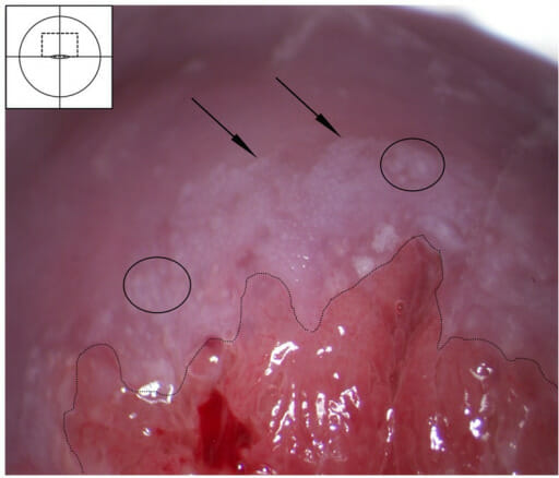
Cervical intraepithelial neoplasia (CI) stage I–II after application of acetic acid: Acetowhite lesion in the transformation zone abutting the squamocolumnar junction (dashed line). The white area is dense and has feathery margins (arrows), possibly with some mosaic pattern (ovals). This finding probably represents CIN stage I–II.
Image: “Cervical intraepithelial neoplasia stage I–II after application of acetic acid” by Norwegian Centre for Imported and Tropical Diseases, Department of Infectious Diseases, Oslo University Hospital Ullevaal. License: CC BY 4.0
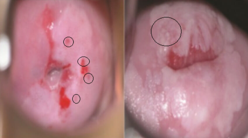
Left (circled): punctations
Right (circled): mosaicism
Image: “mages of VIA negative, VIA positive-cryotherapy eligible and VIA positive-cryotherapy ineligible lesions from women undergoing screening” by Center for Infectious Disease Research in Zambia, Lusaka. License: CC BY 4.0, edited by Lecturio.
BiopsyBiopsyRemoval and pathologic examination of specimens from the living body.Ewing Sarcoma
Done through the speculum using a long, thin instrument that reaches the cervixCervixThe uterus, cervix, and fallopian tubes are part of the internal female reproductive system. The most inferior portion of the uterus is the cervix, which connects the uterine cavity to the vagina. Externally, the cervix is lined by stratified squamous cells; however, the cervical canal is lined by columnar epithelium.Uterus, Cervix, and Fallopian Tubes: Anatomy.
Most commonly used instrument is called a Kevorkian cervical biopsyBiopsyRemoval and pathologic examination of specimens from the living body.Ewing Sarcoma instrument.
Local anestheticsAnestheticsAgents that are capable of inducing a total or partial loss of sensation, especially tactile sensation and pain. They may act to induce general anesthesia, in which an unconscious state is achieved, or may act locally to induce numbness or lack of sensation at a targeted site.Anesthesiology: History and Basic Concepts are nottypically used:
CervixCervixThe uterus, cervix, and fallopian tubes are part of the internal female reproductive system. The most inferior portion of the uterus is the cervix, which connects the uterine cavity to the vagina. Externally, the cervix is lined by stratified squamous cells; however, the cervical canal is lined by columnar epithelium.Uterus, Cervix, and Fallopian Tubes: Anatomy has relatively poor innervation to detect sharp painPainAn unpleasant sensation induced by noxious stimuli which are detected by nerve endings of nociceptive neurons.Pain: Types and Pathways → painPainAn unpleasant sensation induced by noxious stimuli which are detected by nerve endings of nociceptive neurons.Pain: Types and Pathways is felt more as intense visceral cramping
Injection of location anesthesiaAnesthesiaA state characterized by loss of feeling or sensation. This depression of nerve function is usually the result of pharmacologic action and is induced to allow performance of surgery or other painful procedures.Anesthesiology: History and Basic Concepts is typically as uncomfortable as (or is more uncomfortable than) the biopsyBiopsyRemoval and pathologic examination of specimens from the living body.Ewing Sarcoma itself.
Each specimen is individually labeled according to its location on the cervixCervixThe uterus, cervix, and fallopian tubes are part of the internal female reproductive system. The most inferior portion of the uterus is the cervix, which connects the uterine cavity to the vagina. Externally, the cervix is lined by stratified squamous cells; however, the cervical canal is lined by columnar epithelium.Uterus, Cervix, and Fallopian Tubes: Anatomy.
Obtain targeted biopsies of all abnormal areas.
Control bleeding:
Usually stops spontaneously with pressure from a cotton-tipped swab
Can also use:
Silver nitrate sticks
Ferric subsulfate (Monsel’s solution)
Surgical packing
Pelvic rest (no intercourse/anything in the vaginaVaginaThe vagina is the female genital canal, extending from the vulva externally to the cervix uteri internally. The structures have sexual, reproductive, and urinary functions and a rich blood supply, mainly arising from the internal iliac artery.Vagina, Vulva, and Pelvic Floor: Anatomy) for 24‒48 hours
Example of a cervical biopsy forceps
Image by Lecturio.
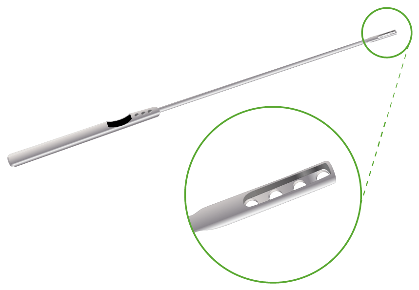
Endocervical curettageCurettageA scraping, usually of the interior of a cavity or tract, for removal of new growth or other abnormal tissue, or to obtain material for tissue diagnosis. It is performed with a curet (curette), a spoon-shaped instrument designed for that purpose.Benign Bone Tumors (ECC)
Considered a “biopsyBiopsyRemoval and pathologic examination of specimens from the living body.Ewing Sarcoma” of the endocervical canal
Contraindicated in pregnancyPregnancyThe status during which female mammals carry their developing young (embryos or fetuses) in utero before birth, beginning from fertilization to birth.Pregnancy: Diagnosis, Physiology, and Care
Uses a curette—a long, thin instrument with a sharp metal “basket” on the end
The curette is introduced into the canal and moved in and out to scrape all 4 quadrants (up, down, left, and right)
The curette is swirled in formalin to remove the tissue.
An endocervical brush is then inserted and rotated to remove any additional exfoliated tissue.
These specimens should be collected and labeled.
Endocervical curette: The tip is referred to as the “basket” and is used to scrape the inside of the endocervical canal.
Image by Lecturio.
Complications
Complications from colposcopyColposcopyThe examination, therapy or surgery of the cervix and vagina by means of a specially designed endoscope introduced vaginally.Cervical Cancer Screening, biopsies, and ECC are all exceedingly rare, but may include:
Severe bleeding
Infection
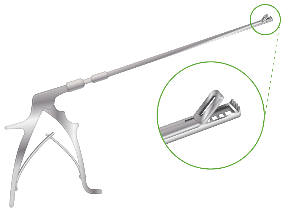
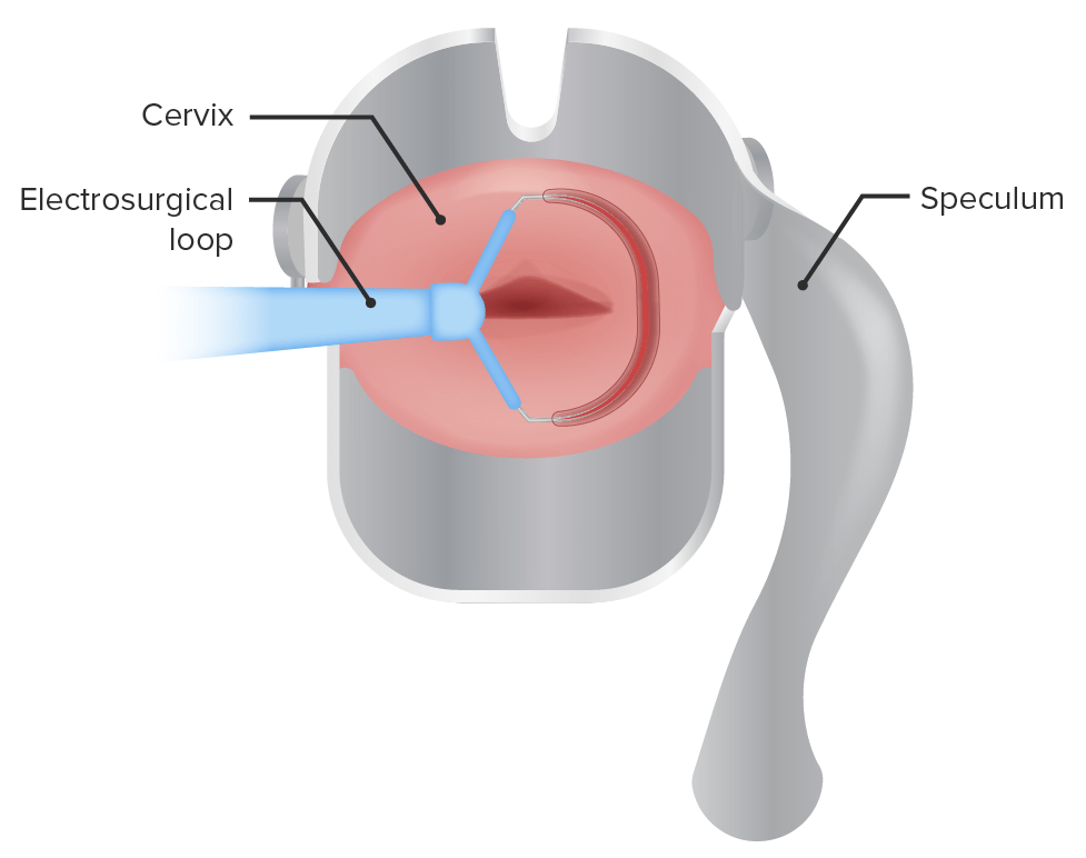
Loop Electrosurgical Excision Procedure
Description
Used to diagnose and treat cervical dysplasia or very-early-stage cervical cancerCervical cancerCervical cancer, or invasive cervical carcinoma (ICC), is the 3rd most common cancer in women in the world, with > 50% of the cases being fatal. In the United States, ICC is the 13th most common cancer and the cause of < 3% of all cancer deaths due to the slow progression of precursor lesions and, more importantly, effective cancer screening. Cervical Cancer
An electrified loop of wire is used to excise the TZ and/or pathologic area on the cervixCervixThe uterus, cervix, and fallopian tubes are part of the internal female reproductive system. The most inferior portion of the uterus is the cervix, which connects the uterine cavity to the vagina. Externally, the cervix is lined by stratified squamous cells; however, the cervical canal is lined by columnar epithelium.Uterus, Cervix, and Fallopian Tubes: Anatomy.
Typically done under local anesthesiaAnesthesiaA state characterized by loss of feeling or sensation. This depression of nerve function is usually the result of pharmacologic action and is induced to allow performance of surgery or other painful procedures.Anesthesiology: History and Basic Concepts in the office, but occasionally is done in the surgical suite under general anesthesiaGeneral anesthesiaProcedure in which patients are induced into an unconscious state through use of various medications so that they do not feel pain during surgery.Anesthesiology: History and Basic Concepts
Schematic representation of cervical conization using an electrosurgical loop
Image by Lecturio.
Indications
Treatment of high-grade cervical dysplasia (primary indication)
Diagnosis:
Suspicion of high-grade cervical dysplasia based on colposcopyColposcopyThe examination, therapy or surgery of the cervix and vagina by means of a specially designed endoscope introduced vaginally.Cervical Cancer Screening exam with inadequate or unclear biopsyBiopsyRemoval and pathologic examination of specimens from the living body.Ewing Sarcoma results
Used as an alternative when pathologic discrepancy arises between high-grade cytology on Pap smearPap smearCytological preparation of cells collected from a mucosal surface and stained with Papanicolaou stain.Cervical Cancer Screening and low-grade histology on cervical biopsyBiopsyRemoval and pathologic examination of specimens from the living body.Ewing Sarcoma
ContraindicationsContraindicationsA condition or factor associated with a recipient that makes the use of a drug, procedure, or physical agent improper or inadvisable. Contraindications may be absolute (life threatening) or relative (higher risk of complications in which benefits may outweigh risks).Noninvasive Ventilation
Multiple recurrent procedures resulting in an abnormally short cervixCervixThe uterus, cervix, and fallopian tubes are part of the internal female reproductive system. The most inferior portion of the uterus is the cervix, which connects the uterine cavity to the vagina. Externally, the cervix is lined by stratified squamous cells; however, the cervical canal is lined by columnar epithelium.Uterus, Cervix, and Fallopian Tubes: Anatomy
CervicitisCervicitisInflammation of the uterine cervix.Gonorrhea (e.g., active chlamydiaChlamydiaChlamydiae are obligate intracellular gram-negative bacteria. They lack a peptidoglycan layer and are best visualized using Giemsa stain. The family of Chlamydiaceae comprises 3 pathogens that can infect humans: Chlamydia trachomatis, Chlamydia psittaci, and Chlamydia pneumoniae.Chlamydia infection)
PregnancyPregnancyThe status during which female mammals carry their developing young (embryos or fetuses) in utero before birth, beginning from fertilization to birth.Pregnancy: Diagnosis, Physiology, and Care (strong but relative contraindication)
Anticoagulant use (relative)
Procedure
A speculum is inserted.
ColposcopyColposcopyThe examination, therapy or surgery of the cervix and vagina by means of a specially designed endoscope introduced vaginally.Cervical Cancer Screening is performed:
Acetic acid is applied and the cervixCervixThe uterus, cervix, and fallopian tubes are part of the internal female reproductive system. The most inferior portion of the uterus is the cervix, which connects the uterine cavity to the vagina. Externally, the cervix is lined by stratified squamous cells; however, the cervical canal is lined by columnar epithelium.Uterus, Cervix, and Fallopian Tubes: Anatomy is visualized using a colposcope.
Extent of dysplasia is noted.
Smallest wire needed to excise the entire lesion should be selected.
Local anesthesiaAnesthesiaA state characterized by loss of feeling or sensation. This depression of nerve function is usually the result of pharmacologic action and is induced to allow performance of surgery or other painful procedures.Anesthesiology: History and Basic Concepts is injected creating a cervical block; typically:
1%–2% lidocaineLidocaineA local anesthetic and cardiac depressant used as an antiarrhythmic agent. Its actions are more intense and its effects more prolonged than those of procaine but its duration of action is shorter than that of bupivacaine or prilocaine.Local Anesthetics with epinephrineEpinephrineThe active sympathomimetic hormone from the adrenal medulla. It stimulates both the alpha- and beta- adrenergic systems, causes systemic vasoconstriction and gastrointestinal relaxation, stimulates the heart, and dilates bronchi and cerebral vessels.Sympathomimetic Drugs
Approximately 10 mL (total) is injected at least at 5:00 and 7:00, and often at 11:00 and 1:00 as well.
Note: Direct injection at 3:00 and 9:00 should be avoided in order to avoid the cervical vessels.
The loop is activated (typically blended cutting and coagulation current at a relatively low voltage)
The loop is carefully passed around and under the TZ, ideally in one continuous movement, thus excising it.
Moving too fast: the loop drags or sticks to the tissue and doesn’t adequately cut it
Moving too slowly: excessive thermal damage causing the loop to stick within the tissue, making additional passes necessary
Endocervical curettageCurettageA scraping, usually of the interior of a cavity or tract, for removal of new growth or other abnormal tissue, or to obtain material for tissue diagnosis. It is performed with a curet (curette), a spoon-shaped instrument designed for that purpose.Benign Bone Tumors is typically performed following removal of the loop electrosurgical excision procedure specimen.
Both loop electrosurgical excision procedure and ECC specimens are sent for histologic evaluation.
A sample of tissue is taken from the vulvaVulvaThe vulva is the external genitalia of the female and includes the mons pubis, labia majora, labia minora, clitoris, vestibule, vestibular bulb, and greater vestibular glands. Vagina, Vulva, and Pelvic Floor: Anatomy.
Indication
Vulvar biopsies are indicated in the evaluation of any abnormal-appearing vulvar lesions to rule out (or identify) neoplasia and to assist in the diagnosis of vulvar dermatitisDermatitisAny inflammation of the skin.Atopic Dermatitis (Eczema).
Vulvar pruritusPruritusAn intense itching sensation that produces the urge to rub or scratch the skin to obtain relief.Atopic Dermatitis (Eczema) that is not due to infectionsInfectionsInvasion of the host organism by microorganisms or their toxins or by parasites that can cause pathological conditions or diseases.Chronic Granulomatous DiseasevulvovaginitisVulvovaginitisThe term vulvovaginitis is used to describe an acute inflammation of the vulva and vagina. Vulvovaginitis can be caused by several infectious and non-infectious etiologies, and results from disruption of the normal vaginal environment. Common signs and symptoms include pain, pruritus, erythema, edema, vaginal discharge and dyspareunia. Vulvovaginitis (e.g., candidaCandidaCandida is a genus of dimorphic, opportunistic fungi. Candida albicans is part of the normal human flora and is the most common cause of candidiasis. The clinical presentation varies and can include localized mucocutaneous infections (e.g., oropharyngeal, esophageal, intertriginous, and vulvovaginal candidiasis) and invasive disease (e.g., candidemia, intraabdominal abscess, pericarditis, and meningitis). Candida/Candidiasis infection)
Worrisome visible lesions (similar to the “ABCDEs” of melanomaMelanomaMelanoma is a malignant tumor arising from melanocytes, the melanin-producing cells of the epidermis. These tumors are most common in fair-skinned individuals with a history of excessive sun exposure and sunburns. Melanoma):
Asymmetry
(Irregular) Borders
Color variation
Diameter (larger lesions are more concerning)
Evolution (lesion is changing)
Others:
Underlying skinSkinThe skin, also referred to as the integumentary system, is the largest organ of the body. The skin is primarily composed of the epidermis (outer layer) and dermis (deep layer). The epidermis is primarily composed of keratinocytes that undergo rapid turnover, while the dermis contains dense layers of connective tissue.Skin: Structure and Functions retraction
Changes in the surrounding vulvar architecture
Nonhealing ulcers
Lesions that do not respond to standard therapy
Abnormal vasculature (Note: Do notattempt to biopsyBiopsyRemoval and pathologic examination of specimens from the living body.Ewing Sarcoma a highly vascular lesion in the office.)
ContraindicationsContraindicationsA condition or factor associated with a recipient that makes the use of a drug, procedure, or physical agent improper or inadvisable. Contraindications may be absolute (life threatening) or relative (higher risk of complications in which benefits may outweigh risks).Noninvasive Ventilation
None
If risk of bleeding is high, biopsies should be done in the OR instead of the office.
Procedure
Perform vulvar colposcopyColposcopyThe examination, therapy or surgery of the cervix and vagina by means of a specially designed endoscope introduced vaginally.Cervical Cancer Screening:
Generously apply acetic acid to the vulvaVulvaThe vulva is the external genitalia of the female and includes the mons pubis, labia majora, labia minora, clitoris, vestibule, vestibular bulb, and greater vestibular glands. Vagina, Vulva, and Pelvic Floor: Anatomy with soaked cotton balls.
Observe with a colposcope.
Avoid taking a biopsyBiopsyRemoval and pathologic examination of specimens from the living body.Ewing Sarcoma sample near the clitorisClitorisAn erectile structure homologous with the penis, situated beneath the anterior labial commissure, partially hidden between the anterior ends of the labia minora.Vagina, Vulva, and Pelvic Floor: Anatomy or the urethral or anal openings.
Prepare the area with antiseptic (e.g., povidone–iodineIodineA nonmetallic element of the halogen group that is represented by the atomic symbol I, atomic number 53, and atomic weight of 126. 90. It is a nutritionally essential element, especially important in thyroid hormone synthesis. In solution, it has anti-infective properties and is used topically.Thyroid Hormones)
Inject 1‒2 mL of local anesthetic (typically 1%‒2% lidocaineLidocaineA local anesthetic and cardiac depressant used as an antiarrhythmic agent. Its actions are more intense and its effects more prolonged than those of procaine but its duration of action is shorter than that of bupivacaine or prilocaine.Local Anesthetics with or without epinephrineEpinephrineThe active sympathomimetic hormone from the adrenal medulla. It stimulates both the alpha- and beta- adrenergic systems, causes systemic vasoconstriction and gastrointestinal relaxation, stimulates the heart, and dilates bronchi and cerebral vessels.Sympathomimetic Drugs)
Obtain the biopsyBiopsyRemoval and pathologic examination of specimens from the living body.Ewing Sarcoma sample:
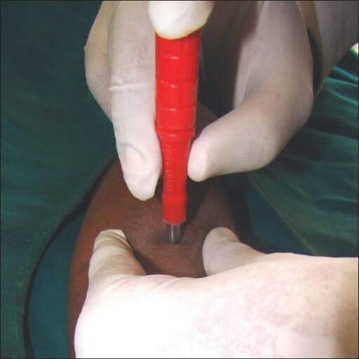
Punch biopsyPunch BiopsyActinic Keratosis: A tool with a 3‒5 mm sharp circular tip is twisted around the site to be excised.
A punch biopsy tool is used to take an excisional biopsy of the skin: This tool is commonly used for vulvar biopsies.
Image: “Proper technique of holding the punch for performing punch biopsy” by Nischal U. License: CC BY 2.0
Endometrial Biopsy
Description
A thin pipelle is used to sample the endometriumEndometriumThe mucous membrane lining of the uterine cavity that is hormonally responsive during the menstrual cycle and pregnancy. The endometrium undergoes cyclic changes that characterize menstruation. After successful fertilization, it serves to sustain the developing embryo.Embryoblast and Trophoblast Development for direct histologic evaluation.
Done in the office, usually without anesthesiaAnesthesiaA state characterized by loss of feeling or sensation. This depression of nerve function is usually the result of pharmacologic action and is induced to allow performance of surgery or other painful procedures.Anesthesiology: History and Basic Concepts
Indication
Evaluation for precancerousPrecancerousPathological conditions that tend eventually to become malignant.Barrett Esophagus and neoplastic conditions of the endometriumEndometriumThe mucous membrane lining of the uterine cavity that is hormonally responsive during the menstrual cycle and pregnancy. The endometrium undergoes cyclic changes that characterize menstruation. After successful fertilization, it serves to sustain the developing embryo.Embryoblast and Trophoblast Development
Abnormal uterine bleedingAbnormal Uterine BleedingAbnormal uterine bleeding is the medical term for abnormalities in the frequency, volume, duration, and regularity of the menstrual cycle. Abnormal uterine bleeding is classified using the acronym PALM-COEIN, with PALM representing the structural causes and COEIN indicating the non-structural causes. Abnormal Uterine Bleeding (AUBAUBAbnormal uterine bleeding is the medical term for abnormalities in the frequency, volume, duration, and regularity of the menstrual cycle. Abnormal uterine bleeding is classified using the acronym palm-coein, with palm representing the structural causes and coein indicating the non-structural causes.Abnormal Uterine Bleeding) in women with risk factors for neoplasia
PregnancyPregnancyThe status during which female mammals carry their developing young (embryos or fetuses) in utero before birth, beginning from fertilization to birth.Pregnancy: Diagnosis, Physiology, and Care
Acute infection (pelvic inflammatory, cervical, or vaginal)
Cervical cancerCervical cancerCervical cancer, or invasive cervical carcinoma (ICC), is the 3rd most common cancer in women in the world, with > 50% of the cases being fatal. In the United States, ICC is the 13th most common cancer and the cause of < 3% of all cancer deaths due to the slow progression of precursor lesions and, more importantly, effective cancer screening. Cervical Cancer
Insert a speculum and identify the cervixCervixThe uterus, cervix, and fallopian tubes are part of the internal female reproductive system. The most inferior portion of the uterus is the cervix, which connects the uterine cavity to the vagina. Externally, the cervix is lined by stratified squamous cells; however, the cervical canal is lined by columnar epithelium.Uterus, Cervix, and Fallopian Tubes: Anatomy.
Prepare the cervixCervixThe uterus, cervix, and fallopian tubes are part of the internal female reproductive system. The most inferior portion of the uterus is the cervix, which connects the uterine cavity to the vagina. Externally, the cervix is lined by stratified squamous cells; however, the cervical canal is lined by columnar epithelium.Uterus, Cervix, and Fallopian Tubes: Anatomy with antiseptic (e.g., povidone–iodineIodineA nonmetallic element of the halogen group that is represented by the atomic symbol I, atomic number 53, and atomic weight of 126. 90. It is a nutritionally essential element, especially important in thyroid hormone synthesis. In solution, it has anti-infective properties and is used topically.Thyroid Hormones)
Insert the endometrial pipelle through the cervical os until resistanceResistancePhysiologically, the opposition to flow of air caused by the forces of friction. As a part of pulmonary function testing, it is the ratio of driving pressure to the rate of air flow.Ventilation: Mechanics of Breathing is felt at the uterine fundusFundusThe superior portion of the body of the stomach above the level of the cardiac notch.Stomach: Anatomy:
Pipelles have rulers on them → note the length of the cavity (average length: 6‒8 cm)
Place a forcepsForcepsSurgical Instruments and Sutures (tenaculum) on the anterior lip of the cervixCervixThe uterus, cervix, and fallopian tubes are part of the internal female reproductive system. The most inferior portion of the uterus is the cervix, which connects the uterine cavity to the vagina. Externally, the cervix is lined by stratified squamous cells; however, the cervical canal is lined by columnar epithelium.Uterus, Cervix, and Fallopian Tubes: Anatomy.
Provide gentle outward traction to straighten the uterocervical angle and keep the cervixCervixThe uterus, cervix, and fallopian tubes are part of the internal female reproductive system. The most inferior portion of the uterus is the cervix, which connects the uterine cavity to the vagina. Externally, the cervix is lined by stratified squamous cells; however, the cervical canal is lined by columnar epithelium.Uterus, Cervix, and Fallopian Tubes: Anatomy in place during insertion of the pipelle.
Pull the piston back on the pipelle to generate suction within the tube
Slide the pipelle in and out several times (while keeping the tip within the endometrial cavity), rotating the pipelle to obtain the most comprehensive sample possible.
Empty the sample into a labeled specimen container.
Control bleeding:
Small amounts of bleeding from the os and tenaculum sites are common and usually respond to pressure.
Severe bleeding would be suspicious for malignancyMalignancyHemothorax or an underlying bleeding disorder.
Complications
Cramping
Uterine perforationPerforationA pathological hole in an organ, blood vessel or other soft part of the body, occurring in the absence of external force.Esophagitis (uncommon with modern plastic pipelles)
Infection
Schematic depiction of an endometrial biopsy using a pipelle device: The pipelle is inserted to the fundus of the uterus; then the piston on the opposite end is pulled back, creating space and generating suction within the tube. This suction pulls endometrial tissue into the tube, which can then be sent for histologic evaluation.
Image by Lecturio.
Sonography (Ultrasound)
Ultrasound is the most common diagnostic procedure used to visualize the internal female reproductive organs.
Allows for the best visualization of female reproductive structures located within the pelvisPelvisThe pelvis consists of the bony pelvic girdle, the muscular and ligamentous pelvic floor, and the pelvic cavity, which contains viscera, vessels, and multiple nerves and muscles. The pelvic girdle, composed of 2 “hip” bones and the sacrum, is a ring-like bony structure of the axial skeleton that links the vertebral column with the lower extremities.Pelvis: Anatomy
TransducerTransducerA device placed on the patient’s body to visualize a targetUltrasound (Sonography) is placed inside the vaginaVaginaThe vagina is the female genital canal, extending from the vulva externally to the cervix uteri internally. The structures have sexual, reproductive, and urinary functions and a rich blood supply, mainly arising from the internal iliac artery.Vagina, Vulva, and Pelvic Floor: Anatomy.
At or below the cervixCervixThe uterus, cervix, and fallopian tubes are part of the internal female reproductive system. The most inferior portion of the uterus is the cervix, which connects the uterine cavity to the vagina. Externally, the cervix is lined by stratified squamous cells; however, the cervical canal is lined by columnar epithelium.Uterus, Cervix, and Fallopian Tubes: Anatomy
Angled slightly upward to visualize the reproductive organs
Transabdominal ultrasound (TAUS):
TransducerTransducerA device placed on the patient’s body to visualize a targetUltrasound (Sonography) is placed on the lower abdomen.
Best for visualizing structures above the true pelvisPelvisThe pelvis consists of the bony pelvic girdle, the muscular and ligamentous pelvic floor, and the pelvic cavity, which contains viscera, vessels, and multiple nerves and muscles. The pelvic girdle, composed of 2 “hip” bones and the sacrum, is a ring-like bony structure of the axial skeleton that links the vertebral column with the lower extremities.Pelvis: Anatomy, for example:
An enlarged uterusUterusThe uterus, cervix, and fallopian tubes are part of the internal female reproductive system. The uterus has a thick wall made of smooth muscle (the myometrium) and an inner mucosal layer (the endometrium). The most inferior portion of the uterus is the cervix, which connects the uterine cavity to the vagina.Uterus, Cervix, and Fallopian Tubes: Anatomy (e.g., during pregnancyPregnancyThe status during which female mammals carry their developing young (embryos or fetuses) in utero before birth, beginning from fertilization to birth.Pregnancy: Diagnosis, Physiology, and Care)
Large cystsCystsAny fluid-filled closed cavity or sac that is lined by an epithelium. Cysts can be of normal, abnormal, non-neoplastic, or neoplastic tissues.Fibrocystic Change or fibroidsFibroidsA benign tumor derived from smooth muscle tissue, also known as a fibroid tumor. They rarely occur outside of the uterus and the gastrointestinal tract but can occur in the skin and subcutaneous tissue, probably arising from the smooth muscle of small blood vessels in these tissues.Infertility extending out of the pelvisPelvisThe pelvis consists of the bony pelvic girdle, the muscular and ligamentous pelvic floor, and the pelvic cavity, which contains viscera, vessels, and multiple nerves and muscles. The pelvic girdle, composed of 2 “hip” bones and the sacrum, is a ring-like bony structure of the axial skeleton that links the vertebral column with the lower extremities.Pelvis: Anatomy
Useful in people who cannot tolerate transvaginal exams
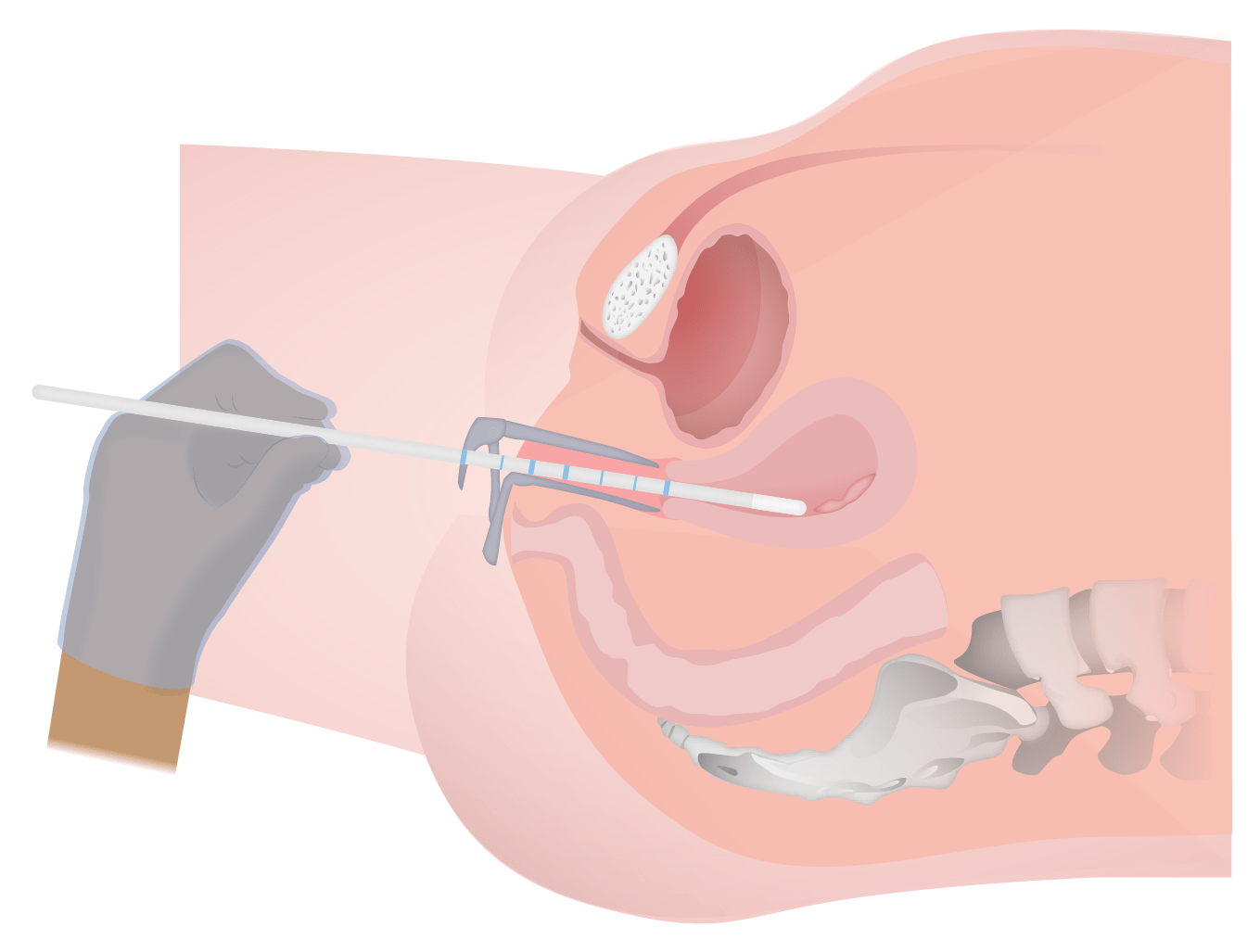
A catheter is placed into the endometrial cavity (same procedure as placing an endometrial pipelle).
Once the catheter is in place, a balloon is inflated to keep the catheter in place and the speculum is removed.
A TVUS probeProbeA device placed on the patient’s body to visualize a targetUltrasound (Sonography) is inserted into the vaginaVaginaThe vagina is the female genital canal, extending from the vulva externally to the cervix uteri internally. The structures have sexual, reproductive, and urinary functions and a rich blood supply, mainly arising from the internal iliac artery.Vagina, Vulva, and Pelvic Floor: Anatomy and the uterusUterusThe uterus, cervix, and fallopian tubes are part of the internal female reproductive system. The uterus has a thick wall made of smooth muscle (the myometrium) and an inner mucosal layer (the endometrium). The most inferior portion of the uterus is the cervix, which connects the uterine cavity to the vagina.Uterus, Cervix, and Fallopian Tubes: Anatomy identified.
While observing on TVUS in real time, sterileSterileBasic Procedures saline is injected into the endometrial cavity
This fluid distends the cavity, allowing for evaluation of intracavitary lesions.
Although the fluid does efflux through the fallopian tubesFallopian tubesThe uterus, cervix, and fallopian tubes are part of the internal female reproductive system. The fallopian tubes receive an ovum after ovulation and help move it and/or a fertilized embryo toward the uterus via ciliated cells lining the tubes and peristaltic movements of its smooth muscle. Uterus, Cervix, and Fallopian Tubes: Anatomy, the tubes are too thin for this to be observed on TVUS (HSG is required; see below)
After images are taken, the balloon is deflated and the catheter and probeProbeA device placed on the patient’s body to visualize a targetUltrasound (Sonography) are removed.
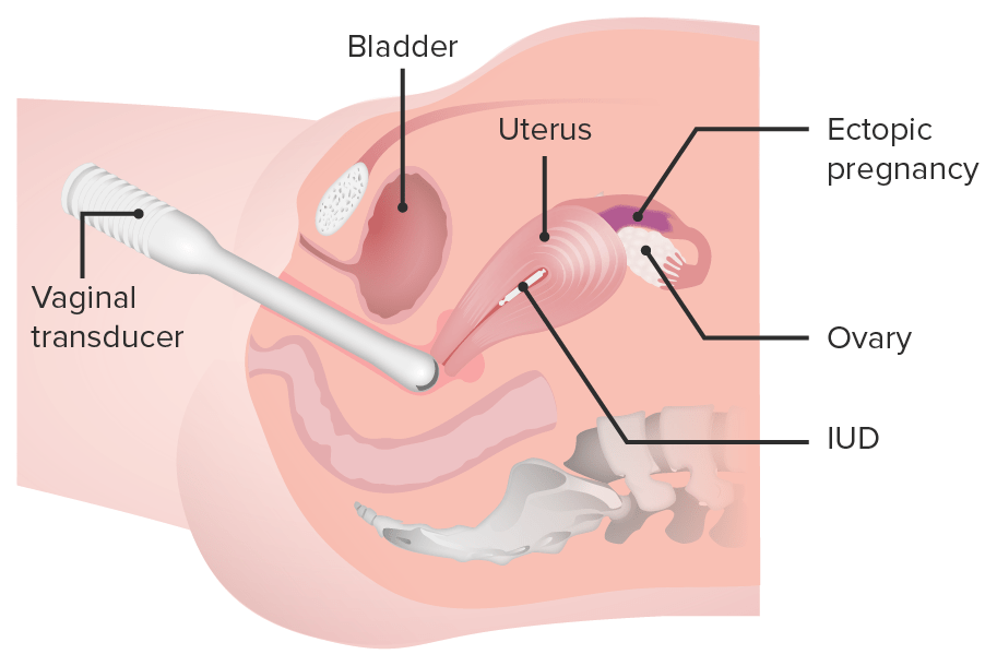
A schematic depiction of the vaginal transducer being placed within the vagina to visualize the ovary and fallopian tube:
In this example case, the transvaginal ultrasound can detect the location of the intrauterine device (IUD) and the presence of an ectopic pregnancy in the fallopian tube.
Image by Lecturio.
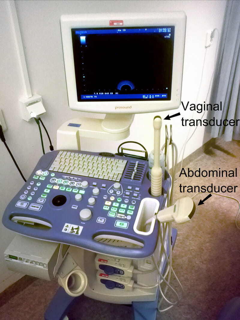
Portable device showing both transducers
Image: “Device for abdominal and vaginal ultrasonography” by Mikael Häggström. License: Public Domain
Indications
TAUS/TVUS:
The indications for TVUS and TAUS are the same and generally include bleeding and/or painPainAn unpleasant sensation induced by noxious stimuli which are detected by nerve endings of nociceptive neurons.Pain: Types and Pathways. Both are typically performed during the same exam to ensure a complete evaluation of the female reproductive organs.
Suspected ovarian or fallopian tubeFallopian TubeA pair of highly specialized canals extending from the uterus to its corresponding ovary. They provide the means for ovum transport from the ovaries and they are the site of the ovum’s final maturation and fertilization. The fallopian tube consists of an interstitium, an isthmus, an ampulla, an infundibulum, and fimbriae. Its wall consists of three layers: serous, muscular, and an internal mucosal layer lined with both ciliated and secretory cells.Uterus, Cervix, and Fallopian Tubes: Anatomy masses:
CystsCystsAny fluid-filled closed cavity or sac that is lined by an epithelium. Cysts can be of normal, abnormal, non-neoplastic, or neoplastic tissues.Fibrocystic Change
Ectopic pregnancyEctopic pregnancyEctopic pregnancy refers to the implantation of a fertilized egg (embryo) outside the uterine cavity. The main cause is disruption of the normal anatomy of the fallopian tube. Ectopic Pregnancy
To assess the uterusUterusThe uterus, cervix, and fallopian tubes are part of the internal female reproductive system. The uterus has a thick wall made of smooth muscle (the myometrium) and an inner mucosal layer (the endometrium). The most inferior portion of the uterus is the cervix, which connects the uterine cavity to the vagina.Uterus, Cervix, and Fallopian Tubes: Anatomy in cases of abnormal bleeding (including menstrual bleeding, postmenopausal bleeding, and bleeding in pregnancyPregnancyThe status during which female mammals carry their developing young (embryos or fetuses) in utero before birth, beginning from fertilization to birth.Pregnancy: Diagnosis, Physiology, and Care), looking especially for:
Endometrial thickness → may indicate hyperplasiaHyperplasiaAn increase in the number of cells in a tissue or organ without tumor formation. It differs from hypertrophy, which is an increase in bulk without an increase in the number of cells.Cellular Adaptation (if thick) or atrophyAtrophyDecrease in the size of a cell, tissue, organ, or multiple organs, associated with a variety of pathological conditions such as abnormal cellular changes, ischemia, malnutrition, or hormonal changes.Cellular Adaptation (if thin)
Signs of adenomyosisAdenomyosisAdenomyosis is a benign uterine condition characterized by the presence of ectopic endometrial glands and stroma within the myometrium. Adenomyosis is a common condition, affecting 20%-35% of women, and typically presents with heavy menstrual bleeding and dysmenorrhea. Adenomyosis:
Enlarged uterusUterusThe uterus, cervix, and fallopian tubes are part of the internal female reproductive system. The uterus has a thick wall made of smooth muscle (the myometrium) and an inner mucosal layer (the endometrium). The most inferior portion of the uterus is the cervix, which connects the uterine cavity to the vagina.Uterus, Cervix, and Fallopian Tubes: Anatomy
Asymmetrical thickening of the myometrium
Myometrial cystsCystsAny fluid-filled closed cavity or sac that is lined by an epithelium. Cysts can be of normal, abnormal, non-neoplastic, or neoplastic tissues.Fibrocystic Change
Linear striations radiating out from the endometriumEndometriumThe mucous membrane lining of the uterine cavity that is hormonally responsive during the menstrual cycle and pregnancy. The endometrium undergoes cyclic changes that characterize menstruation. After successful fertilization, it serves to sustain the developing embryo.Embryoblast and Trophoblast Development
Loss of a clear endomyometrial border
Pelvic painPainAn unpleasant sensation induced by noxious stimuli which are detected by nerve endings of nociceptive neurons.Pain: Types and Pathways (looking for structural causes)
Evaluation of congenital anomalies
InfertilityInfertilityInfertility is the inability to conceive in the context of regular intercourse. The most common causes of infertility in women are related to ovulatory dysfunction or tubal obstruction, whereas, in men, abnormal sperm is a common cause. Infertility assessments
PregnancyPregnancyThe status during which female mammals carry their developing young (embryos or fetuses) in utero before birth, beginning from fertilization to birth.Pregnancy: Diagnosis, Physiology, and Care assessments:
Dating
Cervical length
Anatomic, fluid, and growth assessments of the fetus
Visually assist with other invasive procedures, including:
Aspiration of ova for in vitrofertilizationFertilizationTo undergo fertilization, the sperm enters the uterus, travels towards the ampulla of the fallopian tube, and encounters the oocyte. The zona pellucida (the outer layer of the oocyte) deteriorates along with the zygote, which travels towards the uterus and eventually forms a blastocyst, allowing for implantation to occur. Fertilization and First Week
Aspiration of pelvic fluid
Obstetric uses, including:
AmniocentesisAmniocentesisPercutaneous transabdominal puncture of the uterus during pregnancy to obtain amniotic fluid. It is commonly used for fetal karyotype determination in order to diagnose abnormal fetal conditions.Polyhydramnios
By distending the uterusUterusThe uterus, cervix, and fallopian tubes are part of the internal female reproductive system. The uterus has a thick wall made of smooth muscle (the myometrium) and an inner mucosal layer (the endometrium). The most inferior portion of the uterus is the cervix, which connects the uterine cavity to the vagina.Uterus, Cervix, and Fallopian Tubes: Anatomy, SISSISInfertility allows identificationIdentificationDefense Mechanisms of intracavitary pathology (in addition to everything seen on routine TVUS). A standard TVUS is typically performed before the SISSISInfertility procedure.
Can identify:
Polyps
Submucosal leiomyomas
Adhesions
Septa
Contour of the endometrial cavity
Typically done to evaluate:
InfertilityInfertilityInfertility is the inability to conceive in the context of regular intercourse. The most common causes of infertility in women are related to ovulatory dysfunction or tubal obstruction, whereas, in men, abnormal sperm is a common cause. Infertility
Abnormal uterine bleedingAbnormal Uterine BleedingAbnormal uterine bleeding is the medical term for abnormalities in the frequency, volume, duration, and regularity of the menstrual cycle. Abnormal uterine bleeding is classified using the acronym PALM-COEIN, with PALM representing the structural causes and COEIN indicating the non-structural causes. Abnormal Uterine Bleeding with suspected endometrial pathology
Intrauterine surgical planning
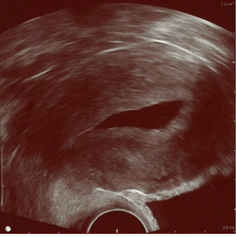
Saline infusion sonogram (SIS): The sterile saline instilled into the cavity of the uterus is anechoic (visible as the dark central portion of the image) and delineates the shape of the endometrial cavity. This image shows a normal endometrium (the hyperechoic/brighter band around the cavity), without any focal changes. The endometrium is surrounded by the myometrium, which stretches almost to the right border of the image.
Image: “Normal hysterosonography” by Mikael Häggström. License: Public Domain
ContraindicationsContraindicationsA condition or factor associated with a recipient that makes the use of a drug, procedure, or physical agent improper or inadvisable. Contraindications may be absolute (life threatening) or relative (higher risk of complications in which benefits may outweigh risks).Noninvasive Ventilation
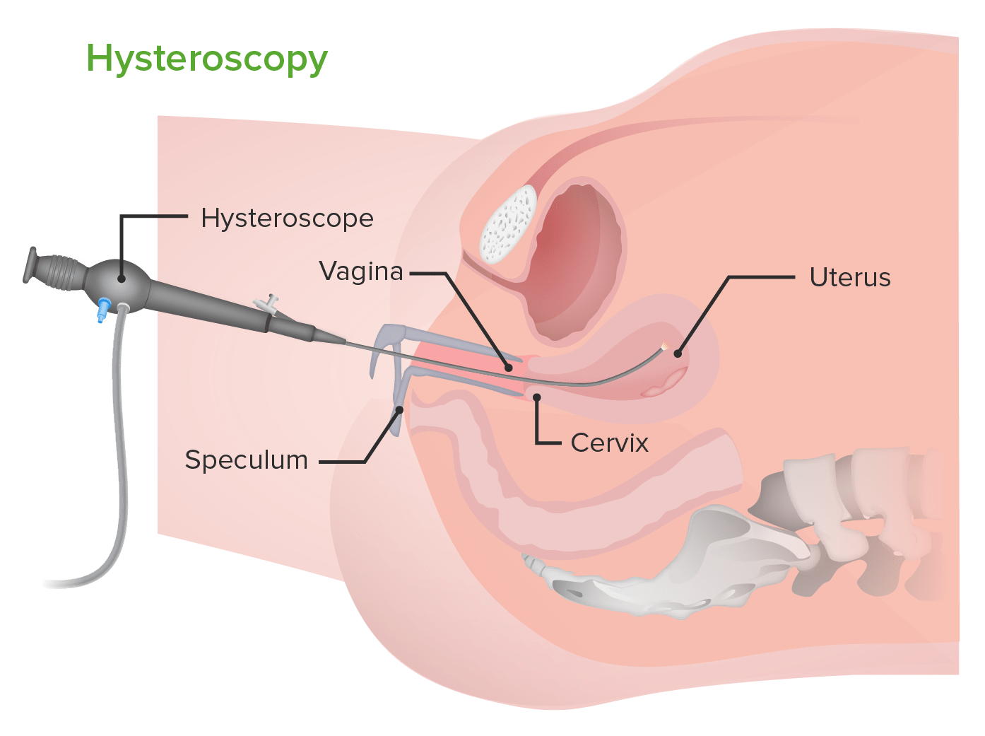
A scope is introduced into the endometrial cavity through the cervixCervixThe uterus, cervix, and fallopian tubes are part of the internal female reproductive system. The most inferior portion of the uterus is the cervix, which connects the uterine cavity to the vagina. Externally, the cervix is lined by stratified squamous cells; however, the cervical canal is lined by columnar epithelium.Uterus, Cervix, and Fallopian Tubes: Anatomy.
Used to diagnose and/or treat intrauterine pathologies
It is a gold standarddiagnostic tool for assessment of:
Endometrial cavity
Tubal ostia (where the tubes enter the endometrial cavity)
Endocervical canal
Can be performed in the office or in a surgical suite
Indications
From a diagnostic standpoint, HSC has similar indications as for SISSISInfertility. The primary advantage of HSC over SISSISInfertility is the ability to directly visualize lesions and treat them simultaneously. HSC is performed to:
Diagnose and investigate causes of:
Abnormal uterine bleedingAbnormal Uterine BleedingAbnormal uterine bleeding is the medical term for abnormalities in the frequency, volume, duration, and regularity of the menstrual cycle. Abnormal uterine bleeding is classified using the acronym PALM-COEIN, with PALM representing the structural causes and COEIN indicating the non-structural causes. Abnormal Uterine Bleeding
Thickened endometriumEndometriumThe mucous membrane lining of the uterine cavity that is hormonally responsive during the menstrual cycle and pregnancy. The endometrium undergoes cyclic changes that characterize menstruation. After successful fertilization, it serves to sustain the developing embryo.Embryoblast and Trophoblast Development seen on sonography; can distinguish between:
Diffusely thickened endometriumEndometriumThe mucous membrane lining of the uterine cavity that is hormonally responsive during the menstrual cycle and pregnancy. The endometrium undergoes cyclic changes that characterize menstruation. After successful fertilization, it serves to sustain the developing embryo.Embryoblast and Trophoblast Development (a concern for hyperplasiaHyperplasiaAn increase in the number of cells in a tissue or organ without tumor formation. It differs from hypertrophy, which is an increase in bulk without an increase in the number of cells.Cellular Adaptation)
Polyps
Submucosal fibroidsFibroidsA benign tumor derived from smooth muscle tissue, also known as a fibroid tumor. They rarely occur outside of the uterus and the gastrointestinal tract but can occur in the skin and subcutaneous tissue, probably arising from the smooth muscle of small blood vessels in these tissues.Infertility
Intrauterine adhesions
Endocervical lesions
InfertilityInfertilityInfertility is the inability to conceive in the context of regular intercourse. The most common causes of infertility in women are related to ovulatory dysfunction or tubal obstruction, whereas, in men, abnormal sperm is a common cause. Infertility
Postmenopausal bleeding
Dislocation of IUDs
Treat lesions via:
Adhesiolysis
Myomectomy/polypectomy
Endometrial ablation (or resection)
Removal of intrauterine foreign bodies
ContraindicationsContraindicationsA condition or factor associated with a recipient that makes the use of a drug, procedure, or physical agent improper or inadvisable. Contraindications may be absolute (life threatening) or relative (higher risk of complications in which benefits may outweigh risks).Noninvasive Ventilation
Viable intrauterine pregnancyPregnancyThe status during which female mammals carry their developing young (embryos or fetuses) in utero before birth, beginning from fertilization to birth.Pregnancy: Diagnosis, Physiology, and Care
Active pelvic infection
Known cervical or uterine cancer
Procedure
CervixCervixThe uterus, cervix, and fallopian tubes are part of the internal female reproductive system. The most inferior portion of the uterus is the cervix, which connects the uterine cavity to the vagina. Externally, the cervix is lined by stratified squamous cells; however, the cervical canal is lined by columnar epithelium.Uterus, Cervix, and Fallopian Tubes: Anatomy and vaginaVaginaThe vagina is the female genital canal, extending from the vulva externally to the cervix uteri internally. The structures have sexual, reproductive, and urinary functions and a rich blood supply, mainly arising from the internal iliac artery.Vagina, Vulva, and Pelvic Floor: Anatomy are prepared with antiseptic (e.g., povidone–iodineIodineA nonmetallic element of the halogen group that is represented by the atomic symbol I, atomic number 53, and atomic weight of 126. 90. It is a nutritionally essential element, especially important in thyroid hormone synthesis. In solution, it has anti-infective properties and is used topically.Thyroid Hormones)
A speculum is inserted and the cervixCervixThe uterus, cervix, and fallopian tubes are part of the internal female reproductive system. The most inferior portion of the uterus is the cervix, which connects the uterine cavity to the vagina. Externally, the cervix is lined by stratified squamous cells; however, the cervical canal is lined by columnar epithelium.Uterus, Cervix, and Fallopian Tubes: Anatomy identified.
The anterior lip of the cervixCervixThe uterus, cervix, and fallopian tubes are part of the internal female reproductive system. The most inferior portion of the uterus is the cervix, which connects the uterine cavity to the vagina. Externally, the cervix is lined by stratified squamous cells; however, the cervical canal is lined by columnar epithelium.Uterus, Cervix, and Fallopian Tubes: Anatomy is grasped with a tenaculum forcepsForcepsSurgical Instruments and Sutures to:
Provide countertraction during dilation and advancement of the scope
Straighten the uterocervical angle with gentle outward pressure
The cervixCervixThe uterus, cervix, and fallopian tubes are part of the internal female reproductive system. The most inferior portion of the uterus is the cervix, which connects the uterine cavity to the vagina. Externally, the cervix is lined by stratified squamous cells; however, the cervical canal is lined by columnar epithelium.Uterus, Cervix, and Fallopian Tubes: Anatomy is dilated up to the diameter of the scope.
The hysteroscope (usually about 4‒5 mm in diameter) is introduced through the cervixCervixThe uterus, cervix, and fallopian tubes are part of the internal female reproductive system. The most inferior portion of the uterus is the cervix, which connects the uterine cavity to the vagina. Externally, the cervix is lined by stratified squamous cells; however, the cervical canal is lined by columnar epithelium.Uterus, Cervix, and Fallopian Tubes: Anatomy.
The uterusUterusThe uterus, cervix, and fallopian tubes are part of the internal female reproductive system. The uterus has a thick wall made of smooth muscle (the myometrium) and an inner mucosal layer (the endometrium). The most inferior portion of the uterus is the cervix, which connects the uterine cavity to the vagina.Uterus, Cervix, and Fallopian Tubes: Anatomy is distended with a gas or fluid medium (most often, normal salineNormal salineA crystalloid solution that contains 9. 0g of sodium chloride per liter of water. It has a variety of uses, including: as a contact lens solution, in ophthalmic solutions and nasal lavage, in wound irrigation, and for fluid therapy.Intravenous Fluids).
Images are taken.
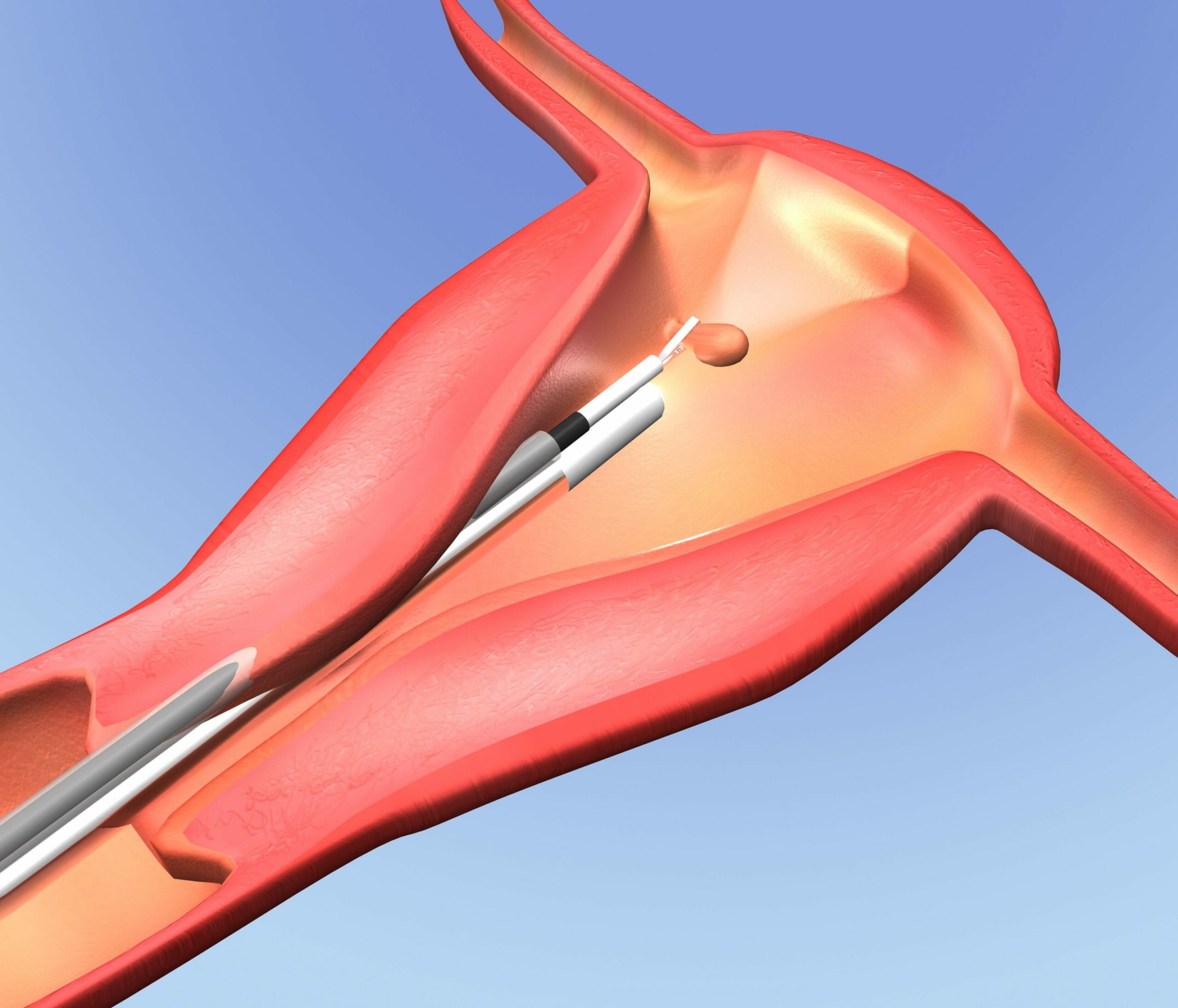
Surgical treatments (e.g., myomectomy) are performed
Camera is contained within a sheath.
These sheaths can also contain operative ports—channelsChannelsThe Cell: Cell Membrane through which surgical instruments can be introduced.
Schematic depiction of a forceps, inserted through the operative channel of a hysteroscope, removing an intrauterine polyp
Image: “Carcinoma Endometrium” by Ruby Hall IVF & Endoscopy Center, Ruby Hall Clinic, Pune, India. License: CC BY 2.0
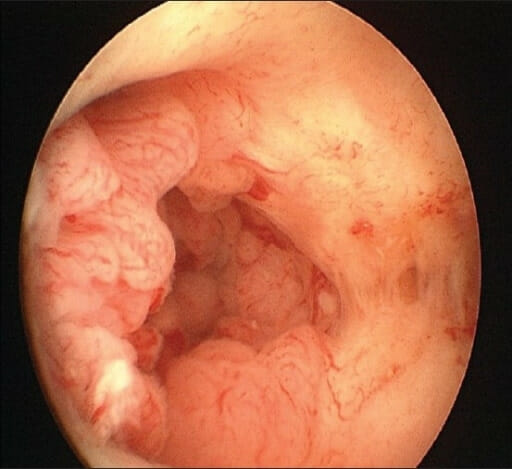
Endometrial polyps on the posterior wall of the uterus, seen on hysteroscopy
Image: “Multiple Endometrial Polyps” by Ruby Hall IVF & Endoscopy Center, Ruby Hall Clinic, Pune, India. License: CC BY 2.0
Complications
Uterine perforationPerforationA pathological hole in an organ, blood vessel or other soft part of the body, occurring in the absence of external force.Esophagitis
Injury to internal organs
Hemorrhage
Infection
Complications from distention media:
Gas embolism
Fluid overload
HyponatremiaHyponatremiaHyponatremia is defined as a decreased serum sodium (sNa+) concentration less than 135 mmol/L. Serum sodium is the greatest contributor to plasma osmolality, which is very tightly controlled via antidiuretic hormone (ADH) release from the hypothalamus and by the thirst mechanism.Hyponatremia
Schematic depiction of hysteroscopy
Image by Lecturio.
Hysterosalpingography (HSG)
Description
A fluoroscopic examination that allows assessment of the uterine cavity shape and patency of the fallopian tubesFallopian tubesThe uterus, cervix, and fallopian tubes are part of the internal female reproductive system. The fallopian tubes receive an ovum after ovulation and help move it and/or a fertilized embryo toward the uterus via ciliated cells lining the tubes and peristaltic movements of its smooth muscle. Uterus, Cervix, and Fallopian Tubes: Anatomy
A catheter is inserted into the uterine cavity → dye is injected → x-rayX-rayPenetrating electromagnetic radiation emitted when the inner orbital electrons of an atom are excited and release radiant energy. X-ray wavelengths range from 1 pm to 10 nm. Hard x-rays are the higher energy, shorter wavelength x-rays. Soft x-rays or grenz rays are less energetic and longer in wavelength. The short wavelength end of the x-ray spectrum overlaps the gamma rays wavelength range. The distinction between gamma rays and x-rays is based on their radiation source.Pulmonary Function Tests images are taken
Indications
Assess tubal patency:
Usually as part of an infertilityInfertilityInfertility is the inability to conceive in the context of regular intercourse. The most common causes of infertility in women are related to ovulatory dysfunction or tubal obstruction, whereas, in men, abnormal sperm is a common cause. Infertility evaluation
Note: The tubal canal is very thin and not visible with other imaging methods.
Suspected congenital uterine anomalies
ContraindicationsContraindicationsA condition or factor associated with a recipient that makes the use of a drug, procedure, or physical agent improper or inadvisable. Contraindications may be absolute (life threatening) or relative (higher risk of complications in which benefits may outweigh risks).Noninvasive Ventilation
PregnancyPregnancyThe status during which female mammals carry their developing young (embryos or fetuses) in utero before birth, beginning from fertilization to birth.Pregnancy: Diagnosis, Physiology, and Care
These procedures are typically done after mensesMensesThe periodic shedding of the endometrium and associated menstrual bleeding in the menstrual cycle of humans and primates. Menstruation is due to the decline in circulating progesterone, and occurs at the late luteal phase when luteolysis of the corpus luteum takes place.Menstrual Cycle but prior to ovulationOvulationThe discharge of an ovum from a rupturing follicle in the ovary.Menstrual Cycle in order to avoid interruption of early viable pregnancies.
For unclear reasons, there is an increased chance of conception during cycles in which HSG is performed.
Active undiagnosed vaginal bleeding
Active pelvic infection
Procedure
A speculum is inserted and the cervixCervixThe uterus, cervix, and fallopian tubes are part of the internal female reproductive system. The most inferior portion of the uterus is the cervix, which connects the uterine cavity to the vagina. Externally, the cervix is lined by stratified squamous cells; however, the cervical canal is lined by columnar epithelium.Uterus, Cervix, and Fallopian Tubes: Anatomy identified.
Balloon is inflated to hold the catheter in place and to prevent leakage of dye.
A tenaculum can be used if needed.
The contrast material is injected slowly and observed on fluoroscopyFluoroscopyProduction of an image when x-rays strike a fluorescent screen.X-rays in real time.
Images are taken.
The balloon is deflated; a final image is usually taken of dye filling the vaginal cavity while the catheter is withdrawn.
Findings
Normal:
Normal uterine cavity contour
Fallopian tubesFallopian tubesThe uterus, cervix, and fallopian tubes are part of the internal female reproductive system. The fallopian tubes receive an ovum after ovulation and help move it and/or a fertilized embryo toward the uterus via ciliated cells lining the tubes and peristaltic movements of its smooth muscle. Uterus, Cervix, and Fallopian Tubes: Anatomy fill with dye → dye is seen spilling out the ends of both tubes into the pelvic cavity (“bilateral fill and spill”)
Dilated fallopian tubeFallopian TubeA pair of highly specialized canals extending from the uterus to its corresponding ovary. They provide the means for ovum transport from the ovaries and they are the site of the ovum’s final maturation and fertilization. The fallopian tube consists of an interstitium, an isthmus, an ampulla, an infundibulum, and fimbriae. Its wall consists of three layers: serous, muscular, and an internal mucosal layer lined with both ciliated and secretory cells.Uterus, Cervix, and Fallopian Tubes: Anatomy → hydrosalpinxHydrosalpinxGynecological Imaging
Normal HSG examination: This radiograph shows a normal uterine contour with bilateral fill and spill of dye from the fallopian tubes.
Image: “Hysterosalpingographic evaluation of primary and secondary infertility” by Muhammad Usman Aziz, MBBS, FCPS (Radiology). Senior Registrar, Department of Radiology, Liaquat National Hospital. License: CC BY 3.0
Complications
Cramping
Leaking contrast material
Vaginal bleeding
FeverFeverFever is defined as a measured body temperature of at least 38°C (100.4°F). Fever is caused by circulating endogenous and/or exogenous pyrogens that increase levels of prostaglandin E2 in the hypothalamus. Fever is commonly associated with chills, rigors, sweating, and flushing of the skin. Fever
Severe painPainAn unpleasant sensation induced by noxious stimuli which are detected by nerve endings of nociceptive neurons.Pain: Types and Pathways
Clinical Relevance
The following conditions are some of the most common gynecologic conditions diagnosed with the procedures discussed on this page:
Cervical cancerCervical cancerCervical cancer, or invasive cervical carcinoma (ICC), is the 3rd most common cancer in women in the world, with > 50% of the cases being fatal. In the United States, ICC is the 13th most common cancer and the cause of < 3% of all cancer deaths due to the slow progression of precursor lesions and, more importantly, effective cancer screening. Cervical Cancer: 3rd most common cancer in women in the world. Owing to effective screeningScreeningPreoperative Care, diagnosis, and treatment, however, most disease can be caught and treated at an early, preinvasive stage. Diagnosis is made using cervical Pap smears with assessing cytology, HPV testingHPV testingCervical Cancer Screening, colposcopyColposcopyThe examination, therapy or surgery of the cervix and vagina by means of a specially designed endoscope introduced vaginally.Cervical Cancer Screening, and biopsies. Preinvasive disease is typically managed with loop electrosurgical excision procedures, while invasive disease requires more significant surgeries.
Abnormal uterine bleedingAbnormal Uterine BleedingAbnormal uterine bleeding is the medical term for abnormalities in the frequency, volume, duration, and regularity of the menstrual cycle. Abnormal uterine bleeding is classified using the acronym PALM-COEIN, with PALM representing the structural causes and COEIN indicating the non-structural causes. Abnormal Uterine Bleeding (AUBAUBAbnormal uterine bleeding is the medical term for abnormalities in the frequency, volume, duration, and regularity of the menstrual cycle. Abnormal uterine bleeding is classified using the acronym palm-coein, with palm representing the structural causes and coein indicating the non-structural causes.Abnormal Uterine Bleeding): collective term for abnormalities in the frequency, volume, duration, and/or regularity of the menstrual cycleMenstrual cycleThe menstrual cycle is the cyclic pattern of hormonal and tissular activity that prepares a suitable uterine environment for the fertilization and implantation of an ovum. The menstrual cycle involves both an endometrial and ovarian cycle that are dependent on one another for proper functioning. There are 2 phases of the ovarian cycle and 3 phases of the endometrial cycle.Menstrual Cycle. Etiologies include Polyps, Adenomyosis, Leiomyomas, Malignancy/hyperplasiaHyperplasiaAn increase in the number of cells in a tissue or organ without tumor formation. It differs from hypertrophy, which is an increase in bulk without an increase in the number of cells.Cellular Adaptation, Coagulopathy, Ovulatory dysfunction, Endometritis and atrophyAtrophyDecrease in the size of a cell, tissue, organ, or multiple organs, associated with a variety of pathological conditions such as abnormal cellular changes, ischemia, malnutrition, or hormonal changes.Cellular Adaptation, Iatrogenic causes (official acronym: PALM-COEINPALM-COEINAbnormal uterine bleeding is the medical term for abnormalities in the frequency, volume, duration, and regularity of the menstrual cycle. Abnormal uterine bleeding is classified using the acronym palm-coein, with palm representing the structural causes and coein indicating the non-structural causes.Abnormal Uterine Bleeding). Diagnosis is done by extensive history taking, exam, lab workup, sonography, and/or endometrial biopsyBiopsyRemoval and pathologic examination of specimens from the living body.Ewing Sarcoma. Treatment is based on the underlying etiology.
Uterine leiomyomaLeiomyomaA benign tumor derived from smooth muscle tissue, also known as a fibroid tumor. They rarely occur outside of the uterus and the gastrointestinal tract but can occur in the skin and subcutaneous tissue, probably arising from the smooth muscle of small blood vessels in these tissues.Infertility:benignBenignFibroadenoma tumors arising from smooth muscle cells in the uterine myometrium. Uterineleiomyomas are usually easily identified as a hypoechoicHypoechoicA structure that produces a low-amplitude echo (darker grays)Ultrasound (Sonography), well-circumscribed, round masses on pelvic sonography. Treatment for leiomyomas may include surgical resection or medical options to reduce bleeding and/or bulk symptoms.
Endometrial polypsEndometrial polypsEndometrial polyps are pedunculated or sessile projections of the endometrium that result from overgrowth of endometrial glands and stroma around a central vascular stalk. Endometrial polyps are a few millimeters to a few centimeters in size, can occur anywhere within the uterine cavity, and, while usually benign, can be malignant, particularly in postmenopausal women. Endometrial Polyps: pedunculated or sessile projections of the endometriumEndometriumThe mucous membrane lining of the uterine cavity that is hormonally responsive during the menstrual cycle and pregnancy. The endometrium undergoes cyclic changes that characterize menstruation. After successful fertilization, it serves to sustain the developing embryo.Embryoblast and Trophoblast Development that result from overgrowth of endometrial glands and stroma around a central vascular stalk. Endometrial polypsEndometrial polypsEndometrial polyps are pedunculated or sessile projections of the endometrium that result from overgrowth of endometrial glands and stroma around a central vascular stalk. Endometrial polyps are a few millimeters to a few centimeters in size, can occur anywhere within the uterine cavity, and, while usually benign, can be malignant, particularly in postmenopausal women. Endometrial Polyps are a few millimeters to a few centimeters in size and can occur anywhere within the uterine cavity. These polyps can become malignant in postmenopausal women. Endometrial polypsEndometrial polypsEndometrial polyps are pedunculated or sessile projections of the endometrium that result from overgrowth of endometrial glands and stroma around a central vascular stalk. Endometrial polyps are a few millimeters to a few centimeters in size, can occur anywhere within the uterine cavity, and, while usually benign, can be malignant, particularly in postmenopausal women. Endometrial Polyps are suspected on a sonogramSonogramChorioretinitis showing a thickened endometriumEndometriumThe mucous membrane lining of the uterine cavity that is hormonally responsive during the menstrual cycle and pregnancy. The endometrium undergoes cyclic changes that characterize menstruation. After successful fertilization, it serves to sustain the developing embryo.Embryoblast and Trophoblast Development and are more definitively diagnosed on SISSISInfertility, HSG, and/or hysteroscopy and are usually treated with hysteroscopic resection.
AdenomyosisAdenomyosisAdenomyosis is a benign uterine condition characterized by the presence of ectopic endometrial glands and stroma within the myometrium. Adenomyosis is a common condition, affecting 20%-35% of women, and typically presents with heavy menstrual bleeding and dysmenorrhea. Adenomyosis:benignBenignFibroadenoma uterine condition characterized by the presence of ectopic endometrial glands and stroma within the myometrium. AdenomyosisAdenomyosisAdenomyosis is a benign uterine condition characterized by the presence of ectopic endometrial glands and stroma within the myometrium. Adenomyosis is a common condition, affecting 20%-35% of women, and typically presents with heavy menstrual bleeding and dysmenorrhea. Adenomyosis typically presents with heavy menstrual bleedingHeavy menstrual bleedingExcessive menstrual blood loss (objectively defined as > 80 mL blood loss/cycle). Can be based on heavy flow, as determined by the patientAbnormal Uterine Bleeding and dysmenorrhea. Diagnosis is often made during history taking, with numerous findings suggestive of adenomyosisAdenomyosisAdenomyosis is a benign uterine condition characterized by the presence of ectopic endometrial glands and stroma within the myometrium. Adenomyosis is a common condition, affecting 20%-35% of women, and typically presents with heavy menstrual bleeding and dysmenorrhea. Adenomyosis on ultrasound. Management is based on the woman’s preference regarding future childbearing and may include hysterectomy (definitive treatment), other surgical options, or medical hormonal suppressionSuppressionDefense Mechanisms.
Ovarian cystsCystsAny fluid-filled closed cavity or sac that is lined by an epithelium. Cysts can be of normal, abnormal, non-neoplastic, or neoplastic tissues.Fibrocystic Change: collections of fluid or semiliquid material, often walled off by a membrane, located in the ovary. When the cystsCystsAny fluid-filled closed cavity or sac that is lined by an epithelium. Cysts can be of normal, abnormal, non-neoplastic, or neoplastic tissues.Fibrocystic Change occur as a result of normal physiologic processes, they are called functional, whereas if there is abnormal growth of ovarian cells, the cyst is referred to as neoplastic. Neoplastic ovarian cystsCystsAny fluid-filled closed cavity or sac that is lined by an epithelium. Cysts can be of normal, abnormal, non-neoplastic, or neoplastic tissues.Fibrocystic Change are subcategorized as either benignBenignFibroadenoma or malignant and are categorized according to their cell of origin (e.g., epithelial, germ cell, or stromal cells).
Vulvar dermatoses:
Lichen sclerosisSclerosisA pathological process consisting of hardening or fibrosis of an anatomical structure, often a vessel or a nerve.Wilms Tumor(LS): chronic, progressive dermatologic condition of the vulvaVulvaThe vulva is the external genitalia of the female and includes the mons pubis, labia majora, labia minora, clitoris, vestibule, vestibular bulb, and greater vestibular glands. Vagina, Vulva, and Pelvic Floor: Anatomy that is characterized by inflammationInflammationInflammation is a complex set of responses to infection and injury involving leukocytes as the principal cellular mediators in the body’s defense against pathogenic organisms. Inflammation is also seen as a response to tissue injury in the process of wound healing. The 5 cardinal signs of inflammation are pain, heat, redness, swelling, and loss of function. Inflammation and epithelial thinning, visible as porcelain-white “parchment-like” vulvar plaques; as LS progresses, scarringScarringInflammation can distort the anatomy. Lichen sclerosisSclerosisA pathological process consisting of hardening or fibrosis of an anatomical structure, often a vessel or a nerve.Wilms Tumor itself is benignBenignFibroadenoma, but it is associated with an increased risk for vulvar squamous cell carcinomaSquamous cell carcinomaCutaneous squamous cell carcinoma (cSCC) is caused by malignant proliferation of atypical keratinocytes. This condition is the 2nd most common skin malignancy and usually affects sun-exposed areas of fair-skinned patients. The cancer presents as a firm, erythematous, keratotic plaque or papule. Squamous Cell Carcinoma (SCC) (SCC). Diagnosis is clinical, but definitive diagnosis requires vulvar biopsyBiopsyRemoval and pathologic examination of specimens from the living body.Ewing Sarcoma, which shows a thinned epidermal layer and homogenization of collagenCollagenA polypeptide substance comprising about one third of the total protein in mammalian organisms. It is the main constituent of skin; connective tissue; and the organic substance of bones (bone and bones) and teeth (tooth).Connective Tissue: Histology in the upper dermisDermisA layer of vascularized connective tissue underneath the epidermis. The surface of the dermis contains innervated papillae. Embedded in or beneath the dermis are sweat glands; hair follicles; and sebaceous glands.Skin: Structure and Functions.
Lichen simplex chronicusLichen Simplex ChronicusA benign vulvar skin disorder characterized by hyperkeratosis (thickening of the skin) that occurs secondary to chronic vulvar irritationBenign Vulvar Conditions: benignBenignFibroadenoma vulvar skinSkinThe skin, also referred to as the integumentary system, is the largest organ of the body. The skin is primarily composed of the epidermis (outer layer) and dermis (deep layer). The epidermis is primarily composed of keratinocytes that undergo rapid turnover, while the dermis contains dense layers of connective tissue.Skin: Structure and Functions disorder characterized by hyperkeratosisHyperkeratosisIchthyosis Vulgaris (thickening of the skinSkinThe skin, also referred to as the integumentary system, is the largest organ of the body. The skin is primarily composed of the epidermis (outer layer) and dermis (deep layer). The epidermis is primarily composed of keratinocytes that undergo rapid turnover, while the dermis contains dense layers of connective tissue.Skin: Structure and Functions) that occurs secondary to chronic vulvar irritation. Lichen simplex chronicusLichen Simplex ChronicusA benign vulvar skin disorder characterized by hyperkeratosis (thickening of the skin) that occurs secondary to chronic vulvar irritationBenign Vulvar Conditions presents with intense pruritusPruritusAn intense itching sensation that produces the urge to rub or scratch the skin to obtain relief.Atopic Dermatitis (Eczema), often worse at night, and well-demarcated, dry patchy plaques; the skinSkinThe skin, also referred to as the integumentary system, is the largest organ of the body. The skin is primarily composed of the epidermis (outer layer) and dermis (deep layer). The epidermis is primarily composed of keratinocytes that undergo rapid turnover, while the dermis contains dense layers of connective tissue.Skin: Structure and Functions is usually thick, scaly, and firm and may appear similar to some vulvar malignancies. Diagnosis is usually clinical (but a biopsyBiopsyRemoval and pathologic examination of specimens from the living body.Ewing Sarcoma may be needed to rule out malignancyMalignancyHemothorax) and shows a hypertrophic epidermal layer.