Peripheral artery disease (PAD) is obstruction of the arterial lumen resulting in decreased blood flow to the distal limbs. The disease can be a result of atherosclerosis or thrombosis. Patients may be asymptomatic or have progressive claudication, skin discoloration, ischemic ulcers, or gangrene. Onset may be insidious (atherosclerosis) or abrupt (thrombosis). Diagnosis is made with the clinical history, exam, and measurement of the ankle-brachial index. Imaging studies can determine the location and extent of the arterial disease. Treatment varies depending on the severity but can include lifestyle modifications, antiplatelet therapy, risk factor modifications, phosphodiesterase inhibitors, and revascularization.
Affects approximately 10% of the population > 55 years of age in the United States
Worldwide prevalencePrevalenceThe total number of cases of a given disease in a specified population at a designated time. It is differentiated from incidence, which refers to the number of new cases in the population at a given time.Measures of Disease Frequency is 3%–12%
Men > women
Higher prevalencePrevalenceThe total number of cases of a given disease in a specified population at a designated time. It is differentiated from incidence, which refers to the number of new cases in the population at a given time.Measures of Disease Frequency among African Americans and Hispanic American women
Higher rates seen in older patientsPatientsIndividuals participating in the health care system for the purpose of receiving therapeutic, diagnostic, or preventive procedures.Clinician–Patient Relationship, families with atherosclerosisAtherosclerosisAtherosclerosis is a common form of arterial disease in which lipid deposition forms a plaque in the blood vessel walls. Atherosclerosis is an incurable disease, for which there are clearly defined risk factors that often can be reduced through a change in lifestyle and behavior of the patient. Atherosclerosis and those with cardiovascular disease
Etiology[1,4,7,11]
Peripheral artery diseasePeripheral artery diseasePeripheral artery disease (PAD) is obstruction of the arterial lumen resulting in decreased blood flow to the distal limbs. The disease can be a result of atherosclerosis or thrombosis. Patients may be asymptomatic or have progressive claudication, skin discoloration, ischemic ulcers, or gangrene. Peripheral Artery Disease (PAD) usually has the same causative factors as coronary and carotid disease.
AtherosclerosisAtherosclerosisAtherosclerosis is a common form of arterial disease in which lipid deposition forms a plaque in the blood vessel walls. Atherosclerosis is an incurable disease, for which there are clearly defined risk factors that often can be reduced through a change in lifestyle and behavior of the patient. Atherosclerosis:
Aging
HypertensionHypertensionHypertension, or high blood pressure, is a common disease that manifests as elevated systemic arterial pressures. Hypertension is most often asymptomatic and is found incidentally as part of a routine physical examination or during triage for an unrelated medical encounter. Hypertension
SmokingSmokingWillful or deliberate act of inhaling and exhaling smoke from burning substances or agents held by hand.Interstitial Lung Diseases and tobacco use
DiabetesDiabetesDiabetes mellitus (DM) is a metabolic disease characterized by hyperglycemia and dysfunction of the regulation of glucose metabolism by insulin. Type 1 DM is diagnosed mostly in children and young adults as the result of autoimmune destruction of β cells in the pancreas and the resulting lack of insulin. Type 2 DM has a significant association with obesity and is characterized by insulin resistance.Diabetes Mellitus mellitus
HypercholesterolemiaHypercholesterolemiaA condition with abnormally high levels of cholesterol in the blood. It is defined as a cholesterol value exceeding the 95th percentile for the population.Lipid Disorders
Hyperhomocysteinemia
ObesityObesityObesity is a condition associated with excess body weight, specifically with the deposition of excessive adipose tissue. Obesity is considered a global epidemic. Major influences come from the western diet and sedentary lifestyles, but the exact mechanisms likely include a mixture of genetic and environmental factors. Obesity
Male sexSexThe totality of characteristics of reproductive structure, functions, phenotype, and genotype, differentiating the male from the female organism.Gender Dysphoria
Family historyFamily HistoryAdult Health Maintenance of atherosclerosisAtherosclerosisAtherosclerosis is a common form of arterial disease in which lipid deposition forms a plaque in the blood vessel walls. Atherosclerosis is an incurable disease, for which there are clearly defined risk factors that often can be reduced through a change in lifestyle and behavior of the patient. Atherosclerosis
Arterial embolism:
CholesterolCholesterolThe principal sterol of all higher animals, distributed in body tissues, especially the brain and spinal cord, and in animal fats and oils.Cholesterol MetabolismatheroembolismAtheroembolismAn embolus is an intravascular solid, liquid, or gaseous material that is carried by the blood to a site distant from its point of origin. Emboli of all types warrant immediate medical attention. The majority of emboli dislodge from a thrombus, forming a thromboembolus. Other less common nonthrombotic types of emboli are cholesterol, fat, air, amniotic fluid, and tumor emboli.Nonthrombotic Embolism
Atrial fibrillationAtrial fibrillationAtrial fibrillation (AF or Afib) is a supraventricular tachyarrhythmia and the most common kind of arrhythmia. It is caused by rapid, uncontrolled atrial contractions and uncoordinated ventricular responses. Atrial Fibrillation
EndocarditisEndocarditisEndocarditis is an inflammatory disease involving the inner lining (endometrium) of the heart, most commonly affecting the cardiac valves. Both infectious and noninfectious etiologies lead to vegetations on the valve leaflets. Patients may present with nonspecific symptoms such as fever and fatigue. Endocarditis
Artificial heart valves
Functional:
Vasospasm
Previous injury to the limb
Risk groups:
Age ≥ 70 years
Age 50–69 years + smokingSmokingWillful or deliberate act of inhaling and exhaling smoke from burning substances or agents held by hand.Interstitial Lung Diseases or diabetesDiabetesDiabetes mellitus (DM) is a metabolic disease characterized by hyperglycemia and dysfunction of the regulation of glucose metabolism by insulin. Type 1 DM is diagnosed mostly in children and young adults as the result of autoimmune destruction of β cells in the pancreas and the resulting lack of insulin. Type 2 DM has a significant association with obesity and is characterized by insulin resistance.Diabetes Mellitus
Age 40–49 + diabetesDiabetesDiabetes mellitus (DM) is a metabolic disease characterized by hyperglycemia and dysfunction of the regulation of glucose metabolism by insulin. Type 1 DM is diagnosed mostly in children and young adults as the result of autoimmune destruction of β cells in the pancreas and the resulting lack of insulin. Type 2 DM has a significant association with obesity and is characterized by insulin resistance.Diabetes Mellitus + 1 other risk factor for atherosclerosisAtherosclerosisAtherosclerosis is a common form of arterial disease in which lipid deposition forms a plaque in the blood vessel walls. Atherosclerosis is an incurable disease, for which there are clearly defined risk factors that often can be reduced through a change in lifestyle and behavior of the patient. Atherosclerosis
AtherosclerosisAtherosclerosisAtherosclerosis is a common form of arterial disease in which lipid deposition forms a plaque in the blood vessel walls. Atherosclerosis is an incurable disease, for which there are clearly defined risk factors that often can be reduced through a change in lifestyle and behavior of the patient. Atherosclerosis at other sites (e.g., coronary, carotid, renal)
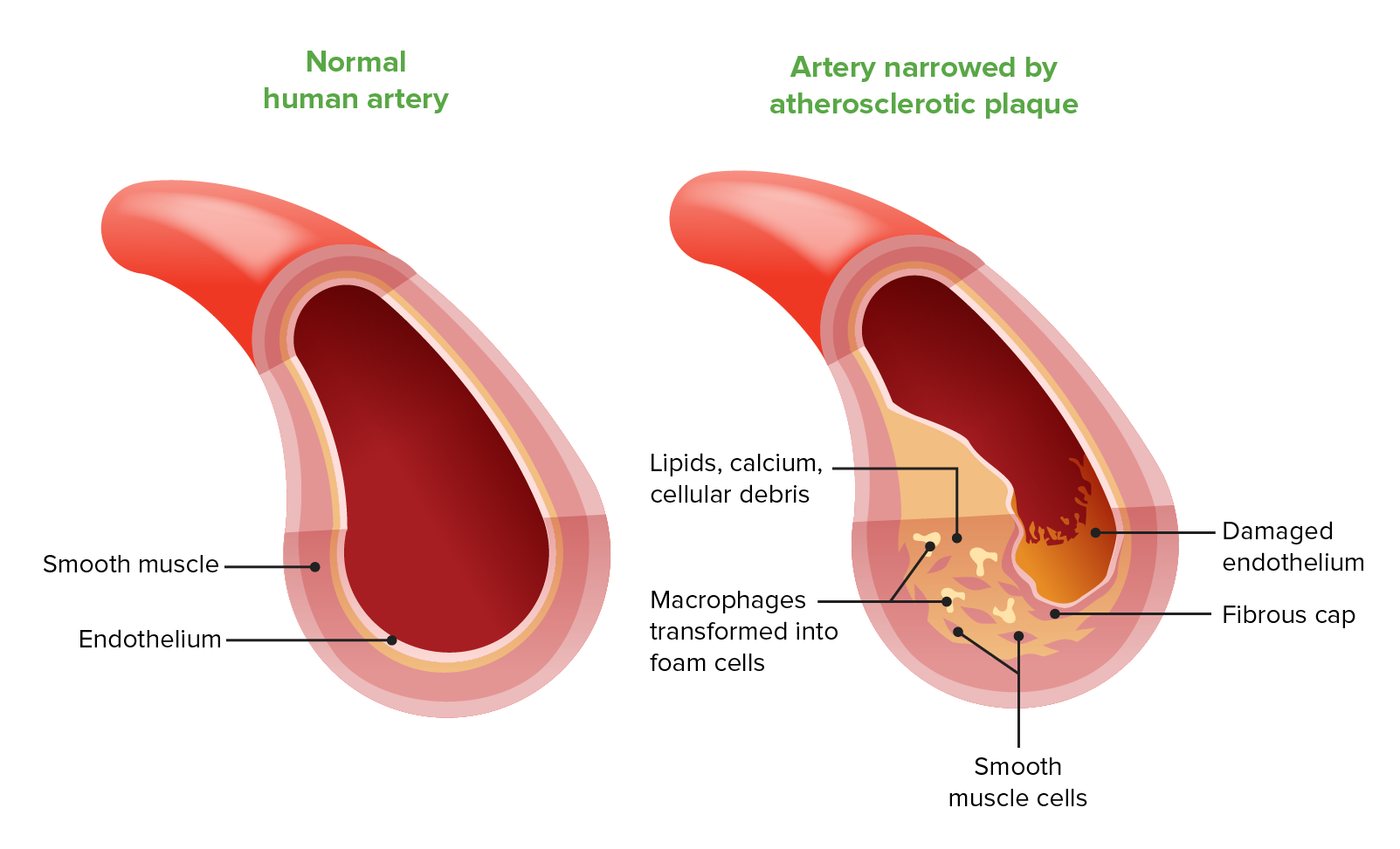
AtherosclerosisAtherosclerosisAtherosclerosis is a common form of arterial disease in which lipid deposition forms a plaque in the blood vessel walls. Atherosclerosis is an incurable disease, for which there are clearly defined risk factors that often can be reduced through a change in lifestyle and behavior of the patient. Atherosclerosis:[6,8]
Endothelial cell dysfunction → macrophage and cholesterolCholesterolThe principal sterol of all higher animals, distributed in body tissues, especially the brain and spinal cord, and in animal fats and oils.Cholesterol Metabolism accumulation → foam cell formation
Platelet-derived growth factorPlatelet-derived growth factorMitogenic peptide growth hormone carried in the alpha-granules of platelets. It is released when platelets adhere to traumatized tissues. Connective tissue cells near the traumatized region respond by initiating the process of replication.Hypertrophic and Keloid Scars (PDGF) and fibroblast growth factorFibroblast growth factorA family of small polypeptide growth factors that share several common features including a strong affinity for heparin, and a central barrel-shaped core region of 140 amino acids that is highly homologous between family members. Although originally studied as proteins that stimulate the growth of fibroblasts this distinction is no longer a requirement for membership in the fibroblast growth factor family.X-linked Hypophosphatemic Rickets (FGF) release → smooth muscle cell migration → proliferation and extracellular matrixExtracellular matrixA meshwork-like substance found within the extracellular space and in association with the basement membrane of the cell surface. It promotes cellular proliferation and provides a supporting structure to which cells or cell lysates in culture dishes adhere.Hypertrophic and Keloid Scars deposition → fibrousFibrousFibrocystic ChangeplaquePlaquePrimary Skin Lesions
Subintimal accumulation of lipid and fibrousFibrousFibrocystic Change material of the arteriesArteriesArteries are tubular collections of cells that transport oxygenated blood and nutrients from the heart to the tissues of the body. The blood passes through the arteries in order of decreasing luminal diameter, starting in the largest artery (the aorta) and ending in the small arterioles. Arteries are classified into 3 types: large elastic arteries, medium muscular arteries, and small arteries and arterioles. Arteries: Histology → vessel lumen narrowing → restricted blood flowBlood flowBlood flow refers to the movement of a certain volume of blood through the vasculature over a given unit of time (e.g., mL per minute).Vascular Resistance, Flow, and Mean Arterial Pressure → chronic ischemiaIschemiaA hypoperfusion of the blood through an organ or tissue caused by a pathologic constriction or obstruction of its blood vessels, or an absence of blood circulation.Ischemic Cell Damage of the affected limb
PlaquePlaquePrimary Skin Lesions rupture or thrombosisThrombosisFormation and development of a thrombus or blood clot in the blood vessel.Epidemic Typhus → abrupt occlusion of downstream vessels → acute ischemiaIschemiaA hypoperfusion of the blood through an organ or tissue caused by a pathologic constriction or obstruction of its blood vessels, or an absence of blood circulation.Ischemic Cell Damage of the limb
Composition of the fibrous plaque in atherosclerosis
PatientsPatientsIndividuals participating in the health care system for the purpose of receiving therapeutic, diagnostic, or preventive procedures.Clinician–Patient Relationship with PAD may be asymptomatic (20%–25%) or present with evidence of chronic or acute limb ischemiaIschemiaA hypoperfusion of the blood through an organ or tissue caused by a pathologic constriction or obstruction of its blood vessels, or an absence of blood circulation.Ischemic Cell Damage.
Intermittent claudicationIntermittent claudicationA symptom complex characterized by pain and weakness in skeletal muscle group associated with exercise, such as leg pain and weakness brought on by walking. Such muscle limpness disappears after a brief rest and is often relates to arterial stenosis; muscle ischemia; and accumulation of lactate.Thromboangiitis Obliterans (Buerger Disease):
Reproducible, painful cramping in the buttock, hips, thighs, calf, or footFootThe foot is the terminal portion of the lower limb, whose primary function is to bear weight and facilitate locomotion. The foot comprises 26 bones, including the tarsal bones, metatarsal bones, and phalanges. The bones of the foot form longitudinal and transverse arches and are supported by various muscles, ligaments, and tendons.Foot: Anatomy with exertion
Relieved with rest
Atypical limb painPainAn unpleasant sensation induced by noxious stimuli which are detected by nerve endings of nociceptive neurons.Pain: Types and Pathways or feeling of “heaviness”
Limb numbness or weakness
Impotence, erectile dysfunctionErectile DysfunctionErectile dysfunction (ED) is defined as the inability to achieve or maintain a penile erection, resulting in difficulty to perform penetrative sexual intercourse. Local penile factors and systemic diseases, including diabetes, cardiac disease, and neurological disorders, can cause ED. Erectile Dysfunction
CyanosisCyanosisA bluish or purplish discoloration of the skin and mucous membranes due to an increase in the amount of deoxygenated hemoglobin in the blood or a structural defect in the hemoglobin molecule.Pulmonary Examination
Weak or absent pulse below a narrowed area of the artery
Bruits over arteriesArteriesArteries are tubular collections of cells that transport oxygenated blood and nutrients from the heart to the tissues of the body. The blood passes through the arteries in order of decreasing luminal diameter, starting in the largest artery (the aorta) and ending in the small arterioles. Arteries are classified into 3 types: large elastic arteries, medium muscular arteries, and small arteries and arterioles. Arteries: Histology heard on auscultation
Decreased blood pressure in the affected limb
Severe disease:
Loss of hair follicles, sweat glandsSweat glandsSweat-producing structures that are embedded in the dermis. Each gland consists of a single tube, a coiled body, and a superficial duct.Soft Tissue Abscess, and sebaceous glands
SkinSkinThe skin, also referred to as the integumentary system, is the largest organ of the body. The skin is primarily composed of the epidermis (outer layer) and dermis (deep layer). The epidermis is primarily composed of keratinocytes that undergo rapid turnover, while the dermis contains dense layers of connective tissue.Skin: Structure and Functions becomes smooth and shiny.
Table: Fontaine classificationFontaine classificationPeripheral Artery Disease of peripheral artery diseasePeripheral artery diseasePeripheral artery disease (PAD) is obstruction of the arterial lumen resulting in decreased blood flow to the distal limbs. The disease can be a result of atherosclerosis or thrombosis. Patients may be asymptomatic or have progressive claudication, skin discoloration, ischemic ulcers, or gangrene. Peripheral Artery Disease
Stage
Symptoms
1
Asymptomatic
2a
Intermittent claudicationIntermittent claudicationA symptom complex characterized by pain and weakness in skeletal muscle group associated with exercise, such as leg pain and weakness brought on by walking. Such muscle limpness disappears after a brief rest and is often relates to arterial stenosis; muscle ischemia; and accumulation of lactate.Thromboangiitis Obliterans (Buerger Disease) after walking > 200 meters (219 yards)
2b
Intermittent claudicationIntermittent claudicationA symptom complex characterized by pain and weakness in skeletal muscle group associated with exercise, such as leg pain and weakness brought on by walking. Such muscle limpness disappears after a brief rest and is often relates to arterial stenosis; muscle ischemia; and accumulation of lactate.Thromboangiitis Obliterans (Buerger Disease) after walking < 200 meters (219 yards)
3
Nocturnal or resting painPainAn unpleasant sensation induced by noxious stimuli which are detected by nerve endings of nociceptive neurons.Pain: Types and Pathways
4
NecrosisNecrosisThe death of cells in an organ or tissue due to disease, injury or failure of the blood supply.Ischemic Cell Damage or gangreneGangreneDeath and putrefaction of tissue usually due to a loss of blood supply.Small Bowel Obstruction of the limb
Rutherford classification:[6,35]
Used to determine the severity of PAD with greater precision
Categories are based on clinical symptoms and objective vascular testing criteria, such as ankle pressure measurements (see table).
Table: Rutherford classification of peripheral artery diseasePeripheral artery diseasePeripheral artery disease (PAD) is obstruction of the arterial lumen resulting in decreased blood flow to the distal limbs. The disease can be a result of atherosclerosis or thrombosis. Patients may be asymptomatic or have progressive claudication, skin discoloration, ischemic ulcers, or gangrene. Peripheral Artery Disease
Grade
Category
Clinical Description
Objective Criteria
0
0
Asymptomatic
Normal treadmill/stress test
I
1
Mild claudication
Completes treadmill test; AP after exercise > 50 mmHg but at least 20 mmHg lower than resting
I
2
Moderate claudication
Between categories 1 and 3
I
3
Severe claudication
Cannot complete standard treadmill test; AP after exercise < 50 mmHg
II
4
Ischemic rest painPainAn unpleasant sensation induced by noxious stimuli which are detected by nerve endings of nociceptive neurons.Pain: Types and Pathways
Resting AP < 40 mmHg, ankle/metatarsal PVR flat or barely pulsatile; TP < 30 mmHg
III
5
Minor tissue loss (non-healing ulcers, focal areas of poor blood flowBlood flowBlood flow refers to the movement of a certain volume of blood through the vasculature over a given unit of time (e.g., mL per minute).Vascular Resistance, Flow, and Mean Arterial Pressure)
Resting AP < 60 mmHg, ankle/metatarsal PVR flat or barely pulsatile; TP < 40 mmHg
IV
6
Major tissue loss (damage extends above TMTmTubular System level, non-salvageable footFootThe foot is the terminal portion of the lower limb, whose primary function is to bear weight and facilitate locomotion. The foot comprises 26 bones, including the tarsal bones, metatarsal bones, and phalanges. The bones of the foot form longitudinal and transverse arches and are supported by various muscles, ligaments, and tendons.Foot: Anatomy)
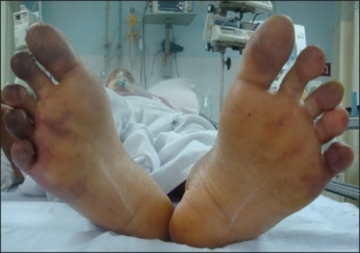
Image: “Bilateral toes showing cyanosis” by Department of Anaesthesia and Intensive Care, Bhubaneswar, India. License: CC BY 2.0
Chronic limb-threatening (critical) ischemiaIschemiaA hypoperfusion of the blood through an organ or tissue caused by a pathologic constriction or obstruction of its blood vessels, or an absence of blood circulation.Ischemic Cell Damage[1,2,6]
Any 1 of the following indicates that blood flowBlood flowBlood flow refers to the movement of a certain volume of blood through the vasculature over a given unit of time (e.g., mL per minute).Vascular Resistance, Flow, and Mean Arterial Pressure no longer meets the metabolic demands of the limb tissues at rest:
Resting painPainAn unpleasant sensation induced by noxious stimuli which are detected by nerve endings of nociceptive neurons.Pain: Types and Pathways:
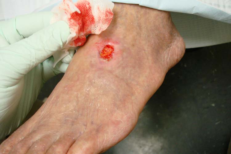
Non-healing arterial ulcer on the dorsum of the foot in a patient with PAD
Image: “Severe peripheral arterial disease” by Jonathan Moore. License: CC BY 3.0
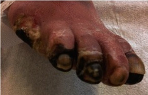
Peripheral arterial disease resulting in necrosis of multiple toes
Image: “Peripheral arterial disease” by Karl-Christian Münter. License: CC BY 4.0
Acute limb ischemiaIschemiaA hypoperfusion of the blood through an organ or tissue caused by a pathologic constriction or obstruction of its blood vessels, or an absence of blood circulation.Ischemic Cell Damage[8]
Acute limb ischemiaIschemiaA hypoperfusion of the blood through an organ or tissue caused by a pathologic constriction or obstruction of its blood vessels, or an absence of blood circulation.Ischemic Cell Damage is most commonly due to an embolism or plaquePlaquePrimary Skin Lesions rupture. PatientsPatientsIndividuals participating in the health care system for the purpose of receiving therapeutic, diagnostic, or preventive procedures.Clinician–Patient Relationship will demonstrate acute onset of:
6 Ps:
Pain
Pallor
Pulseless
Poikilothermia (cold)
Paresthesia
Paralysis
Blue toe syndromeBlue toe syndromeA condition that is caused by recurring atheroembolism in the lower extremities. It is characterized by cyanotic discoloration of the toes, usually the first, fourth, and fifth toes. Discoloration may extend to the lateral aspect of the foot. Despite the gangrene-like appearance, blue toes may respond to conservative therapy without amputation.Peripheral Artery Disease:
Due to cholesterolCholesterolThe principal sterol of all higher animals, distributed in body tissues, especially the brain and spinal cord, and in animal fats and oils.Cholesterol MetabolismatheroembolismAtheroembolismAn embolus is an intravascular solid, liquid, or gaseous material that is carried by the blood to a site distant from its point of origin. Emboli of all types warrant immediate medical attention. The majority of emboli dislodge from a thrombus, forming a thromboembolus. Other less common nonthrombotic types of emboli are cholesterol, fat, air, amniotic fluid, and tumor emboli.Nonthrombotic Embolism to digital arteriesArteriesArteries are tubular collections of cells that transport oxygenated blood and nutrients from the heart to the tissues of the body. The blood passes through the arteries in order of decreasing luminal diameter, starting in the largest artery (the aorta) and ending in the small arterioles. Arteries are classified into 3 types: large elastic arteries, medium muscular arteries, and small arteries and arterioles. Arteries: Histology
Categories of Severity in acute limb ischemiaIschemiaA hypoperfusion of the blood through an organ or tissue caused by a pathologic constriction or obstruction of its blood vessels, or an absence of blood circulation.Ischemic Cell Damage[2,35]
Viable or not immediately threatened (I)
Mild (not continuous)) painPainAn unpleasant sensation induced by noxious stimuli which are detected by nerve endings of nociceptive neurons.Pain: Types and Pathways
Intact capillary refill
No neurologic deficit
Audible arterial dopplerDopplerUltrasonography applying the doppler effect, with frequency-shifted ultrasound reflections produced by moving targets (usually red blood cells) in the bloodstream along the ultrasound axis in direct proportion to the velocity of movement of the targets, to determine both direction and velocity of blood flow.Ultrasound (Sonography)
Threatenedviability(II): reversible limb ischemiaIschemiaA hypoperfusion of the blood through an organ or tissue caused by a pathologic constriction or obstruction of its blood vessels, or an absence of blood circulation.Ischemic Cell Damage (salvageable if arterial obstruction is relieved immediately)
Moderate (not continuous) painPainAn unpleasant sensation induced by noxious stimuli which are detected by nerve endings of nociceptive neurons.Pain: Types and Pathways
Inaudible arterial dopplerDopplerUltrasonography applying the doppler effect, with frequency-shifted ultrasound reflections produced by moving targets (usually red blood cells) in the bloodstream along the ultrasound axis in direct proportion to the velocity of movement of the targets, to determine both direction and velocity of blood flow.Ultrasound (Sonography)
Severe (continuous) painPainAn unpleasant sensation induced by noxious stimuli which are detected by nerve endings of nociceptive neurons.Pain: Types and Pathways
Inaudible arterial dopplerDopplerUltrasonography applying the doppler effect, with frequency-shifted ultrasound reflections produced by moving targets (usually red blood cells) in the bloodstream along the ultrasound axis in direct proportion to the velocity of movement of the targets, to determine both direction and velocity of blood flow.Ultrasound (Sonography)
Nonviable (III): major irreversible ischemic damage; significant neuromuscular damage expected (major amputationAmputationAn amputation is the separation of a portion of the limb or the entire limb from the body, along with the bone. Amputations are generally indicated for conditions that compromise the viability of the limb or promote the spread of a local process that could manifest systemically. Amputation needed)
Absent capillary refill
Neurologic deficit: profound sensorySensoryNeurons which conduct nerve impulses to the central nervous system.Nervous System: Histology loss, + paralysis
Inaudible arterial dopplerDopplerUltrasonography applying the doppler effect, with frequency-shifted ultrasound reflections produced by moving targets (usually red blood cells) in the bloodstream along the ultrasound axis in direct proportion to the velocity of movement of the targets, to determine both direction and velocity of blood flow.Ultrasound (Sonography)
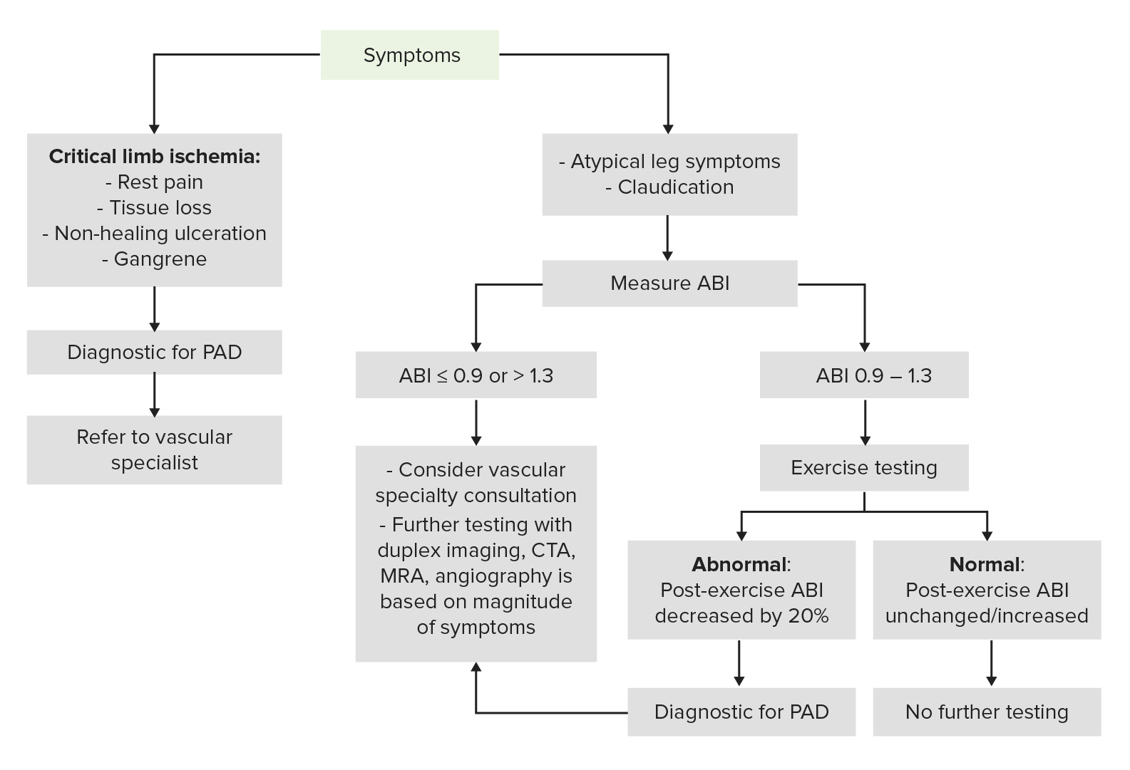
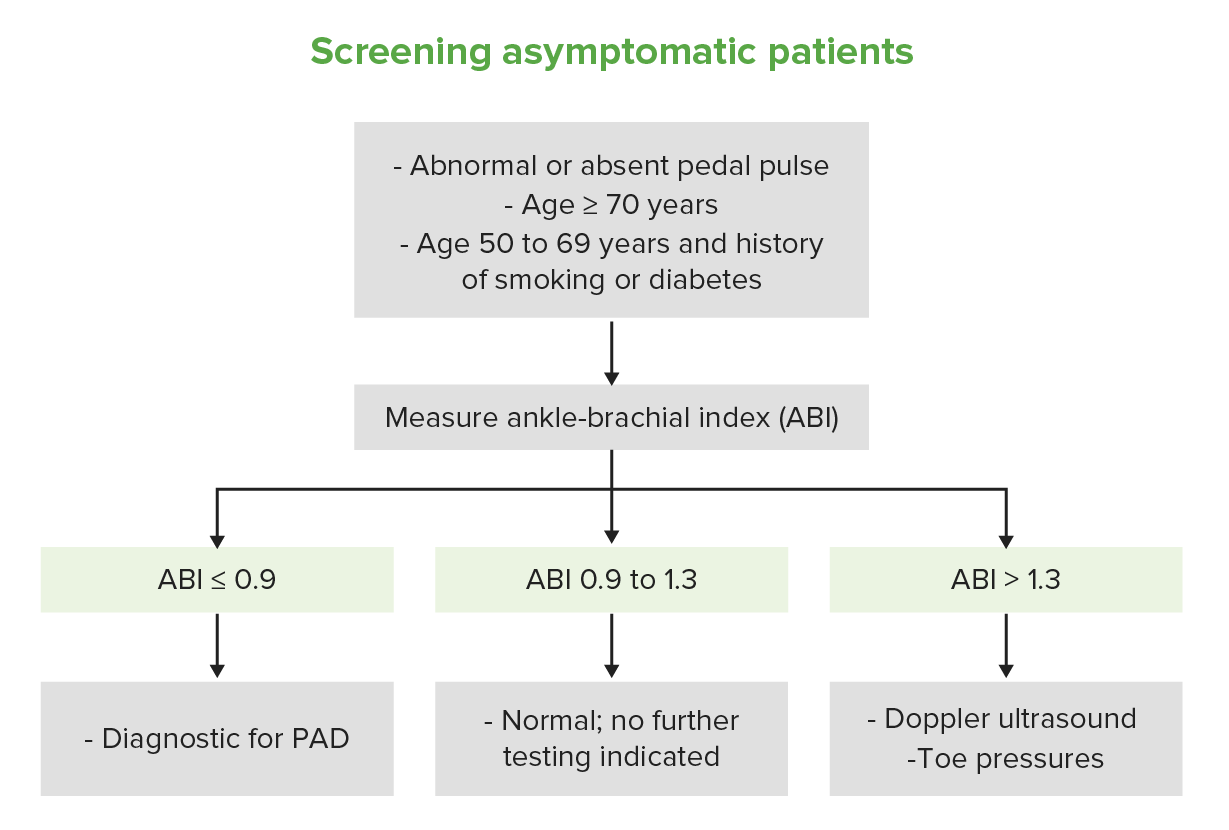
This algorithm demonstrates the diagnostic pathway for a patient presenting with signs or symptoms of PAD:
Diagnostic algorithm for PAD
Image by Lecturio.
The diagnosis is usually established through clinical history, exam, and non-invasive testing techniques (ankle-brachial indexAnkle-brachial indexComparison of the blood pressure between the brachial artery and the posterior tibial artery. It is a predictor of peripheral arterial disease.Cardiovascular Examination (ABI), exercise testing).
Note that those with features of limb-threatening ischemiaIschemiaA hypoperfusion of the blood through an organ or tissue caused by a pathologic constriction or obstruction of its blood vessels, or an absence of blood circulation.Ischemic Cell Damage need urgent vascular surgeryVascular surgeryVascular surgery is the specialized field of medicine that focuses on the surgical management of the pathologies of the peripheral circulation. The main goal of most vascular procedures is to restore circulatory function to the affected vessels by relieving occlusions or by redirecting blood flow (e.g., bypass).Vascular Surgery evaluation. These patientsPatientsIndividuals participating in the health care system for the purpose of receiving therapeutic, diagnostic, or preventive procedures.Clinician–Patient Relationship may still undergo imaging studies to locate the area of vascular obstruction or stenosisStenosisHypoplastic Left Heart Syndrome (HLHS) as part of surgical planning.
Non-invasive techniques[2,11,12,18,27,29]
These studies are used to establish the diagnosis:
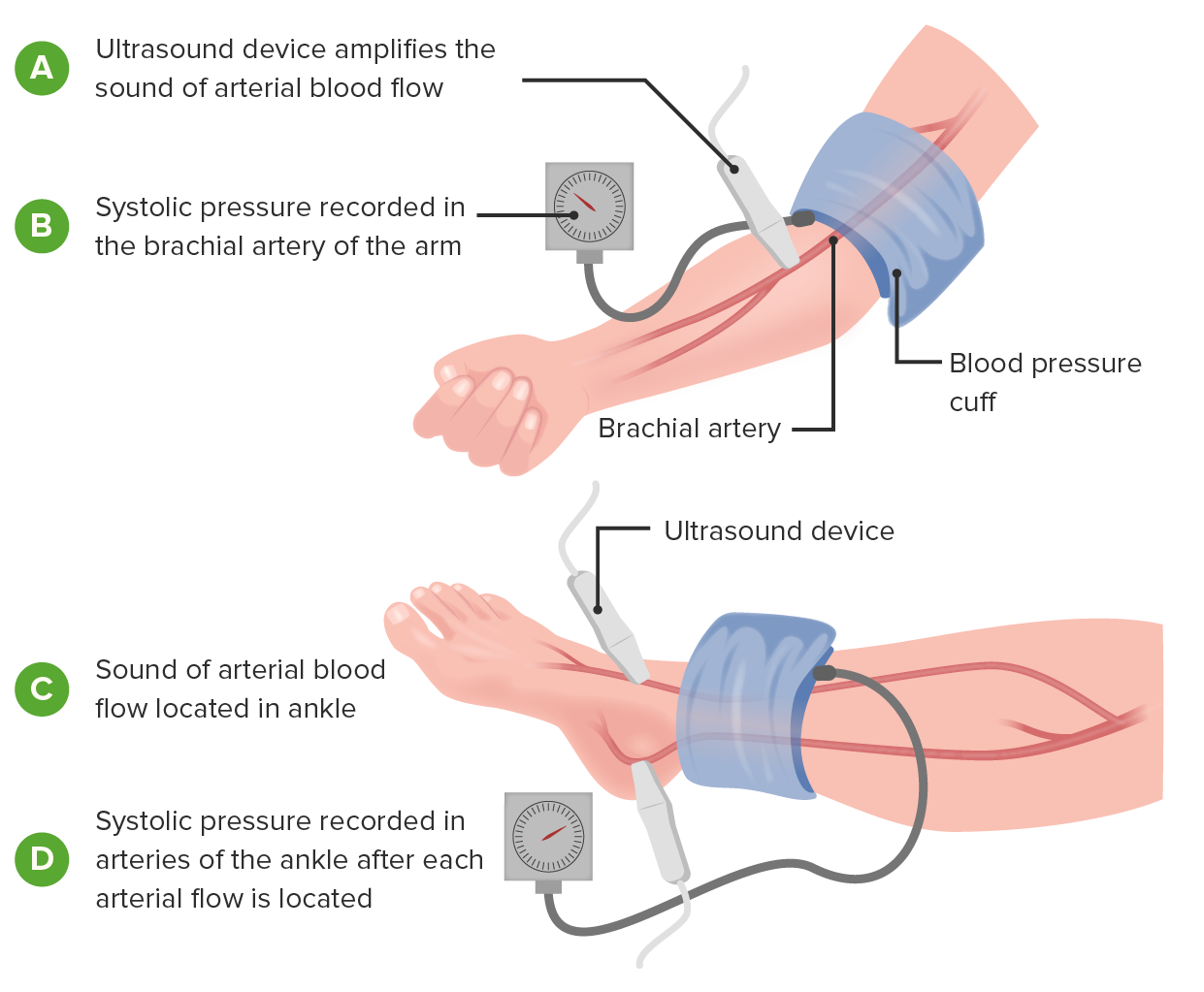
Ankle-brachial indexAnkle-brachial indexComparison of the blood pressure between the brachial artery and the posterior tibial artery. It is a predictor of peripheral arterial disease.Cardiovascular Examination
Usually the first choice in testing to establish a diagnosis
Equals the legLegThe lower leg, or just “leg” in anatomical terms, is the part of the lower limb between the knee and the ankle joint. The bony structure is composed of the tibia and fibula bones, and the muscles of the leg are grouped into the anterior, lateral, and posterior compartments by extensions of fascia.Leg: Anatomy systolic blood pressure divided by the armArmThe arm, or “upper arm” in common usage, is the region of the upper limb that extends from the shoulder to the elbow joint and connects inferiorly to the forearm through the cubital fossa. It is divided into 2 fascial compartments (anterior and posterior).Arm: Anatomy systolic blood pressure:
An ABI < 0.9 indicates PAD.
An ABI > 1.3 may indicate noncompressible vessels due to calcification (common in diabetics).
If the ABI is > 1.3 and suspicion for PAD is high, consider measuring a BP in the 1st toe or proceed to DopplerDopplerUltrasonography applying the doppler effect, with frequency-shifted ultrasound reflections produced by moving targets (usually red blood cells) in the bloodstream along the ultrasound axis in direct proportion to the velocity of movement of the targets, to determine both direction and velocity of blood flow.Ultrasound (Sonography) ultrasound.
Indicated for patientsPatientsIndividuals participating in the health care system for the purpose of receiving therapeutic, diagnostic, or preventive procedures.Clinician–Patient Relationship with a classic history suggesting PAD and normal resting ABI (0.91–1.30)
ABIs are measured before and after exercise:
Normal physiology: ABI should ↑ or stay the same.
In PAD: post-exercise ABI ↓ by ≥ 20%
Table: Interpretation of the ankle-brachial indexAnkle-brachial indexComparison of the blood pressure between the brachial artery and the posterior tibial artery. It is a predictor of peripheral arterial disease.Cardiovascular Examination (ABI)
Parameter
Value
Normal
≥ 0.9
Mild
0.71–0.9
Moderate
0.41–0.7
Severe
≤ 0.4
Image demonstrating how to measure the ankle-brachial index: This assists in the diagnosis and severity determination of PAD.
Image by Lecturio.
Imaging[11,12,17,18,22–24,29]
These studies are used to evaluate the location and severity of disease, particularly if revascularizationRevascularizationThromboangiitis Obliterans (Buerger Disease) is considered. Imaging is typically not warranted for asymptomatic patientsPatientsIndividuals participating in the health care system for the purpose of receiving therapeutic, diagnostic, or preventive procedures.Clinician–Patient Relationship.
DopplerDopplerUltrasonography applying the doppler effect, with frequency-shifted ultrasound reflections produced by moving targets (usually red blood cells) in the bloodstream along the ultrasound axis in direct proportion to the velocity of movement of the targets, to determine both direction and velocity of blood flow.Ultrasound (Sonography) ultrasound
Noninvasive, but operator-dependent
Can determine blood flowBlood flowBlood flow refers to the movement of a certain volume of blood through the vasculature over a given unit of time (e.g., mL per minute).Vascular Resistance, Flow, and Mean Arterial Pressure through arteriesArteriesArteries are tubular collections of cells that transport oxygenated blood and nutrients from the heart to the tissues of the body. The blood passes through the arteries in order of decreasing luminal diameter, starting in the largest artery (the aorta) and ending in the small arterioles. Arteries are classified into 3 types: large elastic arteries, medium muscular arteries, and small arteries and arterioles. Arteries: Histology
Computed tomography with angiographyAngiographyRadiography of blood vessels after injection of a contrast medium.Cardiac Surgery (CTACTAA non-invasive method that uses a ct scanner for capturing images of blood vessels and tissues. A contrast material is injected, which helps produce detailed images that aid in diagnosing vascular diseases.Pulmonary Function Tests)
Quick, but requires contrast administration
Becoming more commonly utilized to locate stenotic lesions
Can avoid ionizing radiationRadiationEmission or propagation of acoustic waves (sound), electromagnetic energy waves (such as light; radio waves; gamma rays; or x-rays), or a stream of subatomic particles (such as electrons; neutrons; protons; or alpha particles).Osteosarcoma
High diagnostic accuracy
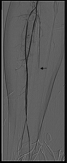
Digital subtraction arteriography
Reference standard
Intravenous contrast is injected imaged with a series of radiographs.
Has the highest diagnostic accuracy
Arteriography showing occlusion of the posterior tibial artery (arrow)
Image: “Posterior tibial artery occlusion” by C. Voiculeț et al. License: CC BY 2.0
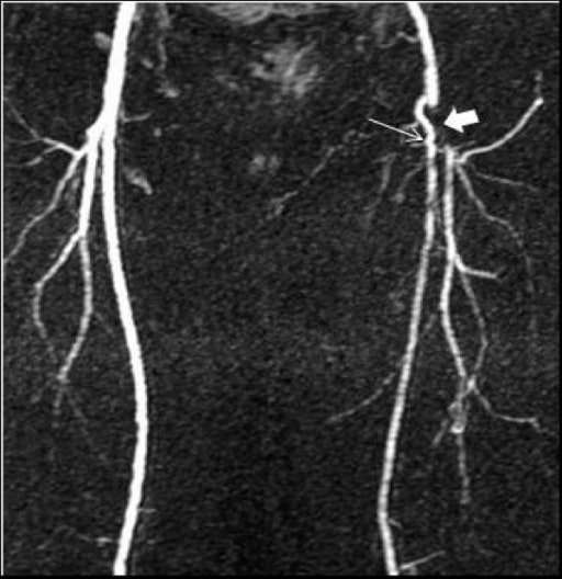
An MRA demonstrating occluded left distal common and proximal superficial (thin arrow) and profunda (thick arrow) femoral arteries, with a collateral artery connecting the 2
Image: “F1” by Bariatric and Metabolic Institute & Dept. of Surgery, Cleveland Clinic, 9500 Euclid Avenue, Cleveland 44950, Ohio, USA. License: CC BY 2.0
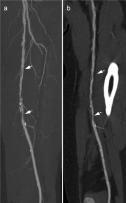
These CTA images demonstrate stenosis (arrows) in the left superficial femoral artery.
Image: “Example of a run-off CTA” by Klinik für Radiologie, Charité Universitätsmedizin Berlin, Berlin, Germany. License: CC BY 4.0
Laboratory testing[14]
These studies are not used for the diagnosis of PAD, but can help evaluate risk factors or organ injury:
Hemoglobin A1c → diabetesDiabetesDiabetes mellitus (DM) is a metabolic disease characterized by hyperglycemia and dysfunction of the regulation of glucose metabolism by insulin. Type 1 DM is diagnosed mostly in children and young adults as the result of autoimmune destruction of β cells in the pancreas and the resulting lack of insulin. Type 2 DM has a significant association with obesity and is characterized by insulin resistance.Diabetes Mellitus
PatientsPatientsIndividuals participating in the health care system for the purpose of receiving therapeutic, diagnostic, or preventive procedures.Clinician–Patient Relationship with evidence of atherosclerotic disease in other areas (e.g. cerebrovascular)
Important in preventing progression and complications
Can be used to identify patientsPatientsIndividuals participating in the health care system for the purpose of receiving therapeutic, diagnostic, or preventive procedures.Clinician–Patient Relationship at risk for other types of cardiovascular disease
Again, ABI is the test of choice for establishing the diagnosis.
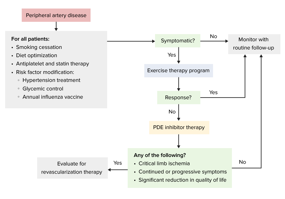
Management can be location-specific. The following information is based on US, European, and UK guidelines and literature.
Goals
Prevent disease progression.
Claudication painPainAn unpleasant sensation induced by noxious stimuli which are detected by nerve endings of nociceptive neurons.Pain: Types and Pathways management
Limb-salvage surgery as last resort
Simplified management algorithm for patients with peripheral artery disease
PDE: phosphodiesterase
Image by Lecturio.
Lifestyle modification[3,10–12,18,29]
Lifestyle modification is the 1st line of therapy:
SmokingSmokingWillful or deliberate act of inhaling and exhaling smoke from burning substances or agents held by hand.Interstitial Lung Diseases cessation
35‒60 minutes of track or treadmill walking 3‒4 times a week
Recumbent stepper, upper-body ergometer, or lower-extremity cycle can also be used.
Goal is to progressively increase the interval to reach near-maximal claudication painPainAn unpleasant sensation induced by noxious stimuli which are detected by nerve endings of nociceptive neurons.Pain: Types and Pathways.
Helps develop collateral circulationCirculationThe movement of the blood as it is pumped through the cardiovascular system.ABCDE Assessment
Reduces blood cell aggregationAggregationThe attachment of platelets to one another. This clumping together can be induced by a number of agents (e.g., thrombin; collagen) and is part of the mechanism leading to the formation of a thrombus.Coagulation Studies and blood viscosityBlood viscosityThe internal resistance of the blood to shear forces. The in vitro measure of whole blood viscosity is of limited clinical utility because it bears little relationship to the actual viscosity within the circulation, but an increase in the viscosity of circulating blood can contribute to morbidity in patients suffering from disorders such as sickle cell anemia and polycythemia.Vascular Resistance, Flow, and Mean Arterial Pressure
Medications[3,10–12,18,29]
Antiplatelet and antithrombotic therapy:
Initiate with lifestyle modifications
Benefits:
Used for prevention of cardiovascular events in patientsPatientsIndividuals participating in the health care system for the purpose of receiving therapeutic, diagnostic, or preventive procedures.Clinician–Patient Relationship with PAD, even in asymptomatic individuals
AspirinAspirinThe prototypical analgesic used in the treatment of mild to moderate pain. It has anti-inflammatory and antipyretic properties and acts as an inhibitor of cyclooxygenase which results in the inhibition of the biosynthesis of prostaglandins. Aspirin also inhibits platelet aggregation and is used in the prevention of arterial and venous thrombosis.Nonsteroidal Antiinflammatory Drugs (NSAIDs) 75‒325 mg per day
ClopidogrelClopidogrelA ticlopidine analog and platelet purinergic p2y receptor antagonist that inhibits adenosine diphosphate-mediated platelet aggregation. It is used to prevent thromboembolism in patients with arterial occlusive diseases; myocardial infarction; stroke; or atrial fibrillation.Antiplatelet Drugs 75 mg by mouth daily (improved efficacy over aspirinAspirinThe prototypical analgesic used in the treatment of mild to moderate pain. It has anti-inflammatory and antipyretic properties and acts as an inhibitor of cyclooxygenase which results in the inhibition of the biosynthesis of prostaglandins. Aspirin also inhibits platelet aggregation and is used in the prevention of arterial and venous thrombosis.Nonsteroidal Antiinflammatory Drugs (NSAIDs))
RivaroxabanRivaroxabanA morpholine and thiophene derivative that functions as a factor Xa inhibitor and is used in the treatment and prevention of deep-vein thrombosis and pulmonary embolism. It is also used for the prevention of stroke and systemic embolization in patients with non-valvular atrial fibrillation, and for the prevention of atherothrombotic events in patients after an acute coronary syndrome.Anticoagulants (low dose ~2.5 mg twice daily) taken with low-dose aspirinAspirinThe prototypical analgesic used in the treatment of mild to moderate pain. It has anti-inflammatory and antipyretic properties and acts as an inhibitor of cyclooxygenase which results in the inhibition of the biosynthesis of prostaglandins. Aspirin also inhibits platelet aggregation and is used in the prevention of arterial and venous thrombosis.Nonsteroidal Antiinflammatory Drugs (NSAIDs) (81 mg daily) in severely symptomatic PAD or after revascularizationRevascularizationThromboangiitis Obliterans (Buerger Disease):
Reduces major adverse cardiovascular events and major adverse limb events
Recommended for those who are not at increased risk of bleeding
Risk-factor modification:
Lipid disordersLipid disordersAn abnormal amount of lipid in blood is called dyslipidemia, which includes abnormal levels of cholesterol, triglycerides, and/or lipoproteins. Dyslipidemia may be primary (familial) or secondary (acquired). Both primary and secondary causes can lead to the development of premature cardiovascular (atherosclerosis) disease.Lipid Disorders:
Statin therapy (high intensity) is recommended for all patientsPatientsIndividuals participating in the health care system for the purpose of receiving therapeutic, diagnostic, or preventive procedures.Clinician–Patient Relationship.
Lower LDL-C to levels <70 mg/dL.
HypertensionHypertensionHypertension, or high blood pressure, is a common disease that manifests as elevated systemic arterial pressures. Hypertension is most often asymptomatic and is found incidentally as part of a routine physical examination or during triage for an unrelated medical encounter. Hypertension: antihypertensive treatment
Used for patientsPatientsIndividuals participating in the health care system for the purpose of receiving therapeutic, diagnostic, or preventive procedures.Clinician–Patient Relationship with hypertensionHypertensionHypertension, or high blood pressure, is a common disease that manifests as elevated systemic arterial pressures. Hypertension is most often asymptomatic and is found incidentally as part of a routine physical examination or during triage for an unrelated medical encounter. Hypertension (goal < 130/80)
ACEisACEIsA class of drugs whose main indications are the treatment of hypertension and heart failure. They exert their hemodynamic effect mainly by inhibiting the renin-angiotensin system. They also modulate sympathetic nervous system activity and increase prostaglandin synthesis. They cause mainly vasodilation and mild natriuresis without affecting heart rate and contractility.Heart Failure and Chronic Coronary Syndrome Medication or ARBsARBsAgents that antagonize angiotensin receptors. Many drugs in this class specifically target the angiotensin type 1 receptor.Heart Failure and Chronic Coronary Syndrome Medication are associated with reduced cardiovascular events.
DiabetesDiabetesDiabetes mellitus (DM) is a metabolic disease characterized by hyperglycemia and dysfunction of the regulation of glucose metabolism by insulin. Type 1 DM is diagnosed mostly in children and young adults as the result of autoimmune destruction of β cells in the pancreas and the resulting lack of insulin. Type 2 DM has a significant association with obesity and is characterized by insulin resistance.Diabetes Mellitus:
GLP-1GLP-1A peptide of 36 or 37 amino acids that is derived from proglucagon and mainly produced by the intestinal l cells. Glp-1(1-37 or 1-36) is further n-terminally truncated resulting in glp-1(7-37) or glp-1-(7-36) which can be amidated. These glp-1 peptides are known to enhance glucose-dependent insulin release, suppress glucagon release and gastric emptying, lower blood glucose, and reduce food intake.InsulinomasreceptorReceptorReceptors are proteins located either on the surface of or within a cell that can bind to signaling molecules known as ligands (e.g., hormones) and cause some type of response within the cell.Receptors agonists (liraglutide, semaglutide, dulaglutide) and SGLT2 inhibitorsSGLT2 inhibitorsNon-insulinotropic Diabetes Drugs provide cardiovascular benefit in PAD patientsPatientsIndividuals participating in the health care system for the purpose of receiving therapeutic, diagnostic, or preventive procedures.Clinician–Patient Relationship with diabetesDiabetesDiabetes mellitus (DM) is a metabolic disease characterized by hyperglycemia and dysfunction of the regulation of glucose metabolism by insulin. Type 1 DM is diagnosed mostly in children and young adults as the result of autoimmune destruction of β cells in the pancreas and the resulting lack of insulin. Type 2 DM has a significant association with obesity and is characterized by insulin resistance.Diabetes Mellitus.
Poor glycemic control increases risk of amputationAmputationAn amputation is the separation of a portion of the limb or the entire limb from the body, along with the bone. Amputations are generally indicated for conditions that compromise the viability of the limb or promote the spread of a local process that could manifest systemically. Amputation.
InfluenzaInfluenzaInfluenza viruses are members of the Orthomyxoviridae family and the causative organisms of influenza, a highly contagious febrile respiratory disease. There are 3 primary influenza viruses (A, B, and C) and various subtypes, which are classified based on their virulent surface antigens, hemagglutinin (HA) and neuraminidase (NA). Influenza typically presents with a fever, myalgia, headache, and symptoms of an upper respiratory infection. Influenza Viruses/Influenza: Annual influenzaInfluenzaInfluenza viruses are members of the Orthomyxoviridae family and the causative organisms of influenza, a highly contagious febrile respiratory disease. There are 3 primary influenza viruses (A, B, and C) and various subtypes, which are classified based on their virulent surface antigens, hemagglutinin (HA) and neuraminidase (NA). Influenza typically presents with a fever, myalgia, headache, and symptoms of an upper respiratory infection. Influenza Viruses/InfluenzavaccinationVaccinationVaccination is the administration of a substance to induce the immune system to develop protection against a disease. Unlike passive immunization, which involves the administration of pre-performed antibodies, active immunization constitutes the administration of a vaccine to stimulate the body to produce its own antibodies.Vaccination
Phosphodiesterase inhibitorsPhosphodiesterase inhibitorsPhosphodiesterase (PDE) inhibitors are a group of drugs that act by inhibiting PDE enzymes. Phosphodiesterase inhibitors have various mechanisms of action depending on the subtype of PDE targeted, but their main action is increasing the amount of intracellular cAMP or cGMP, which in turn results in physiologic effects such as reducing inflammation, promoting smooth muscle relaxation, and vasodilation.Phosphodiesterase Inhibitors:
May be added to antiplatelet therapy for patientsPatientsIndividuals participating in the health care system for the purpose of receiving therapeutic, diagnostic, or preventive procedures.Clinician–Patient Relationship with intermittent claudicationIntermittent claudicationA symptom complex characterized by pain and weakness in skeletal muscle group associated with exercise, such as leg pain and weakness brought on by walking. Such muscle limpness disappears after a brief rest and is often relates to arterial stenosis; muscle ischemia; and accumulation of lactate.Thromboangiitis Obliterans (Buerger Disease) that has not responded to exercise therapy and lifestyle modifications
Benefits:
Improves pain-free walking distances
Most effective pharmaceutical treatment for improving claudication symptoms
Indicated after failure of conservative measures
Reduces platelet aggregationPlatelet aggregationThe attachment of platelets to one another. This clumping together can be induced by a number of agents (e.g., thrombin; collagen) and is part of the mechanism leading to the formation of a thrombus.Hemostasis and allows arterial vasodilationVasodilationThe physiological widening of blood vessels by relaxing the underlying vascular smooth muscle.Pulmonary Hypertension Drugs
Options:
CilostazolCilostazolA quinoline and tetrazole derivative that acts as a phosphodiesterase type 3 inhibitor, with anti-platelet and vasodilating activity. It is used in the treatment of peripheral vascular diseases; ischemic heart disease; and in the prevention of stroke.Phosphodiesterase Inhibitors (US) 100 mg by mouth twice daily
Naftidrofuryl (Europe) 600 mg by mouth daily[12]
Treatments that have not been found to be effective:
PentoxifyllinePentoxifyllineA methylxanthine derivative that inhibits phosphodiesterase and affects blood rheology. It improves blood flow by increasing erythrocyte and leukocyte flexibility. It also inhibits platelet aggregation. Pentoxifylline modulates immunologic activity by stimulating cytokine production.Phosphodiesterase Inhibitors
Chelation therapy
Vitamins (folateFolateFolate and vitamin B12 are 2 of the most clinically important water-soluble vitamins. Deficiencies can present with megaloblastic anemia, GI symptoms, neuropsychiatric symptoms, and adverse pregnancy complications, including neural tube defects. Folate and Vitamin B12 and vitamin B12) for hyperhomocysteinemia
Vasodilator prostaglandinsProstaglandinsA group of compounds derived from unsaturated 20-carbon fatty acids, primarily arachidonic acid, via the cyclooxygenase pathway. They are extremely potent mediators of a diverse group of physiological processes.Eicosanoids
Salvage the limb tissue and prevent amputationAmputationAn amputation is the separation of a portion of the limb or the entire limb from the body, along with the bone. Amputations are generally indicated for conditions that compromise the viability of the limb or promote the spread of a local process that could manifest systemically. Amputation
This is best accomplished with an interdisciplinary care team of individuals specializing in endovascular/surgical repair, wound healingWound healingWound healing is a physiological process involving tissue repair in response to injury. It involves a complex interaction of various cell types, cytokines, and inflammatory mediators. Wound healing stages include hemostasis, inflammation, granulation, and remodeling. Wound Healing, footFootThe foot is the terminal portion of the lower limb, whose primary function is to bear weight and facilitate locomotion. The foot comprises 26 bones, including the tarsal bones, metatarsal bones, and phalanges. The bones of the foot form longitudinal and transverse arches and are supported by various muscles, ligaments, and tendons.Foot: Anatomy surgery, and medical management.[11]
Not for patientsPatientsIndividuals participating in the health care system for the purpose of receiving therapeutic, diagnostic, or preventive procedures.Clinician–Patient Relationship with intermittent claudicationIntermittent claudicationA symptom complex characterized by pain and weakness in skeletal muscle group associated with exercise, such as leg pain and weakness brought on by walking. Such muscle limpness disappears after a brief rest and is often relates to arterial stenosis; muscle ischemia; and accumulation of lactate.Thromboangiitis Obliterans (Buerger Disease) or where tissue is immediately threatened or irreversibly damaged
May be catheter-directed
Procedures:
Indications:
Chronic Limb-Threatening IschemiaIschemiaA hypoperfusion of the blood through an organ or tissue caused by a pathologic constriction or obstruction of its blood vessels, or an absence of blood circulation.Ischemic Cell Damage (CLTI)
Failure to improve with lifestyle modification and medications
Significant disabilityDisabilityDetermination of the degree of a physical, mental, or emotional handicap. The diagnosis is applied to legal qualification for benefits and income under disability insurance and to eligibility for social security and workman’s compensation benefits.ABCDE Assessment due to symptoms
Post-revascularization:
Antiplatelet therapy (aspirinAspirinThe prototypical analgesic used in the treatment of mild to moderate pain. It has anti-inflammatory and antipyretic properties and acts as an inhibitor of cyclooxygenase which results in the inhibition of the biosynthesis of prostaglandins. Aspirin also inhibits platelet aggregation and is used in the prevention of arterial and venous thrombosis.Nonsteroidal Antiinflammatory Drugs (NSAIDs) or clopidogrelClopidogrelA ticlopidine analog and platelet purinergic p2y receptor antagonist that inhibits adenosine diphosphate-mediated platelet aggregation. It is used to prevent thromboembolism in patients with arterial occlusive diseases; myocardial infarction; stroke; or atrial fibrillation.Antiplatelet Drugs) should be continued after any procedures.
Low-dose rivaroxabanRivaroxabanA morpholine and thiophene derivative that functions as a factor Xa inhibitor and is used in the treatment and prevention of deep-vein thrombosis and pulmonary embolism. It is also used for the prevention of stroke and systemic embolization in patients with non-valvular atrial fibrillation, and for the prevention of atherothrombotic events in patients after an acute coronary syndrome.Anticoagulants (2.5 mg BID) + aspirinAspirinThe prototypical analgesic used in the treatment of mild to moderate pain. It has anti-inflammatory and antipyretic properties and acts as an inhibitor of cyclooxygenase which results in the inhibition of the biosynthesis of prostaglandins. Aspirin also inhibits platelet aggregation and is used in the prevention of arterial and venous thrombosis.Nonsteroidal Antiinflammatory Drugs (NSAIDs) are recommended in eligible patientsPatientsIndividuals participating in the health care system for the purpose of receiving therapeutic, diagnostic, or preventive procedures.Clinician–Patient Relationship.
Short-term dual antiplatelet therapy is also reasonable.
Options:
Percutaneous transluminal angioplastyAngioplastyReconstruction or repair of a blood vessel, which includes the widening of a pathological narrowing of an artery or vein by the removal of atheromatous plaque material and/or the endothelial lining as well, or by dilatation (balloon angioplasty) to compress an atheroma. Except for endarterectomy, usually these procedures are performed via catheterization as minimally invasive endovascular procedures.Cardiac Surgery (PTAPTAA peritonsillar abscess (PTA), also called quinsy, is a collection of pus between the capsule of the palatine tonsil and the pharyngeal muscles. A pta is usually a complication of acute tonsillitis, an infection caused by group a streptococci. Patients often present with a sore throat, trismus, and a muffled voice.Peritonsillar Abscess):
Can be used for in-stent restenosis or in areas where stent placement is not feasible
Surgical procedures:
EndarterectomyEndarterectomySurgical excision, performed under general anesthesia, of the atheromatous tunica intima of an artery. When reconstruction of an artery is performed as an endovascular procedure through a catheter, it is called atherectomy.Intestinal Ischemia (direct removal of obstructive plaquePlaquePrimary Skin Lesions)
Both are indicated for individuals with CLTI for whom medical therapy and exercise therapy have failed.
Both have been shown to be good treatments for critical limb ischemiaIschemiaA hypoperfusion of the blood through an organ or tissue caused by a pathologic constriction or obstruction of its blood vessels, or an absence of blood circulation.Ischemic Cell Damage.
“Hybrid” procedures (endovascular + open) are increasingly used.
Choice comes down to:
Nature of the lesion
Patient preference
Ability to tolerate monitored anesthesiaAnesthesiaA state characterized by loss of feeling or sensation. This depression of nerve function is usually the result of pharmacologic action and is induced to allow performance of surgery or other painful procedures.Anesthesiology: History and Basic Concepts (endovascular) versus general anesthesiaGeneral anesthesiaProcedure in which patients are induced into an unconscious state through use of various medications so that they do not feel pain during surgery.Anesthesiology: History and Basic Concepts (open)
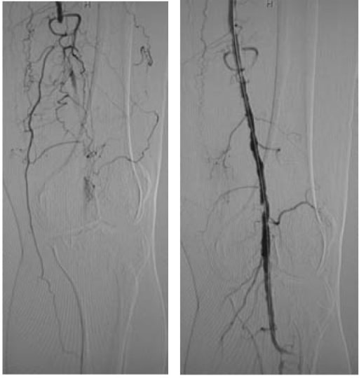
Left: left popliteal artery occlusion Right: return of blood flow after PTA with stenting
Image: “Popliteal arterial long occlusion case” by Department of Radiology, the First Hospital of China Medical University, Shenyang, Liaoning, People’s Republic of China. License: CC BY 2.0
Additional considerations[11,29,34]
For patientsPatientsIndividuals participating in the health care system for the purpose of receiving therapeutic, diagnostic, or preventive procedures.Clinician–Patient Relationship with PAD and diabetesDiabetesDiabetes mellitus (DM) is a metabolic disease characterized by hyperglycemia and dysfunction of the regulation of glucose metabolism by insulin. Type 1 DM is diagnosed mostly in children and young adults as the result of autoimmune destruction of β cells in the pancreas and the resulting lack of insulin. Type 2 DM has a significant association with obesity and is characterized by insulin resistance.Diabetes Mellitus:
Instruct about the importance of footFootThe foot is the terminal portion of the lower limb, whose primary function is to bear weight and facilitate locomotion. The foot comprises 26 bones, including the tarsal bones, metatarsal bones, and phalanges. The bones of the foot form longitudinal and transverse arches and are supported by various muscles, ligaments, and tendons.Foot: Anatomy self-examinations.
Encourage proper footFootThe foot is the terminal portion of the lower limb, whose primary function is to bear weight and facilitate locomotion. The foot comprises 26 bones, including the tarsal bones, metatarsal bones, and phalanges. The bones of the foot form longitudinal and transverse arches and are supported by various muscles, ligaments, and tendons.Foot: Anatomy care.
Annual or biannual footFootThe foot is the terminal portion of the lower limb, whose primary function is to bear weight and facilitate locomotion. The foot comprises 26 bones, including the tarsal bones, metatarsal bones, and phalanges. The bones of the foot form longitudinal and transverse arches and are supported by various muscles, ligaments, and tendons.Foot: Anatomy examinations by a clinicianClinicianA physician, nurse practitioner, physician assistant, or another health professional who is directly involved in patient care and has a professional relationship with patients.Clinician–Patient Relationship are encouraged.
Wounds should be closely monitored and treated.
InfectionsInfectionsInvasion of the host organism by microorganisms or their toxins or by parasites that can cause pathological conditions or diseases.Chronic Granulomatous Disease should be promptly diagnosed and treated to reduce the risk of amputationAmputationAn amputation is the separation of a portion of the limb or the entire limb from the body, along with the bone. Amputations are generally indicated for conditions that compromise the viability of the limb or promote the spread of a local process that could manifest systemically. Amputation.
Follow-up[11,29,32]
PatientsPatientsIndividuals participating in the health care system for the purpose of receiving therapeutic, diagnostic, or preventive procedures.Clinician–Patient Relationship should follow up with a clinicianClinicianA physician, nurse practitioner, physician assistant, or another health professional who is directly involved in patient care and has a professional relationship with patients.Clinician–Patient Relationship periodically.
Threatened limbThreatened limbMinimal sensory loss, mild-to-moderate muscle weakness, absent arterial doppler tones, requires urgent intervention.Acute Limb Ischemia from acute limb ischemiaIschemiaA hypoperfusion of the blood through an organ or tissue caused by a pathologic constriction or obstruction of its blood vessels, or an absence of blood circulation.Ischemic Cell Damage[11,29]
Surgical embolectomy should be performed in most circumstances.
Intraoperative angiographyAngiographyRadiography of blood vessels after injection of a contrast medium.Cardiac Surgery follow-up to ensure normal flowFlowBlood flows through the heart, arteries, capillaries, and veins in a closed, continuous circuit. Flow is the movement of volume per unit of time. Flow is affected by the pressure gradient and the resistance fluid encounters between 2 points. Vascular resistance is the opposition to flow, which is caused primarily by blood friction against vessel walls.Vascular Resistance, Flow, and Mean Arterial Pressure
Compartment syndromeCompartment SyndromeCompartment syndrome is a surgical emergency usually occurring secondary to trauma. The condition is marked by increased pressure within a compartment that compromises the circulation and function of the tissues within that space.Compartment Syndrome[13]
Increased risk after prolonged ischemiaIschemiaA hypoperfusion of the blood through an organ or tissue caused by a pathologic constriction or obstruction of its blood vessels, or an absence of blood circulation.Ischemic Cell Damage (> 6 hours)
Occurs due to reperfusion injuryReperfusion injuryAdverse functional, metabolic, or structural changes in ischemic tissues resulting from the restoration of blood flow to the tissue (reperfusion), including swelling; hemorrhage; necrosis; and damage from free radicals. The most common instance is myocardial reperfusion injury.Ischemic Cell Damage → leads to swellingSwellingInflammation and increased pressure (may result in further ischemiaIschemiaA hypoperfusion of the blood through an organ or tissue caused by a pathologic constriction or obstruction of its blood vessels, or an absence of blood circulation.Ischemic Cell Damage, necrosisNecrosisThe death of cells in an organ or tissue due to disease, injury or failure of the blood supply.Ischemic Cell Damage, and limb loss)
PatientsPatientsIndividuals participating in the health care system for the purpose of receiving therapeutic, diagnostic, or preventive procedures.Clinician–Patient Relationship will develop severe painPainAn unpleasant sensation induced by noxious stimuli which are detected by nerve endings of nociceptive neurons.Pain: Types and Pathways, paresthesiasParesthesiasSubjective cutaneous sensations (e.g., cold, warmth, tingling, pressure, etc.) that are experienced spontaneously in the absence of stimulation.Posterior Cord Syndrome, and muscle weakness.
Limb may feel tense.
Diagnosis is made by measuring compartment pressures.
Requires 4-compartment fasciotomyFasciotomySurgical incision on the fascia. It is used to decompress compartment pressure (e.g. in compartment syndromes; circumferential burns and extremity injuries) or to release contractures (e.g. in dupuytren’s contracture).Compartment Syndrome
Subclavian stealSubclavian stealA clinically significant reduction in blood supply to the brain stem and cerebellum (i.e., vertebrobasilar insufficiency) resulting from reversal of blood flow through the vertebral artery from occlusion or stenosis of the proximal subclavian or brachiocephalic artery. Common symptoms include vertigo, syncope, and intermittent claudication of the involved upper extremity. Subclavian steal may also occur in asymptomatic individuals.Subclavian Steal Syndrome syndrome[11]
Retrograde vertebral arteryVertebral arteryThe first branch of the subclavian artery with distribution to muscles of the neck; vertebrae; spinal cord; cerebellum; and interior of the cerebrum.Lateral Medullary Syndrome (Wallenberg Syndrome)flowFlowBlood flows through the heart, arteries, capillaries, and veins in a closed, continuous circuit. Flow is the movement of volume per unit of time. Flow is affected by the pressure gradient and the resistance fluid encounters between 2 points. Vascular resistance is the opposition to flow, which is caused primarily by blood friction against vessel walls.Vascular Resistance, Flow, and Mean Arterial Pressure due to subclavian artery stenosisStenosisHypoplastic Left Heart Syndrome (HLHS) or occlusion
Usually asymptomatic, but upper extremity ischemiaIschemiaA hypoperfusion of the blood through an organ or tissue caused by a pathologic constriction or obstruction of its blood vessels, or an absence of blood circulation.Ischemic Cell Damage and neurologic symptoms (from vertebrobasilar ischemiaVertebrobasilar ischemiaLocalized or diffuse reduction in blood flow through the vertebrobasilar arterial system, which supplies the brain stem; cerebellum; occipital lobe; medial temporal lobe; and thalamus. Characteristic clinical features include syncope; lightheadedness; visual disturbances; and vertigo. Brain stem infarctions or other brain infarction may be associated.Subclavian Steal Syndrome) indicate severe disease.
Management is similar to general PAD treatment.
GangreneGangreneDeath and putrefaction of tissue usually due to a loss of blood supply.Small Bowel Obstruction or limb loss[11,13,18]
AmputationAmputationAn amputation is the separation of a portion of the limb or the entire limb from the body, along with the bone. Amputations are generally indicated for conditions that compromise the viability of the limb or promote the spread of a local process that could manifest systemically. Amputation is performed when:
There is progressive gangreneGangreneDeath and putrefaction of tissue usually due to a loss of blood supply.Small Bowel Obstruction
Uncontrolled infection
Unrelenting painPainAn unpleasant sensation induced by noxious stimuli which are detected by nerve endings of nociceptive neurons.Pain: Types and Pathways
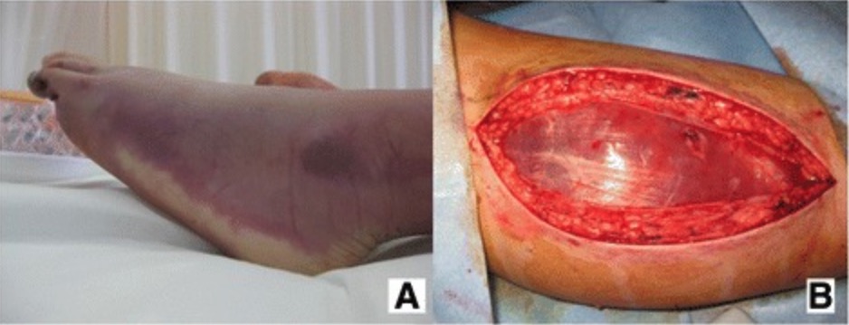
Images demonstrating an ischemic limb (A) that developed compartment syndrome: A fasciotomy (B) was required.
Arterial aneurysmAneurysmAn aneurysm is a bulging, weakened area of a blood vessel that causes an abnormal widening of its diameter > 1.5 times the size of the native vessel. Aneurysms occur more often in arteries than in veins and are at risk of dissection and rupture, which can be life-threatening. Thoracic Aortic Aneurysms: abnormal dilation of the arteriesArteriesArteries are tubular collections of cells that transport oxygenated blood and nutrients from the heart to the tissues of the body. The blood passes through the arteries in order of decreasing luminal diameter, starting in the largest artery (the aorta) and ending in the small arterioles. Arteries are classified into 3 types: large elastic arteries, medium muscular arteries, and small arteries and arterioles. Arteries: Histology due to weakening of the arterial wall. ThrombosisThrombosisFormation and development of a thrombus or blood clot in the blood vessel.Epidemic Typhus of a popliteal arteryPopliteal ArteryThe continuation of the femoral artery coursing through the popliteal fossa; it divides into the anterior and posterior tibial arteries.Popliteal Fossa: AnatomyaneurysmAneurysmAn aneurysm is a bulging, weakened area of a blood vessel that causes an abnormal widening of its diameter > 1.5 times the size of the native vessel. Aneurysms occur more often in arteries than in veins and are at risk of dissection and rupture, which can be life-threatening. Thoracic Aortic Aneurysms can result in symptoms of lower extremity ischemiaIschemiaA hypoperfusion of the blood through an organ or tissue caused by a pathologic constriction or obstruction of its blood vessels, or an absence of blood circulation.Ischemic Cell Damage. PatientsPatientsIndividuals participating in the health care system for the purpose of receiving therapeutic, diagnostic, or preventive procedures.Clinician–Patient Relationship will have a cold, pale legLegThe lower leg, or just “leg” in anatomical terms, is the part of the lower limb between the knee and the ankle joint. The bony structure is composed of the tibia and fibula bones, and the muscles of the leg are grouped into the anterior, lateral, and posterior compartments by extensions of fascia.Leg: Anatomy with absent distal pulses and paresthesiasParesthesiasSubjective cutaneous sensations (e.g., cold, warmth, tingling, pressure, etc.) that are experienced spontaneously in the absence of stimulation.Posterior Cord Syndrome. Physical exam may reveal a large, pulsatile popliteal arteryPopliteal ArteryThe continuation of the femoral artery coursing through the popliteal fossa; it divides into the anterior and posterior tibial arteries.Popliteal Fossa: Anatomy. Imaging will confirm the diagnosis and differentiate this diagnosis from PAD. Treatment involves surgical repair of the artery.
Arterial dissectionArterial dissectionArterial dissection is a violation of the structural integrity of the arterial wall that results in blood accumulating between the layers.Dissection of the Carotid and Vertebral Arteries: disruption in the medial layer of the arterial wall, resulting in bleeding into the vessel wall (creating a “false lumenFalse lumenAortic Dissection”), which may be due to connective tissueConnective tissueConnective tissues originate from embryonic mesenchyme and are present throughout the body except inside the brain and spinal cord. The main function of connective tissues is to provide structural support to organs. Connective tissues consist of cells and an extracellular matrix.Connective Tissue: Histology disorders or from vascular interventions. Occlusion of the “true” lumen may result, causing symptoms of ischemiaIschemiaA hypoperfusion of the blood through an organ or tissue caused by a pathologic constriction or obstruction of its blood vessels, or an absence of blood circulation.Ischemic Cell Damage (such as limb-threatening ischemiaIschemiaA hypoperfusion of the blood through an organ or tissue caused by a pathologic constriction or obstruction of its blood vessels, or an absence of blood circulation.Ischemic Cell Damage). Ultrasound or CTACTAA non-invasive method that uses a ct scanner for capturing images of blood vessels and tissues. A contrast material is injected, which helps produce detailed images that aid in diagnosing vascular diseases.Pulmonary Function Tests may be used to establish the diagnosis and will differentiate this condition from PAD. Treatment may require surgical repair of the artery.
Thromboembolic disease: vascular occlusion due to a dislodged thrombus from a more proximal source. The presentation depends on the source, size, and location of the embolism, but includes acute limb ischemiaIschemiaA hypoperfusion of the blood through an organ or tissue caused by a pathologic constriction or obstruction of its blood vessels, or an absence of blood circulation.Ischemic Cell Damage and blue toe syndromeBlue toe syndromeA condition that is caused by recurring atheroembolism in the lower extremities. It is characterized by cyanotic discoloration of the toes, usually the first, fourth, and fifth toes. Discoloration may extend to the lateral aspect of the foot. Despite the gangrene-like appearance, blue toes may respond to conservative therapy without amputation.Peripheral Artery Disease. The clinical history, hypercoagulableHypercoagulableHypercoagulable states (also referred to as thrombophilias) are a group of hematologic diseases defined by an increased risk of clot formation (i.e., thrombosis) due to either an increase in procoagulants, a decrease in anticoagulants, or a decrease in fibrinolysis. Hypercoagulable States workup, and imaging aid in the diagnosis, and a source will typically be suspected or found on workup, which differentiates thromboembolic disease from PAD. Management includes anticoagulationAnticoagulationPulmonary Hypertension Drugs and revascularizationRevascularizationThromboangiitis Obliterans (Buerger Disease).
Popliteal arteryPopliteal ArteryThe continuation of the femoral artery coursing through the popliteal fossa; it divides into the anterior and posterior tibial arteries.Popliteal Fossa: Anatomy entrapment syndrome: an uncommon condition in which an abnormally positioned, or enlarged, calf muscle compresses the popliteal arteryPopliteal ArteryThe continuation of the femoral artery coursing through the popliteal fossa; it divides into the anterior and posterior tibial arteries.Popliteal Fossa: Anatomy. The compressionCompressionBlunt Chest Trauma leads to obstruction of blood flowBlood flowBlood flow refers to the movement of a certain volume of blood through the vasculature over a given unit of time (e.g., mL per minute).Vascular Resistance, Flow, and Mean Arterial Pressure to the lower extremity, causing distal extremity ischemiaIschemiaA hypoperfusion of the blood through an organ or tissue caused by a pathologic constriction or obstruction of its blood vessels, or an absence of blood circulation.Ischemic Cell Damage, ulcerationUlcerationCorneal Abrasions, Erosion, and Ulcers, or necrosisNecrosisThe death of cells in an organ or tissue due to disease, injury or failure of the blood supply.Ischemic Cell Damage. The diagnosis is made with imaging, which will differentiate this condition from PAD. Management includes avoiding any inciting exercise and vascular surgeryVascular surgeryVascular surgery is the specialized field of medicine that focuses on the surgical management of the pathologies of the peripheral circulation. The main goal of most vascular procedures is to restore circulatory function to the affected vessels by relieving occlusions or by redirecting blood flow (e.g., bypass).Vascular Surgery evaluation.
Spinal stenosisStenosisHypoplastic Left Heart Syndrome (HLHS):compressionCompressionBlunt Chest Trauma of nerve roots due to narrowing of the spinal canalSpinal CanalThe cavity within the spinal column through which the spinal cord passes.Spinal Cord Injuries. PatientsPatientsIndividuals participating in the health care system for the purpose of receiving therapeutic, diagnostic, or preventive procedures.Clinician–Patient Relationship may have positional back painPainAn unpleasant sensation induced by noxious stimuli which are detected by nerve endings of nociceptive neurons.Pain: Types and Pathways and exertional lower extremity painPainAn unpleasant sensation induced by noxious stimuli which are detected by nerve endings of nociceptive neurons.Pain: Types and Pathways. The painPainAn unpleasant sensation induced by noxious stimuli which are detected by nerve endings of nociceptive neurons.Pain: Types and Pathways will not be relieved with rest. Other symptoms include weakness, paresthesiasParesthesiasSubjective cutaneous sensations (e.g., cold, warmth, tingling, pressure, etc.) that are experienced spontaneously in the absence of stimulation.Posterior Cord Syndrome, and diminished reflexes. Unlike PAD, pulses will be intact. The diagnosis is made based on clinical exam and MRI imaging of the spineSpineThe human spine, or vertebral column, is the most important anatomical and functional axis of the human body. It consists of 7 cervical vertebrae, 12 thoracic vertebrae, and 5 lumbar vertebrae and is limited cranially by the skull and caudally by the sacrum.Vertebral Column: Anatomy. Treatment includes physical therapyPhysical TherapyBecker Muscular Dystrophy, painPainAn unpleasant sensation induced by noxious stimuli which are detected by nerve endings of nociceptive neurons.Pain: Types and Pathways management, and surgery for severe cases.
Thromboangiitis obliteransThromboangiitis ObliteransA non-atherosclerotic, inflammatory thrombotic disease that commonly involves small and medium-sized arteries or veins in the extremities. It is characterized by occlusive thrombosis and fibrosis in the vascular wall leading to digital and limb ischemia and ulcerations. Thromboangiitis obliterans is highly associated with tobacco smoking.Thromboangiitis Obliterans (Buerger Disease) (Buerger’s disease): nonatherosclerotic segmental inflammatory disease that affects small to medium-sized vessels of the extremities. PatientsPatientsIndividuals participating in the health care system for the purpose of receiving therapeutic, diagnostic, or preventive procedures.Clinician–Patient Relationship are typically young smokers presenting with distal extremity ischemiaIschemiaA hypoperfusion of the blood through an organ or tissue caused by a pathologic constriction or obstruction of its blood vessels, or an absence of blood circulation.Ischemic Cell Damage, ulcers, or gangreneGangreneDeath and putrefaction of tissue usually due to a loss of blood supply.Small Bowel Obstruction. Diagnosis is based on clinical findings, vascular testing, and angiographyAngiographyRadiography of blood vessels after injection of a contrast medium.Cardiac Surgery. Other potential diagnoses must be ruled out, including PAD. SmokingSmokingWillful or deliberate act of inhaling and exhaling smoke from burning substances or agents held by hand.Interstitial Lung Diseases cessation is an essential part of management and decreases the risk of amputationAmputationAn amputation is the separation of a portion of the limb or the entire limb from the body, along with the bone. Amputations are generally indicated for conditions that compromise the viability of the limb or promote the spread of a local process that could manifest systemically. Amputation.
VasculitisVasculitisInflammation of any one of the blood vessels, including the arteries; veins; and rest of the vasculature system in the body.Systemic Lupus Erythematosus: a vascular inflammatory disease, often resulting in ischemiaIschemiaA hypoperfusion of the blood through an organ or tissue caused by a pathologic constriction or obstruction of its blood vessels, or an absence of blood circulation.Ischemic Cell Damage, necrosisNecrosisThe death of cells in an organ or tissue due to disease, injury or failure of the blood supply.Ischemic Cell Damage, and organ damage. Any vessel can be involved. Etiologies include autoimmune disorders, drugs, and infectionsInfectionsInvasion of the host organism by microorganisms or their toxins or by parasites that can cause pathological conditions or diseases.Chronic Granulomatous Disease. PatientsPatientsIndividuals participating in the health care system for the purpose of receiving therapeutic, diagnostic, or preventive procedures.Clinician–Patient Relationship present with feverFeverFever is defined as a measured body temperature of at least 38°C (100.4°F). Fever is caused by circulating endogenous and/or exogenous pyrogens that increase levels of prostaglandin E2 in the hypothalamus. Fever is commonly associated with chills, rigors, sweating, and flushing of the skin. Fever, arthralgias, and arthritisArthritisAcute or chronic inflammation of joints.Osteoarthritis, as well as potential end-organ damage. Diagnosis involves inflammatory markers, autoimmune serologySerologyThe study of serum, especially of antigen-antibody reactions in vitro.Yellow Fever Virus, infectious workup, and biopsyBiopsyRemoval and pathologic examination of specimens from the living body.Ewing Sarcoma, which will differentiate this disease from PAD. Management depends on the underlying cause.
Billing and Coding
Diagnosis Codes:
These codes are used to diagnose Peripheral Artery DiseasePeripheral artery diseasePeripheral artery disease (PAD) is obstruction of the arterial lumen resulting in decreased blood flow to the distal limbs. The disease can be a result of atherosclerosis or thrombosis. Patients may be asymptomatic or have progressive claudication, skin discoloration, ischemic ulcers, or gangrene. Peripheral Artery Disease (PAD), which is atherosclerosisAtherosclerosisAtherosclerosis is a common form of arterial disease in which lipid deposition forms a plaque in the blood vessel walls. Atherosclerosis is an incurable disease, for which there are clearly defined risk factors that often can be reduced through a change in lifestyle and behavior of the patient. Atherosclerosis of the arteriesArteriesArteries are tubular collections of cells that transport oxygenated blood and nutrients from the heart to the tissues of the body. The blood passes through the arteries in order of decreasing luminal diameter, starting in the largest artery (the aorta) and ending in the small arterioles. Arteries are classified into 3 types: large elastic arteries, medium muscular arteries, and small arteries and arterioles. Arteries: Histology supplying the limbs. The codes specify the location (e.g., lower extremities) and presence of symptoms like claudication.
Coding System
Code
Description
ICD-10-CM
I73.9
Peripheral vascular disease, unspecified
ICD-10-CM
I70.211
AtherosclerosisAtherosclerosisAtherosclerosis is a common form of arterial disease in which lipid deposition forms a plaque in the blood vessel walls. Atherosclerosis is an incurable disease, for which there are clearly defined risk factors that often can be reduced through a change in lifestyle and behavior of the patient. Atherosclerosis of native arteriesArteriesArteries are tubular collections of cells that transport oxygenated blood and nutrients from the heart to the tissues of the body. The blood passes through the arteries in order of decreasing luminal diameter, starting in the largest artery (the aorta) and ending in the small arterioles. Arteries are classified into 3 types: large elastic arteries, medium muscular arteries, and small arteries and arterioles. Arteries: Histology of extremities with intermittent claudicationIntermittent claudicationA symptom complex characterized by pain and weakness in skeletal muscle group associated with exercise, such as leg pain and weakness brought on by walking. Such muscle limpness disappears after a brief rest and is often relates to arterial stenosis; muscle ischemia; and accumulation of lactate.Thromboangiitis Obliterans (Buerger Disease), right legLegThe lower leg, or just “leg” in anatomical terms, is the part of the lower limb between the knee and the ankle joint. The bony structure is composed of the tibia and fibula bones, and the muscles of the leg are grouped into the anterior, lateral, and posterior compartments by extensions of fascia.Leg: Anatomy
Evaluation & Workup:
This CPT code is for the Ankle-Brachial IndexAnkle-brachial indexComparison of the blood pressure between the brachial artery and the posterior tibial artery. It is a predictor of peripheral arterial disease.Cardiovascular Examination (ABI), a simple, non-invasive test that compares the blood pressure in the ankles to the blood pressure in the arms and is the primary screeningScreeningPreoperative Care tool for PAD.
Coding System
Code
Description
CPT
93922
Limited bilateral noninvasive physiologic studies of upper or lower extremity arteriesArteriesArteries are tubular collections of cells that transport oxygenated blood and nutrients from the heart to the tissues of the body. The blood passes through the arteries in order of decreasing luminal diameter, starting in the largest artery (the aorta) and ending in the small arterioles. Arteries are classified into 3 types: large elastic arteries, medium muscular arteries, and small arteries and arterioles. Arteries: Histology (eg, for ABI)
Procedures/Interventions:
This code is for a percutaneous transluminal angioplastyAngioplastyReconstruction or repair of a blood vessel, which includes the widening of a pathological narrowing of an artery or vein by the removal of atheromatous plaque material and/or the endothelial lining as well, or by dilatation (balloon angioplasty) to compress an atheroma. Except for endarterectomy, usually these procedures are performed via catheterization as minimally invasive endovascular procedures.Cardiac Surgery with stenting, a common endovascular procedure to open a blocked peripheral artery and restore blood flowBlood flowBlood flow refers to the movement of a certain volume of blood through the vasculature over a given unit of time (e.g., mL per minute).Vascular Resistance, Flow, and Mean Arterial Pressure.
Coding System
Code
Description
CPT
37226
RevascularizationRevascularizationThromboangiitis Obliterans (Buerger Disease), endovascular, open or percutaneous, femoral/popliteal arteryPopliteal ArteryThe continuation of the femoral artery coursing through the popliteal fossa; it divides into the anterior and posterior tibial arteries.Popliteal Fossa: Anatomy(s), unilateral; with transluminal angioplastyAngioplastyReconstruction or repair of a blood vessel, which includes the widening of a pathological narrowing of an artery or vein by the removal of atheromatous plaque material and/or the endothelial lining as well, or by dilatation (balloon angioplasty) to compress an atheroma. Except for endarterectomy, usually these procedures are performed via catheterization as minimally invasive endovascular procedures.Cardiac Surgery and stent placement(s)
Medications:
These codes are for medical management of PAD. Antiplatelet agentsAntiplatelet agentsAntiplatelet agents are medications that inhibit platelet aggregation, a critical step in the formation of the initial platelet plug. Abnormal, or inappropriate, platelet aggregation is a key step in the pathophysiology of arterial ischemic events. The primary categories of antiplatelet agents include aspirin, ADP inhibitors, phosphodiesterase/adenosine uptake inhibitors, and glycoprotein IIb/IIIa inhibitors. Antiplatelet Drugs like aspirinAspirinThe prototypical analgesic used in the treatment of mild to moderate pain. It has anti-inflammatory and antipyretic properties and acts as an inhibitor of cyclooxygenase which results in the inhibition of the biosynthesis of prostaglandins. Aspirin also inhibits platelet aggregation and is used in the prevention of arterial and venous thrombosis.Nonsteroidal Antiinflammatory Drugs (NSAIDs) or clopidogrelClopidogrelA ticlopidine analog and platelet purinergic p2y receptor antagonist that inhibits adenosine diphosphate-mediated platelet aggregation. It is used to prevent thromboembolism in patients with arterial occlusive diseases; myocardial infarction; stroke; or atrial fibrillation.Antiplatelet Drugs are essential, and cilostazolCilostazolA quinoline and tetrazole derivative that acts as a phosphodiesterase type 3 inhibitor, with anti-platelet and vasodilating activity. It is used in the treatment of peripheral vascular diseases; ischemic heart disease; and in the prevention of stroke.Phosphodiesterase Inhibitors is a specific medication used to improve symptoms of intermittent claudicationIntermittent claudicationA symptom complex characterized by pain and weakness in skeletal muscle group associated with exercise, such as leg pain and weakness brought on by walking. Such muscle limpness disappears after a brief rest and is often relates to arterial stenosis; muscle ischemia; and accumulation of lactate.Thromboangiitis Obliterans (Buerger Disease).
Coding System
Code
Description
RxNorm
32953
ClopidogrelClopidogrelA ticlopidine analog and platelet purinergic p2y receptor antagonist that inhibits adenosine diphosphate-mediated platelet aggregation. It is used to prevent thromboembolism in patients with arterial occlusive diseases; myocardial infarction; stroke; or atrial fibrillation.Antiplatelet Drugs (ingredient)
RxNorm
32535
CilostazolCilostazolA quinoline and tetrazole derivative that acts as a phosphodiesterase type 3 inhibitor, with anti-platelet and vasodilating activity. It is used in the treatment of peripheral vascular diseases; ischemic heart disease; and in the prevention of stroke.Phosphodiesterase Inhibitors (ingredient)
Treat-Jacobson, D., et al. (2019). Implementation of supervised exercise therapy for patients with symptomatic peripheral artery disease: a science advisory from the American Heart Association. Circulation, 140(13), e700–e710. https://doi.org/10.1161/CIR.0000000000000727
Guyatt, G. H., et al. (2012) Executive summary: antithrombotic therapy and prevention of thrombosis (9th ed.). Chest, 141(2), 7S–47S. https://doi.org/10.1378/chest.1412S3
Gornik, H. L., Aronow, H. D., Goodney, P. P., Arya, S., Brewster, L. P., Byrd, L., Chandra, V., Drachman, D. E., Eaves, J. M., Ehrman, J. K., Evans, J. N., Getchius, T. S. D., Gutiérrez, J. A., Hawkins, B. M., Hess, C. N., Ho, K. J., Jones, W. S., Kim, E. S. H., Kinlay, S., Kirksey, L., … Peer Review Committee Members (2024). 2024 ACC/AHA/AACVPR/APMA/ABC/SCAI/SVM/SVN/SVS/SIR/VESS Guideline for the Management of Lower Extremity Peripheral Artery Disease: A Report of the American College of Cardiology/American Heart Association Joint Committee on Clinical Practice Guidelines. Circulation, 149(24), e1313–e1410. https://doi.org/10.1161/CIR.0000000000001251
National Institute for Health and Care Excellence. (2014). Peripheral arterial disease: quality standard. Retrieved Sept 30, 2025, from https://www.nice.org.uk/guidance/qs52
National Institute for Health and Care Excellence. (2012). Percutaneous laser atherectomy as an adjunct to balloon angioplasty (with or without stenting) for peripheral arterial disease. Retrieved Sept 30, 2025, from https://www.nice.org.uk/guidance/ipg433
Jaff, M. R., et al. (2015). An update on methods for revascularization and expansion of the TASC lesion classification to include below-the-knee arteries: a supplement to the Inter-Society Consensus for the management of peripheral arterial disease (TASC II). Vascular Medicine, 20(5), 465–478. https://journals.sagepub.com/doi/pdf/10.1177/1358863X15597877
American College of Radiology. (2022). ACR appropriateness criteria: lower extremity arterial claudication-imaging assessment for revascularization. Retrieved June 5, 2023, from https://acsearch.acr.org/docs/69411/Narrative/
Criqui, M. H., et al. (2021). Lower extremity peripheral artery disease: contemporary epidemiology, management gaps, and future directions: a scientific statement from the American Heart Association. Circulation, 144(9), e171–e191. https://www.ahajournals.org/doi/epub/10.1161/CIR.0000000000001005
Treat-Jacobson, D., et al. (2019). Optimal exercise programs for patients with peripheral artery disease: a scientific statement from the American Heart Association. Circulation, 139(4), e10–e33. https://www.ahajournals.org/doi/epub/10.1161/CIR.0000000000000623
American College of Radiology. (2022). ACR appropriateness criteria: Lower Extremity Arterial Claudication-Imaging Assessment for Revascularization Retrieved Sept 30, 2025 from https://acsearch.acr.org/docs/3102394/Narrative/
Klein, A. J., et al. (2017). SCAI appropriate use criteria for peripheral arterial interventions: an update. Catheterization and Cardiovascular Interventions, 90(4), E90–E110. https://onlinelibrary.wiley.com/doi/full/10.1002/ccd.27141
Dhanoa, D., et al. (2016). Position statement on noninvasive imaging of peripheral arterial disease by the Society of Interventional Radiology and the Canadian Interventional Radiology Association. Journal of Vascular Interventional Radiology, 27, 947–951. https://www.jvir.org/article/S1051-0443(16)30027-6/pdf
Smith, S.C., Jr., et al. (2011). AHA/ACCF secondary prevention and risk reduction therapy for patients with coronary and other atherosclerotic vascular disease: 2011 update. Circulation, 124, 2458–2473. https://www.ahajournals.org/doi/10.1161/cir.0b013e318235eb4d
Aboyans, V., et al. (2021). Antithrombotic therapies in aortic and peripheral arterial diseases in 2021: a consensus document from the ESC working group on aorta and peripheral vascular diseases, the ESC working group on thrombosis, and the ESC working group on cardiovascular pharmacotherapy. European Heart Journal, 42(39), 4013–4024. https://academic.oup.com/eurheartj/article/42/39/4013/6323985?login=false
Venermo, M., et al. (2019). Follow-up of patients after revascularisation for peripheral arterial diseases: a consensus document from the European Society of Cardiology working group on aorta and peripheral vascular diseases and the European Society for Vascular Surgery. European Journal of Preventive Cardiology, 26(18), 1971–1984. https://journals.sagepub.com/doi/pdf/10.1177/2047487319846999
Aboyans, V., et al. (2018). Editor’s choice—2017 ESC guidelines on the diagnosis and treatment of peripheral arterial diseases, in collaboration with the European Society for Vascular Surgery (ESVS). European Journal of Vascular and Endovascular Surgery, 55, 305–368. https://www.ejves.com/article/S1078-5884(17)30454-9/pdf
Rutherford, R. B., Baker, J. D., Ernst, C., Johnston, K. W., Porter, J. M., Ahn, S., & Jones, D. N. (1997). Recommended standards for reports dealing with lower extremity ischemia: revised version. Journal of vascular surgery, 26(3), 517–538. https://doi.org/10.1016/s0741-5214(97)70045-4
Create your free account or log in to continue reading!