Croup (Clinical)

Croup Croup Croup, also known as laryngotracheobronchitis, is a disease most commonly caused by a viral infection that leads to severe inflammation of the upper airway. It usually presents in children < 5 years of age. Patients develop a hoarse, "seal-like" barking cough and inspiratory stridor. Croup, also known as laryngotracheobronchitis Laryngotracheobronchitis Croup, also known as laryngotracheobronchitis, is a disease most commonly caused by a viral infection that leads to severe inflammation of the upper airway. It usually presents in children < 5 years of age. Patients develop a hoarse, "seal-like" barking cough and inspiratory stridor. Croup, is a disease most commonly caused by a viral infection that leads to severe inflammation Inflammation Inflammation is a complex set of responses to infection and injury involving leukocytes as the principal cellular mediators in the body's defense against pathogenic organisms. Inflammation is also seen as a response to tissue injury in the process of wound healing. The 5 cardinal signs of inflammation are pain, heat, redness, swelling, and loss of function. Inflammation of the upper airway Airway ABCDE Assessment. It usually presents in children < 5 years of age. Patients Patients Individuals participating in the health care system for the purpose of receiving therapeutic, diagnostic, or preventive procedures. Clinician–Patient Relationship develop a hoarse, “seal-like” barking cough and inspiratory stridor Stridor Laryngomalacia and Tracheomalacia. Human parainfluenza viruses Human parainfluenza viruses Human parainfluenza viruses (HPIVs) are single-stranded, linear, negative-sense RNA viruses of the family paramyxoviridae and the genus paramyxovirus. Human parainfluenza viruses are the 2nd most common cause of lower respiratory disease in children, after the respiratory syncytial virus. Parainfluenza Virus account for the majority of cases, followed by respiratory syncytial virus Respiratory Syncytial Virus Respiratory syncytial virus (RSV) is an enveloped, single-stranded, linear, negative-sense RNA virus of the family Paramyxoviridae and the genus Orthopneumovirus. Two subtypes (A and B) are present in outbreaks, but type A causes more severe disease. Respiratory syncytial virus causes infections of the lungs and respiratory tract. Respiratory Syncytial Virus, adenovirus Adenovirus Adenovirus (member of the family Adenoviridae) is a nonenveloped, double-stranded DNA virus. Adenovirus is transmitted in a variety of ways, and it can have various presentations based on the site of entry. Presentation can include febrile pharyngitis, conjunctivitis, acute respiratory disease, atypical pneumonia, and gastroenteritis. Adenovirus, rhinovirus Rhinovirus Rhinovirus is an acid-labile, positive-sense RNA virus of the Picornavirus family. The virus, which causes the common cold, is most often acquired through the airway via the inhalation of aerosols containing rhinovirus and fomites. Rhinovirus, and enteroviruses. Croup Croup Croup, also known as laryngotracheobronchitis, is a disease most commonly caused by a viral infection that leads to severe inflammation of the upper airway. It usually presents in children < 5 years of age. Patients develop a hoarse, "seal-like" barking cough and inspiratory stridor. Croup is usually diagnosed clinically or with the aid of X-ray X-ray Penetrating electromagnetic radiation emitted when the inner orbital electrons of an atom are excited and release radiant energy. X-ray wavelengths range from 1 pm to 10 nm. Hard x-rays are the higher energy, shorter wavelength x-rays. Soft x-rays or grenz rays are less energetic and longer in wavelength. The short wavelength end of the x-ray spectrum overlaps the gamma rays wavelength range. The distinction between gamma rays and x-rays is based on their radiation source. Pulmonary Function Tests imaging, which may show a narrowing of the air column in the trachea Trachea The trachea is a tubular structure that forms part of the lower respiratory tract. The trachea is continuous superiorly with the larynx and inferiorly becomes the bronchial tree within the lungs. The trachea consists of a support frame of semicircular, or C-shaped, rings made out of hyaline cartilage and reinforced by collagenous connective tissue. Trachea: Anatomy called the “ steeple sign Steeple Sign Pediatric Chest Abnormalities”. Treatment consists of steroids Steroids A group of polycyclic compounds closely related biochemically to terpenes. They include cholesterol, numerous hormones, precursors of certain vitamins, bile acids, alcohols (sterols), and certain natural drugs and poisons. Steroids have a common nucleus, a fused, reduced 17-carbon atom ring system, cyclopentanoperhydrophenanthrene. Most steroids also have two methyl groups and an aliphatic side-chain attached to the nucleus. Benign Liver Tumors and epinephrine Epinephrine The active sympathomimetic hormone from the adrenal medulla. It stimulates both the alpha- and beta- adrenergic systems, causes systemic vasoconstriction and gastrointestinal relaxation, stimulates the heart, and dilates bronchi and cerebral vessels. Sympathomimetic Drugs.

Last updated: Apr 26, 2025

Editorial responsibility: Stanley Oiseth, Lindsay Jones, Evelin Maza

Overview

Epidemiology[1,2,3,5]

  • Primarily affects children aged 6–36 months
    • Incidence Incidence The number of new cases of a given disease during a given period in a specified population. It also is used for the rate at which new events occur in a defined population. It is differentiated from prevalence, which refers to all cases in the population at a given time. Measures of Disease Frequency: peaks in the second year of life
    • Potential affected age range: 6 months to 15 years
  • 7% of emergency hospitalizations in children < 5 years of age are caused by croup Croup Croup, also known as laryngotracheobronchitis, is a disease most commonly caused by a viral infection that leads to severe inflammation of the upper airway. It usually presents in children < 5 years of age. Patients develop a hoarse, "seal-like" barking cough and inspiratory stridor. Croup.
  • More prevalent in the fall and early winter Winter Pityriasis Rosea
  • More common in boys, with a male:female ratio of 1.4:1 
  • 5–6 cases per 100 toddlers in the second year of life in the United States

Transmission[3,5]

  • Contagious within the first few days, particularly the first or second day 
  • Transmitted in the form of aerosol droplets Droplets Varicella-Zoster Virus/Chickenpox released by sneezing Sneezing The sudden, forceful, involuntary expulsion of air from the nose and mouth caused by irritation to the mucous membranes of the upper respiratory tract. Rhinovirus and coughing and via direct contact with infected secretions

Risk factors[1,5]

Etiology and Pathophysiology

Etiology[1,2,3,5]

Croup Croup Croup, also known as laryngotracheobronchitis, is a disease most commonly caused by a viral infection that leads to severe inflammation of the upper airway. It usually presents in children < 5 years of age. Patients develop a hoarse, "seal-like" barking cough and inspiratory stridor. Croup is commonly caused by a virus Virus Viruses are infectious, obligate intracellular parasites composed of a nucleic acid core surrounded by a protein capsid. Viruses can be either naked (non-enveloped) or enveloped. The classification of viruses is complex and based on many factors, including type and structure of the nucleoid and capsid, the presence of an envelope, the replication cycle, and the host range. Virology (75% of cases) and less commonly by bacteria Bacteria Bacteria are prokaryotic single-celled microorganisms that are metabolically active and divide by binary fission. Some of these organisms play a significant role in the pathogenesis of diseases. Bacteriology.

  • Viral:
    • Most common: parainfluenza virus Parainfluenza virus Human parainfluenza viruses (HPIVs) are single-stranded, linear, negative-sense RNA viruses of the family Paramyxoviridae and the genus Paramyxovirus. Human parainfluenza viruses are the 2nd most common cause of lower respiratory disease in children, after the respiratory syncytial virus. Parainfluenza Virus types 1 and 2 (66% of cases)
      • Parainfluenza type 1 Type 1 Spinal Muscular Atrophy: most commonly causes acute laryngotracheitis (during winter Winter Pityriasis Rosea and fall)
      • 3 virulence Virulence The degree of pathogenicity within a group or species of microorganisms or viruses as indicated by case fatality rates and/or the ability of the organism to invade the tissues of the host. The pathogenic capacity of an organism is determined by its virulence factors. Proteus proteins Proteins Linear polypeptides that are synthesized on ribosomes and may be further modified, crosslinked, cleaved, or assembled into complex proteins with several subunits. The specific sequence of amino acids determines the shape the polypeptide will take, during protein folding, and the function of the protein. Energy Homeostasis: fusion protein Fusion protein Proteins that catalyze membrane fusion. Measles Virus, hemagglutinin Hemagglutinin Agents that cause agglutination of red blood cells. They include antibodies, blood group antigens, lectins, autoimmune factors, bacterial, viral, or parasitic blood agglutinins, etc. Measles Virus, neuraminidase Neuraminidase An enzyme that catalyzes the hydrolysis of alpha-2, 3, alpha-2, 6-, and alpha-2, 8-glycosidic linkages (at a decreasing rate, respectively) of terminal sialic residues in oligosaccharides, glycoproteins, glycolipids, colominic acid, and synthetic substrate. Antivirals for Influenza
    • Others: respiratory syncytial virus Respiratory Syncytial Virus Respiratory syncytial virus (RSV) is an enveloped, single-stranded, linear, negative-sense RNA virus of the family Paramyxoviridae and the genus Orthopneumovirus. Two subtypes (A and B) are present in outbreaks, but type A causes more severe disease. Respiratory syncytial virus causes infections of the lungs and respiratory tract. Respiratory Syncytial Virus (second most common cause), adenovirus Adenovirus Adenovirus (member of the family Adenoviridae) is a nonenveloped, double-stranded DNA virus. Adenovirus is transmitted in a variety of ways, and it can have various presentations based on the site of entry. Presentation can include febrile pharyngitis, conjunctivitis, acute respiratory disease, atypical pneumonia, and gastroenteritis. Adenovirus, coronavirus Coronavirus Coronaviruses are a group of related viruses that contain positive-sense, single-stranded RNA. Coronavirus derives its name from “κορώνη korṓnē” in Greek, which translates as “crown,” after the small club-shaped proteins visible as a ring around the viral envelope in electron micrographs. Coronavirus, measles Measles Measles (also known as rubeola) is caused by a single-stranded, linear, negative-sense RNA virus of the family Paramyxoviridae. It is highly contagious and spreads by respiratory droplets or direct-contact transmission from an infected person. Typically a disease of childhood, measles classically starts with cough, coryza, and conjunctivitis, followed by a maculopapular rash. Measles Virus, influenza Influenza Influenza viruses are members of the Orthomyxoviridae family and the causative organisms of influenza, a highly contagious febrile respiratory disease. There are 3 primary influenza viruses (A, B, and C) and various subtypes, which are classified based on their virulent surface antigens, hemagglutinin (HA) and neuraminidase (NA). Influenza typically presents with a fever, myalgia, headache, and symptoms of an upper respiratory infection. Influenza Viruses/Influenza virus Virus Viruses are infectious, obligate intracellular parasites composed of a nucleic acid core surrounded by a protein capsid. Viruses can be either naked (non-enveloped) or enveloped. The classification of viruses is complex and based on many factors, including type and structure of the nucleoid and capsid, the presence of an envelope, the replication cycle, and the host range. Virology, rhinovirus Rhinovirus Rhinovirus is an acid-labile, positive-sense RNA virus of the Picornavirus family. The virus, which causes the common cold, is most often acquired through the airway via the inhalation of aerosols containing rhinovirus and fomites. Rhinovirus, enterovirus Enterovirus A genus of the family picornaviridae whose members preferentially inhabit the intestinal tract of a variety of hosts. The genus contains many species. Newly described members of human enteroviruses are assigned continuous numbers with the species designated ‘human enterovirus’. Coxsackievirus, herpes simplex Herpes Simplex A group of acute infections caused by herpes simplex virus type 1 or type 2 that is characterized by the development of one or more small fluid-filled vesicles with a raised erythematous base on the skin or mucous membrane. It occurs as a primary infection or recurs due to a reactivation of a latent infection. Congenital TORCH Infections virus Virus Viruses are infectious, obligate intracellular parasites composed of a nucleic acid core surrounded by a protein capsid. Viruses can be either naked (non-enveloped) or enveloped. The classification of viruses is complex and based on many factors, including type and structure of the nucleoid and capsid, the presence of an envelope, the replication cycle, and the host range. Virology, metapneumovirus, SARS-CoV-2
  • Bacterial:
    • Usually divided into:
      • Bacterial tracheitis
      • Laryngotracheobronchitis Laryngotracheobronchitis Croup, also known as laryngotracheobronchitis, is a disease most commonly caused by a viral infection that leads to severe inflammation of the upper airway. It usually presents in children < 5 years of age. Patients develop a hoarse, "seal-like" barking cough and inspiratory stridor. Croup
      • Laryngeal diphtheria Diphtheria Diphtheria is an infectious disease caused by Corynebacterium diphtheriae that most often results in respiratory disease with membranous inflammation of the pharynx, sore throat, fever, swollen glands, and weakness. The hallmark sign is a sheet of thick, gray material covering the back of the throat. Diphtheria caused by Corynebacterium diphtheriae Corynebacterium diphtheriae Diphtheria is an infectious disease caused by corynebacterium diphtheriae that most often results in respiratory disease with membranous inflammation of the pharynx, sore throat, fever, swollen glands, and weakness. The hallmark sign is a sheet of thick, gray material covering the back of the throat. Diphtheria
      • Laryngotracheal bronchopneumonitis
    • Other bacterial agents:
      • Mycoplasma pneumoniae Mycoplasma pneumoniae Short filamentous organism of the genus mycoplasma, which binds firmly to the cells of the respiratory epithelium. It is one of the etiologic agents of non-viral primary atypical pneumonia in man. Mycoplasma
      • Staphylococcus aureus Staphylococcus aureus Potentially pathogenic bacteria found in nasal membranes, skin, hair follicles, and perineum of warm-blooded animals. They may cause a wide range of infections and intoxications. Brain Abscess
      • Haemophilus influenzae Haemophilus Influenzae A species of Haemophilus found on the mucous membranes of humans and a variety of animals. The species is further divided into biotypes I through viii. Haemophilus
      • Streptococcus Streptococcus Streptococcus is one of the two medically important genera of gram-positive cocci, the other being Staphylococcus. Streptococci are identified as different species on blood agar on the basis of their hemolytic pattern and sensitivity to optochin and bacitracin. There are many pathogenic species of streptococci, including S. pyogenes, S. agalactiae, S. pneumoniae, and the viridans streptococci. Streptococcus pneumonia Pneumonia Pneumonia or pulmonary inflammation is an acute or chronic inflammation of lung tissue. Causes include infection with bacteria, viruses, or fungi. In more rare cases, pneumonia can also be caused through toxic triggers through inhalation of toxic substances, immunological processes, or in the course of radiotherapy. Pneumonia
      • Moraxella catarrhalis Moraxella catarrhalis Gram-negative aerobic cocci of low virulence that colonize the nasopharynx and occasionally cause meningitis; bacteremia; empyema; pericarditis; and pneumonia. Moraxella
Transmission electron micrograph of a parainfluenza virus

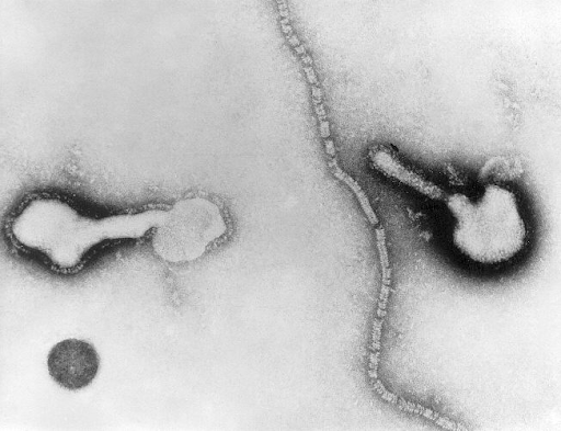
Transmission electron micrograph of parainfluenza virus

Image: “Transmission electron micrograph” by Public Health Image Library. License: CDC/Public Domain

Pathophysiology[1,5]

  • Viruses Viruses Minute infectious agents whose genomes are composed of DNA or RNA, but not both. They are characterized by a lack of independent metabolism and the inability to replicate outside living host cells. Virology that cause croup Croup Croup, also known as laryngotracheobronchitis, is a disease most commonly caused by a viral infection that leads to severe inflammation of the upper airway. It usually presents in children < 5 years of age. Patients develop a hoarse, "seal-like" barking cough and inspiratory stridor. Croup infect the nasal and pharyngeal mucosal epithelia through aerosol droplets Droplets Varicella-Zoster Virus/Chickenpox.
  • Infection spreads to the larynx Larynx The larynx, also commonly called the voice box, is a cylindrical space located in the neck at the level of the C3-C6 vertebrae. The major structures forming the framework of the larynx are the thyroid cartilage, cricoid cartilage, and epiglottis. The larynx serves to produce sound (phonation), conducts air to the trachea, and prevents large molecules from reaching the lungs. Larynx: Anatomy and trachea Trachea The trachea is a tubular structure that forms part of the lower respiratory tract. The trachea is continuous superiorly with the larynx and inferiorly becomes the bronchial tree within the lungs. The trachea consists of a support frame of semicircular, or C-shaped, rings made out of hyaline cartilage and reinforced by collagenous connective tissue. Trachea: Anatomy via respiratory epithelium Epithelium The epithelium is a complex of specialized cellular organizations arranged into sheets and lining cavities and covering the surfaces of the body. The cells exhibit polarity, having an apical and a basal pole. Structures important for the epithelial integrity and function involve the basement membrane, the semipermeable sheet on which the cells rest, and interdigitations, as well as cellular junctions. Surface Epithelium: Histology.
  • Infection triggers the infiltration of white blood cells, especially histiocytes Histiocytes Macrophages found in the tissues, as opposed to those found in the blood (monocytes) or serous cavities (serous membrane). Chronic Granulomatous Disease, neutrophils Neutrophils Granular leukocytes having a nucleus with three to five lobes connected by slender threads of chromatin, and cytoplasm containing fine inconspicuous granules and stainable by neutral dyes. Innate Immunity: Phagocytes and Antigen Presentation, plasma Plasma The residual portion of blood that is left after removal of blood cells by centrifugation without prior blood coagulation. Transfusion Products cells, and lymphocytes Lymphocytes Lymphocytes are heterogeneous WBCs involved in immune response. Lymphocytes develop from the bone marrow, starting from hematopoietic stem cells (HSCs) and progressing to common lymphoid progenitors (CLPs). B and T lymphocytes and natural killer (NK) cells arise from the lineage. Lymphocytes: Histology.
  • Leads to swelling Swelling Inflammation and edema Edema Edema is a condition in which excess serous fluid accumulates in the body cavity or interstitial space of connective tissues. Edema is a symptom observed in several medical conditions. It can be categorized into 2 types, namely, peripheral (in the extremities) and internal (in an organ or body cavity). Edema inside the trachea Trachea The trachea is a tubular structure that forms part of the lower respiratory tract. The trachea is continuous superiorly with the larynx and inferiorly becomes the bronchial tree within the lungs. The trachea consists of a support frame of semicircular, or C-shaped, rings made out of hyaline cartilage and reinforced by collagenous connective tissue. Trachea: Anatomy, larynx Larynx The larynx, also commonly called the voice box, is a cylindrical space located in the neck at the level of the C3-C6 vertebrae. The major structures forming the framework of the larynx are the thyroid cartilage, cricoid cartilage, and epiglottis. The larynx serves to produce sound (phonation), conducts air to the trachea, and prevents large molecules from reaching the lungs. Larynx: Anatomy, and large bronchi Bronchi The larger air passages of the lungs arising from the terminal bifurcation of the trachea. They include the largest two primary bronchi which branch out into secondary bronchi, and tertiary bronchi which extend into bronchioles and pulmonary alveoli. Bronchial Tree: Anatomy
  • Swelling Swelling Inflammation partially obstructs/narrows the subglottic airway Airway ABCDE Assessment (anatomic hallmark of croup Croup Croup, also known as laryngotracheobronchitis, is a disease most commonly caused by a viral infection that leads to severe inflammation of the upper airway. It usually presents in children < 5 years of age. Patients develop a hoarse, "seal-like" barking cough and inspiratory stridor. Croup)
  • Heavy breathing during exercise and other vigorous physical activities results in stridor Stridor Laryngomalacia and Tracheomalacia.

Clinical Presentation

  • Signs and symptoms vary:[1]
  • Typical course (viral croup Croup Croup, also known as laryngotracheobronchitis, is a disease most commonly caused by a viral infection that leads to severe inflammation of the upper airway. It usually presents in children < 5 years of age. Patients develop a hoarse, "seal-like" barking cough and inspiratory stridor. Croup)[2,5,6]:
    • Initial symptoms: coryza Coryza Inflammation of the nasal mucosa, the mucous membrane lining the nasal cavities. Rhinitis, nasal congestion
    • 12–48 hours: fever Fever Fever is defined as a measured body temperature of at least 38°C (100.4°F). Fever is caused by circulating endogenous and/or exogenous pyrogens that increase levels of prostaglandin E2 in the hypothalamus. Fever is commonly associated with chills, rigors, sweating, and flushing of the skin. Fever, barking cough, hoarseness Hoarseness An unnaturally deep or rough quality of voice. Parapharyngeal Abscess, stridor Stridor Laryngomalacia and Tracheomalacia
    • As disease progresses, respiratory distress (noted by tachypnea Tachypnea Increased respiratory rate. Pulmonary Examination, dyspnea Dyspnea Dyspnea is the subjective sensation of breathing discomfort. Dyspnea is a normal manifestation of heavy physical or psychological exertion, but also may be caused by underlying conditions (both pulmonary and extrapulmonary). Dyspnea at rest, thoracic retractions, mental status changes) can occur.
    • Disease lasts around 3–7 days (self-limited).
  • Spasmodic croup Croup Croup, also known as laryngotracheobronchitis, is a disease most commonly caused by a viral infection that leads to severe inflammation of the upper airway. It usually presents in children < 5 years of age. Patients develop a hoarse, "seal-like" barking cough and inspiratory stridor. Croup[5,6]:
    • More abrupt onset
    • Often without fever Fever Fever is defined as a measured body temperature of at least 38°C (100.4°F). Fever is caused by circulating endogenous and/or exogenous pyrogens that increase levels of prostaglandin E2 in the hypothalamus. Fever is commonly associated with chills, rigors, sweating, and flushing of the skin. Fever 
    • Symptoms are episodic and occur at night, with periods of daytime improvement.
    • May be seen more frequently in those with a family history Family History Adult Health Maintenance of allergies Allergies A medical specialty concerned with the hypersensitivity of the individual to foreign substances and protection from the resultant infection or disorder. Selective IgA Deficiency

Diagnosis

The diagnosis is based on the presence of a “seal-like” barking cough and inspiratory stridor Stridor Laryngomalacia and Tracheomalacia.[6]

  • Neither radiographs nor laboratory tests are necessary to make the diagnosis.
  • The severity of croup Croup Croup, also known as laryngotracheobronchitis, is a disease most commonly caused by a viral infection that leads to severe inflammation of the upper airway. It usually presents in children < 5 years of age. Patients develop a hoarse, "seal-like" barking cough and inspiratory stridor. Croup can be detected by the following laboratory tests:
    • Lateral neck Neck The part of a human or animal body connecting the head to the rest of the body. Peritonsillar Abscess X-ray X-ray Penetrating electromagnetic radiation emitted when the inner orbital electrons of an atom are excited and release radiant energy. X-ray wavelengths range from 1 pm to 10 nm. Hard x-rays are the higher energy, shorter wavelength x-rays. Soft x-rays or grenz rays are less energetic and longer in wavelength. The short wavelength end of the x-ray spectrum overlaps the gamma rays wavelength range. The distinction between gamma rays and x-rays is based on their radiation source. Pulmonary Function Tests to rule out any other cause, such as a peritonsillar abscess Abscess Accumulation of purulent material in tissues, organs, or circumscribed spaces, usually associated with signs of infection. Chronic Granulomatous Disease
    • Anteroposterior X-ray usually shows a “steeple sign,” which represents subglottic narrowing
  • Bronchoscopy Bronchoscopy Endoscopic examination, therapy or surgery of the bronchi. Laryngomalacia and Tracheomalacia may be indicated in patients Patients Individuals participating in the health care system for the purpose of receiving therapeutic, diagnostic, or preventive procedures. Clinician–Patient Relationship with recurrent episodes of croup Croup Croup, also known as laryngotracheobronchitis, is a disease most commonly caused by a viral infection that leads to severe inflammation of the upper airway. It usually presents in children < 5 years of age. Patients develop a hoarse, "seal-like" barking cough and inspiratory stridor. Croup.
  • Severity of the disease can be assessed by the Westley score Westley score Croup (calculator):
Feature Number of points assigned for this feature
0 1 2 3 4 5
Chest wall Chest wall The chest wall consists of skin, fat, muscles, bones, and cartilage. The bony structure of the chest wall is composed of the ribs, sternum, and thoracic vertebrae. The chest wall serves as armor for the vital intrathoracic organs and provides the stability necessary for the movement of the shoulders and arms. Chest Wall: Anatomy retraction None Mild Moderate Severe
Stridor Stridor Laryngomalacia and Tracheomalacia None With agitation Agitation A feeling of restlessness associated with increased motor activity. This may occur as a manifestation of nervous system drug toxicity or other conditions. St. Louis Encephalitis Virus At rest
Cyanosis Cyanosis A bluish or purplish discoloration of the skin and mucous membranes due to an increase in the amount of deoxygenated hemoglobin in the blood or a structural defect in the hemoglobin molecule. Pulmonary Examination None With agitation Agitation A feeling of restlessness associated with increased motor activity. This may occur as a manifestation of nervous system drug toxicity or other conditions. St. Louis Encephalitis Virus At rest
Level of consciousness Normal Disoriented
Air entry Normal Decreased Markedly decreased

Tip: While a chest X-ray X-ray Penetrating electromagnetic radiation emitted when the inner orbital electrons of an atom are excited and release radiant energy. X-ray wavelengths range from 1 pm to 10 nm. Hard x-rays are the higher energy, shorter wavelength x-rays. Soft x-rays or grenz rays are less energetic and longer in wavelength. The short wavelength end of the x-ray spectrum overlaps the gamma rays wavelength range. The distinction between gamma rays and x-rays is based on their radiation source. Pulmonary Function Tests will show a narrowing of the air column in the trachea Trachea The trachea is a tubular structure that forms part of the lower respiratory tract. The trachea is continuous superiorly with the larynx and inferiorly becomes the bronchial tree within the lungs. The trachea consists of a support frame of semicircular, or C-shaped, rings made out of hyaline cartilage and reinforced by collagenous connective tissue. Trachea: Anatomy ( steeple sign Steeple Sign Pediatric Chest Abnormalities), a chest X-ray X-ray Penetrating electromagnetic radiation emitted when the inner orbital electrons of an atom are excited and release radiant energy. X-ray wavelengths range from 1 pm to 10 nm. Hard x-rays are the higher energy, shorter wavelength x-rays. Soft x-rays or grenz rays are less energetic and longer in wavelength. The short wavelength end of the x-ray spectrum overlaps the gamma rays wavelength range. The distinction between gamma rays and x-rays is based on their radiation source. Pulmonary Function Tests is rarely done in practice and is always the wrong answer if a question stem asks what the most appropriate next step in diagnosis is, since the diagnosis is based on the clinical signs of a “seal-like” barking cough and inspiratory stridor Stridor Laryngomalacia and Tracheomalacia.

Steeple sign

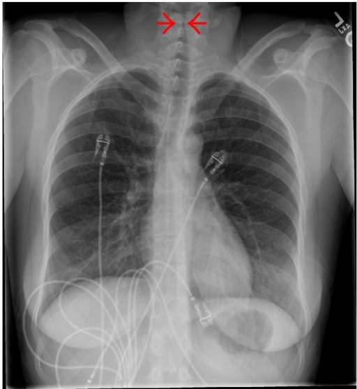
Chest X-ray showing subglottic stenosis, known as steeple sign, commonly seen on anteroposterior X-ray in patients with croup.

Image: “Steeple sign” by Jayshil J. Patel, Emily Kitchin, and Kurt Pfeifer. License: CC BY 4.0

Management

Management may vary based on practice location. The following information is based on US, Canada, and UK-based guidelines.

Overview of treatment[4]

  • Supportive care: supplemental oxygen Supplemental Oxygen Respiratory Failure, antipyretics, fluid intake
  • Oxygen administration:
    • The patient should inhale oxygen via the “blow-by” method (holding an oxygen source near the child’s face).
    • To avoid distress in children, a mask or nasal cannula Nasal Cannula Respiratory Failure should not be used.
    • Hot steam or humidified air use does not provide effective relief.
    • Endotracheal intubation Intubation Peritonsillar Abscess is required in < 3% of cases.
  • Medications:
    • Corticosteroids Corticosteroids Chorioretinitis (e.g., dexamethasone Dexamethasone An anti-inflammatory 9-fluoro-glucocorticoid. Antiemetics, budesonide Budesonide A glucocorticoid used in the management of asthma, the treatment of various skin disorders, and allergic rhinitis. Asthma Drugs) are given orally or by injection; improvement in airway Airway ABCDE Assessment inflammation Inflammation Inflammation is a complex set of responses to infection and injury involving leukocytes as the principal cellular mediators in the body’s defense against pathogenic organisms. Inflammation is also seen as a response to tissue injury in the process of wound healing. The 5 cardinal signs of inflammation are pain, heat, redness, swelling, and loss of function. Inflammation/symptoms is usually seen in 6–8 hours.
    • Racemic epinephrine Epinephrine The active sympathomimetic hormone from the adrenal medulla. It stimulates both the alpha- and beta- adrenergic systems, causes systemic vasoconstriction and gastrointestinal relaxation, stimulates the heart, and dilates bronchi and cerebral vessels. Sympathomimetic Drugs can be given by nebulization Nebulization Asthma Drugs to control the severity of the disease.
    • Antibiotics are prescribed only in cases of primary or secondary bacterial infection ( vancomycin Vancomycin Antibacterial obtained from streptomyces orientalis. It is a glycopeptide related to ristocetin that inhibits bacterial cell wall assembly and is toxic to kidneys and the inner ear. Glycopeptides and cefotaxime Cefotaxime Semisynthetic broad-spectrum cephalosporin. Cephalosporins are recommended in these cases).
    • Antiviral Antiviral Antivirals for Hepatitis B neuraminidase inhibitors Neuraminidase Inhibitors Antivirals for Influenza should be given to patients Patients Individuals participating in the health care system for the purpose of receiving therapeutic, diagnostic, or preventive procedures. Clinician–Patient Relationship with influenza Influenza Influenza viruses are members of the Orthomyxoviridae family and the causative organisms of influenza, a highly contagious febrile respiratory disease. There are 3 primary influenza viruses (A, B, and C) and various subtypes, which are classified based on their virulent surface antigens, hemagglutinin (HA) and neuraminidase (NA). Influenza typically presents with a fever, myalgia, headache, and symptoms of an upper respiratory infection. Influenza Viruses/Influenza.
    • Cough medicines with dextromethorphan or guaifenesin should be avoided.
    • Antipyretics as needed
    • Oxygen support as needed
  • Observe 3–4 hours after initial treatment and pharmacologic intervention. 
  • Hospitalization Hospitalization The confinement of a patient in a hospital. Delirium should be considered for patients Patients Individuals participating in the health care system for the purpose of receiving therapeutic, diagnostic, or preventive procedures. Clinician–Patient Relationship with ongoing severe symptoms after ≥ 4 hours of initial treatment.

Management details (based on severity)

Mild: no stridor Stridor Laryngomalacia and Tracheomalacia at rest ( Westley score Westley score Croup ≤ 2)[1-3]

  • Medication options:
    • Dexamethasone Dexamethasone An anti-inflammatory 9-fluoro-glucocorticoid. Antiemetics 0.15‒0.6 mg/kg by mouth up to 16 mg once
    • Prednisolone Prednisolone A glucocorticoid with the general properties of the corticosteroids. It is the drug of choice for all conditions in which routine systemic corticosteroid therapy is indicated, except adrenal deficiency states. Immunosuppressants 1 mg/kg by mouth once
  • May discharge home with caregiver education on disease course and when to seek medical attention Attention Focusing on certain aspects of current experience to the exclusion of others. It is the act of heeding or taking notice or concentrating. Psychiatric Assessment (e.g., stridor Stridor Laryngomalacia and Tracheomalacia at rest, difficulty breathing, drooling Drooling Peritonsillar Abscess, etc ETC The electron transport chain (ETC) sends electrons through a series of proteins, which generate an electrochemical proton gradient that produces energy in the form of adenosine triphosphate (ATP). Electron Transport Chain (ETC).)

Moderate: stridor Stridor Laryngomalacia and Tracheomalacia at rest without agitation Agitation A feeling of restlessness associated with increased motor activity. This may occur as a manifestation of nervous system drug toxicity or other conditions. St. Louis Encephalitis Virus ( Westley score Westley score Croup 3–7)[1-3]

  • Minimize patient discomfort.
  • Allow parents to comfort the child by holding on their lap.
  • Dexamethasone Dexamethasone An anti-inflammatory 9-fluoro-glucocorticoid. Antiemetics:
    • 0.15‒0.6 mg/kg by mouth once
    • 0.6 mg/kg IM once
    • 0.6 mg/kg IV once (do not start IV access for medication administration to limit Limit A value (e.g., pressure or time) that should not be exceeded and which is specified by the operator to protect the lung Invasive Mechanical Ventilation the distress of the child; use IV access only if necessary because of poor oral intake and volume depletion Volume depletion Volume status is a balance between water and solutes, the majority of which is Na. Volume depletion refers to a loss of both water and Na, whereas dehydration refers only to a loss of water. Volume depletion can be caused by GI losses, renal losses, bleeding, poor oral Na intake, or third spacing of fluids. Volume Depletion and Dehydration)
    • Budesonide Budesonide A glucocorticoid used in the management of asthma, the treatment of various skin disorders, and allergic rhinitis. Asthma Drugs 2 mg nebulized once (alternative to dexamethasone Dexamethasone An anti-inflammatory 9-fluoro-glucocorticoid. Antiemetics if patient is vomiting Vomiting The forcible expulsion of the contents of the stomach through the mouth. Hypokalemia and IV access is hard to establish)
  • Nebulized epinephrine Epinephrine The active sympathomimetic hormone from the adrenal medulla. It stimulates both the alpha- and beta- adrenergic systems, causes systemic vasoconstriction and gastrointestinal relaxation, stimulates the heart, and dilates bronchi and cerebral vessels. Sympathomimetic Drugs:
    • Racemic epinephrine Epinephrine The active sympathomimetic hormone from the adrenal medulla. It stimulates both the alpha- and beta- adrenergic systems, causes systemic vasoconstriction and gastrointestinal relaxation, stimulates the heart, and dilates bronchi and cerebral vessels. Sympathomimetic Drugs (2.25% solution)
      • 0.05‒0.1 mL/kg/dose inhaled
      • Maximum dose: 0.5 mL
    • L-epinephrine (1 mg/mL parenteral solution)
      • 0.5 mL/kg/dose of 1:1,000 solution
      • Maximum dose: 5 mL
  • Oxygen support as needed

Severe: stridor Stridor Laryngomalacia and Tracheomalacia at rest with agitation Agitation A feeling of restlessness associated with increased motor activity. This may occur as a manifestation of nervous system drug toxicity or other conditions. St. Louis Encephalitis Virus or lethargy Lethargy A general state of sluggishness, listless, or uninterested, with being tired, and having difficulty concentrating and doing simple tasks. It may be related to depression or drug addiction. Hyponatremia ( Westley score Westley score Croup 8–11)[1,2]

  • Follow guidelines for moderate symptoms. 
  • Add supplemental O2 if cyanosis Cyanosis A bluish or purplish discoloration of the skin and mucous membranes due to an increase in the amount of deoxygenated hemoglobin in the blood or a structural defect in the hemoglobin molecule. Pulmonary Examination or SpO2 < 92% is present.[1,3]
    • Blow-by at 8‒10 L/min
    • Nonrebreather, 100% O2

For moderate to severe symptoms, observe for 3‒4 hours after initial treatment.[1-3]

  • If symptoms are resolving and Westley score Westley score Croup < 3:
    • Discharge home with precautions and caregiver education.
    • Follow-up with pediatrician after 24 hours.
  • If patient continues to have moderate symptoms ( Westley score Westley score Croup 3‒7):
    • Repeat nebulized epinephrine Epinephrine The active sympathomimetic hormone from the adrenal medulla. It stimulates both the alpha- and beta- adrenergic systems, causes systemic vasoconstriction and gastrointestinal relaxation, stimulates the heart, and dilates bronchi and cerebral vessels. Sympathomimetic Drugs.
    • Treat with antipyretics if needed.
    • Optimize fluid status orally or IV if needed.
    • Consider hospital admission.
  • If the patient continues with or progresses to severe symptoms ( Westley score Westley score Croup 8‒11):
    • Repeat nebulized epinephrine Epinephrine The active sympathomimetic hormone from the adrenal medulla. It stimulates both the alpha- and beta- adrenergic systems, causes systemic vasoconstriction and gastrointestinal relaxation, stimulates the heart, and dilates bronchi and cerebral vessels. Sympathomimetic Drugs.
    • Treat with antipyretics if needed.
    • Optimize fluid status orally or IV if needed.
    • Consider alternative diagnoses (e.g., bacterial infection).
    • Antibiotics prescribed only in cases of primary or secondary bacterial infection ( vancomycin Vancomycin Antibacterial obtained from streptomyces orientalis. It is a glycopeptide related to ristocetin that inhibits bacterial cell wall assembly and is toxic to kidneys and the inner ear. Glycopeptides and cefotaxime Cefotaxime Semisynthetic broad-spectrum cephalosporin. Cephalosporins recommended in these cases)
    • Admit to ICU ICU Hospital units providing continuous surveillance and care to acutely ill patients. West Nile Virus

Impending respiratory failure Respiratory failure Respiratory failure is a syndrome that develops when the respiratory system is unable to maintain oxygenation and/or ventilation. Respiratory failure may be acute or chronic and is classified as hypoxemic, hypercapnic, or a combination of the two. Respiratory Failure ( Westley score Westley score Croup ≥ 12)[1-3]

  • Dexamethasone Dexamethasone An anti-inflammatory 9-fluoro-glucocorticoid. Antiemetics:
    • 0.6 mg/kg IM or IV; maximum dose: 16 mg
  • Nebulized epinephrine Epinephrine The active sympathomimetic hormone from the adrenal medulla. It stimulates both the alpha- and beta- adrenergic systems, causes systemic vasoconstriction and gastrointestinal relaxation, stimulates the heart, and dilates bronchi and cerebral vessels. Sympathomimetic Drugs:
    • Racemic epinephrine Epinephrine The active sympathomimetic hormone from the adrenal medulla. It stimulates both the alpha- and beta- adrenergic systems, causes systemic vasoconstriction and gastrointestinal relaxation, stimulates the heart, and dilates bronchi and cerebral vessels. Sympathomimetic Drugs (2.25% solution)
      • 0.05‒0.1 mL/kg/dose inhaled
      • Maximum dose: 0.5 mL
    • L-epinephrine (1 mg/mL parenteral solution)
      • 0.5 mL/kg/dose of 1:1,000 solution
      • Maximum dose: 5 mL
  • Intubation Intubation Peritonsillar Abscess:
    • Controlled environment with a skilled provider (anesthesiologist or otolaryngologist) assisting, if possible
    • If possible, perform in an operating room for advanced airway Airway ABCDE Assessment management.
    • Endotracheal tube (ETT) 0.5‒1 mm smaller than normal (normal pediatric ETT formula = (age in years/4) + 4)
    • Transfer to ICU ICU Hospital units providing continuous surveillance and care to acutely ill patients. West Nile Virus for ventilation Ventilation The total volume of gas inspired or expired per unit of time, usually measured in liters per minute. Ventilation: Mechanics of Breathing management.
    • Endotracheal intubation Intubation Peritonsillar Abscess is required in < 3% of cases.

Discharge criteria[2,3]

Prognosis

  • Croup Croup Croup, also known as laryngotracheobronchitis, is a disease most commonly caused by a viral infection that leads to severe inflammation of the upper airway. It usually presents in children < 5 years of age. Patients develop a hoarse, "seal-like" barking cough and inspiratory stridor. Croup is a self-limiting Self-Limiting Meningitis in Children disease that usually resolves within 3 days (80% of cases).[1]
  • Most cases improve in 3–7 days.
  • Hospitalization Hospitalization The confinement of a patient in a hospital. Delirium is needed in only 8%–15% of cases.
  • Croup Croup Croup, also known as laryngotracheobronchitis, is a disease most commonly caused by a viral infection that leads to severe inflammation of the upper airway. It usually presents in children < 5 years of age. Patients develop a hoarse, "seal-like" barking cough and inspiratory stridor. Croup is a life-threatening illness but rarely progresses to death; mortality Mortality All deaths reported in a given population. Measures of Health Status occurs in < 1% of intubated patients Patients Individuals participating in the health care system for the purpose of receiving therapeutic, diagnostic, or preventive procedures. Clinician–Patient Relationship; out-of-hospital cardiac arrest Cardiac arrest Cardiac arrest is the sudden, complete cessation of cardiac output with hemodynamic collapse. Patients present as pulseless, unresponsive, and apneic. Rhythms associated with cardiac arrest are ventricular fibrillation/tachycardia, asystole, or pulseless electrical activity. Cardiac Arrest and death may occur.
  • Complications (uncommon)
    • Pneumonia Pneumonia Pneumonia or pulmonary inflammation is an acute or chronic inflammation of lung tissue. Causes include infection with bacteria, viruses, or fungi. In more rare cases, pneumonia can also be caused through toxic triggers through inhalation of toxic substances, immunological processes, or in the course of radiotherapy. Pneumonia
    • Pulmonary edema Pulmonary edema Pulmonary edema is a condition caused by excess fluid within the lung parenchyma and alveoli as a consequence of a disease process. Based on etiology, pulmonary edema is classified as cardiogenic or noncardiogenic. Patients may present with progressive dyspnea, orthopnea, cough, or respiratory failure. Pulmonary Edema
    • Secondary bacterial tracheitis (see high fever Fever Fever is defined as a measured body temperature of at least 38°C (100.4°F). Fever is caused by circulating endogenous and/or exogenous pyrogens that increase levels of prostaglandin E2 in the hypothalamus. Fever is commonly associated with chills, rigors, sweating, and flushing of the skin. Fever, toxic appearance, mucopurulent exudates in the trachea Trachea The trachea is a tubular structure that forms part of the lower respiratory tract. The trachea is continuous superiorly with the larynx and inferiorly becomes the bronchial tree within the lungs. The trachea consists of a support frame of semicircular, or C-shaped, rings made out of hyaline cartilage and reinforced by collagenous connective tissue. Trachea: Anatomy)
    • Respiratory failure Respiratory failure Respiratory failure is a syndrome that develops when the respiratory system is unable to maintain oxygenation and/or ventilation. Respiratory failure may be acute or chronic and is classified as hypoxemic, hypercapnic, or a combination of the two. Respiratory Failure
    • Pneumothorax Pneumothorax A pneumothorax is a life-threatening condition in which air collects in the pleural space, causing partial or full collapse of the lung. A pneumothorax can be traumatic or spontaneous. Patients present with a sudden onset of sharp chest pain, dyspnea, and diminished breath sounds on exam. Pneumothorax
    • Pneumomediastinum Pneumomediastinum Mediastinitis
    • Recurrent symptoms (5% of cases)

Differential Diagnosis

  • Pneumonia Pneumonia Pneumonia or pulmonary inflammation is an acute or chronic inflammation of lung tissue. Causes include infection with bacteria, viruses, or fungi. In more rare cases, pneumonia can also be caused through toxic triggers through inhalation of toxic substances, immunological processes, or in the course of radiotherapy. Pneumonia: infection that inflames the alveoli Alveoli Small polyhedral outpouchings along the walls of the alveolar sacs, alveolar ducts and terminal bronchioles through the walls of which gas exchange between alveolar air and pulmonary capillary blood takes place. Acute Respiratory Distress Syndrome (ARDS) resulting in fluid or pus (purulent material) accumulation. Pneumonia Pneumonia Pneumonia or pulmonary inflammation is an acute or chronic inflammation of lung tissue. Causes include infection with bacteria, viruses, or fungi. In more rare cases, pneumonia can also be caused through toxic triggers through inhalation of toxic substances, immunological processes, or in the course of radiotherapy. Pneumonia clinically manifests as a productive cough with phlegm or pus (not barking cough), fever Fever Fever is defined as a measured body temperature of at least 38°C (100.4°F). Fever is caused by circulating endogenous and/or exogenous pyrogens that increase levels of prostaglandin E2 in the hypothalamus. Fever is commonly associated with chills, rigors, sweating, and flushing of the skin. Fever, chills Chills The sudden sensation of being cold. It may be accompanied by shivering. Fever, and difficulty breathing. Lung exam would include crackles. This is a lower respiratory infection and Xray shows infiltrates, consolidation Consolidation Pulmonary Function Tests or an interstitial pattern.
  • Secondary bacterial tracheitis: caused by a secondary bacterial infection of the trachea Trachea The trachea is a tubular structure that forms part of the lower respiratory tract. The trachea is continuous superiorly with the larynx and inferiorly becomes the bronchial tree within the lungs. The trachea consists of a support frame of semicircular, or C-shaped, rings made out of hyaline cartilage and reinforced by collagenous connective tissue. Trachea: Anatomy. The development of mucopurulent exudates and edema Edema Edema is a condition in which excess serous fluid accumulates in the body cavity or interstitial space of connective tissues. Edema is a symptom observed in several medical conditions. It can be categorized into 2 types, namely, peripheral (in the extremities) and internal (in an organ or body cavity). Edema leads to upper airway obstruction Upper Airway Obstruction Airway Obstruction. Lateral Xray would show inglammation of the trachea Trachea The trachea is a tubular structure that forms part of the lower respiratory tract. The trachea is continuous superiorly with the larynx and inferiorly becomes the bronchial tree within the lungs. The trachea consists of a support frame of semicircular, or C-shaped, rings made out of hyaline cartilage and reinforced by collagenous connective tissue. Trachea: Anatomy.
  • Epiglottitis Epiglottitis Epiglottitis (or “supraglottitis”) is an inflammation of the epiglottis and adjacent supraglottic structures. The majority of cases are caused by bacterial infection. Symptoms are rapid in onset and severe. Epiglottitis: inflammation Inflammation Inflammation is a complex set of responses to infection and injury involving leukocytes as the principal cellular mediators in the body’s defense against pathogenic organisms. Inflammation is also seen as a response to tissue injury in the process of wound healing. The 5 cardinal signs of inflammation are pain, heat, redness, swelling, and loss of function. Inflammation of the epiglottis Epiglottis A thin leaf-shaped cartilage that is covered with laryngeal mucosa and situated posterior to the root of the tongue and hyoid bone. During swallowing, the epiglottis folds back over the larynx inlet thus prevents foods from entering the airway. Larynx: Anatomy caused by Haemophilus Haemophilus Haemophilus is a genus of Gram-negative coccobacilli, all of whose strains require at least 1 of 2 factors for growth (factor V [NAD] and factor X [heme]); therefore, it is most often isolated on chocolate agar, which can supply both factors. The pathogenic species are H. influenzae and H. ducreyi. Haemophilus, streptococcal, or staphylococcal infections Staphylococcal Infections Infections with bacteria of the genus staphylococcus. Glycopeptides, causing dyspnea Dyspnea Dyspnea is the subjective sensation of breathing discomfort. Dyspnea is a normal manifestation of heavy physical or psychological exertion, but also may be caused by underlying conditions (both pulmonary and extrapulmonary). Dyspnea, stridor Stridor Laryngomalacia and Tracheomalacia, and cyanosis Cyanosis A bluish or purplish discoloration of the skin and mucous membranes due to an increase in the amount of deoxygenated hemoglobin in the blood or a structural defect in the hemoglobin molecule. Pulmonary Examination, ultimately leading to death due to obstruction of airways. Dysphagia Dysphagia Dysphagia is the subjective sensation of difficulty swallowing. Symptoms can range from a complete inability to swallow, to the sensation of solids or liquids becoming “stuck.” Dysphagia is classified as either oropharyngeal or esophageal, with esophageal dysphagia having 2 sub-types: functional and mechanical. Dysphagia and drooling Drooling Peritonsillar Abscess are seen, but barking cough is not common.
  • Laryngeal diphtheria: manifests as swollen neck and throat, or “bull neck” accompanied by the following clinical symptoms: “barking” cough, stridor Stridor Laryngomalacia and Tracheomalacia, hoarseness Hoarseness An unnaturally deep or rough quality of voice. Parapharyngeal Abscess, difficulty breathing, and diphtheritic croup Croup Croup, also known as laryngotracheobronchitis, is a disease most commonly caused by a viral infection that leads to severe inflammation of the upper airway. It usually presents in children < 5 years of age. Patients develop a hoarse, "seal-like" barking cough and inspiratory stridor. Croup
  • Foreign body Foreign Body Foreign Body Aspiration aspiration: Aspirated food may lodge in the larynx Larynx The larynx, also commonly called the voice box, is a cylindrical space located in the neck at the level of the C3-C6 vertebrae. The major structures forming the framework of the larynx are the thyroid cartilage, cricoid cartilage, and epiglottis. The larynx serves to produce sound (phonation), conducts air to the trachea, and prevents large molecules from reaching the lungs. Larynx: Anatomy or trachea Trachea The trachea is a tubular structure that forms part of the lower respiratory tract. The trachea is continuous superiorly with the larynx and inferiorly becomes the bronchial tree within the lungs. The trachea consists of a support frame of semicircular, or C-shaped, rings made out of hyaline cartilage and reinforced by collagenous connective tissue. Trachea: Anatomy, which can lead to choking and potentially death.
  • Peritonsillar, parapharyngeal, or retropharyngeal abscesses: Abscesses of the pharynx Pharynx The pharynx is a component of the digestive system that lies posterior to the nasal cavity, oral cavity, and larynx. The pharynx can be divided into the oropharynx, nasopharynx, and laryngopharynx. Pharyngeal muscles play an integral role in vital processes such as breathing, swallowing, and speaking. Pharynx: Anatomy present with fever Fever Fever is defined as a measured body temperature of at least 38°C (100.4°F). Fever is caused by circulating endogenous and/or exogenous pyrogens that increase levels of prostaglandin E2 in the hypothalamus. Fever is commonly associated with chills, rigors, sweating, and flushing of the skin. Fever, lymphadenopathy Lymphadenopathy Lymphadenopathy is lymph node enlargement (> 1 cm) and is benign and self-limited in most patients. Etiologies include malignancy, infection, and autoimmune disorders, as well as iatrogenic causes such as the use of certain medications. Generalized lymphadenopathy often indicates underlying systemic disease. Lymphadenopathy, painful neck Neck The part of a human or animal body connecting the head to the rest of the body. Peritonsillar Abscess, neck stiffness Neck Stiffness Meningitis, drooling Drooling Peritonsillar Abscess, uvula Uvula A fleshy extension at the back of the soft palate that hangs above the opening of the throat. Peritonsillar Abscess deviation, “hot potato” voice that is muffled, and hoarse, and trouble swallowing Swallowing The act of taking solids and liquids into the gastrointestinal tract through the mouth and throat. Gastrointestinal Motility. Barking cough is not noted.
  • Angioedema Angioedema Angioedema is a localized, self-limited (but potentially life-threatening), nonpitting, asymmetrical edema occurring in the deep layers of the skin and mucosal tissue. The common underlying pathophysiology involves inflammatory mediators triggering significant vasodilation and increased capillary permeability. Angioedema or allergic reaction: rapid onset of airway Airway ABCDE Assessment obstruction, which can manifest as inspiratory stridor Stridor Laryngomalacia and Tracheomalacia. There is no associated fever Fever Fever is defined as a measured body temperature of at least 38°C (100.4°F). Fever is caused by circulating endogenous and/or exogenous pyrogens that increase levels of prostaglandin E2 in the hypothalamus. Fever is commonly associated with chills, rigors, sweating, and flushing of the skin. Fever. There can be lip swelling Swelling Inflammation, or rash Rash Rocky Mountain Spotted Fever. History of allergies Allergies A medical specialty concerned with the hypersensitivity of the individual to foreign substances and protection from the resultant infection or disorder. Selective IgA Deficiency or similar attack/s may be present.

References

  1. Bjornson C., Johnson D. (2025). Croup: symptoms, diagnosis, and treatment. BMJ Best Practice. https://bestpractice.bmj.com/topics/en-gb/681
  2. Smith, D. K., McDermott, A. J., & Sullivan, J. F. (2018). Croup: Diagnosis and Management. American family physician, 97(9), 575–580. https://www.aafp.org/pubs/afp/issues/2018/0501/p575.html
  3. Defendi, G. (2024) Croup. Medscape. Retrieved April 22, 2025 from https://emedicine.medscape.com/article/962972-overview
  4. Woods, C. R., Goldman, M., Tovar Padua, L. (2025). Management of Croup. UpToDate. Retrieved April 22, 2025, from https://www.uptodate.com/contents/croup-management
  5. Woods, C.R. ,Goldman, M, Tovar Padua, L.(2025) Croup: clinical features, evaluation, and diagnosis. UpToDate. Retrieved April 22, 2025, from https://www.uptodate.com/contents/croup-clinical-features-evaluation-and-diagnosis
  6. Bower, J., McBride, J.T. (2015). Croup in children (acute laryngotracheobronchitis). Mandell, Douglas, and Bennett’s Principles and Practice of Infectious Diseases, 8th ed. pp. 762–766.e1. https://doi.org/10.1016/B978-1-4557-4801-3.00061-8

User Reviews

Details