Chronic obstructive pulmonary diseasePulmonary diseaseDiseases involving the respiratory system.Blastomyces/Blastomycosis (COPD) is a lung disease characterized by progressive, largely irreversible airflow obstruction. The condition usually presents in middle-aged or elderly persons with a history of cigarette smokingSmokingWillful or deliberate act of inhaling and exhaling smoke from burning substances or agents held by hand.Interstitial Lung Diseases. Symptoms include progressive dyspneaDyspneaDyspnea is the subjective sensation of breathing discomfort. Dyspnea is a normal manifestation of heavy physical or psychological exertion, but also may be caused by underlying conditions (both pulmonary and extrapulmonary). Dyspnea and chronic cough. Prolonged expirationExpirationVentilation: Mechanics of Breathing, wheezingWheezingWheezing is an abnormal breath sound characterized by a whistling noise that can be relatively high-pitched and shrill (more common) or coarse. Wheezing is produced by the movement of air through narrowed or compressed small (intrathoracic) airways. Wheezing, and/or diminished breath sounds may be noted on physical exam. The diagnosis is confirmed with a pulmonary function testPulmonary function testPulmonary function tests are a group of diagnostic procedures yielding useful, quantifiable information about the rate of the flow of air through the individual's airways, lung capacity, and the efficiency of gas exchange in relation to time. The most commonly utilized tests include spirometry (before and after bronchodilator use), lung volumes, and quantitation of diffusing capacity for carbon monoxide (CO). The tests can be influenced by the individual's effort/fatigue, disease state, or anatomical malformation.Pulmonary Function Tests. Management includes smokingSmokingWillful or deliberate act of inhaling and exhaling smoke from burning substances or agents held by hand.Interstitial Lung Diseases cessation, pulmonary rehabilitation, and pharmacotherapy.
Chronic obstructive pulmonary diseasePulmonary diseaseDiseases involving the respiratory system.Blastomyces/Blastomycosis (COPD) is a lung disease characterized by airflow limitation resulting from airwayAirwayABCDE Assessment disease and/or parenchymal destruction.
Types
The subtypes may have differing presentations and response to therapy. PatientsPatientsIndividuals participating in the health care system for the purpose of receiving therapeutic, diagnostic, or preventive procedures.Clinician–Patient Relationship may have any combination of both.
Chronic bronchitisChronic bronchitisA subcategory of chronic obstructive pulmonary disease. The disease is characterized by hypersecretion of mucus accompanied by a chronic (more than 3 months in 2 consecutive years) productive cough. Infectious agents are a major cause of chronic bronchitis.Rhinovirus:
Clinically defined
Productive cough > 3 months per year for at least 2 consecutive years
Must be in the absence of other causes of chronic cough
Emphysema:
Pathologically or radiologically defined
Destruction and permanent dilation of alveolar sacs
Epidemiology
Worldwide:
PrevalencePrevalenceThe total number of cases of a given disease in a specified population at a designated time. It is differentiated from incidence, which refers to the number of new cases in the population at a given time.Measures of Disease Frequency: 11.7% (expected to rise)
Annual deaths: 3 million
4th leading cause of death (soon to be 3rd)
In the United States:
PrevalencePrevalenceThe total number of cases of a given disease in a specified population at a designated time. It is differentiated from incidence, which refers to the number of new cases in the population at a given time.Measures of Disease Frequency: 16 million people
Annual deaths: > 140,000
3rd leading cause of death
Age:
PrevalencePrevalenceThe total number of cases of a given disease in a specified population at a designated time. It is differentiated from incidence, which refers to the number of new cases in the population at a given time.Measures of Disease Frequency peaks around 50–60 years of age
Age of onset is lower for heavy smokers.
SexSexThe totality of characteristics of reproductive structure, functions, phenotype, and genotype, differentiating the male from the female organism.Gender Dysphoria:
Cigarette smokingSmokingWillful or deliberate act of inhaling and exhaling smoke from burning substances or agents held by hand.Interstitial Lung Diseases (90% of cases)
2nd-hand smoke
Air pollutionPollutionThe presence of contaminants or pollutant substances in the air (air pollutants) that interfere with human health or welfare, or produce other harmful environmental effects. The substances may include gases; particulate matter; or volatile organic chemicals.Asthma
PrematurePrematureChildbirth before 37 weeks of pregnancy (259 days from the first day of the mother’s last menstrual period, or 245 days after fertilization).Necrotizing Enterocolitis birth
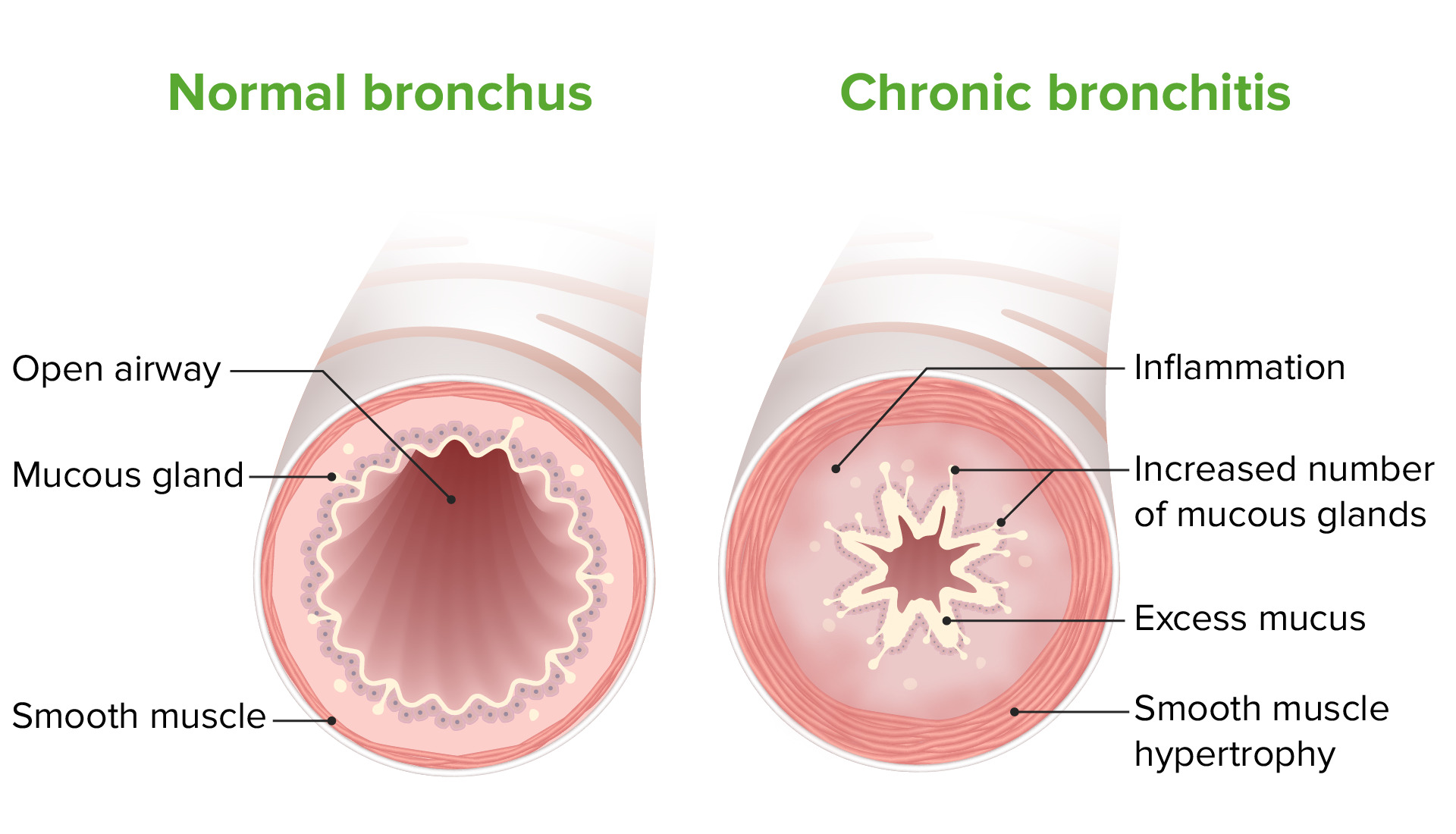
Chronic bronchitisChronic bronchitisA subcategory of chronic obstructive pulmonary disease. The disease is characterized by hypersecretion of mucus accompanied by a chronic (more than 3 months in 2 consecutive years) productive cough. Infectious agents are a major cause of chronic bronchitis.Rhinovirus
Damage to endothelial cells → ↓ mucocilliary clearance
Mucous gland hyperplasiaHyperplasiaAn increase in the number of cells in a tissue or organ without tumor formation. It differs from hypertrophy, which is an increase in bulk without an increase in the number of cells.Cellular Adaptation → mucous hypersecretion and plugging
AirwayAirwayABCDE AssessmentedemaEdemaEdema is a condition in which excess serous fluid accumulates in the body cavity or interstitial space of connective tissues. Edema is a symptom observed in several medical conditions. It can be categorized into 2 types, namely, peripheral (in the extremities) and internal (in an organ or body cavity). Edema and smooth muscle hyperplasiaHyperplasiaAn increase in the number of cells in a tissue or organ without tumor formation. It differs from hypertrophy, which is an increase in bulk without an increase in the number of cells.Cellular Adaptation and hypertrophyHypertrophyGeneral increase in bulk of a part or organ due to cell enlargement and accumulation of fluids and secretions, not due to tumor formation, nor to an increase in the number of cells (hyperplasia).Cellular Adaptation → luminal narrowing
Peribronchial fibrosisFibrosisAny pathological condition where fibrous connective tissue invades any organ, usually as a consequence of inflammation or other injury.Bronchiolitis Obliterans → bronchial distortionDistortionDefense Mechanisms
Chronic bronchitis pathophysiology: Inflammation, smooth muscle hypertrophy, and excess mucus production lead to progressive airway obstruction.
Image by Lecturio.
Emphysema
In normal lungsLungsLungs are the main organs of the respiratory system. Lungs are paired viscera located in the thoracic cavity and are composed of spongy tissue. The primary function of the lungs is to oxygenate blood and eliminate CO2. Lungs: Anatomy, there is a balance between:
ProteasesProteasesProteins and Peptides → break down elastin and connective tissueConnective tissueConnective tissues originate from embryonic mesenchyme and are present throughout the body except inside the brain and spinal cord. The main function of connective tissues is to provide structural support to organs. Connective tissues consist of cells and an extracellular matrix.Connective Tissue: Histology as part of normal tissue repairtissue repairThe process of generating a scar or less functional tissue with a different form and/or composition of the original tissue.Wound Healing:
Neutrophil elastaseElastaseA protease of broad specificity, obtained from dried pancreas. Molecular weight is approximately 25, 000. The enzyme breaks down elastin, the specific protein of elastic fibers, and digests other proteins such as fibrin, hemoglobin, and albumin.Proteins and Peptides
Matrix metalloproteinaseMatrix metalloproteinaseA family of zinc-dependent metalloendopeptidases that is involved in the degradation of extracellular matrix components.Pulmonary Fibrosis (MMP)
Cathepsins
Antiproteases → balance proteaseProteaseEnzyme of the human immunodeficiency virus that is required for post-translational cleavage of gag and gag-pol precursor polyproteins into functional products needed for viral assembly. HIV protease is an aspartic protease encoded by the amino terminus of the pol gene.HIV Infection and AIDS activity:
AAT
Secretory leukoprotease inhibitor derived from airwayAirwayABCDE AssessmentepitheliumEpitheliumThe epithelium is a complex of specialized cellular organizations arranged into sheets and lining cavities and covering the surfaces of the body. The cells exhibit polarity, having an apical and a basal pole. Structures important for the epithelial integrity and function involve the basement membrane, the semipermeable sheet on which the cells rest, and interdigitations, as well as cellular junctions. Surface Epithelium: Histology
ProteaseProteaseEnzyme of the human immunodeficiency virus that is required for post-translational cleavage of gag and gag-pol precursor polyproteins into functional products needed for viral assembly. HIV protease is an aspartic protease encoded by the amino terminus of the pol gene.HIV Infection and AIDS activity exceeds antiprotease activity → tissue destruction
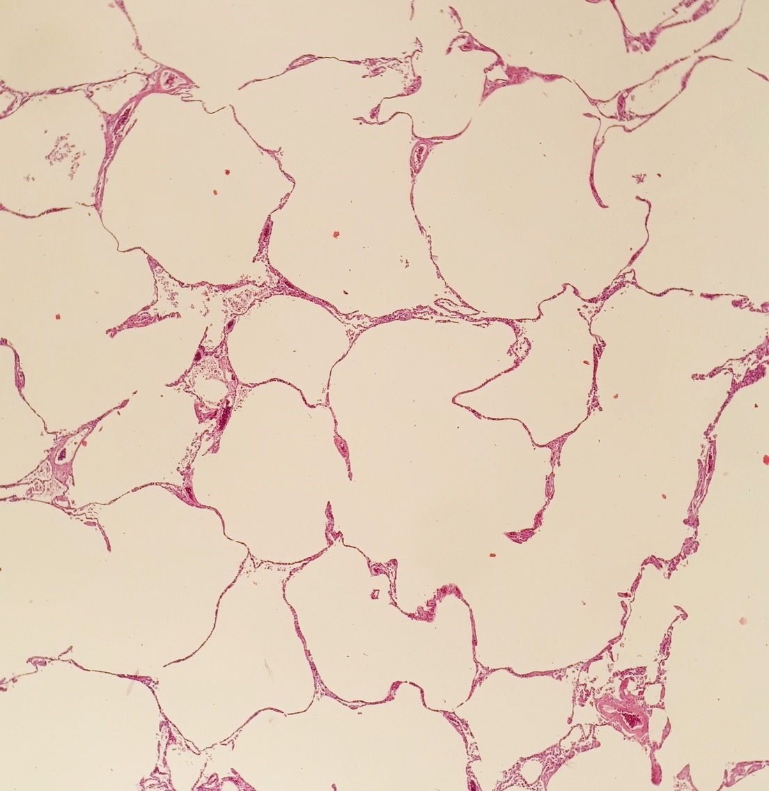
Alveolar destruction leads to:
Enlarged alveoliAlveoliSmall polyhedral outpouchings along the walls of the alveolar sacs, alveolar ducts and terminal bronchioles through the walls of which gas exchange between alveolar air and pulmonary capillary blood takes place.Acute Respiratory Distress Syndrome (ARDS)
↑ ComplianceComplianceDistensibility measure of a chamber such as the lungs (lung compliance) or bladder. Compliance is expressed as a change in volume per unit change in pressure.Veins: Histology
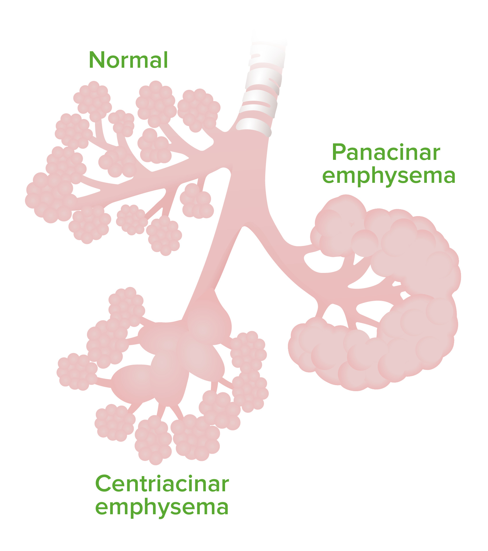
Centriacinar emphysema (associated with cigarette smokingSmokingWillful or deliberate act of inhaling and exhaling smoke from burning substances or agents held by hand.Interstitial Lung Diseases):
Destruction of the respiratory bronchiolesBronchiolesThe small airways branching off the tertiary bronchi. Terminal bronchioles lead into several orders of respiratory bronchioles which in turn lead into alveolar ducts and then into pulmonary alveoli.Bronchial Tree: Anatomy and a central portion of the acini
More severe in the apical lung fields
Panacinar emphysema (associated with AAT deficiency):
Destruction of all parts of the acinus
More severe in the basal lung fields
Emphysema: The image demonstrates the difference in affected areas in centriacinar vs panacinar emphysema.
Image by Lecturio.
Histopathology of severe emphysema showing enlarged alveoli (white spaces)
Image: “Histopathology of emphysema” by Mikael Häggström, M.D. License: Public Domain
Effects of the pulmonary vasculature
Tissue destruction → ↓ ability to oxygenate blood
HypoxemiaHypoxemiaNeonatal Respiratory Distress Syndrome → vasoconstrictionVasoconstrictionThe physiological narrowing of blood vessels by contraction of the vascular smooth muscle.Vascular Resistance, Flow, and Mean Arterial Pressure in small pulmonary arteriesArteriesArteries are tubular collections of cells that transport oxygenated blood and nutrients from the heart to the tissues of the body. The blood passes through the arteries in order of decreasing luminal diameter, starting in the largest artery (the aorta) and ending in the small arterioles. Arteries are classified into 3 types: large elastic arteries, medium muscular arteries, and small arteries and arterioles. Arteries: Histology → ↑ vascular resistanceResistancePhysiologically, the opposition to flow of air caused by the forces of friction. As a part of pulmonary function testing, it is the ratio of driving pressure to the rate of air flow.Ventilation: Mechanics of Breathing
Chronic hypoxemiaHypoxemiaNeonatal Respiratory Distress Syndrome → vascular remodelingVascular remodelingThe active alterations of vascular wall structures, often leading to elevated vascular resistance. It is associated with aging; atherosclerosis; diabetes mellitus; hypertension; pregnancy; pulmonary hypertension; and stroke, but is also a normal part of embryogenesis.Cor Pulmonale → irreversible pulmonary hypertensionPulmonary HypertensionPulmonary hypertension (PH) or pulmonary arterial hypertension (PAH) is characterized by elevated pulmonary arterial pressure, which can lead to chronic progressive right heart failure. Pulmonary hypertension is grouped into 5 categories based on etiology, which include primary PAH, and PH due to cardiac disease, lung or hypoxic disease, chronic thromboembolic disease, and multifactorial or unclear etiologies. Pulmonary Hypertension
Clinical Presentation
Symptoms
PatientsPatientsIndividuals participating in the health care system for the purpose of receiving therapeutic, diagnostic, or preventive procedures.Clinician–Patient Relationship suffer from chronic, progressive symptoms with acute exacerbations.
General:
Progressive dyspneaDyspneaDyspnea is the subjective sensation of breathing discomfort. Dyspnea is a normal manifestation of heavy physical or psychological exertion, but also may be caused by underlying conditions (both pulmonary and extrapulmonary). Dyspnea (particularly with exertion)
Chronic cough
Sputum production
Chest tightness
Weight gain or loss
FatigueFatigueThe state of weariness following a period of exertion, mental or physical, characterized by a decreased capacity for work and reduced efficiency to respond to stimuli.Fibromyalgia
Acute exacerbation:
Worsening dyspneaDyspneaDyspnea is the subjective sensation of breathing discomfort. Dyspnea is a normal manifestation of heavy physical or psychological exertion, but also may be caused by underlying conditions (both pulmonary and extrapulmonary). Dyspnea
Increased cough
Purulent sputum production
WheezingWheezingWheezing is an abnormal breath sound characterized by a whistling noise that can be relatively high-pitched and shrill (more common) or coarse. Wheezing is produced by the movement of air through narrowed or compressed small (intrathoracic) airways. Wheezing
FeverFeverFever is defined as a measured body temperature of at least 38°C (100.4°F). Fever is caused by circulating endogenous and/or exogenous pyrogens that increase levels of prostaglandin E2 in the hypothalamus. Fever is commonly associated with chills, rigors, sweating, and flushing of the skin. Fever may or may not be present.
Physical examination
When examining a patient with possible COPD, look for the following findings:
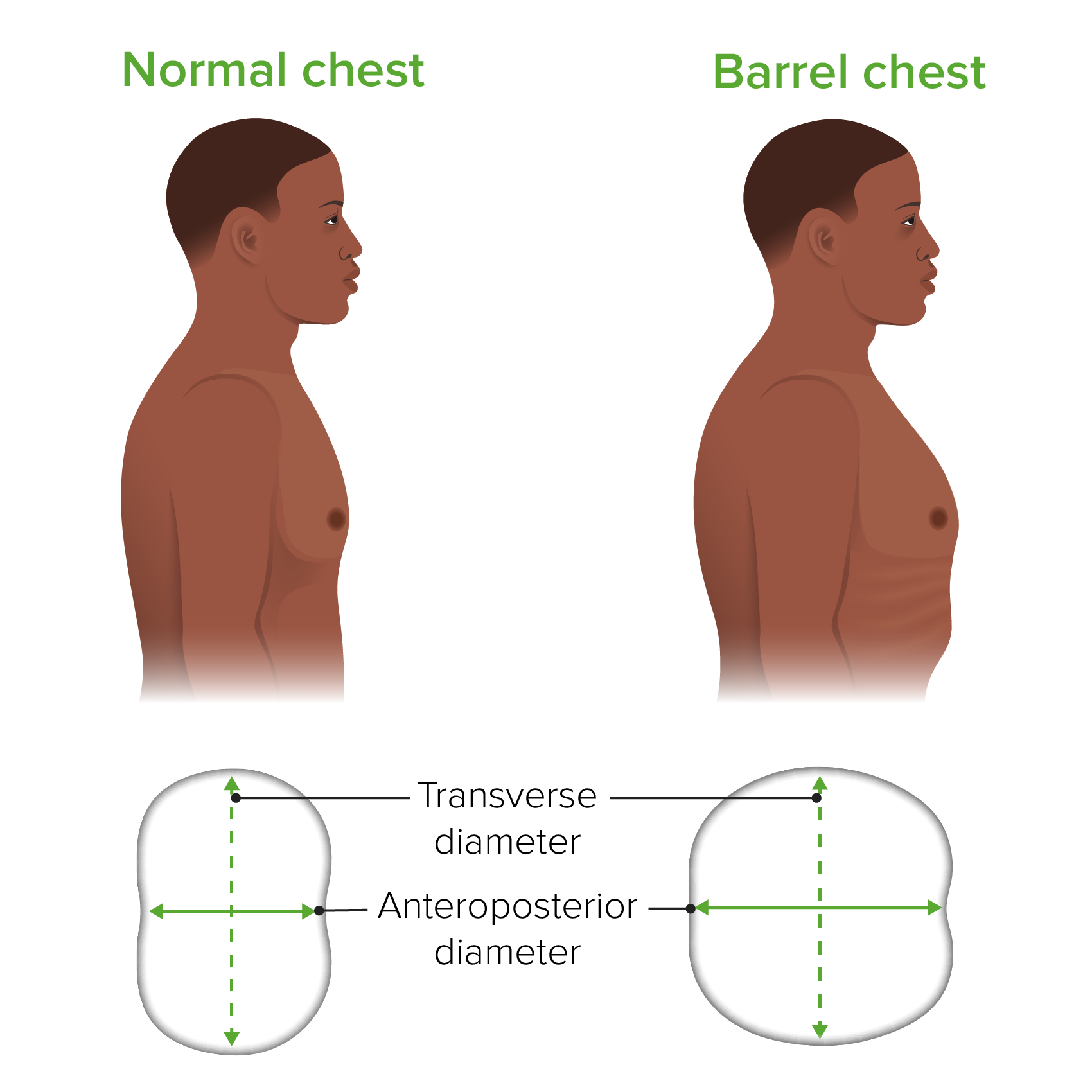
Barrel chest: increased anteroposterior chest wallChest wallThe chest wall consists of skin, fat, muscles, bones, and cartilage. The bony structure of the chest wall is composed of the ribs, sternum, and thoracic vertebrae. The chest wall serves as armor for the vital intrathoracic organs and provides the stability necessary for the movement of the shoulders and arms. Chest Wall: Anatomy diameter from hyperinflationHyperinflationImaging of the Lungs and Pleura
WheezingWheezingWheezing is an abnormal breath sound characterized by a whistling noise that can be relatively high-pitched and shrill (more common) or coarse. Wheezing is produced by the movement of air through narrowed or compressed small (intrathoracic) airways. Wheezing
Diminished breath sounds
PalpationPalpationApplication of fingers with light pressure to the surface of the body to determine consistency of parts beneath in physical diagnosis; includes palpation for determining the outlines of organs.Dermatologic Examination and percussionPercussionAct of striking a part with short, sharp blows as an aid in diagnosing the condition beneath the sound obtained.Pulmonary Examination:
Hyperresonance on percussionPercussionAct of striking a part with short, sharp blows as an aid in diagnosing the condition beneath the sound obtained.Pulmonary Examination
Reduced chest wallChest wallThe chest wall consists of skin, fat, muscles, bones, and cartilage. The bony structure of the chest wall is composed of the ribs, sternum, and thoracic vertebrae. The chest wall serves as armor for the vital intrathoracic organs and provides the stability necessary for the movement of the shoulders and arms. Chest Wall: Anatomy expansion
Extremities:
CyanosisCyanosisA bluish or purplish discoloration of the skin and mucous membranes due to an increase in the amount of deoxygenated hemoglobin in the blood or a structural defect in the hemoglobin molecule.Pulmonary Examination
Presence should raise suspicion for other comorbiditiesComorbiditiesThe presence of co-existing or additional diseases with reference to an initial diagnosis or with reference to the index condition that is the subject of study. Comorbidity may affect the ability of affected individuals to function and also their survival; it may be used as a prognostic indicator for length of hospital stay, cost factors, and outcome or survival.St. Louis Encephalitis Virus (e.g., lung cancerLung cancerLung cancer is the malignant transformation of lung tissue and the leading cause of cancer-related deaths. The majority of cases are associated with long-term smoking. The disease is generally classified histologically as either small cell lung cancer or non-small cell lung cancer. Symptoms include cough, dyspnea, weight loss, and chest discomfort. Lung Cancer, bronchiectasisBronchiectasisBronchiectasis is a chronic disease of the airways that results from permanent bronchial distortion. This results from a continuous cycle of inflammation, bronchial damage and dilation, impaired clearance of secretions, and recurrent infections. Bronchiectasis, interstitial lung disease)
Findings suggestive of cor pulmonaleCor PulmonaleCor pulmonale is right ventricular (RV) dysfunction caused by lung disease that results in pulmonary artery hypertension. The most common cause of cor pulmonale is chronic obstructive pulmonary disease. Dyspnea is the usual presenting symptom. Cor Pulmonale:
Peripheral edemaPeripheral edemaPeripheral edema is the swelling of the lower extremities, namely, legs, feet, and ankles.Edema
Barrel chest as seen in emphysema: Note the increase in the anteroposterior diameter.
Image by Lecturio.
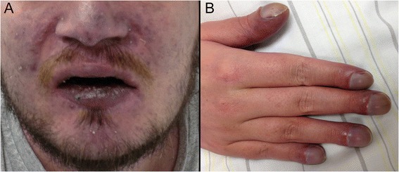
Clinical signs of hypoxemia: A: Cyanosis B: Digital clubbing
Image: “Clinical signs of chronic hypoxaemia” by Maximilian Patzig et al. License: CC BY 4.0
Clinical phenotypes
Signs and symptoms are associated more frequently with either chronic bronchitisChronic bronchitisA subcategory of chronic obstructive pulmonary disease. The disease is characterized by hypersecretion of mucus accompanied by a chronic (more than 3 months in 2 consecutive years) productive cough. Infectious agents are a major cause of chronic bronchitis.Rhinovirus or emphysema. However, patientsPatientsIndividuals participating in the health care system for the purpose of receiving therapeutic, diagnostic, or preventive procedures.Clinician–Patient Relationship often present with a mixture of features.
Chronic bronchitisChronic bronchitisA subcategory of chronic obstructive pulmonary disease. The disease is characterized by hypersecretion of mucus accompanied by a chronic (more than 3 months in 2 consecutive years) productive cough. Infectious agents are a major cause of chronic bronchitis.Rhinovirus (“blue bloaterBlue bloaterA subcategory of chronic obstructive pulmonary disease. The disease is characterized by hypersecretion of mucus accompanied by a chronic (more than 3 months in 2 consecutive years) productive cough. Infectious agents are a major cause of chronic bronchitis.Rhinovirus”):
PatientsPatientsIndividuals participating in the health care system for the purpose of receiving therapeutic, diagnostic, or preventive procedures.Clinician–Patient Relationship are generally overweight.
Frequent, productive cough
Peripheral edemaPeripheral edemaPeripheral edema is the swelling of the lower extremities, namely, legs, feet, and ankles.Edema
CyanosisCyanosisA bluish or purplish discoloration of the skin and mucous membranes due to an increase in the amount of deoxygenated hemoglobin in the blood or a structural defect in the hemoglobin molecule.Pulmonary Examination
Emphysema (“pink puffer”):
PatientsPatientsIndividuals participating in the health care system for the purpose of receiving therapeutic, diagnostic, or preventive procedures.Clinician–Patient Relationship are generally thin.
Barrel chest
Infrequent cough
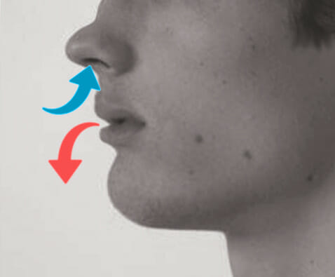
Pursed lip breathing
Accessory muscle use
Tripod positioning
Hyperresonant chest
Demonstrating pursed lip breathing, which is seen in emphysema
Image: “Pursed lip breathing” by O.Chaigasame. License: CC BY 4.0
↑ Residual volumeResidual volumeThe volume of air remaining in the lungs at the end of a maximal expiration. Common abbreviation is rv.Ventilation: Mechanics of Breathing and total lung capacityTotal lung capacityThe volume of air contained in the lungs at the end of a maximal inspiration. It is the equivalent to each of the following sums: vital capacity plus residual volume; inspiratory capacity plus functional residual capacity; tidal volume plus inspiratory reserve volume plus functional residual capacity; or tidal volume plus inspiratory reserve volume plus expiratory reserve volume plus residual volume.Ventilation: Mechanics of Breathing (air trapping)
Emphysema:
↓ Diffusing capacity for CO:
Also known as transfer factor
Due to loss of surface area for gas exchangeGas exchangeHuman cells are primarily reliant on aerobic metabolism. The respiratory system is involved in pulmonary ventilation and external respiration, while the circulatory system is responsible for transport and internal respiration. Pulmonary ventilation (breathing) represents movement of air into and out of the lungs. External respiration, or gas exchange, is represented by the O2 and CO2 exchange between the lungs and the blood.Gas Exchange
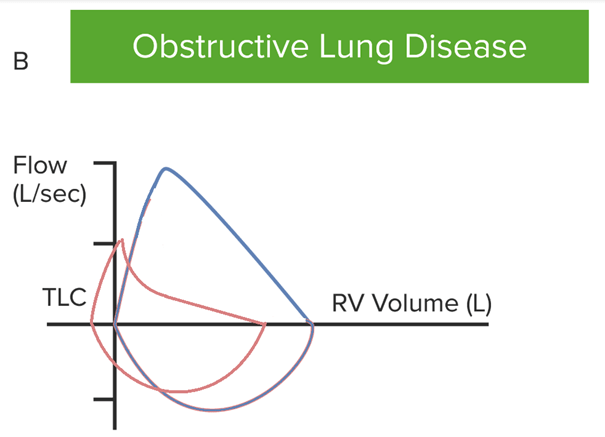
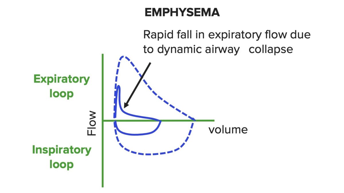
Rapid fall in expiratory flowFlowBlood flows through the heart, arteries, capillaries, and veins in a closed, continuous circuit. Flow is the movement of volume per unit of time. Flow is affected by the pressure gradient and the resistance fluid encounters between 2 points. Vascular resistance is the opposition to flow, which is caused primarily by blood friction against vessel walls.Vascular Resistance, Flow, and Mean Arterial Pressure (dynamic airwayAirwayABCDE Assessment collapse) → produces a concave pattern
Post-bronchodilator test:
Used to assess the reversibility of the obstructive condition
Minimal reversibility in COPD
Flow-volume curve in a case of obstructive lung disease. Note that both flows are reduced due to obstruction. Dynamic airway collapse causes a rapid fall in expiratory flow, leading to a concave contour. Residual volume is increased due to air trapping, causing the curve to shift to the left.
Image by Lecturio.
Flow-volume curve in obstructive lung disease: Both flows are reduced due to obstruction; dynamic airway collapse causes a rapid fall in expiratory flow, which leads to a concave contour.
In addition to COPD diagnosis, spirometrySpirometryMeasurement of volume of air inhaled or exhaled by the lung.Pulmonary Function Tests results may be used in conjunction with symptoms to help stage severity. The Global Initiative for Chronic Obstructive Lung Disease (GOLD) criteria are as follows:
Table: The Global Initiative for Chronic Obstructive Lung Disease (GOLD) criteria
↑ BNPBNPA peptide that is secreted by the brain and the heart atria, stored mainly in cardiac ventricular myocardium. It can cause natriuresis; diuresis; vasodilation; and inhibits secretion of renin and aldosterone. It improves heart function. It contains 32 amino acids.Renal Sodium and Water Regulation in cor pulmonaleCor PulmonaleCor pulmonale is right ventricular (RV) dysfunction caused by lung disease that results in pulmonary artery hypertension. The most common cause of cor pulmonale is chronic obstructive pulmonary disease. Dyspnea is the usual presenting symptom. Cor Pulmonale
AAT testing: Consider if COPD symptoms are present (not in typical demographic):
Younger
Nonsmoker
Concomitant, unexplained liverLiverThe liver is the largest gland in the human body. The liver is found in the superior right quadrant of the abdomen and weighs approximately 1.5 kilograms. Its main functions are detoxification, metabolism, nutrient storage (e.g., iron and vitamins), synthesis of coagulation factors, formation of bile, filtration, and storage of blood. Liver: Anatomy disease
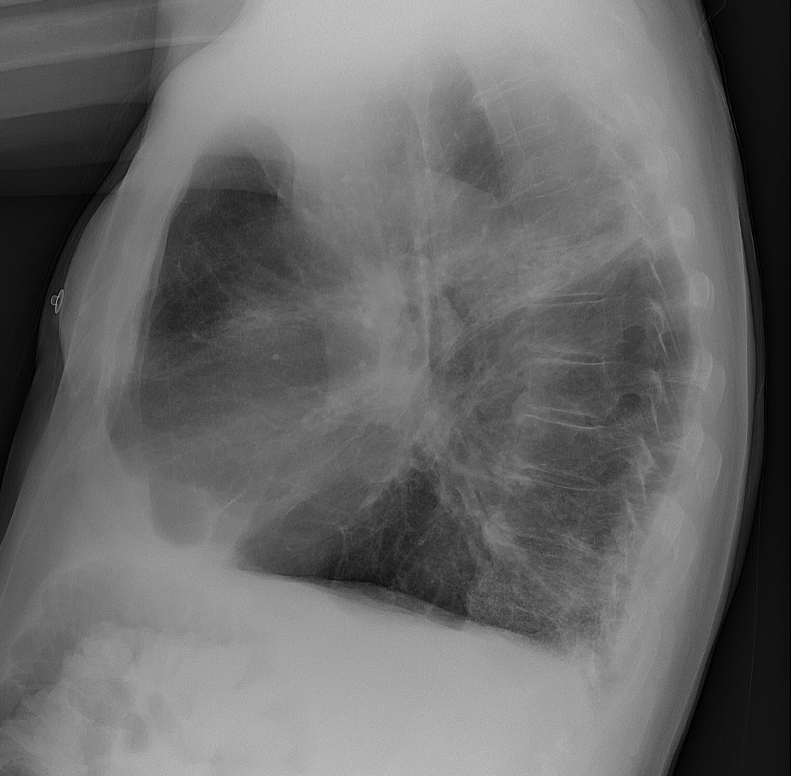
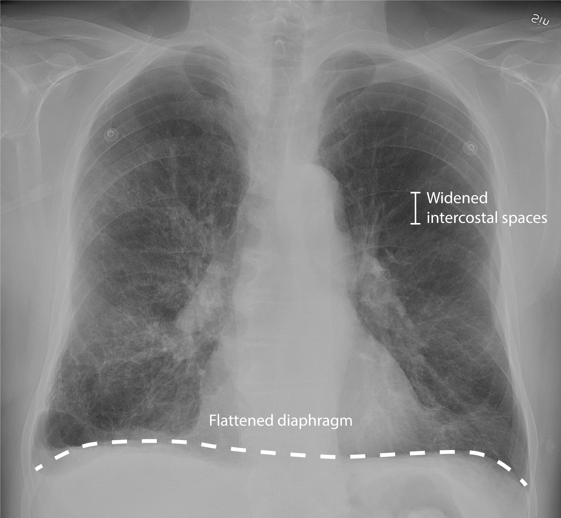
Chest X-rayX-rayPenetrating electromagnetic radiation emitted when the inner orbital electrons of an atom are excited and release radiant energy. X-ray wavelengths range from 1 pm to 10 nm. Hard x-rays are the higher energy, shorter wavelength x-rays. Soft x-rays or grenz rays are less energetic and longer in wavelength. The short wavelength end of the x-ray spectrum overlaps the gamma rays wavelength range. The distinction between gamma rays and x-rays is based on their radiation source.Pulmonary Function Tests:
Horizontal ribsRibsA set of twelve curved bones which connect to the vertebral column posteriorly, and terminate anteriorly as costal cartilage. Together, they form a protective cage around the internal thoracic organs.Chest Wall: Anatomy
Flattened, low diaphragmDiaphragmThe diaphragm is a large, dome-shaped muscle that separates the thoracic cavity from the abdominal cavity. The diaphragm consists of muscle fibers and a large central tendon, which is divided into right and left parts. As the primary muscle of inspiration, the diaphragm contributes 75% of the total inspiratory muscle force.Diaphragm: Anatomy
Hyperlucency
Attenuated peripheral vascular markings (due to parenchymal destruction)
Chest X-ray of an 81-year-old man with chronic obstructive pulmonary disease (COPD) and concomitant pneumonia
Image: “Chest X-ray of an 81-year-old man with chronic obstructive pulmonary disease (COPD), and concomitant pneumonia” by Mikael Häggström, M.D. License: CC0 1.0
Chest X-ray of an 81-year-old man with chronic obstructive pulmonary disease (COPD) presenting mainly with productive cough: Note the widened intercostal spaces, flattened diaphragm, and opacity in the inferior right upper lobe, which suggests concomitant pneumonia.
Improve qualityQualityActivities and programs intended to assure or improve the quality of care in either a defined medical setting or a program. The concept includes the assessment or evaluation of the quality of care; identification of problems or shortcomings in the delivery of care; designing activities to overcome these deficiencies; and follow-up monitoring to ensure effectiveness of corrective steps.Quality Measurement and Improvement of life.
General management
SmokingSmokingWillful or deliberate act of inhaling and exhaling smoke from burning substances or agents held by hand.Interstitial Lung Diseases cessation (critical in slowing lung function decline)
Vaccinations for:
Pneumococcal pneumoniaPneumoniaPneumonia or pulmonary inflammation is an acute or chronic inflammation of lung tissue. Causes include infection with bacteria, viruses, or fungi. In more rare cases, pneumonia can also be caused through toxic triggers through inhalation of toxic substances, immunological processes, or in the course of radiotherapy.Pneumonia
InfluenzaInfluenzaInfluenza viruses are members of the Orthomyxoviridae family and the causative organisms of influenza, a highly contagious febrile respiratory disease. There are 3 primary influenza viruses (A, B, and C) and various subtypes, which are classified based on their virulent surface antigens, hemagglutinin (HA) and neuraminidase (NA). Influenza typically presents with a fever, myalgia, headache, and symptoms of an upper respiratory infection. Influenza Viruses/Influenza
Pulmonary rehabilitation:
Guided exercise and behavioral interventions
Goal is to improve functional capacity.
O2 therapy:
If O2 saturation is < 88% in a stable patient (PO₂ < 55 mm Hg)
If concurrent pulmonary hypertensionPulmonary HypertensionPulmonary hypertension (PH) or pulmonary arterial hypertension (PAH) is characterized by elevated pulmonary arterial pressure, which can lead to chronic progressive right heart failure. Pulmonary hypertension is grouped into 5 categories based on etiology, which include primary PAH, and PH due to cardiac disease, lung or hypoxic disease, chronic thromboembolic disease, and multifactorial or unclear etiologies. Pulmonary Hypertension, right-sided heart failureRight-Sided Heart FailureEbstein’s Anomaly, or polycythemiaPolycythemiaAn increase in the total red cell mass of the blood.Renal Cell Carcinoma
Beta-2 adrenergic agonistsAdrenergic agonistsSympathomimetic drugs, also known as adrenergic agonists, mimic the action of the stimulators (î±, β, or dopamine receptors) of the sympathetic autonomic nervous system. Sympathomimetic drugs are classified based on the type of receptors the drugs act on (some agents act on several receptors but 1 is predominate).Sympathomimetic Drugs (e.g., albuterolAlbuterolA short-acting beta-2 adrenergic agonist that is primarily used as a bronchodilator agent to treat asthma.Sympathomimetic Drugs)
AnticholinergicsAnticholinergicsAnticholinergic drugs block the effect of the neurotransmitter acetylcholine at the muscarinic receptors in the central and peripheral nervous systems. Anticholinergic agents inhibit the parasympathetic nervous system, resulting in effects on the smooth muscle in the respiratory tract, vascular system, urinary tract, GI tract, and pupils of the eyes. Anticholinergic Drugs (e.g., ipratropium bromideIpratropium BromideAsthma Drugs)
Long acting:
Beta-2 adrenergic agonistsAdrenergic agonistsSympathomimetic drugs, also known as adrenergic agonists, mimic the action of the stimulators (î±, β, or dopamine receptors) of the sympathetic autonomic nervous system. Sympathomimetic drugs are classified based on the type of receptors the drugs act on (some agents act on several receptors but 1 is predominate).Sympathomimetic Drugs (e.g., salmeterolSalmeterolAsthma Drugs, formoterolFormoterolAsthma Drugs, indacaterol)
AnticholinergicsAnticholinergicsAnticholinergic drugs block the effect of the neurotransmitter acetylcholine at the muscarinic receptors in the central and peripheral nervous systems. Anticholinergic agents inhibit the parasympathetic nervous system, resulting in effects on the smooth muscle in the respiratory tract, vascular system, urinary tract, GI tract, and pupils of the eyes. Anticholinergic Drugs (e.g., tiotropium, aclidiniumAclidiniumAnticholinergic Drugs, umeclidinium)
Reduces inflammationInflammationInflammation is a complex set of responses to infection and injury involving leukocytes as the principal cellular mediators in the body’s defense against pathogenic organisms. Inflammation is also seen as a response to tissue injury in the process of wound healing. The 5 cardinal signs of inflammation are pain, heat, redness, swelling, and loss of function. Inflammation
Options: budesonideBudesonideA glucocorticoid used in the management of asthma, the treatment of various skin disorders, and allergic rhinitis.Asthma Drugs, fluticasoneFluticasoneA steroid with glucocorticoid receptor activity that is used to manage the symptoms of asthma; allergic rhinitis, and atopic dermatitis.Glucocorticoids
Can produce both marginal improvements and adverse effects
TheophyllineTheophyllineA methyl xanthine derivative from tea with diuretic, smooth muscle relaxant, bronchial dilation, cardiac and central nervous system stimulant activities. Theophylline inhibits the 3.Asthma Drugs(oral bronchodilator)
Mucolytics
Surgical intervention
Surgery is reserved for severe cases not controlled with medical therapy to improve qualityQualityActivities and programs intended to assure or improve the quality of care in either a defined medical setting or a program. The concept includes the assessment or evaluation of the quality of care; identification of problems or shortcomings in the delivery of care; designing activities to overcome these deficiencies; and follow-up monitoring to ensure effectiveness of corrective steps.Quality Measurement and Improvement of life.
Systemic steroidsSteroidsA group of polycyclic compounds closely related biochemically to terpenes. They include cholesterol, numerous hormones, precursors of certain vitamins, bile acids, alcohols (sterols), and certain natural drugs and poisons. Steroids have a common nucleus, a fused, reduced 17-carbon atom ring system, cyclopentanoperhydrophenanthrene. Most steroids also have two methyl groups and an aliphatic side-chain attached to the nucleus.Benign Liver Tumors
Antibiotics are indicated for:
Purulent sputum
Evidence of pneumoniaPneumoniaPneumonia or pulmonary inflammation is an acute or chronic inflammation of lung tissue. Causes include infection with bacteria, viruses, or fungi. In more rare cases, pneumonia can also be caused through toxic triggers through inhalation of toxic substances, immunological processes, or in the course of radiotherapy.Pneumonia
PatientsPatientsIndividuals participating in the health care system for the purpose of receiving therapeutic, diagnostic, or preventive procedures.Clinician–Patient Relationship requiring hospitalizationHospitalizationThe confinement of a patient in a hospital.Delirium
Controlled O2 therapy for acute respiratory failureRespiratory failureRespiratory failure is a syndrome that develops when the respiratory system is unable to maintain oxygenation and/or ventilation. Respiratory failure may be acute or chronic and is classified as hypoxemic, hypercapnic, or a combination of the two. Respiratory Failure:
Severe respiratory failureRespiratory failureRespiratory failure is a syndrome that develops when the respiratory system is unable to maintain oxygenation and/or ventilation. Respiratory failure may be acute or chronic and is classified as hypoxemic, hypercapnic, or a combination of the two. Respiratory Failure
May be difficult to wean patientsPatientsIndividuals participating in the health care system for the purpose of receiving therapeutic, diagnostic, or preventive procedures.Clinician–Patient Relationship with severe COPD
Complications
Respiratory failureRespiratory failureRespiratory failure is a syndrome that develops when the respiratory system is unable to maintain oxygenation and/or ventilation. Respiratory failure may be acute or chronic and is classified as hypoxemic, hypercapnic, or a combination of the two. Respiratory Failure
Respiratory infectionsInfectionsInvasion of the host organism by microorganisms or their toxins or by parasites that can cause pathological conditions or diseases.Chronic Granulomatous Disease (pneumoniaPneumoniaPneumonia or pulmonary inflammation is an acute or chronic inflammation of lung tissue. Causes include infection with bacteria, viruses, or fungi. In more rare cases, pneumonia can also be caused through toxic triggers through inhalation of toxic substances, immunological processes, or in the course of radiotherapy.Pneumonia)
Pulmonary hypertensionPulmonary HypertensionPulmonary hypertension (PH) or pulmonary arterial hypertension (PAH) is characterized by elevated pulmonary arterial pressure, which can lead to chronic progressive right heart failure. Pulmonary hypertension is grouped into 5 categories based on etiology, which include primary PAH, and PH due to cardiac disease, lung or hypoxic disease, chronic thromboembolic disease, and multifactorial or unclear etiologies. Pulmonary Hypertension → cor pulmonaleCor PulmonaleCor pulmonale is right ventricular (RV) dysfunction caused by lung disease that results in pulmonary artery hypertension. The most common cause of cor pulmonale is chronic obstructive pulmonary disease. Dyspnea is the usual presenting symptom. Cor Pulmonale
Long-term complications of steroidsSteroidsA group of polycyclic compounds closely related biochemically to terpenes. They include cholesterol, numerous hormones, precursors of certain vitamins, bile acids, alcohols (sterols), and certain natural drugs and poisons. Steroids have a common nucleus, a fused, reduced 17-carbon atom ring system, cyclopentanoperhydrophenanthrene. Most steroids also have two methyl groups and an aliphatic side-chain attached to the nucleus.Benign Liver Tumors → osteoporosisOsteoporosisOsteoporosis refers to a decrease in bone mass and density leading to an increased number of fractures. There are 2 forms of osteoporosis: primary, which is commonly postmenopausal or senile; and secondary, which is a manifestation of immobilization, underlying medical disorders, or long-term use of certain medications. Osteoporosis
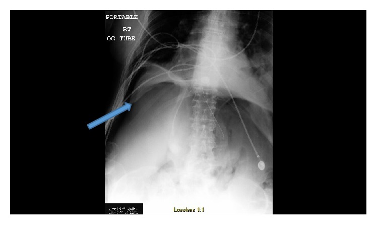
X-ray showing a right-sided pneumothorax in a patient with chronic obstructive pulmonary disease (COPD): Pneumoperitoneum (arrow points to air under the diaphragm) is noted due to a diaphragmatic defect, which allows air from the pneumothorax to escape into the abdominal cavity.
Image:“Chest/abdomen X-ray showing right pneumothorax and also air under diaphragm suggesting perforated viscus.” by Fernanda Duarte et al. License: CC BY 4.0
AsthmaAsthmaAsthma is a chronic inflammatory respiratory condition characterized by bronchial hyperresponsiveness and airflow obstruction. The disease is believed to result from the complex interaction of host and environmental factors that increase disease predisposition, with inflammation causing symptoms and structural changes. Patients typically present with wheezing, cough, and dyspnea. Asthma: a chronic, inflammatory condition characterized by reversible airflow obstruction in the lower airways. PatientsPatientsIndividuals participating in the health care system for the purpose of receiving therapeutic, diagnostic, or preventive procedures.Clinician–Patient Relationship present with intermittent or persistent wheezingWheezingWheezing is an abnormal breath sound characterized by a whistling noise that can be relatively high-pitched and shrill (more common) or coarse. Wheezing is produced by the movement of air through narrowed or compressed small (intrathoracic) airways. Wheezing, cough, and dyspneaDyspneaDyspnea is the subjective sensation of breathing discomfort. Dyspnea is a normal manifestation of heavy physical or psychological exertion, but also may be caused by underlying conditions (both pulmonary and extrapulmonary). Dyspnea. Diagnosis is usually confirmed with a pulmonary function testPulmonary function testPulmonary function tests are a group of diagnostic procedures yielding useful, quantifiable information about the rate of the flow of air through the individual’s airways, lung capacity, and the efficiency of gas exchange in relation to time. The most commonly utilized tests include spirometry (before and after bronchodilator use), lung volumes, and quantitation of diffusing capacity for carbon monoxide (CO). The tests can be influenced by the individual’s effort/fatigue, disease state, or anatomical malformation.Pulmonary Function Tests showing a reversible, obstructive pattern. Management varies based on severity and includes bronchodilatorsBronchodilatorsAsthma Drugs and inhaled corticosteroidsCorticosteroidsChorioretinitis for inflammationInflammationInflammation is a complex set of responses to infection and injury involving leukocytes as the principal cellular mediators in the body’s defense against pathogenic organisms. Inflammation is also seen as a response to tissue injury in the process of wound healing. The 5 cardinal signs of inflammation are pain, heat, redness, swelling, and loss of function. Inflammation control.
BronchiectasisBronchiectasisBronchiectasis is a chronic disease of the airways that results from permanent bronchial distortion. This results from a continuous cycle of inflammation, bronchial damage and dilation, impaired clearance of secretions, and recurrent infections. Bronchiectasis: a chronic condition with bronchial dilation and destruction as a result of inflammationInflammationInflammation is a complex set of responses to infection and injury involving leukocytes as the principal cellular mediators in the body’s defense against pathogenic organisms. Inflammation is also seen as a response to tissue injury in the process of wound healing. The 5 cardinal signs of inflammation are pain, heat, redness, swelling, and loss of function. Inflammation and infection. Symptoms include dyspneaDyspneaDyspnea is the subjective sensation of breathing discomfort. Dyspnea is a normal manifestation of heavy physical or psychological exertion, but also may be caused by underlying conditions (both pulmonary and extrapulmonary). Dyspnea, chronic cough, and purulent sputum. The diagnosis is made with imaging (X-rayX-rayPenetrating electromagnetic radiation emitted when the inner orbital electrons of an atom are excited and release radiant energy. X-ray wavelengths range from 1 pm to 10 nm. Hard x-rays are the higher energy, shorter wavelength x-rays. Soft x-rays or grenz rays are less energetic and longer in wavelength. The short wavelength end of the x-ray spectrum overlaps the gamma rays wavelength range. The distinction between gamma rays and x-rays is based on their radiation source.Pulmonary Function Tests and CT). Management includes bronchodilatorsBronchodilatorsAsthma Drugs and antibiotics for acute exacerbations.
BronchiolitisBronchiolitisInflammation of the bronchioles.Pediatric Chest Abnormalities obliterans: a chronic, obstructive disease of the small airways usually caused by repeated cycles of inflammationInflammationInflammation is a complex set of responses to infection and injury involving leukocytes as the principal cellular mediators in the body’s defense against pathogenic organisms. Inflammation is also seen as a response to tissue injury in the process of wound healing. The 5 cardinal signs of inflammation are pain, heat, redness, swelling, and loss of function. Inflammation and scarringScarringInflammation. PatientsPatientsIndividuals participating in the health care system for the purpose of receiving therapeutic, diagnostic, or preventive procedures.Clinician–Patient Relationship present with cough and persistent, progressive dyspneaDyspneaDyspnea is the subjective sensation of breathing discomfort. Dyspnea is a normal manifestation of heavy physical or psychological exertion, but also may be caused by underlying conditions (both pulmonary and extrapulmonary). Dyspnea. Pulmonary function tests are used for diagnosis. Management includes steroidsSteroidsA group of polycyclic compounds closely related biochemically to terpenes. They include cholesterol, numerous hormones, precursors of certain vitamins, bile acids, alcohols (sterols), and certain natural drugs and poisons. Steroids have a common nucleus, a fused, reduced 17-carbon atom ring system, cyclopentanoperhydrophenanthrene. Most steroids also have two methyl groups and an aliphatic side-chain attached to the nucleus.Benign Liver Tumors and bronchodilatorsBronchodilatorsAsthma Drugs.
Heart failureHeart FailureA heterogeneous condition in which the heart is unable to pump out sufficient blood to meet the metabolic need of the body. Heart failure can be caused by structural defects, functional abnormalities (ventricular dysfunction), or a sudden overload beyond its capacity. Chronic heart failure is more common than acute heart failure which results from sudden insult to cardiac function, such as myocardial infarction.Total Anomalous Pulmonary Venous Return (TAPVR): an inability to produce normal cardiac outputCardiac outputThe volume of blood passing through the heart per unit of time. It is usually expressed as liters (volume) per minute so as not to be confused with stroke volume (volume per beat).Cardiac Mechanics to meet metabolic needs. PatientsPatientsIndividuals participating in the health care system for the purpose of receiving therapeutic, diagnostic, or preventive procedures.Clinician–Patient Relationship present with dyspneaDyspneaDyspnea is the subjective sensation of breathing discomfort. Dyspnea is a normal manifestation of heavy physical or psychological exertion, but also may be caused by underlying conditions (both pulmonary and extrapulmonary). Dyspnea, hypoxiaHypoxiaSub-optimal oxygen levels in the ambient air of living organisms.Ischemic Cell Damage, and peripheral edemaPeripheral edemaPeripheral edema is the swelling of the lower extremities, namely, legs, feet, and ankles.Edema. BNPBNPA peptide that is secreted by the brain and the heart atria, stored mainly in cardiac ventricular myocardium. It can cause natriuresis; diuresis; vasodilation; and inhibits secretion of renin and aldosterone. It improves heart function. It contains 32 amino acids.Renal Sodium and Water Regulation will be elevated and pulmonary edemaPulmonary edemaPulmonary edema is a condition caused by excess fluid within the lung parenchyma and alveoli as a consequence of a disease process. Based on etiology, pulmonary edema is classified as cardiogenic or noncardiogenic. Patients may present with progressive dyspnea, orthopnea, cough, or respiratory failure.Pulmonary Edema may be seen on X-rayX-rayPenetrating electromagnetic radiation emitted when the inner orbital electrons of an atom are excited and release radiant energy. X-ray wavelengths range from 1 pm to 10 nm. Hard x-rays are the higher energy, shorter wavelength x-rays. Soft x-rays or grenz rays are less energetic and longer in wavelength. The short wavelength end of the x-ray spectrum overlaps the gamma rays wavelength range. The distinction between gamma rays and x-rays is based on their radiation source.Pulmonary Function Tests. EchocardiographyEchocardiographyUltrasonic recording of the size, motion, and composition of the heart and surrounding tissues. The standard approach is transthoracic.Tricuspid Valve Atresia (TVA) confirms the diagnosis. Management relies on diuresis and medical optimization of cardiac function with beta blockers and ACE inhibitorsACE inhibitorsTruncus Arteriosus.
CysticCysticFibrocystic ChangefibrosisFibrosisAny pathological condition where fibrous connective tissue invades any organ, usually as a consequence of inflammation or other injury.Bronchiolitis Obliterans: an autosomal recessiveAutosomal recessiveAutosomal inheritance, both dominant and recessive, refers to the transmission of genes from the 22 autosomal chromosomes. Autosomal recessive diseases are only expressed when 2 copies of the recessive allele are inherited.Autosomal Recessive and Autosomal Dominant Inheritance disorder leading to dysfunction of chlorideChlorideInorganic compounds derived from hydrochloric acid that contain the Cl- ion.ElectrolyteschannelsChannelsThe Cell: Cell Membrane, which results in hyperviscous mucus and the accumulation of secretions. PatientsPatientsIndividuals participating in the health care system for the purpose of receiving therapeutic, diagnostic, or preventive procedures.Clinician–Patient Relationship often have chronic respiratory infectionsInfectionsInvasion of the host organism by microorganisms or their toxins or by parasites that can cause pathological conditions or diseases.Chronic Granulomatous Disease, failure to thriveFailure to ThriveFailure to thrive (FTT), or faltering growth, describes suboptimal weight gain and growth in children. The majority of cases are due to inadequate caloric intake; however, genetic, infectious, and oncological etiologies are also common. Failure to Thrive, and pancreatic insufficiency. The gold standard for diagnosis is the sweat chlorideChlorideInorganic compounds derived from hydrochloric acid that contain the Cl- ion.Electrolytes test, which can be complemented by genetic testingGenetic TestingDetection of a mutation; genotype; karyotype; or specific alleles associated with genetic traits, heritable diseases, or predisposition to a disease, or that may lead to the disease in descendants. It includes prenatal genetic testing.Myotonic Dystrophies. Management includes cysticCysticFibrocystic ChangefibrosisFibrosisAny pathological condition where fibrous connective tissue invades any organ, usually as a consequence of inflammation or other injury.Bronchiolitis Obliterans transmembrane conductance regulator (CFTR) modulator therapy and system-specific strategies for supportive care.
Pulmonary embolismPulmonary EmbolismPulmonary embolism (PE) is a potentially fatal condition that occurs as a result of intraluminal obstruction of the main pulmonary artery or its branches. The causative factors include thrombi, air, amniotic fluid, and fat. In PE, gas exchange is impaired due to the decreased return of deoxygenated blood to the lungs. Pulmonary Embolism: obstruction of the pulmonary arteriesArteriesArteries are tubular collections of cells that transport oxygenated blood and nutrients from the heart to the tissues of the body. The blood passes through the arteries in order of decreasing luminal diameter, starting in the largest artery (the aorta) and ending in the small arterioles. Arteries are classified into 3 types: large elastic arteries, medium muscular arteries, and small arteries and arterioles. Arteries: Histology most often due to thrombus migration from the deep venous system. Signs and symptoms include pleuritic chest painPainAn unpleasant sensation induced by noxious stimuli which are detected by nerve endings of nociceptive neurons.Pain: Types and Pathways, dyspneaDyspneaDyspnea is the subjective sensation of breathing discomfort. Dyspnea is a normal manifestation of heavy physical or psychological exertion, but also may be caused by underlying conditions (both pulmonary and extrapulmonary). Dyspnea, tachypneaTachypneaIncreased respiratory rate.Pulmonary Examination, and tachycardiaTachycardiaAbnormally rapid heartbeat, usually with a heart rate above 100 beats per minute for adults. Tachycardia accompanied by disturbance in the cardiac depolarization (cardiac arrhythmia) is called tachyarrhythmia.Sepsis in Children. Severe cases can result in hemodynamic instability or cardiopulmonary arrestCardiopulmonary arrestCardiac arrest is the sudden, complete cessation of cardiac output with hemodynamic collapse. Patients present as pulseless, unresponsive, and apneic. Rhythms associated with cardiac arrest are ventricular fibrillation/tachycardia, asystole, or pulseless electrical activity.Cardiac Arrest. Chest CTACTAA non-invasive method that uses a ct scanner for capturing images of blood vessels and tissues. A contrast material is injected, which helps produce detailed images that aid in diagnosing vascular diseases.Pulmonary Function Tests is the primary method of diagnosis. Management includes oxygenation, anticoagulationAnticoagulationPulmonary Hypertension Drugs, and thrombolytic therapy for unstable patientsUnstable PatientsBlunt Chest Trauma.
Mosenifar, Z, Harrington, A., Nikhanj, N.S., and Kamangar, N. (2020). Chronic obstructive pulmonary disease (COPD). In Oppenheimer, J.J. (Ed.), Medscape. Retrieved March 31, 2021, from https://emedicine.medscape.com/article/297664-overview
Agarwarl, A.K., Raja, A., and Brown, B.D. (2021). Chronic obstructive pulmonary disease. [online] StatPearls. Retrieved March 31, 2021, from https://www.ncbi.nlm.nih.gov/books/NBK559281/
Pauwels, R.A., Buist, A.S., Ma, P., Jenkins, C.R., Hurd, S.S. GOLD Scientific Committee. Global strategy for the diagnosis, management, and prevention of chronic obstructive pulmonary disease: National Heart, Lung, and Blood Institute and World Health Organization Global Initiative for Chronic Obstructive Lung Disease (GOLD): executive summary. Respir Care. 2001;46(8):798-825.
Create your free account or log in to continue reading!