COVID-19
(Coronavirus disease 2019)

Coronavirus disease 2019 (COVID-19) is an infectious disease caused by the severe acute respiratory syndrome coronavirus 2 (SARS-CoV-2) that mainly affects the respiratory system but can also cause damage to other body systems (cardiovascular, gastrointestinal, renal, and central nervous systems). Direct person-to-person transmission is the primary means of spread, mainly through close-range contact via respiratory droplets. Direct contact and inhalation of aerosolized virus particles are also methods of transmission. The infection may present asymptomatically, as a mild “flu-like” illness, or severely, with shortness of breath and life-threatening complications. Individuals who are over 65 years of age, immunosuppressed, or have preexisting conditions have a much higher risk of developing severe symptoms and complications. Management is based on supportive care and antiviral and immunomodulatory medications.

Updated December 2022

Coronaviruses
Coronaviruses viewed under an electron microscope. Note the characteristic crown-like (corona) appearance
Image: by CDC/ Dr. Fred Murphy, License: Public Domain Files

Table of Contents

Share this concept:

Table of Contents

Etiology

Coronaviruses (CoVs) are a family of enveloped, positive-sense, single-stranded RNA viruses. They tend to cause mild upper respiratory diseases in humans. Of the 7 known species of CoV, only 3 are known to cause severe infections in humans, all within the Betacoronavirus genus:

  • Severe acute respiratory disease coronavirus (SARS-CoV):
    • Emerged in 2002 in southern China from civet cats
    • The viral genome sequences obtained were 99.8% identical to the human SARS virus, and civet traders had the highest serologic positivity rates (> 70%).
  • Middle East respiratory syndrome coronavirus (MERS-CoV):
    • Emerged in 2012 in Saudi Arabia from dromedary camels
    • Contemporary and decades-old archived camel blood specimens tested positive for MERS-CoV antibodies, and the highest seropositive rates were in camel shepherds and slaughterhouse workers (15 to 23 times higher than in the general population).
  • SARS-CoV-2: emerged in November 2019 in Wuhan, a city in the Hubei province of China
    • The genome is 96.2% identical to the bat coronavirus RaTG13. (This is still a relatively large evolutionary gap, e.g., humans also share 96% of their DNA with chimpanzees).
    • The initial and still widely held theory is that the virus originated in the Huanan seafood “wet” market in Wuhan, China, which is known for the sale of wild animals and their meat, some of which may have been infected with the virus.
    • Another theory, the Mojiang Miners Passage (MMP) hypothesis, claims that SARS-CoV-2 evolved from RaTG13, a pathogenic coronavirus that infected 6 mineshaft workers in April 2012.
      • It is proposed that in situ viral passaging enabled the rapid evolution of the virus without the need for an intermediate host.
      • These patients presented with a mysterious, severe respiratory illness closely resembling COVID-19; 3 of the miners eventually passed away.
      • The virus from the infected miners had reportedly been sent to a high-level laboratory at the Wuhan Institute of Virology (WIV). This facility received the highest laboratory biosafety accreditation (BSL-4) in 2018, which coincides with the beginning of its active research on the virus.
      • The virus is believed to have escaped from the WIV where it was being actively studied and manipulated, per their own published research.
    • On February 2, 2021, the head of the World Health Organization (WHO), Dr. Tedros Adhanom Ghebreyesus, noted that all hypotheses on the origins of COVID-19 remain on the table.” The debate is ongoing. 
    • To scientifically determine the true origin of SARS-CoV-2, the following information would be needed:
      • Data from serological sampling in 2019 and early 2020 that includes information on occupation and location and that encompasses both Wuhan animal market employees and Wuhan laboratory research employees
      • More information regarding the research on SARS-related coronaviruses performed by Wuhan researchers and their collaborators in 2015–2019 about virus samples and sequences (Kahn, 2022, Ref. 80)

Selected diseases caused by coronaviruses

 Common coldGI tract infectionSevere acute respiratory syndrome (SARS)COVID-19 (Wuhan, China)
Incubation3 days3 days4–6 days2–14 days
IncidenceMost commonRareRareCurrent pandemic
PrognosisComplete resolutionComplete resolution
(up to 25% fatal for NEC)
30% resolution
70% severe infection
10% fatal
80% resolution
15% severe case
5% critical case
2.2% case fatality rate (varies widely)
Clinical manifestationSneezing, rhinorrhea, headache, sore throat, malaise, fever, chillsDiarrhea, gastroenteritis, neonatal necrotizing enterocolitis

Fever > 37,8°C (100,0°F), muscle pain, lethargy, cough, sore throat, malaise

Shortness of breath/ pneumonia (direct viral or secondary bacterial)

Asymptomatic

Mild infection: fever, dry cough, malaise, dehydration

Severe infection: high fever, shortness of breath, chest pain, hemoptysis

Complications: pneumonia, ARDS, sepsis, multi-organ failure

Diseases caused by Coronaviruses

 Common ColdGI tract infection
Incubation3 days3 days
IncidenceMost commonRare
PrognosisComplete resolutionComplete resolution(up to 25% fatal for NEC)
Clinical manifestationSneezing, rhinorrhea, headache, sore throat, malaise, fever, chillsDiarrhea, gastroenteritis, neonatal necrotizing enterocolitis
 Severe acute respiratory syndrome (SARS)2019 nCoV (Wuhan City, China)
Incubation4–6 days2–14 days
IncidenceRareCurrent pandemic
Prognosis30% resolution
70% severe infection
10% fatal

80% resolution
15% severe case
5% critical case
2.2% fatal

Clinical manifestation

Fever > 37,8°C (100,0°F), muscle pain, lethargy, cough, sore throat, malaise

Shortness of breath/ pneumonia (direct viral or sencondary bacterial)

Asymptomatic
Mild infection: fever, dry cough, muscle pain, lethargy, dehydration

Severe infection: high fever, shortness of breath, chest pain, hemoptysis

Complications: pneumonia, ARDS, sepsis, multi-organ failure

NEC: Necrotizing enterocolitis ARDS: Acute respiratory distress syndrome

The SARS-CoV-2 virion is approximately 125 nm in diameter and its genome ranges from 26–32 kb, the largest of all RNA viruses. It has 4 structural proteins: spike (S), envelope (E), membrane (M), and nucleocapsid (N).

  • S, E, and M proteins create the viral envelope. 
  • The N protein forms a complex with RNA (nucleocapsid) and aids in the regulation of viral RNA synthesis.
  • The M protein projects on the external surface of the envelope, spans the envelope 3 times, and is important for viral assembly. 
  • The E protein has an unclear function, although it may aid in viral release.
  • The S protein is a club-shaped surface projection that gives the virus its characteristic crown-like appearance on electron microscopy. This protein is responsible for receptor binding and fusion with the host cell membrane.
Coronavirus 2
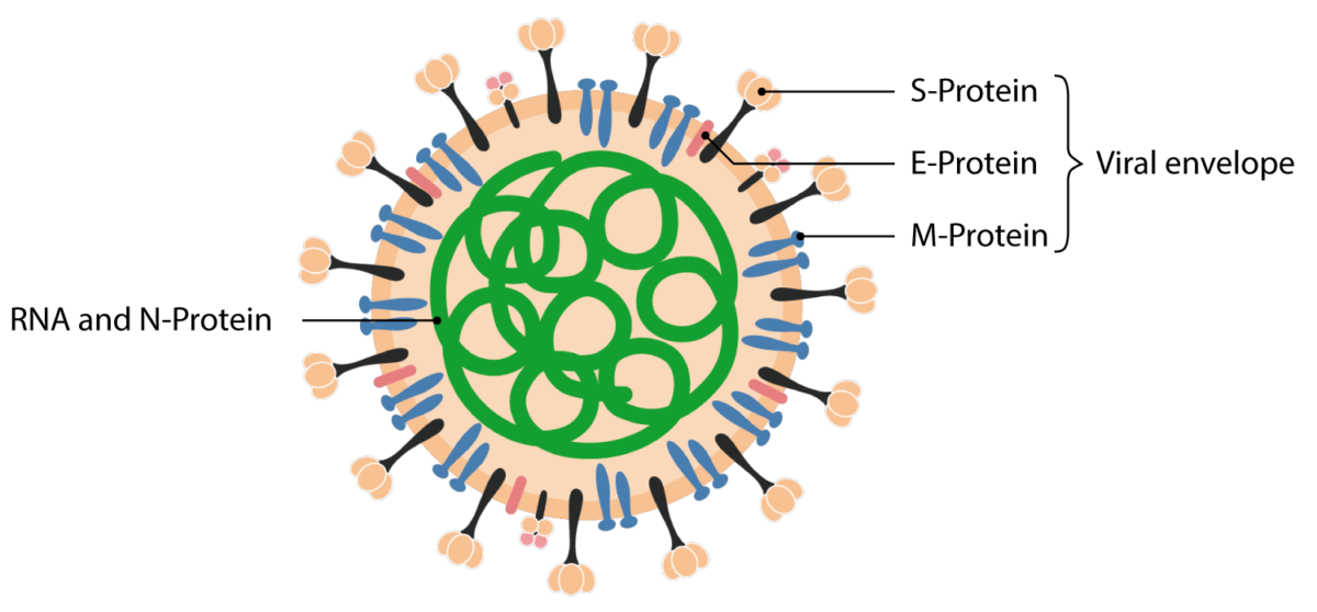
Structural proteins of the SARS-CoV 2 virion.
Image: by Lecturio

Lecturio resources

Transmission

Routes of infection

Coronaviruses are zoonotic; i.e., they are transmitted to humans through animals. It is hypothesized that horseshoe bats are the natural reservoir of SARS-CoV-2, since the virus’s genome is 96.2% identical to that of a bat coronavirus. Although the intermediate host is still unknown, the MMP theory of origin of the SARS-CoV-2 does not require the existence of one.

Once in humans, the virus is transmitted when respiratory droplets or aerosolized particles from infected individuals come into direct contact with the mucous membranes of another individual, including in the eyes, nose, or mouth. In the air, larger droplets tend to drop toward the ground within 1 meter (3 feet) of the infected person, while smaller droplets can travel over 2 meters (6 feet) and remain viable in the air for up to 3 hours under certain conditions. Other forms of transmission include the following:

  • Direct transmission through hand-to-face contact from infected surfaces
  • Transmission in bodily fluids. Although SARS-CoV-2 has been detected in stool specimens, blood, ocular secretions, and semen, the possibility of transmission through these routes remains uncertain.
  • Vertical transmission (mother to child):
    • Has been reported in several cases of peripartum maternal infection in the 3rd trimester
    • Most neonatal infections (asymptomatic and/or mild) are thought to result from postnatal exposure through respiratory droplets from an infected mother or caregiver. 
    • A case of transplacental transmission of SARS-CoV-2 has been reported due to maternal infection in the 3rd trimester. The neonate required neonatal resuscitation and presented with neurological compromise.

Droplet or contact transmission is defined as the transmission of infectious viral particles from an infected person to a new host through large respiratory droplets (> 5 μm in diameter), either through the touching of a contaminated surface or contact with these droplets in the air. 

Airborne transmission differs in that it involves small respiratory droplets (< 5 μm in diameter; also called droplet nuclei) that can remain suspended in the air for long periods, negating the need for close contact between individuals.

The main difference between these modes of transmission is the size of the droplets, which dictates how long they can remain in the air, how far they can travel, and how many virions they can carry (making the droplets more or less infectious). As a general rule, larger droplets fall to the ground within 1–2 meters (3–6 feet) of their source, while aerosol droplets can overcome the force of gravity and travel farther. It is also commonly believed that coughing and sneezing generate large-droplet emission, while only aerosol-generating procedures can emit small droplets.

The distinctions between airborne and droplet transmission are not as rigid as once believed. Once emitted into the air, large droplets can evaporate to form small aerosol droplets. Virus particles can reach 0.84 meters (2.75 feet) in 5.5 seconds. How far these droplets (both large and small) can travel is affected by environmental factors such as humidity, temperature, ventilation pattern and rate, the velocity of emission, and droplet composition.

Although COVID-19 can be carried long distances (> 2 meters (6 feet)) by the airborne route, the extent to which this has contributed to the pandemic is controversial. Outbreaks are rare in outdoor environments, as these spaces do not favor the dispersion of small aerosol particles. It is also possible that some asymptomatic individuals—so-called “superspreaders”—have an increased capacity to generate aerosols through normal talking and breathing.

Explanation mark green

Many factors can extend the range of respiratory droplet dispersion past 2 meters (6 feet). Certain actions, such as forced expiration during yelling, singing, and exercise, can increase both the volume of droplets emitted and the distance these droplets can travel.

Period and capacity of infectivity

The detection of viral RNA in the air does not guarantee the possibility of human infection.  Small aerosol doses are less likely to cause illness, even if the exact infectious dose for COVID-19 is still unknown. Certain medical procedures, such as intubation, can generate virus-laden aerosol clouds that put healthcare personnel at greater risk of becoming infected (see “Prevention” for more information on aerosol-generating procedures).

The reproductive number (R0) for COVID-19, or the number of secondary infections in a non-immune population generated from 1 infected individual, is 2–2.5, higher than for influenza (0.9–2.1).

  • The R0 can vary widely (it was calculated as 5.7 in the early phases of the pandemic), as it depends on both host and viral factors.
  • If the R0 < 1, an epidemic will eventually die out because each infected person generates less than 1 new infection.
  • If the R0 > 1, an epidemic could continue to grow. 
  • The R0 can change dramatically over time due to non-pharmaceutical interventions. For example, closing schools, physical distancing, and mask use can reduce the R0. The decrease can be dramatic if other constraints are added, such as workplace closures, bans on public events and large gatherings, internal movement limits, and stay-at-home requirements
    • In mid-February 2021, the R0 for the United Kingdom (UK) was 0.6–0.9 after strict lockdown measures had been in place for multiple weeks.

COVID-19 is highly contagious for the following reasons: 

  • Production of high viral loads
  • Efficient and prolonged shedding of virions from the upper respiratory tract
  • The infectious nature of asymptomatic individuals poses a significant challenge for contagion prevention.
    • Viral loads peak before symptom onset, leading to asymptomatic or presymptomatic spreading of the virus and making symptom-based detection and isolation ineffective. 
    • Asymptomatic patients can produce high viral loads through secretions of the upper respiratory tract and can shed the virus for the same amount of time as symptomatic patients.
  • SARS-CoV-2 can remain infectious on surfaces outside of a host from a few hours to a few days.
    • The viral lifespan depends on the type of surface, temperature, and humidity levels.
    • There is currently no evidence to suggest that COVID-19 can be acquired from mail or packaged goods (such as groceries), but caution is still advised when handling these materials.
Explanation mark green

The period of infectivity for symptomatic cases is currently believed to range from 3 days before the onset of symptoms up to 3 days after their resolution if symptom onset was > 10 days prior. The exact limits of infectivity are still under investigation.

The median duration of viral RNA shedding from the upper respiratory tract is 20 days. Viral shedding can outlast the resolution of symptoms. Some studies have reported viral detection in stool samples up to 35 days after the onset of symptoms (approximately 25 days after symptom resolution). However, the detection of viral RNA does not necessarily indicate the presence of an infectious virus. It has been shown that the more severe the case of COVID-19, the longer the patient will continue to shed the virus after recovery.

Studies have also shown that recovered patients generate a significant CD4+ and  CD8+ T cell response against SARS-CoV-2. Seroconversion, or the production of COVID-19–specific antibodies, occurs after 7 days in 50% of patients and by day 14 in all patients.

Epidemiology

The first case of COVID-19 was traced back to the city of Wuhan, China, in late November 2019, with an outbreak developing in December. The virus quickly spread, with widespread ongoing transmission occurring globally. The COVID-19 outbreak was declared a Public Health Emergency of International Concern on January 30, 2020, and a pandemic on March 11, 2020, by the WHO. COVID-19 cases have been reported on every continent.

COVID-19 global cases by Johns Hopkins Center for Systems Science and Engineering. https://www.arcgis.com/apps/opsdashboard/index.html#/bda7594740fd40299423467b48e9ecf6

The case fatality rate (CFR) of COVID-19 varies across different countries and age groups. See “Case Fatality Rate of COVID-19” in the graph below. However, the CFR is not a biological constant and is a poor measure to rely upon during an epidemic for the following reasons:

  • The CFR only indicates the mortality rate among documented cases.
  • Since many cases are asymptomatic and not tested, the infection fatality rate (IFR, the estimated mortality rate among all individuals with infection) is considerably lower, and may be between 0.5% and 1%.
  • Since many fatal infections are undiagnosed, the reported CFRs may be underestimated. 
  • Neither the CFR nor the IFR can account for the full burden of a pandemic while it is still active, as they do not include mortality caused by the delayed care of other conditions, overburdened health care systems, or the worsening of the social determinants of health.

Pathophysiology

Two main processes drive the pathogenesis of COVID-19.

  • Early infection: driven by the replication of SARS-CoV-2
  • Later infection: driven by an exaggerated immune/inflammatory response to the virus that results in tissue damage

SARS-CoV-2 attaches to the host cell by binding its S protein to the receptor protein, angiotensin-converting enzyme 2 (ACE2), which is expressed by epithelial cells of the intestine, kidney, blood vessels, and, most abundantly, in type II alveolar cells of the lungs. The human enzyme transmembrane protease, serine 2 (TMPRSS2), is also used by the virus for S protein priming and to aid in membrane fusion. The virus then enters the host cell via endocytosis.

SARS-CoV-2 affects the expression and presentation of ACE2, contributing to its pathogenesis in the following ways:

  • Viral entry causes internalization of the receptor, leading to its reduced availability on the cell surface. 
  • ACE2 inhibition induces ADAM17 gene expression, leading to the release of tumor necrosis factor α (TNFα) and cytokines such as interleukin 4 (IL-4) and interferon γ (IFNγ).
  • Increased cytokine concentrations activate further pro-inflammatory pathways, leading to a cytokine storm.
  • ADAM-17 also promotes the cleavage of ACE2 receptors.
  • SARS-CoV-2’s affinity for ACE2 also results in direct and acute injury to the lung, heart, endothelial cells, and, potentially, other organs.
    • ACE2 is a negative regulator of the renin-angiotensin-aldosterone system (RAAS), and its downregulation directly affects cardiovascular function.
    • ACE2 has a direct protective role in alveolar epithelial cells; its reduction leads to alveolar cell damage.
Explanation mark green

High levels of ACE2 expression are associated with certain chronic conditions, especially cardiovascular disease, and are linked to a higher risk of severe cases of COVID-19.

The expression of ACE2 is significantly increased through the use of ACE inhibitors or angiotensin II receptor blockers (ARBs). Contrary to initial reports, it has now been proven that the use of ACE inhibitors and ARBs is not associated with a risk of hospitalization or mortality among those infected with SARS-CoV-2. Rather, there is an approximately 40% lower risk of hospitalization associated with their use in the Medicare population in the United States, leading to the theory that the use of ACE inhibitors may help reduce the risk of hospitalization among elderly patients.

Although COVID-19 is a respiratory disease, clinical and pathology reports suggest that severe cases reflect a confluence of vascular dysfunction, thrombosis, and dysregulated inflammation. The development of complications and organ damage may be due not only to direct organ damage caused by the viral infection and local inflammation but also by indirect pathogenic mechanisms, including: 

  • Widespread endothelial damage (endothelialitis) with microangiopathy involving the vascular beds of the lungs, heart, kidneys, liver, and intestines
    • An autopsy study found that the lungs of a patient with COVID-19 had 9 times as many clots as those who died of the H1N1 flu.
  • Thrombosis and disseminated intravascular coagulation (DIC)
  • An atypical inflammatory response
  • Autoimmune phenomena, such as Guillain-Barré syndrome and pediatric inflammatory multisystem syndrome, which is an inflammatory state with clinical features similar to those of Kawasaki disease and toxic shock syndrome


COVID-19 differs from the pathogenesis of influenza in that it produces pulmonary angiogenesis. The lungs of COVID-19 patients have shown distorted vascularity with structurally deformed capillaries. This ability has also been associated with the human cytomegalovirus (HMCV). This virus produces increased endothelial cell proliferation and capillary tube formation and is associated with various vascular diseases (e.g., atherosclerosis, transplant vascular sclerosis, and coronary restenosis). This similarity suggests that there may be a link between the pathogenic mechanisms of SARS-CoV-2 and HCMV.

COVID-19 also triggers cell-mediated immune responses. Studies have identified SARS-CoV-2–specific CD8 and CD4 T cells in 70% and 100% of COVID-19 convalescent patients, respectively. CD4 cells seem to target the M, spike, and N proteins, while CD8 cells mainly target spike and M proteins.

Explanation mark green

T cell proliferation has been linked to the immune system’s production of viral-specific neutralizing antibodies, which not only provides a protective effect but also aids vaccine design and evaluation.

However, SARS-CoV-2–reactive T cells have also been detected in individuals who have no known exposure to the virus and who tested negative for the virus by the reverse transcriptase-polymerase chain reaction (RT-PCR) method, which is the most used type of nucleic acid amplification tests (NAATs). Thus, cross-reactive T-cell recognition must exist between SARS-CoV-2 and other types of seasonal coronaviruses that cause the “common cold.” This suggests that common cold coronaviruses may provide a small residual immunity, but the effect is believed to be minimal. 

COVID-19 Virus Variants

The virus has been mutating since the pandemic began, with the initial variants being named after the place where they were first identified; since then, the WHO has named the variants using the Greek alphabet. A variant is a form of the virus that, after many rounds of replication, has mutated into a form that is genetically distinct from its original form.

There are 3 categories of variants in the WHO System (definitions by WHO):

  • Variant Under Monitoring (VUM): a variant that may be associated with future risk, but current evidence is unclear
  • Variant of Interest (VOI): a variant with genetic changes similar to those of VOCs and which are increasing in prevalence in multiple countries
  • Variant of Concern (VOC): a variant that is increasing in prevalence in multiple countries and is associated with one or more of the following changes:
    • Increase in transmissibility or detrimental change in COVID-19 epidemiology
    • Increase in virulence or change in clinical disease presentation
    • Decrease in the effectiveness of public health and social measures or available diagnostics, vaccines, and/or therapeutics

A fourth type of variant retained by the CDC is a “variant of high consequence (VOHC)”, if there is evidence that prevention measures or medical countermeasures have significantly reduced effectiveness compared to previously circulating variants. No variant has been classified as a VOHC to date.

Each variant classification includes the possible features of lower classes. For example, VOC includes the possible features of VOI. Variant status may be escalated or de-escalated, as determined by the most recent scientific evidence. US classifications may differ from those of WHO because the impact of variants may differ by location.

COVID-19 Variants of Concern (CDC & WHO)
Omicron is the predominant variant of concern that is circulating globally. (December 2022)

WHO Label Scientific Name (Pango Lineage) Region First Identified Transmissibility Severity Vaccine Protection
Omicron* B.1.1.529 Botswana/South Africa (Nov. 2021) Appears to be higher Lower risk of severe disease Prevents severe disease, but attenuated effectiveness in preventing symptomatic
infection
Omicron Subvariants BA.1 BA.1.1 BA.2
BA.3 BA.4 BA.5
B1/2 recombinant
Multiple countries As above As above As above

*Omicron B.1.1.529 and its sublineage BA.2 gained much attention in late 2021 and early 2022 because they have many more mutations in their spike protein genes than the other variants, as well as apparently being more transmissible. They also demonstrate the ability to escape the humoral immunity provided by prior infection by another variant or by vaccination. However, severe disease is less commonly seen than in other variants. A booster dose is associated with greater vaccine effectiveness against severe disease than a primary series alone.

COVID-19 Variants Being Monitored which were Downgraded from Being Variants of
Concern Earlier in the Pandemic
(December 2022)

WHO LabelScientific Name (Pango Lineage)Region First IdentifiedTransmissibilitySeverityVaccine Protection
AlphaB.1.1.7United Kingdom (Dec. 2020)HigherPossibly more severeGood protection
BetaB.1.351
B.1.351.2
B.1.351.3
South Africa (Dec. 2020)HigherPossibly more severeLess protection
GammaP.1
P.1.1
P.1.2
Brazil
(Jan. 2021)
HigherPossibly more severeLikely protection against severe disease
DeltaB.1.617.2
AY.1
AY.2
AY.3
India
(May 2021)
Much higherPossibly more severeLikely protection against severe disease

No variants of interest are listed by either the WHO or the CDC. 

Clinical Presentation

The incubation period for COVID-19 ranges from 2–14 days, with an average of 5 days. 

  • 80% of infections are mild or asymptomatic.
  • 15% of infections are severe (requiring oxygen therapy).
  • 5% of infections are critical (requiring intensive care unit (ICU) admission and ventilation)

The proportion of severe and critical-to-mild cases is higher than in influenza infections.

Explanation mark green

The rate of severe, critical, and fatal cases varies depending on country and age group. Children are symptomatic in < 5% of cases and critical in < 1%, while approximately 30%–60% of the oldest patients develop critical infections.

It has been recently shown that children < 5 years old with mild-to-moderate COVID-19 symptoms have high amounts of SARS-CoV-2 viral RNA in their nasopharynx compared with older children and adults. Even if this study was limited to the detection of viral nucleic acid rather than an infectious virus, it is clear that there is a correlation between higher nucleic acid levels and the ability to culture infectious virus.

Asymptomatic cases

  • These individuals can transmit the virus.
  • These cases represent > 50% of all infections (still under investigation).
  • These individuals do not develop any noticeable symptoms.
  • Anosmia, hyposmia, and dysgeusia have been reported in many laboratory-confirmed cases of otherwise asymptomatic patients.
  • It has not been determined how long asymptomatic individuals remain contagious after the initial infection.
  • These individuals can present radiological and laboratory findings characteristically found in symptomatic COVID-19 patients (see “Diagnostics”).

Mild cases

  • May present with a dry cough, moderate fever, anosmia, and dysgeusia
  • Include common flu-like symptoms such as fatigue, malaise, myalgia, runny nose, nasal congestion, and sore throat
  • Less frequently, diarrhea, nausea, vomiting, diffuse abdominal pain, productive cough, headache, and muscle or joint pain are seen.
  • Have a recovery time of approximately 2 weeks

Dermatologic symptoms have now been associated with 5%–20% of COVID-19 patients in recent reports. Symptoms include maculopapular rashes involving mainly the trunk and which are associated with viremia, urticarial and vesicular lesions, petechiae/purpura, chilblains, livedo reticularis, and distal ischemia or necrosis. Notably, chilblain-type lesions of the fingers and toes that last 3–4 weeks have also been seen; these lesions have come to be called “COVID toes.”

Explanation mark green

There are no specific clinical features that can reliably distinguish COVID-19 from other viral respiratory infections such as influenza, SARS, pneumonia, or tuberculosis.

Clinical presentation of covid 19
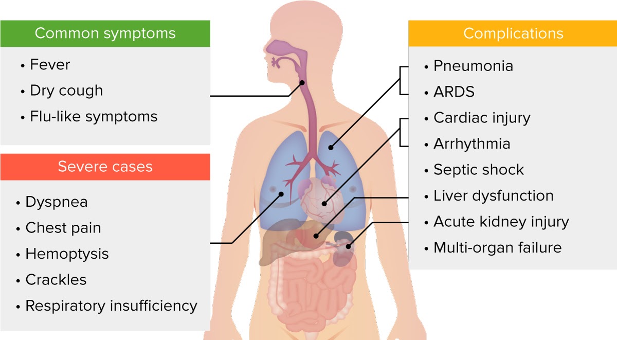
Clinical presentation of COVID-19.
Image: by Lecturio

Severe cases and complications

  • Approximately 1 in 6 people with COVID-19 experience clinical deterioration and/or develop complications in the 2nd week of illness. This is usually marked by the appearance and worsening of dyspnea.
  • The median time from onset of symptoms to the onset of critical care/ICU transfer is 8–9 days.
  • Patients develop dyspnea, high fever, chest pain, hemoptysis, anorexia, and/or respiratory crackles, which indicates the development of pneumonia (the most frequent complication in severe cases).
  • Respiratory failure from acute respiratory distress syndrome (ARDS) is the most common finding in critical cases.
  • Recovery time is approximately 3–6 weeks.
Explanation mark green

The most common complications of COVID-19 include viral pneumonia, respiratory failure and ARDS, sepsis and septic shock, cardiomyopathy, acute kidney injury, and pulmonary thromboembolism. Other complications include acute cardiac injury, deep vein thrombosis, arrhythmia, stroke, liver dysfunction, and multi-organ failure.

Risk factors for a severe infection and development of complications from COVID-19 (from highest to lowest risk) include the following:

  • Age > 65 years 
    • The mortality rate for patients < 65 is < 3%. However, this rises to 3%–11% for individuals aged 65–84 and 10%–27% for individuals ≥ 85 years of age.
  • Living in a nursing home or long-term care facility
  • Chronic diseases:
    • Chronic lung disease or moderate-to-severe asthma
    • Cardiovascular disease
    • Immunosuppression (from long-term steroid use, cancer, AIDS/HIV infection, congenital immunodeficiency, organ transplants, immunosuppressants, etc.)
    • Severe obesity (BMI > 40)
    • Diabetes mellitus, chronic kidney disease undergoing dialysis, cerebrovascular disease, and liver disease
  • Pregnancy
    • The risk of infection is the same as in nonpregnant individuals.
    • A higher risk of severe illness in pregnant individuals is assumed due to the behavior of similar respiratory infections, such as SARS and influenza.
  • Smoking
    • Recent evidence suggests that smoking is associated with increased severity of disease and death in hospitalized COVID-19 patients.

Refractory cases and persistent symptoms

Nearly 50% of COVID-19 patients do not achieve clinical and radiological remission within 10 days of hospitalization. Men, older patients, individuals with anorexia, and those with no/low fever at the time of admission have a higher risk of presenting with a refractory progression. 

  • This phenomenon is also known as “post-COVID-19 syndrome” or “long COVID.” 
  • 83% of 43 Italian patients hospitalized for COVID-19 continued to have at least 1 symptom 60 days after discharge; fatigue and dyspnea were most commonly reported.
  • 32% of 669 COVID-19-positive Swiss patients continued to have at least 1 symptom at a mean of 43 days after diagnosis; fatigue, dyspnea, and dysgeusia or anosmia were the main persistent symptoms.
  • Less common symptoms include joint pain, headache, sicca syndrome, vertigo, and psychologic and neurocognitive symptoms (e.g., anxiety, depression, post-traumatic stress disorder, poor memory and concentration).
  • Survivors of these infections are likely to be at high risk for pulmonary fibrosis; antifibrotic therapies may be beneficial both in the acute phase of the illness and in preventing long-term complications.

COVID-19 phenotypes

The wide range of possible clinical presentations among COVID-19 patients depends on the interaction between 3 basic factors:

  1. The severity of the infection and the host’s immune response, physiological reserve, and comorbidities
  2. The patient’s responsiveness to ventilatory support for hypoxemia
  3. The period of time between symptom onset and evaluation in a hospital

No new ARDS phenotypes are associated with COVID-19. Early in the pandemic, it was reported that COVID-19 produced a new type of ARDS, type L, which has low elastance (i.e., high compliance), in contrast with the high-elastance (low-compliance) type H.

  • If this is true, type L would derive little benefit from PEEP and higher tidal volumes could be used, while type H could be managed with proven tried-and-true ARDS net strategies.
  • This theory was disproved, however, by a review of a 2014 study showing that 12% of ARDS patients showed low elastance (high compliance), the so-called type L phenotype.

COVID-19 in children

The clinical presentation and severity of cases of COVID-19 in patients < 18 years old is different from that of adults. Children are at a lower risk of developing severe or critical infections, and complications appear to be milder.

Range of clinical presentations in children (the percentages are approximate):  

  • 66% are asymptomatic.
  • 27% are mild (COVID-19-related symptoms).
  • 5% are moderate (pneumonia, gastroenteritis, dehydration).
  • 2% are severe (respiratory failure requiring mechanical ventilation, shock, multi-organ failure requiring intensive care unit admission.

Multisystem inflammatory syndrome in children (MIS-C) is a rare but serious condition associated with COVID-19. The clinical features of MIS-C may resemble those of Kawasaki disease or toxic shock syndrome:

  • Persistent fever, hypotension, gastrointestinal symptoms, rash, myocarditis
  • Laboratory evidence of increased inflammation 
  • Respiratory symptoms may be lacking. 

Lecturio resources

Diagnostics

The RT-PCR test is currently the only test being used to confirm cases of acute COVID-19 infection. This test should be performed once a person under investigation (PUI) is identified according to the priorities outlined below. A positive test for SARS-CoV-2 generally confirms the diagnosis of COVID-19, regardless of the patient’s clinical status. The specimens used for testing include the following:

  • Nasopharyngeal (NP) or oropharyngeal (OP) swab
  • NP swabs are the 1st choice. Oropharyngeal swabs are acceptable only if NP swabs are not available.
  • Nasal mid-turbinate swab or swab of anterior nares (nasal swab)
  • Nasopharyngeal wash/aspirate or nasal wash/aspirate specimen
  • Sputum (for patients with productive cough; inducing is not recommended)


Bronchoalveolar lavage, tracheal aspirate, pleural fluid, and lung biopsy
(for patients with critical infections receiving invasive mechanical ventilation)

Reverse transcriptase-polymerase chain reaction testing can be negative initially. If suspicion of COVID-19 remains, the patient should be retested every 2–3 days. In severe cases, swabs from the upper respiratory tract may be negative, while specimens from the lower respiratory tract are positive. These tests can also yield false negatives in 20%–30% of cases. (For further information see the Centers for Disease Control and Prevention’s [CDC’s] Interim Guidelines for Collecting, Handling, and Testing Clinical Specimens from Persons for Coronavirus Disease 2019 (COVID-19).”)

Swabs 3 new
Image: by Lecturio

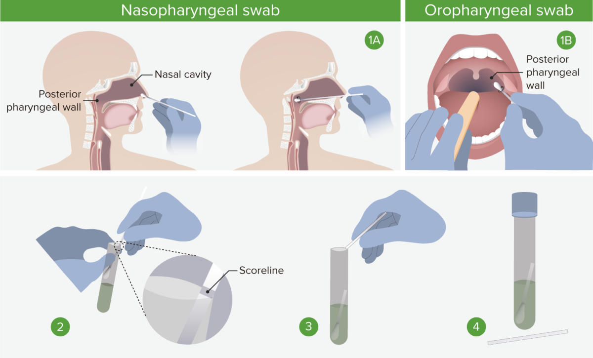
1A. Nasopharyngeal swab: Insert the swab into a nostril parallel to the palate, and carefully slide it forward until a soft resistance is felt. The swab should reach a depth equal to the distance from the nostrils to the outer opening of the ear. Rotate for several seconds to absorb secretions, and then slowly remove. 1B. Oropharyngeal swab: Insert swab into the oral cavity without touching the gums, teeth, or tongue. A tongue depressor may be used. Swab the posterior pharyngeal wall using a rotatory motion. 2. Place swabs immediately into sterile tubes containing 23 mL of viral transport media. If both swabs are collected, they should be combined into a single vial. 3. Carefully leverage the swab against the tube rim to break the shaft at the scoreline. 4. Store specimens at 2°C–8°C (35.6°F–46.4°F) for up to 72 hours after collection. If a delay in testing/shipping is expected, store specimens at –70°C (–94°C) or below. Use only synthetic fiber swabs with plastic shafts. Calcium alginate swabs or swabs with wooden shafts may inactivate the virus and inhibit PCR testing. 

Explanation mark green

Due to the limited availability of testing in certain countries, a diagnosis of COVID-19 can be made presumptively in the presence of a compatible clinical presentation with an exposure risk, particularly when there is no other evident cause of the symptoms. Testing for other causes of respiratory illness, such as influenza, is strongly encouraged in these cases. However, a positive test result for another respiratory agent does not rule out coinfection.

During an ongoing COVID-19 outbreak, patients with suspected infection who do not present with severe symptoms are encouraged to call before presenting to a healthcare facility for evaluation and testing. Laboratory testing of a PUI should be prioritized as follows according to the CDC (only in a state of emergency due to shortages or limited testing capacity):

  • High priority:
    • Hospitalized patients with symptoms
    • Healthcare facility workers, workers in congregate living settings, and first responders with symptoms
    • Residents in long-term care facilities or other congregate living settings, including prisons and shelters, with symptoms
  • Priority:
    • Persons with signs and symptoms compatible with COVID-19
    • Persons without symptoms who are prioritized by health departments or clinicians for any reason, including but not limited to:
      • Public health monitoring
      • Sentinel surveillance
      • Screening of other asymptomatic individuals according to state and local plans

Reverse transcriptase-polymerase chain reaction assays are based on nucleic acid amplification, and various types of assays being used around the world to detect and amplify different regions of the genome of SARS-CoV-2. The viral genes being targeted include the N, E, S, and RNA-dependent RNA polymerase (RdRp) gene. Both the RdRP and E genes have high analytical sensitivity for detection, whereas the N gene provides poorer analytical sensitivity. The use of at least 2 molecular targets is required to avoid cross-reaction with other endemic coronaviruses. The RT-PCR assay has very high sensitivity and specificity.

Rapid antigen testing, on the other hand, detects specific viral antigens and has the additional advantage of being able to be performed at the point of care. It is particularly useful in the early stages of infection, especially in cases of known exposures. The 2 rapid antigen tests approved by the Food and Drug Administration (FDA) have a specificity of 100%; thus, false-positives are unlikely. However, the sensitivity of these 2 tests is 84% and 97%, respectively, significantly lower than the RT-PCR test (> 95%). In general, positive antigen tests should be confirmed via an RT-PCR test performed at least 2 days later.

Explanation mark green

All PUI and confirmed cases should be reported according to the regulations stipulated by local health authorities and the national surveillance center.

Patients with COVID-19 present with the following laboratory and radiological findings. These findings are more pronounced and common in severe and critical cases but can also be present in asymptomatic infections:

  • WBC count: leukopenia, leukocytosis, and lymphopenia (most common)
  • Inflammatory markers: ↑ D-dimer, ferritin, C-reactive protein, IL-6
  • Liver markers: ↑ AST and ALT 
  • Chest X-ray and CT: 
    • Not recommended for initial evaluation; reserved for hospitalized patients or symptomatic patients with specific clinical indications
    • Common findings include ground-glass opacities (GGOs), multiple areas of consolidation, “crazy paving appearance” (GGOs + inter-/intralobular septal thickening), and bronchovascular thickening.
    • Lesions usually have bilateral, peripheral, and lower lobe distribution.
Explanation mark green

Radiological findings associated with COVID-19 are not specific to the infection; they overlap with other respiratory illnesses including influenza, H1N1, SARS, and MERS.

In hospitalized COVID-19 patients with severe infections, regular laboratory testing and imaging are necessary for the assessment of disease progression and complications:

  • CBC: Severe cases present with advanced lymphocytopenia and thrombocytopenia. 
  • Arterial blood gases: to assess levels of hypoxia and acid-base balance
    • ARDS presents initially as hypoxemic respiratory failure with low PaO2 and respiratory alkalosis, later progressing into hypercapnic respiratory failure.
  • Inflammatory markers: High levels suggest immune dysregulation and progression to cytokine storm.
    • ↑ IL-6 and C-reactive protein in severe cases
    • ↑ Procalcitonin in bacterial coinfection with pneumonia and/or sepsis
    • ↑ Lactate in sepsis and septic shock
  • Hemostasis tests: 
    • Prolonged PT and PTT times
    • ↑ D-dimer in cardiac injury and septic shock (associated with high mortality)
  • Assessment of organ function: Abnormal findings may indicate multi-organ failure.
    • Creatinine, urea, and BUN used to assess renal function
    • Aspartate aminotransferase, alanine aminotransferase, gamma-glutamyl transferase, and bilirubin used to assess hepatic function
    • Cardiac enzymes (troponin and NT-proBNP) and ECG used to assess cardiac function
  • Chest X-ray and CT: severe infections may also present with the following:
    • Pleural thickening and effusion
    • Lymphadenopathy
    • Air bronchograms and atelectasis
    • Solid white consolidation

Serologic testing is not recommended for acute infections. Serology measures the host response to infection by the production of antibodies to SARS-CoV-2 and is an indirect measure of infection that is best utilized retrospectively. IgM responses are mainly nonspecific; specific IgG responses require weeks to develop; thus, serology detection plays an important role in surveillance, not in active case detection and management. It is vital in determining the immunity of healthcare workers as the outbreak progresses and the true mortality rate once the outbreak resolves.

Explanation mark green

The sensitivities and specificities of different serologic tests are highly variable. IgG antibodies are usually detectable by 14 days after onset of symptoms, and usually─but not always─remain detectable for up to 8 months after infection.

These antibodies have also been shown to have neutralizing effects on SARS-CoV-2, which explains the current use of convalescent plasma as an investigational therapy. However, studies have shown that not all patients with COVID-19 develop neutralizing antibodies, and antibody response may be associated with the severity of the disease. One study showed that titers of SARS-CoV-2–specific neutralizing antibodies increase in parallel with the rise in IgG antibodies. The titers are low for the first 7–10 days after symptom onset, increase after 2–3 weeks, then decline gradually over several months after infection.

Differential Diagnoses

 COVID-19InfluenzaCommon cold
Incubation period2–14 days1–4 days< 3 days
OnsetGradualSuddenSudden
FeverVery commonVery commonRare
Dry coughVery common (mild to severe)Very common (mild to severe)Common (usually mild, can be productive)
FatigueCommonVery commonRare or mild
MyalgiaCommonVery commonMild
SneezingSometimesRare or mildVery common
Nasal congestionRare or mildCommonVery common
HeadacheSometimesVery commonRare or mild
Sore throatSometimesSometimesVery common
DiarrheaSometimesSometimesRare
DyspneaCommonRareNever

Lecturio resources

Management

Management in early infection (≤ 5 days) in adults with COVID-19-specific therapy (nirmatrevir/ritonavir (Paxlovid)*:

Only recommended for an adult with any of the following:

  • ≥ 65 years old
  • Immunocompromised 
  • Multiple comorbidities
  • ≥ 50 years old and unvaccinated
  • Does not have Stage 4 kidney disease (eGFR < 30 ml/min) or severe liver disease (Child-Pugh Class C)

If the patient has severe kidney or liver disease OR is seen only within after 5 days but within 6–8 days, then Remdesivir (≤ 5 days) OR convalescent plasma (≤ 8 days) may be considered.

Monoclonal antibodies are no longer routinely recommended because of the high prevalence of resistance associated with the Omicron variants, which allows them to escape neutralization. The Omicron variants are the predominant circulating variants of concern globally.

*Nirmatrevir/ritonavir (Paxlovid):

  • Associated with a lower rate of emergency department visits, hospitalization, and death
  • Nirmatrelvir is a peptidomimetic inhibitor of the SARS-CoV-2 main protease, thereby resulting in inhibition of viral replication.
  • Ritonavir inhibits CYP3A-mediated metabolism of nirmatrelvir, resulting in increased nirmatrelvir plasma concentrations.

In the later stages of infection, immunosuppressive/anti-inflammatory therapies are likely to be more beneficial.

Explanation mark green

All healthcare personnel and patients with confirmed or possible SARS-CoV-2 infection should wear personal protective equipment (PPE). (See the CDC’sInfection Control Guidance for Healthcare Professionals about Coronavirus (COVID-19)and Using Personal Protective Equipment (PPE).”)

According to clinical status and laboratory and radiological findings, COVID-19 patients can be grouped into the following illness categories:

  • Mild disease:
    • Fever, malaise, cough, upper respiratory symptoms, and/or less common features of COVID-19, without dyspnea
    • Most of these patients do not need hospitalization.
  • Moderate disease: 
    • Dyspnea is present.
    • Often warrants hospitalization
    • Infiltrates on chest imaging may still be considered as moderate disease
  • Severe disease, if any of the following are present (FDA definitions):
    • Hypoxia (oxygen saturation ≤ 94% on room air)
    • Need for oxygen or ventilatory support
    • Additional features used in studies to define severe disease:
      • Tachypnea
      • Respiratory distress
      • >50% involvement of the lung parenchyma on chest imaging
  • Critical disease:
    • Individuals who have respiratory failure requiring mechanical ventilation or extracorporeal membrane oxygenation (ECMO), septic shock, and/or multiple organ dysfunction

Management of mild, asymptomatic, or presymptomatic cases 

  • Self-isolate and maintain quarantine for 14 days.
  • Supportive at-home care, including antipyretics (e.g., ibuprofen)
  • One exception would be to administer anti-COVID-19 monoclonal antibodies in the very early stage (< 72 hours) of symptomatic infection to older adults living in nursing facilities
  • In the outpatient setting, it is important to have professional medical assistance if any of the following emergency warning signs develop:
    • Difficulty breathing or shortness of breath
    • Persistent pain or pressure in the chest
    • Confusion or inability to arouse
    • Cyanosis (bluish tint to the lips or face or in warm extremities)

Management of moderate-to-severe cases

  • Most of these cases will require hospitalization, with critical cases requiring admission to an ICU. However, the decision to monitor a patient in the inpatient setting should be made on a case-by-case basis.
  • Once hospitalized, supportive care and acute measures should be applied as necessary and should include the following:
    • Breathing support is crucial in treating severe COVID-19 cases or any respiratory complication in order to alleviate and/or prevent respiratory distress, hypoxemia, or shock. The key elements include oxygen therapy by:
      • Nasal cannula or high-flow nasal oxygen
      • Prone mechanical ventilation
      • ECMO for rescue in select cases
    • Empiric antimicrobials if sepsis or secondary pneumonia is suspected
    • Pharmacologic prophylaxis of venous thromboembolism:
      • COVID-19 patients who experience a thromboembolic event or have a high risk of thromboembolic disease should be managed with therapeutic doses of anticoagulant therapy as per standard of care.
      • A retrospective study showed reduced mortality in hospitalized patients with COVID-19 who received prophylactic anticoagulation.
      • Low-molecular-weight heparin or unfractionated heparin is preferred in hospitalized, critically ill patients.
    • Inhaled corticosteroids: continued use is recommended for patients who are already receiving steroids for another indication, such as asthma
    • Advanced oxygen therapy, ventilatory support, and conservative fluid management in the case of ARDS or respiratory failure
    • Fluid bolus and vasopressors in the case of septic shock
      • The COVID-19 Treatment Guidelines Panel recommends norepinephrine as the 1st-choice vasopressor.
    • Antifibrotic therapy may be beneficial both in the acute phase of the illness and in preventing long-term pulmonary fibrosis.
    • Clinical management of other comorbidities and nosocomial complications

Specific anti–COVID-19 therapy for patients with moderate to severe COVID-19 infection

The optimal approach to treatment has not been established with certainty, and treatment options are still being developed.

Treatment depends on the presence of risk factors and if oxygen is required: 

  • If no risk factors for progression to severe disease: supportive care only 
  • If there are factors for progression to severe disease: Remdesivir 
    • Note: Dexamethasone should not be used at this point since it may worsen the disease.
  • If oxygen is required: Treatment depends on the specific therapy given.
    • Low-flow oxygen (e.g., 1–2 L/min): Remdesivir +/- low-dose dexamethasone, especially if elevated inflammatory markers (e.g., D-dimer, ferritin, C-reactive protein)
      • If markers remain elevated and are within 96 hours of hospitalization, add either baricitinib (an inhibitor of janus kinase [JAK] or tocilizumab (a monoclonal antibody against the interleukin-6 receptor; sarilumab is an alternative).
      • Besides having immunomodulatory effects*, baricitinib may interfere with viral entry.
    • High-flow oxygen or non-invasive ventilation: low-dose dexamethasone and remdesivir
      • If within 96 hours of hospitalization, add baricitinib or tocilizumab. 
    • Mechanical ventilation or extracorporeal membrane oxygenation (ECMO): low-dose dexamethasone 
      • If within 96 hours of hospitalization, add baricitinib or tocilizumab.
      • Remdesivir is valid here IF patient has been intubated < 48 hours.

Guidelines for treatment of COVID-19 are available from the Infectious Diseases Society of America (IDSA).

Enrollment into a clinical trial of additional medications is encouraged, such as those listed at https://clinicaltrials.gov/ct2/covid_view and the WHO website.

*Other immunomodulators: IL-1 inhibitors (e.g., anakinra) have shown reduced mortality in certain patients with severe COVID-19 who also have elevated plasma soluble urokinase plasminogen activator receptor (suPAR), a biomarker associated with disease progression in some studie. Not routinely used because measurement of suPAR is not widely available, and it is uncertain if anakinra offers advantages over other immunomodulatory agents

Other COVID-19-specific therapies:

  • Monoclonal antibodies: These are no longer routinely recommended for either outpatients or hospitalized patients because of the high prevalence of resistance associated with most of the recent circulating virus subvariants of Omicron (e.g., BQ.1 and BQ1.1), which allows them to escape neutralization. 
  • Convalescent plasma, from patients who have recovered from COVID-19:
    • Not recommended for most hospitalized patients, but may be useful in immunocompromised patients (but not proven in randomized trials)
    • Studies in outpatients with non-severe COVID-19 are ongoing.
    • Not recommended for hospitalized patients
  • Other therapies under evaluation:
      • Sabizabulin, a microtubule disruptor which has antiviral and anti-inflammatory effects, showed reduced mortality in phase 3 study of hospitalized patients with moderate to severe COVID-19 (further study needed).
      • Other immunomodulatory agents of different classes are being evaluated (e.g., cytokine inhibitors, other kinase inhibitors, complement inhibitors, bradykinin pathway inhibitors, and hematopoietic colony-stimulating agonists and antagonists).
  • Therapies without clear benefits identified in trials:
    • Hyperimmune globulin
    • Ivermectin, an antiparasitic drug: 
      • Only validated use is for prevention of Strongyloides reactivation in patients receiving glucocorticoids.
      • Initial studies in vitro showed activity against SARS-CoV-2 but the high drug concentrations used cannot be achieved in vivo.
    • Vitamin D: only given to treat known deficiency or to maintain recommended intake
    • Hydroxychloroquine 
    • Colchicine
    • Lopinavir-ritonavir
    • Interferon beta
    • Azithromycin
Explanation mark green

Causes of death in COVID-19 patients include respiratory failure, multi-organ failure, and hypotensive shock.

Lecturio resources

Prevention

It is now a global recommendation that all individuals should help prevent the spread of COVID-19 infection. General recommendations include the following:

  • Face masks:
    • Help prevent the wearer from becoming infected and, just as importantly, prevent the wearer from transmitting the disease (also known as “source control”)
    • For healthcare personnel, PPE and National Institute for Occupational Safety and Health–approved N95 disposable filtering facepieces or higher-level respirators, such as a powered air-purifying respirator, are recommended when providing care for patients with suspected or confirmed COVID-19 due to higher exposure to infected individuals as well as aerosol-generating procedures. These procedures include the following:
      • Open suctioning of airways
      • Sputum induction
      • Cardiopulmonary resuscitation
      • Endotracheal intubation and extubation
      • Non-invasive ventilation 
      • Bronchoscopy
      • Manual ventilation
  • Caps: It is still unclear how long SARS-CoV-2 can survive on human hair, but healthcare personnel are encouraged to cover their hair with a disposable or surgical cap.
  • Home isolation and quarantine:
    • Avoid public/crowded areas whenever possible, particularly within closed buildings, to minimize the chance of exposure or transmission.
    • Before leaving and upon returning from travel, individuals should practice home isolation for 14 days, monitor the possible onset of symptoms, and be tested for COVID-19 if testing is available (especially if traveling to and from an area with a high infection rate).
  • Respiratory hygiene: Coughs and sneezes should be covered with a tissue or the inner elbow.
  • Wash hands regularly for at least 20 seconds with soap and water or with an alcohol-based hand sanitizer that contains at least 60% alcohol.
  • Avoid touching face or eyes or shaking hands: The virus can remain viable for up to 9 hours on unwashed skin.
  • Social distancing:
    • Maintain 1–2 meters (approximately 3–6 feet) distance from other people.
    • Avoid certain actions, such as forced expiration during yelling, singing, and exercise, as these can increase the volume and distance that respiratory droplets can travel.
  • Regular cleaning of all “high-touch” surfaces within the home or workplace (see the CDC’s “Cleaning and Disinfection for Households”)

Isolation and quarantine can be discontinued only after the following criteria have been met:

  • At-home and hospitalized symptomatic cases: 
    • Negative results of PCR testing obtained from at least 2 consecutive NP swab specimens collected ≥ 24 hours apart OR
    • At least 3 days have passed since the resolution of fever without the use of antipyretics and improvement in respiratory symptoms AND
    • At least 10 days have passed since the onset of symptoms
  • Asymptomatic cases:
    • Negative results of PCR testing obtained from at least 2 consecutive NP swab specimens collected ≥ 24 hours apart OR
    • At least 10 days have passed since the first positive COVID-19 diagnostic test 

See the CDC’s “Healthcare Infection Prevention and Control FAQs for COVID-19.

Vaccines

Vaccination is recommended for those individuals who are eligible, according to local guidelines. 

  • Multiple vaccines have been and are being developed in different countries.
  • Each country has the autonomy to issue emergency use authorizations (EUAs) for any health product.
  • The WHO has given Emergency Use Listing to 9 vaccines (see ref. 83).
  • The USA has given EUAs to vaccines from 4 different manufacturers: Pfizer, Moderna, Janssen, and Novavax.
  • A vaccine must be at least 50% effective to meet regulatory approval.
  • No enhanced disease with wild-type virus was reported in vaccinated people in any human studies; this was feared due to initial animal studies of vaccines for SARS-CoV-1 and MERS-CoV.
  • The different vaccines employ 9 or more mechanisms or “platforms” for delivery, and have different efficacies, against the initial viral variants and the subsequent variants. 
    • Viral based (protein subunits or virus-like particles)
    • Inactivated or weakened virus
    • Using a different virus (e.g., adenovirus) as vector
    • DNA- or RNA-based vaccines
    • Indirect immune stimulation (by BCG or polio vaccines or other methods) 

Mechanism of mRNA (messenger RNA) vaccines:  

  1. A non-infective segment of the viral mRNA that creates the outer spike protein used to enter human cells is wrapped in lipid nanoparticles. 
  2. After injection, the vaccine particles encounter human macrophages and dendritic cells, fuse to them, and release the mRNA into their cytoplasm. (The mRNA does not enter the nucleus or interact with the host’s DNA.)
  3. The host cells’ cytoplasmic processes read the new mRNA sequence and build spike proteins. 
  4. The mRNA from the vaccine is eventually destroyed by the cell, leaving no permanent trace.
  5. Some of the spike proteins and fragments of spike proteins migrate to the surface of the cell and stick out on the surface, allowing them to be recognized by the immune system.
  6. Additional spike proteins and protein fragments stimulate the immune system when they are taken up by antigen-presenting cells (APCs). 
  7. APCs present spike protein fragments to helper T cells, which activate and coordinate with B cells that multiply and produce protective antibodies against the spike proteins and future infection by the live virus.
  8. APCs also activate killer T cells, which destroy any coronavirus-infected cells that display spike protein fragments on their surfaces.
  9. Although the amount of antibodies and killer T cells may drop within months after vaccination, memory B cells and memory T cells exist; these can retain information about the coronavirus for many years.

Selected list of vaccines:

  • Pfizer-BioNTech (BNT162b2) and Moderna (mRNA 1273)
    • Platform: mRNA vaccines that received emergency use authorization in the United States in December 2020
    • Each mRNA vaccine has a monovalent formulation (based on the original SARS-CoV-2 strain) and a bivalent formulation (based on the original SARS-CoV-2 strain and the BA.4/BA.5 Omicron subvariants), used as booster for those > 12 years.
    • Each is given in 2 intramuscular doses 3–4 weeks apart. The bivalent booster is given 8 weeks after the second monovalent dose. 
    • Showed 95% and 94% efficacy, respectively, in preventing laboratory-confirmed symptomatic COVID-19 after the second dose, in large (44,000 and 30,000, respectively) placebo-controlled trials
    • Pfizer vaccine can be held at normal medical freezer temperatures of –15°C to –25°C (–5°F to –13°F) for up to 2 weeks, compared with the previously required storage conditions of between –60°C and –80°C (–76°F to –112°F).
    • Adverse effects: 
      • Non-severe local and systemic adverse effects are common (e.g., pain, fever, fatigue, headache). 
      • Severe adverse effects are exceedingly rare (e.g., anaphylaxis, thrombosis, myocarditis/pericarditis in young males (risk: 52–71 cases/million doses)). 
  • Novavax (NVX-CoV2373)
    • Platform: a recombinant protein nanoparticle vaccine composed of trimeric spike glycoproteins and a potent adjuvant
    • Given in 2 intramuscular doses 21 days apart
    • Showed 89% efficacy against the UK variant (B.1.1.7) but only 49% against the South African variant in a phase III trial 
    • Side effects: fatigue, headache, myalgias, and/or malaise, possible risk of myocarditis, pericarditis 
  • AstraZeneca (ChAdOx1 nCoV-19/AZD1222, University of Oxford and the Serum Institute of India)
    • Platform: a replication-incompetent adenovirus vector that expresses the spike protein
    • Given in 2 intramuscular doses 4–12 weeks apart 
    • Showed 62%–90% efficacy in phase III studies, but further investigation is pending
    • Authorized for use in the EU and other countries, including the UK and India
    • Adverse effects: 
      • Fatigue, headache, and fever were relatively common after vaccine receipt and were more severe in up to 8% of patients.
      • Serious adverse effects are rare (thrombosis with thrombocytopenia: 17 cases/million doses). 
  • Janssen (Ad26.COV2.S)
    • Platform: a replication-incompetent adenovirus 26 vector that expresses a stabilized spike protein
    • Given in 1 intramuscular dose or 2 doses 56 days apart
    • A phase I/II trial showed high rates of neutralizing and binding antibodies after a single vaccine dose in healthy individuals 18–85 years of age
    • Showed 66% efficacy in preventing moderate-to-severe COVID-19 starting at 14 days following vaccination; efficacy against severe disease was 85%
    • Adverse effects: 
      • Fever, fatigue, headache, and myalgia were common.
      • Grade 3 systemic adverse effects ranged from 9%–20%, depending on the vaccine dose.
      • Serious adverse effects are very rare: thrombotic complications, with thrombocytopenia (12.4 cases/million doses), Guillain-Barré syndrome 8 cases/million doses) 
  • CanSino Biologics (Ad5-based COVID-19 vaccine)
    • Platform: a replication-incompetent adenovirus 5 vector that expresses the spike protein
    • Given as 1 intramuscular dose
    • Immunogenic at 28 days, but both pre-existing immunity to adenovirus 5 and older age were associated with lower titers of binding and neutralizing antibodies, possibly limiting its utility in an area with high pre-existing immunity 
    • Adverse effects: only mild-to-moderate local and systemic reactions
    • 58% efficacy
    • Licensed in China, Mexico, Turkey 
  • Gam-COVID-Vac/Sputnik V (Gamaleya Institute) 
    • Platform: 2 replication-incompetent adenovirus vectors that express a full-length spike glycoprotein
    • Given as an intramuscular adenovirus 26 vector dose followed by an adenovirus 5 vector-boosting dose 21 days later
    • Available in Russia and several other countries, including Mexico
    • Efficacy: 91.6% at 21 days following the 1st dose 
    • Side effects: local and systemic flu-like reactions were more common, with no serious adverse events noted
  • BBIBP-CorV (Sinopharm)
    • Platform: an inactivated vaccine based on a COVID-19 isolate from a patient in China, with an aluminum hydroxide adjuvant
    • Given in 2 intramuscular doses 28 days apart
    • Showed 86% efficacy in phase III studies in the United Arab Emirates (UAE) against COVID-19 infection, with 100% effectiveness in preventing moderate and severe cases of the disease
    • Side effects: no serious safety concerns
    • This vaccine is available in China and some other countries, including the UAE.
  • CoronaVac (Sinovac)
    • Platform: an inactivated COVID-19 virus developed in China, with an aluminum hydroxide adjuvant
    • Given in 2 intramuscular doses 28 days apart
    • Efficacy: differs in reports from various countries
      • 50.4% effective in Brazilian clinical trials
      • 91.3% effective in Turkey
      • 65.3% effective in Indonesia
    • Side effects: appeared safe in various trials
    • Available in China and some other countries, including Brazil, Turkey, and Indonesia
  • Covaxin (Bharat Biotech/Indian Council of Medical Research)
      • Platform: inactivated vaccine with aluminum hydroxide and a toll-like receptor agonist adjuvant
      • Given intramuscularly in 2 doses 29 days apart
      • Efficacy against symptomatic COVID-19: 78%; one possibly related case of immune thrombocytopenic purpura
  • ZyCoV-D (Zydus Cadila)
    • Platform: the first DNA COVID-19 vaccine, first authorized in India in August 2021
    • Given subcutaneously by a needleless device with a high-pressure stream, 3 doses, each given 28 days apart
    • Efficacy against symptomatic COVID-19: 67%

The type of vaccine being administered, its availability, and the progress of its distribution vary from country to country and from region to region.

Lecturio resources

Additional Resources:

  1. World Health Organization. Coronavirus. Retrieved December 21, 2022, from https://www.who.int/health-topics/coronavirus#tab=tab_1
  2. Centers for Disease Control and Prevention. COVID-19. Retrieved March 10, 2020, from https://www.cdc.gov/coronavirus/2019-nCoV/hcp/index.html
  3. Fang L., Karakiulakis G., & Roth M. (2020), Are patients with hypertension and diabetes mellitus at increased risk for COVID-19 infection? Lancet Respir Med.  https://doi.org/10.1016/S2213-2600(20)30116-8
  4. Kuba K., Imai Y., Rao S., et al. (2005). A crucial role of angiotensin converting enzyme 2 (ACE2) in SARS coronavirus–induced lung injury. Nat Med 11, 875–879. https://doi.org/10.1038/nm1267
  5. Wang K., Chen W., Zhou Y., et al. (2020). SARS-CoV-2 invades host cells via a novel route: CD147-spike protein. bioRxiv. https://doi.org/10.1101/2020.03.14.988345
  6. Liu P., Chen W., & Chen J. P. (2019). Viral metagenomics revealed Sendai virus and coronavirus infection of Malayan pangolins (Manis javanica). Viruses, 11(11), 979. https://doi.org/10.3390/v11110979
  7. Fehr A.R., Perlman S. (2015). Coronaviruses: An Overview of Their Replication and Pathogenesis. In: Maier H., Bickerton E., Britton P. (eds.). Coronaviruses. Methods in Molecular Biology, vol 1282. Humana Press, New York, NY. https://doi.org/10.1007/978-1-4939-2438-7_1
  8. Kampf G., Todt D., Pfaender S., & Steinmann, E. (2020). Persistence of coronaviruses on inanimate surfaces and their inactivation with biocidal agents. The Healthcare Infection Society. https://doi.org/10.1016/j.jhin.2020.01.022
  9. Guo Y.R., Cao Q.D., Hong Z.S., et al. (2020). The origin, transmission and clinical therapies on coronavirus disease 2019 (COVID-19) outbreak – an update on the status. Military Med Res 7, 11. https://doi.org/10.1186/s40779-020-00240-0
  10. Yang L., Yan L., Wan L., Xiang T., Le A., Liu J., et al. Viral dynamics in mild and severe cases of COVID-19. Lancet Inf Dis. https://doi.org/10.1016/S1473-3099(20)30232-2
  11. Guan W.J., Ni Z.Y., Hu Y., Liang W.H., et al. (2020). China Medical Treatment Expert Group for Covid-19. clinical characteristics of coronavirus disease 2019 in China. The New England Journal of Medicine, 382(18), 1708–1720. https://doi.org/10.1056/NEJMoa2002032
  12. Mo P., Xing Y., Xiao Y., et al. (2020). Clinical characteristics of refractory COVID-19 pneumonia in Wuhan, China, Clinical Infectious Diseases, https://doi.org/10.1093/cid/ciaa270
  13. Hoffmann M., Kleine-Weber H., Schroeder S., Müller M., Drosten C., & Pöhlmann S. (2020). SARS-CoV-2 cell entry depends on ACE2 and TMPRSS2 and is blocked by a clinically proven protease inhibitor. Cell. https://doi.org/10.1016/j.cell.2020.02.052
  14. Tufekci Z., Howard J., & Greenhalgh T. (2020). The Real Reason to Wear a Mask. The Atlantic. https://medium.com/the-atlantic/the-real-reason-to-wear-a-mask-e6405abbc484
  15. Tang X., Wu C., Li X., et al. (2020). On the origin and continuing evolution of SARS-CoV-2. National Science Review, Volume 7, Issue 6, June 2020, Pages 1012–1023, https://doi.org/10.1093/nsr/nwaa036
  16. Groß S., Jahn C., Cushman S., Bär C., & Thum T. (2020). SARS-CoV-2 receptor ACE2-dependent implications on the cardiovascular system: From basic science to clinical implications. Journal of Molecular and Cellular Cardiology, Volume 144, July 2020, Pages 47-53, https://doi.org/10.1016/j.yjmcc.2020.04.031
  17. Zheng Y.Y., Ma Y.T., Zhang J.Y., et al. (2020). COVID-19 and the cardiovascular system. Nat Rev Cardiol 17, 259–260. https://doi.org/10.1038/s41569-020-0360-5
  18. Gandhi M., Yokoe D.S., & Havlir D.V. (2020). Asymptomatic transmission, the Achilles’ heel of current strategies to control Covid-19. N Engl J Med 2020; 382:2158-2160 https://doi.org/10.1056/NEJMe2009758
  19. Wölfel R., Corman V.M., Guggemos W., et al. (2020). Virological assessment of hospitalized patients with COVID-2019. Nature 581, 465–469. https://doi.org/10.1038/s41586-020-2196-x
  20. Basu-Ray I., Almaddah N.K., Adeboye A, et al. (2021). Cardiac Manifestations Of Coronavirus (COVID-19) [Updated 2021 Jan 13]. In: StatPearls [Internet]. Treasure Island (FL): StatPearls Publishing. Available from: https://www.ncbi.nlm.nih.gov/books/NBK556152/
  21. Shen C., Wang Z., Zhao F., et al. (2020). Treatment of 5 critically ill patients with COVID-19 with convalescent plasma. JAMA, 323(16), 1582–1589. https://doi.org/10.1001/jama.2020.4783
  22. Wang D., Hu B., Hu C., et al. (2020). Clinical characteristics of 138 hospitalized patients with 2019 novel coronavirus-infected pneumonia in Wuhan, China. JAMA, 323(11), 1061–1069. https://doi.org/10.1001/jama.2020.1585
  23. https://www.cdc.gov/coronavirus/2019-nCoV/lab/guidelines-clinical-specimens.html
  24. Zhao J., Yuan Q., Wang H., et al. (2020). Antibody Responses to SARS-CoV-2 in Patients With Novel Coronavirus Disease 2019. Clinical infectious diseases: an official publication of the Infectious Diseases Society of America, 71(16), 2027–2034. https://doi.org/10.1093/cid/ciaa344
  25. National Institutes of Health (2020). Clinical Spectrum of SARS-CoV-2 Infection https://www.covid19treatmentguidelines.nih.gov/overview/clinical-spectrum/
  26. Bhimraj A., Morgan R.L., Shumaker A.H., et al. (2020). IDSA Guidelines on the Treatment and Management of Patients with COVID-19. Infectious Diseases Society of America. https://www.idsociety.org/practice-guideline/covid-19-guideline-treatment-and-management/
  27. Centers for Disease Control and Prevention. (2020). Interim Clinical Guidance for Management of Patients with Confirmed Coronavirus Disease (COVID-19). https://www.cdc.gov/coronavirus/2019-ncov/hcp/clinical-guidance-management-patients.html
  28. Moorlag S.J.C.F.M., Arts R.J.W., van Crevel R., Netea M.G. (2019). Non-specific effects of BCG vaccine on viral infections. Clinical Microbiology and Infection. Elsevier. vol 25, issue 12, pp. 1473-1478, Dec 01, 2019. doi: https://doi.org/10.1016/j.cmi.2019.04.020
  29. Makiello P., Svirpliene S., Finlay L., et al. Paediatric inflammatory multisystem syndrome temporally associated with COVID-19: A new virus and a new case presentation. BMJ Case Reports CP 2020;13:e238531.
  30. WHO Headquarters (HQ). Smoking and COVID-19 Scientific Brief. (2020). https://www.who.int/publications/i/item/WHO-2019-nCoV-Sci_Brief-Smoking-2020.2
  31. Khera R., Clark C., Lu Y., et al. (2020) Association of angiotensin-converting enzyme inhibitors and angiotensin receptor blockers with the risk of hospitalization and death in hypertensive patients with coronavirus disease-19. medRxiv 2020.05.17.20104943; doi: https://doi.org/10.1101/2020.05.17.20104943
  32. Grifoni A., Weiskopf D., Ramirez S.I., et al. (2020). Targets of T cell responses to SARS-CoV-2 coronavirus in humans with COVID-19 disease and unexposed individuals. Cell, vol 181, issue 7, pp. 489-1501.e15, ISSN 0092-8674, doi: https://doi.org/10.1016/j.cell.2020.05.015
  33. Gattinoni L., Chiumello D., Caironi P. et al. COVID-19 pneumonia: Different respiratory treatments for different phenotypes?. Intensive Care Med 46, 1099–1102 (2020). https://doi.org/10.1007/s00134-020-06033-2
  34. Halpin D.M.G., Singh D., Hadfield R.M. (2020). Inhaled corticosteroids and COVID-19: A systematic review and clinical perspective. European Respiratory Journal, Jan 2020, 2001009; DOI: 10.1183/13993003.01009-2020
  35. National Institutes of Health. Antithrombotic Therapy in Patients With COVID-19. (2021). https://www.covid19treatmentguidelines.nih.gov/antithrombotic-therapy/
  36. Bentz G.L., Yurochko A.D. (2008). Human CMV infection of endothelial cells induces an angiogenic response through viral binding to EGF receptor and β1 and β3 integrins. Proceedings of the National Academy of Sciences Apr 2008, 105 (14) 5531-5536; DOI: 10.1073/pnas.0800037105
  37. Ackermann M., Verleden S.T. Kuehnel, M., et al. (2020). Pulmonary vascular endothelialitis, thrombosis, and angiogenesis in Covid-19. N Engl J Med; 383:120-128. DOI: 10.1056/NEJMoa2015432
  38. World Health Organization. Draft landscape and tracker of COVID-19 candidate vaccines. (2022). https://www.who.int/publications/m/item/draft-landscape-of-covid-19-candidate-vaccines
  39. Tang Y., Schmitz J.E., Persing D.H., Stratton C.W. (2020). Laboratory diagnosis of COVID-19: Current issues and challenges. Journal of Clinical Microbiology May 2020, 58 (6) e00512-20; DOI: 10.1128/JCM.00512-20
  40. Wang K., Long Q.X., Deng H.J., et al. (2020). Longitudinal dynamics of the neutralizing antibody response to SARS-CoV-2 infection. Clin Infect Dis. Aug 3:ciaa1143. doi: 10.1093/cid/ciaa1143
  41. Latham J., Wilson A. (2020). A Proposed Origin for SARS-CoV-2 and the COVID-19 Pandemic. Biotechnology, Commentaries, Health July 15, 2020. https://www.independentsciencenews.org/commentaries/a-proposed-origin-for-sars-cov-2-and-the-covid-19-pandemic/
  42. Sisman J., Mambarambath J.A., Wilmer M., et al. (2020). Intrauterine transmission of SARS-COV-2 infection in a preterm infant, The Pediatric Infectious Disease Journal: Sept 2020, vol 39, issue 9, pp. e265-e267. doi: 10.1097/INF.0000000000002815
  43. Vivanti, A.J., Vauloup-Fellous, C., Prevot, S. et al. Transplacental transmission of SARS-CoV-2 infection. Nat Commun 11, 3572 (2020). https://doi.org/10.1038/s41467-020-17436-6
  44. Leslie, M. (2020). T cells found in COVID-19 patients ‘bode well’ for long-term immunity. American Association for the Advancement of Science (AAAS) https://www.sciencemag.org/news/2020/05/t-cells-found-covid-19-patients-bode-well-long-term-immunity
  45. Wang J.X., Li Y.Y., Liu X.D., Cao X. (2020). Virus transmission from urinals. Physics of Fluids 32, 081703 (2020); https://doi.org/10.1063/5.0021450
  46. Evans, M.J. (2020). Avoiding COVID-19: Aerosol Guidelines. medRxiv 2020.05.21.20108894; doi: https://doi.org/10.1101/2020.05.21.20108894
  47. Heald-Sargent T., Muller W.J., Zheng X., Rippe J., Patel A.B., Kociolek L.K. Age-related differences in nasopharyngeal severe acute respiratory syndrome coronavirus 2 (SARS-CoV-2) levels in patients with mild to moderate coronavirus disease 2019 (COVID-19). (2020). JAMA Pediatr. 2020;174(9):902–903. doi:10.1001/jamapediatrics.2020.3651
  48. Huang, Y., Yang, C., Xu, X., Xu, W., & Liu, S. (2020). Structural and functional properties of SARS-CoV-2 spike protein: Potential antivirus drug development for COVID-19. Acta Pharmacologica Sinica, 41(9), 1141–1149. https://doi.org/10.1038/s41401-020-0485-4
  49. McIntosh, K. (2021). Coronavirus disease 2019 (COVID-19): Epidemiology, virology, and prevention. UpToDate. Retrieved December 19, 2022, from: disease-2019-covid-19-epidemiology-virology-and-prevention
  50. Kim, A.Y., Gandhi, R.T. (2021). Coronavirus disease 2019 (COVID-19): Management in hospitalized adults. UpToDate. Retrieved December 19, 2022, from: https://www.uptodate.com/contents/coronavirus-disease-2019-covid-19-management-in-hospitalized-adults
  51. Cui, J., Li, F., & Shi, Z.-L. (2019). Origin and evolution of pathogenic coronaviruses. Nature Reviews Microbiology, 17(3), 181–192. https://doi.org/10.1038/s41579-018-0118-9
  52. Piller, C., ServickJun. 4, K., 2020, & Pm, 5:30. (2020, June 4). Two elite medical journals retract coronavirus papers over data integrity questions. Science | AAAS. https://www.sciencemag.org/news/2020/06/two-elite-medical-journals-retract-coronavirus-papers-over-data-integrity-questions
  53. Grady D., Mazzei, P. (2021). Doctor’s Death After Covid Vaccine Is Being Investigated. The New York Times, January 12, 2012. Retrieved February 20, 2021, from https://www.ncbi.nlm.nih.gov/search/research-news/12431
  54. Corum, J., & Zimmer, C. (2021). How the Pfizer-Biontech Vaccine Works. The New York Times. Updated Jan. 21, 2021. Retrieved February 20, 2021, from https://www.nytimes.com/interactive/2020/health/pfizer-biontech-covid-19-vaccine.html
  55. Corum, J., & Zimmer, C. (n.d.). How Moderna’s Vaccine Works. The New York Times. Updated Jan. 21, 2021. Retrieved February 20, 2021, from https://www.nytimes.com/interactive/2020/health/moderna-covid-19-vaccine.html
  56. Wang, W., Zhao, X., Wei, W., Fan, W., Gao, K., He, S., & Zhuang, X. (2021). Angiotensin-converting enzyme inhibitors (Acei) or angiotensin receptor blockers (Arbs) may be safe for COVID-19 patients. BMC Infectious Diseases, 21(1), 114. https://doi.org/10.1186/s12879-021-05821-5
  57. Status of COVID-19 Vaccines within WHO EUL/PQ evaluation process https://extranet.who.int/pqweb/sites/default/files/documents/Status_COVID_VAX_16Feb2021.pdf
  58. Mcgill University: Covid19 vaccine tracker. (2021). Retrieved February 20, 2021, from https://covid19.trackvaccines.org/
  59. Zimmer, C., Corum, J., & Wee, S.-L. (n.d.). Coronavirus Vaccine Tracker. The New York Times. Updated Feb. 18, 2021. Retrieved February 20, 2021, from https://www.nytimes.com/interactive/2020/science/coronavirus-vaccine-tracker.html
  60. Schmidt, C. (2021). Booster Shots Against Scary Covid Virus Variants Are In The Works. Scientific American. Feb. 18, 2021. Retrieved February 20, 2021, from https://www.scientificamerican.com/article/booster-shots-against-scary-covid-virus-variants-are-in-the-works1/
  61. Reardon, S. (2021). The most worrying mutations in five emerging coronavirus variants. Scientific American. Jan. 29, 2021. Retrieved February 20, 2021, from https://www.scientificamerican.com/article/the-most-worrying-mutations-in-five-emerging-coronavirus-variants/
  62. The truth about “type l” covid-19 ards phenotype. (n.d.). JournalFeed. Retrieved February 21, 2021, from https://journalfeed.org/article-a-day/2020/the-truth-about-type-l-covid-19-ards-phenotype
  63. Panwar, R., Madotto, F., Laffey, J. G., & van Haren, F. (2020). Compliance phenotypes in early acute respiratory distress syndrome before the COVID-19 pandemic. American journal of respiratory and critical care medicine, 202(9), 1244–1252. https://doi.org/10.1164/rccm.202005-2046OC
  64. NIH. (2021). COVID-19 Treatment Guidelines. Updated Feb. 11, 2021. Retrieved February 21, 2021, from https://www.covid19treatmentguidelines.nih.gov/therapeutic-management/
  65. Nvss—Provisional death counts for covid-19—Executive summary. (2021, January 27). https://www.cdc.gov/nchs/covid19/mortality-overview.htm
  66. Undurraga, E. A., Chowell, G., & Mizumoto, K. (2021). COVID-19 case fatality risk by age and gender in a high testing setting in Latin America: Chile, March–August 2020. Infectious Diseases of Poverty, 10(1), 11. https://doi.org/10.1186/s40249-020-00785-1
  67. Yanez, N. D., Weiss, N. S., Romand, J.-A., & Treggiari, M. M. (2020). COVID-19 mortality risk for older men and women. BMC Public Health, 20(1), 1–7. https://doi.org/10.1186/s12889-020-09826-8
  68. Clinical Trials (2007). Retrieved Feb. 21, 2021 from: https://www.fda.gov/media/73679/download
  69. Sinovac: Brazil results show Chinese vaccine 50.4% effective. (2021, January 13). BBC News. https://www.bbc.com/news/world-latin-america-55642648
  70. Amit, S., Regev-Yochay, G., Afek, A. et al. (2021). Early rate reductions of SARS-CoV-2 infection and COVID-19 in BNT162b2 vaccine recipients. The Lancet. Published online February 18, 2021. Retrieved on Feb. 20, 2021 from: https://doi.org/10.1016/S0140-6736(21)00448-7
  71. Hunter, P.R., Brainard, J. (2021). Estimating the effectiveness of the Pfizer COVID-19 BNT162b2 vaccine after a single dose. A reanalysis of a study of ‘real-world’ vaccination outcomes from Israel. medRxiv preprint. Retrieved December 19, 2022, from: https://doi.org/10.1101/2021.02.01.21250957
  72. Kim, A.Y., Gandhi, R.T. (2021). Coronavirus disease 2019 (COVID-19): Management in hospitalized adults. UpToDate. Retrieved December 22, 2022, from: https://www.uptodate.com/contents/coronavirus-disease-2019-covid-19-management-in-hospitalized-adults
  73. Makary, M. (2021). We’ll Have Herd Immunity by April. Wall Street Journal. February 18, 2021. Retrieved on Feb. 21, 2021 from: https://www.wsj.com/articles/well-have-herd-immunity-by-april-11613669731
  74. Coronavirus: What is the R number and how is it calculated? (2021, February 19). BBC News. https://www.bbc.com/news/health-52473523
  75. Carfì A., Bernabei R., Landi F., for the Gemelli Against COVID-19 Post-Acute Care Study Group. (2020). Persistent symptoms in patients after acute COVID-19. JAMA. 2020;324(6):603–605. doi:10.1001/jama.2020.12603
  76. Nehme, M., Braillard, O., Alcoba, G., Aebischer Perone, S., Courvoisier, D., Chappuis, F., & Guessous, I. (2020). COVID-19 symptoms: Longitudinal evolution and persistence in outpatient settings. Annals of internal medicine, M20-5926. Advance online publication. https://doi.org/10.7326/M20-5926
  77. Type, proportion, and duration of persistent COVID-19 symptoms. UpToDate. (2021). Retrieved December 19, 2022, from https://www.uptodate.com/contents/image?imageKey=PULM%2F130356&topicKey=PULM%2F129312&
  78. McIntosh, K. COVID-19: Epidemiology, virology, and prevention. UpToDate. Retrieved 26 Feb, 2022, from: https://www.uptodate.com/contents/covid-19-epidemiology-virology-and-prevention.
  79. World Health Organization. Tracking SARS-CoV-2 Variants. Retrieved December 21, 2022, from: https://www.who.int/activities/tracking-SARS-CoV-2-variants
  80. Kahn, L. H. (2022). The origins of SARS-CoV-2: still to be determined. Bulletin of the Atomic Scientists, March 10, 2022. Retrieved December 22, 2022, from: https://thebulletin.org/2022/03/the-origins-of-sars-cov-2-still-to-be-determined/
  81. Barnette, K. Gary, et al. (2022). Oral Sabizabulin for High-Risk, Hospitalized Adults with Covid-19: Interim Analysis. NEJM Evidence, vol. 1, no. 9, Aug. 2022, p. EVIDoa2200145. evidence.nejm.org (Atypon), https://doi.org/10.1056/EVIDoa2200145.
  82. Newey, Colleen R., et al. (2022). Presence and Stability of SARS-CoV-2 on Environmental Currency and Money Cards in Utah Reveals a Lack of Live Virus. PLOS ONE, vol. 17, no. 1, Jan. 2022, p. e0263025. PLoS Journals, https://doi.org/10.1371/journal.pone.0263025.
  83. Coronavirus Disease (COVID-19): Vaccines. Retrieved December 21, 2022, from: https://www.who.int/news-room/questions-and-answers/item/coronavirus-disease-(covid-19)-vaccines.