COVID-19 Complications

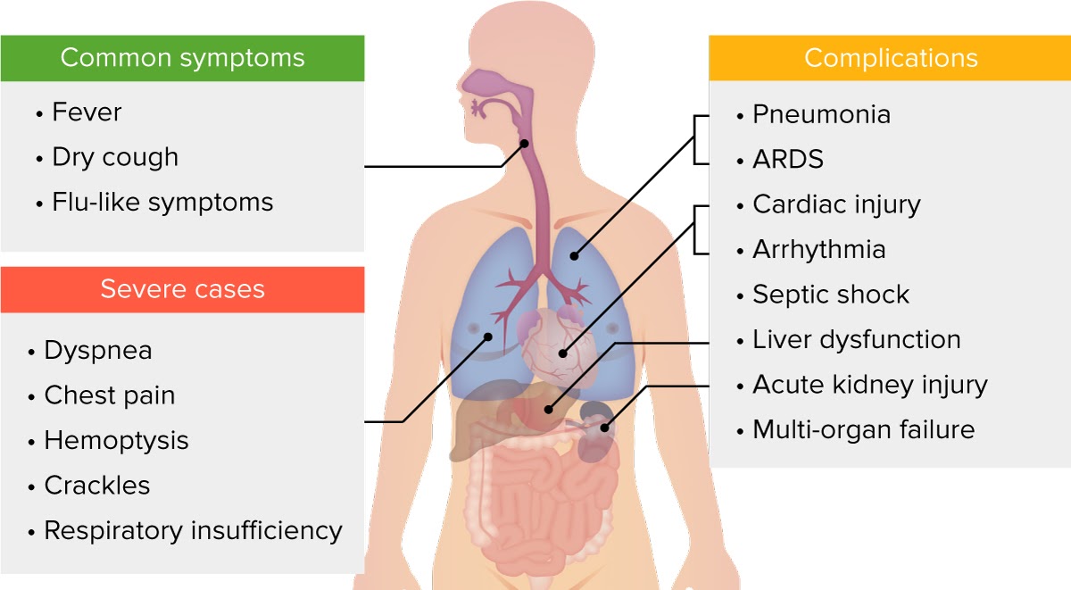
Infection by severe acute respiratory syndrome coronavirus 2 (SARS-CoV-2), now classified as coronavirus disease 2019 (COVID-19), was first identified in December 2019 in Wuhan, China, and has now spread to all continents. It was assigned pandemic status by the World Health Organization in March 2020.  An estimated 80% of cases have been mild or asymptomatic, while the remaining 20% of cases (mostly in older adults and those with comorbidities) develop severe and critical presentations that require hospitalization and are associated with a myriad of complications. Although COVID-19 is a respiratory disease, clinical reports suggest that severe cases reflect a confluence of vascular dysfunction, thrombosis, and dysregulated inflammation. The most common complications include pneumonia, respiratory failure and acute respiratory distress syndrome (ARDS), sepsis and septic shock, cardiomyopathy, acute kidney injury (AKI), and pulmonary thromboembolism. Other complications include acute stroke, arrhythmias, acute cardiac injury, and dermatologic manifestations.

Updated January 2023

Chest radiography demonstrating bilateral hilar opacities

Chest X-ray showing bilateral patchy infiltrates suggestive of ARDS

Image: “Chest radiography demonstrating bilateral hilar opacities” by Ologun G O, Ridley D, Chea N D, et al. (September 08, 2017). License: CC BY 4.0

Table of Contents

Share this page:

Risk Factors and Disease Progression

Anyone can be infected with SARS-CoV-2 and develop symptoms of COVID-19. However, certain population groups are at higher risk of progressing to severe or critical infection, complications, or death. Risk factors (from highest to lowest risk) include the following:

  • Age > 65 years 
    • The mortality rate for patients < 65 is < 3%. However, this rises to 3%–11% for individuals aged 65–84 and 10%–27% for individuals ≥ 85 years of age.
  • Chronic diseases
    • Chronic lung disease 
    • Cardiovascular disease
    • Immunosuppression 
    • Severe obesity (body mass index > 40)
    • Diabetes mellitus, chronic kidney disease requiring dialysis, cerebrovascular disease, and liver disease
  • Pregnancy 
    • Higher risk of severe illness, but same risk of infection as non-pregnant patients 

Symptoms appear an average of 5 days after infection. Initial presentation can vary greatly, although COVID-19 usually presents as a respiratory syndrome. Common signs and symptoms include fever, fatigue, dry cough, anosmia (loss of smell), dysgeusia (loss of taste), anorexia, congestion, headache, myalgia, and gastrointestinal upset.

Explanation mark green

Clinical deterioration and/or the development of complications mostly occurs in the second week of illness and is usually marked by the appearance and worsening of dyspnea.

Although COVID-19 is a respiratory disease, emerging data and clinical reports increasingly suggest that severe cases reflect a confluence of vascular dysfunction, thrombosis, and dysregulated inflammation. The development of complications and organ damage may be due not only to direct organ damage caused by the viral infection and local inflammation but also by indirect pathogenic mechanisms, including widespread endothelial damage (endothelialitis) with microangiopathy involving the vascular beds of the lungs, heart, kidneys, liver, and intestines; thrombosis and disseminated intravascular coagulation; an atypical inflammatory response; and autoimmune phenomena such as Guillain-Barré syndrome and pediatric inflammatory multisystem syndrome, an inflammatory state with clinical features similar to those of Kawasaki disease and toxic shock syndrome.

Explanation mark green

The most common complications include pneumonia, respiratory failure and acute respiratory distress syndrome (ARDS), sepsis and septic shock, cardiomyopathy, acute kidney injury (AKI), and pulmonary thromboembolism.

Other complications include

  • Acute stroke
  • Cardiac arrhythmias
  • Acute cardiac injury
  • Various dermatologic manifestations:
    • Papulovesicular eruption involving mainly the trunk associated with active viremia
    • Chilblain-type lesions of the fingers and toes (“COVID toes”) which last 3 or 4 weeks, most likely due to an immune response

Survivors of severe infections are likely to be at high risk for pulmonary fibrosis, and therefore antifibrotic therapies may be beneficial both in the acute phase of the illness and in preventing long-term complications.

Lecturio resources

Pneumonia

Viral/interstitial pneumonia

  • Not a complication, but the most frequent manifestation of severe infection
  • Characterized in most cases by high fever, dry or productive cough, chest pain, and moderate to severe dyspnea
    • Dyspnea develops approximately 5 days after the onset of symptoms, and hospital admission usually occurs after 7 days of symptoms. 
  • Laboratory findings: lymphocytopenia and elevated C-reactive protein
  • Imaging: 
    • Bilateral ground-glass opacities that can progress to solid white consolidation
    • “Crazy paving” pattern (ground-glass opacities with superimposed interlobular and intralobular septal thickening)
    • Lesions have a bilateral, peripheral, and lower lung zone distribution.

Lecturio resources

Acute Respiratory Failure and ARDS

ARDS is characterized by the following:

  1. Acute hypoxemic respiratory failure
  2. Presents within 1 week of worsening respiratory symptoms
  3. Bilateral opacities on chest X-ray, computed tomography, or ultrasound that are not fully explained by effusions, lobar or lung collapse, or nodules
  4. Cardiac failure ruled out

The histologic manifestation of ARDS is diffuse alveolar damage, which may occur either due to an injury to the airway (alveolar component) or the endothelium (hematologic component) of the alveolar membrane. Interstitial pneumonitis is typically seen in the early stages, and ARDS commonly follows in severe cases. The SARS-CoV-2 virus is unusual because it attacks cells with angiotensin-converting enzyme 2 (ACE2) receptors on both sides of the alveolar membrane and thereby provokes an intense inflammatory response, with subsequent accumulation of intra-alveolar fluid and formation of hyaline membranes. 

The vascular pathology of ARDS in COVID-19 patients is dramatic, as seen in one autopsy study which found that the lungs of a patient with COVID-19 had 9 times as many clots as those who died of the H1N1 flu. Pathological studies have shown that the main pathological features in the lungs of patients who died from COVID-19 include extensive impairment of type I alveolar epithelial cells and atypical hyperplasia of type II alveolar cells. ACE2 has a direct protective role in alveolar epithelial cells and its loss by viral infection leads to alveolar cell damage and an increase in angiotensin II levels, which triggers the IL-6 amplifier inflammatory pathway and release of cytokines similar to a cytokine storm. Reports also show the formation of hyaline membranes, focal hemorrhages, exudation and pulmonary edema, pulmonary consolidation, and the direct binding of macrophages to the S protein of SARS-CoV-2.  

Resolution of the injury is impeded by a marked degree of epithelial necrosis and inflammatory damage and probably accounts for the reportedly more compliant ARDS lungs and severe (and often “silent”) hypoxemia seen in the early stages of COVID-19 cases compared with the classic ARDS lung. The virus also gains wide access to the systemic circulation, leading to the infection of endothelial cells throughout the body and the infection of many other organs with ACE2 receptors. The virus has also been found in T lymphocytes, macrophages, and monocyte-derived dendritic cells. Direct viral destruction of lymphocytes could contribute to the observed lymphopenia in patients. Viral infection in immune cells, such as monocytes and macrophages, can result in aberrant cytokine production, even if viral infection is not productive. Extension of the pathologic process in the lung results in progressive hypoxemia and respiratory failure.

Explanation mark green

Acute respiratory failure is the leading cause of mortality in patients with COVID-19.

Respiratory failure from ARDS is the most common finding in critical COVID-19 cases.

  • Develops in 15%–30% of total cases and 60%–80% of those requiring intensive care unit (ICU) care
  • Progresses quickly after the onset of dyspnea, with a median time to intubation of 8.5 days after symptom onset
  • Mortality rate of 25%–50% (higher in patients who receive mechanical ventilation)
  • Respiratory rate and oxygen saturation (SpO2) are important parameters for patient assessment and allow for early recognition of ARDS. The following conditions indicate severe disease:
    • Respiratory rate ≥ 30 bpm
    • SpO2 ≤ 92%
    • PaO2/FiO2 ≤ 300 mmHg
  • Imaging and laboratory findings are similar to those in COVID-19 pneumonia.

Breathing support is very important in treating any COVID-19 respiratory complication. Key elements include oxygen therapy by nasal cannula, high-flow nasal oxygen, prone mechanical ventilation, and extracorporeal membrane oxygenation for rescue. Most guidelines recommend early intubation and mechanical ventilation. Survivors of severe infections are likely to be at high risk for pulmonary fibrosis, and antifibrotic therapies may therefore be beneficial both in the acute phase of the illness and in preventing long-term complications.

Explanation mark green

Non-invasive ventilation (CPAP and BiPAP) are aerosol-generating procedures and should only be used when appropriate measures of infection prevention and control are in place.

Lecturio resources

Sepsis and Septic Shock

Sepsis is defined as a life-threatening organ dysfunction caused by a dysregulated host response to infection. Septic shock is the presence of sepsis with circulatory, cellular, and metabolic abnormalities that significantly increase mortality. The two main characteristics of septic shock are persisting hypotension requiring vasopressors to maintain MAP ≥ 65 mm Hg and a serum lactate level > 2 mmol/L (18 mg/dL) despite adequate volume resuscitation. Organ dysfunction is assessed via the Sequential Organ Failure Assessment (SOFA) score (see table below). The SOFA score cannot diagnose sepsis or guide treatment. It is used to help to identify patients who have a high risk of death from infection. If a patient is on vasopressors and has a lactate level > 2 mmol/L despite adequate fluid resuscitation, the predicted mortality is 40%, if the SOFA score is ≥ 2.

Sequential Organ Failure Assessment score (SOFA score)

Organ or System Parameter 0 +1 +2 +3 +4
Repiration System PaO2/FiO2 (mmHg) ≥ 400 300–399 200–299 100–199 + mechanically ventilated < 100 + mechanically ventilated
Nervous system Glasgow Coma Scale 15 13–14 10–12 6–9 < 6
Cardiovascular System Mean arterial pressure (MAP) OR need for vasopressors MAP ≥ 70 mmHG MAP < 70 mmHG Dopamine
≤ 5 μg/kg/min,
or dobutamine (any dose)
Dopamine
> 5 μg/kg/min,
or
epinephrine
≤ 0.1 μg/kg/min, or norepinephrine ≤ 0.1 μg/kg/min
Dopamine
> 15 μg/kg/min,
or
epinephrine
> 0.1 μg/kg/min, or
norepinephrine > 0.1 μg/kg/min
Liver Bilirubin (mg/dL) < 1.2 1.2–1.9 2–5.9 6–11.9 > 12
Coagulation Platelets x 103/μl ≥ 150 100–149 50–99 20–49 < 20
Kidney Creatinine (mg/dL) or urine output < 1.2 1.2–1.9 2–3.4 3.5–4.9
or
< 500 mL/day
> 5
or
< 200 mL/day
Organ/System SOFA Score Indication
Repiration system PaO2/FiO2 (mmHg) 0 ≥ 400
+1 300–399
+2 200–299
+3 100–199 + mechanically ventilated
+4 < 100 + mechanically ventilated
Nervous system Glascow Coma Scale 0 15
+1 13–14
+2 10–12
+3 6–9
+4 < 6
Cardiovascular system Mean arterial pressure (MAP) OR need for vasopressors 0 MAP ≥ 70 mmHg
+1 MAP < 70 mmHg
+2 Dopamine ≤ 5 µg/kg/min or dobutamine (any dose)
+3 Dopamine > 5 µg/kg/min or epinephrine ≤ 0.1 µg/kg/min or norepinephrine ≤ 0.1 µg/kg/min
+4 Dopamine > 15 µg/kg/min or epinephrine > 0.1 µg/kg/min or norepinephrine > 0.1 μg/kg/min
Liver Bilirubin (mg/dL) 0 < 1.2
+1 1.2–1.9
+2 2–5.9
+3 6–1.9
+4 ≥ 12
Coagulation Platelets × 103/µl 0 ≥ 150
+1 100–149
+2 50–99
+3 20–49
+4 < 20
Kidneys Creatinine (mg/dL) or urine output 0 < 1.2
+1 1.2–1.9
+2 2–3.4
+3 3.4–4.9 or < 500 mL/day
+4 > 5 or < 200 mL/day

In COVID-19 patients, sepsis and septic shock are thought to result from the cytokine storm that develops in addition to organ damage caused by direct ACE2 attachment and/or progressive hypoxemia.

  • Septic shock is reported in 4%–8% of cases (less common than pneumonia or ARDS)
  • Can quickly deteriorate to multi-organ failure and death
  • Aside from the signs and symptoms normally associated with COVID-19, sepsis and septic shock can present with:
    • Persistently high fever, chills, and diaphoresis OR hypothermia
    • Altered mental status in cases of central nervous system impairment
    • Hypotension in cases of hemodynamic failure
    • Petechiae and purpura in cases of coagulopathy
    • Jaundice in cases of liver failure
    • Oliguria or anuria in cases of renal failure

For the latest step-by-step management guidelines, see the “WHO interim guidance on clinical management of COVID-19” and the “Surviving Sepsis Campaign: International Guidelines for Management of Sepsis and Septic Shock: 2016.”

Lecturio resources

Cardiac Complications

Patients with COVID-19 commonly present with signs of cardiac disease. This seems to be a late complication, at times developing after the respiratory illness improves. The range of cardiac complications is extensive and includes myocarditis, acute myocardial injury, arrhythmias, heart failure, and sudden death. 

The exact cause of COVID-19–related cardiac complications is still under investigation and can vary from case to case. Possible etiologies include direct myocardial and pericardial damage due to ACE2 attachment, ischemic or stress-induced damage due to persistent hypoxemia, inflammation due to cytokine storm, and sequelae of organ dysfunction induced by septic shock.

Explanation mark green

Cardiac complications are also more frequent in severe and critical COVID-19 cases as these patients are usually older, have preexisting cardiovascular diseases, and take ACE inhibitors or angiotensin II receptor blockers, but there is no evidence that stopping ACE inhibitors or ARBs reduces the severity of COVID-19, so they should NOT be discontinued. 

The exact incidence and mortality rate of COVID-19-related cardiac complications are unknown.

Myocardial injury (indicated by elevated cardiac troponin levels) is present in 10%–30% of hospitalized COVID-19 patients, and natriuretic peptide (BNP) is often elevated and associated with increased mortality.
Not all patients with abnormal cardiac tests are symptomatic.

The specific cause of myocardial injury is often not identified, but may be: myocarditis (clinical but not usually histological), hypoxia, stress (takotsubo) cardiomyopathy, microvascular damage, myocardial infarction (mostly type 2, due to oxygen supply/demand mismatch, but also type 1, due to c.a. occlusion), arrhythmias, cor pulmonale, and systemic inflammatory response syndrome (cytokine storm).

COVID-19 may also precipitate heart failure in patients who have preexisting heart disease.

If a cardiac complication is suspected, additional laboratory tests should be performed. Cardiac enzymes (troponin and NT-proBNP) are usually elevated, but negative troponin cannot exclude cardiac disease. Electrocardiogram (ECG) abnormalities vary greatly and can include ST elevation, PR depression, new-onset bundle branch block, QT prolongation, pseudoinfarct pattern, premature beats, and bradyarrhythmia with atrioventricular block.

For further information, see “Recognizing COVID-19-related myocarditis: The possible pathophysiology and proposed guideline for diagnosis and management.”

Lecturio resources

Thrombotic Complications

Many patients with COVID-19 develop abnormal coagulation profiles along with venous and arterial thrombosis of the large and small vessels. Thromboembolic complications, including pulmonary embolism and acute stroke, have also been reported. Up to 40% of deaths from COVID-19 are related to cardiovascular complications. These findings suggest a hypercoagulable state called COVID-19–associated coagulopathy (CAC).

Explanation mark green

Pulmonary embolism and deep vein thrombosis have been reported in 20%–30% of severe cases in the ICU.

The pathogenesis of CAC is still unclear but may be due to hypoxia and systemic inflammation secondary to COVID-19, which leads to the release of proinflammatory cytokines and activation of the coagulation pathway. It is also theorized that the SARS-CoV-2 virus can cause direct vascular damage by targeting and spreading through endothelial cells in the blood vessels. Endothelial injury and dysfunction would not only hinder the physiological inhibition of clot formation but also provide the first factor of Virchow’s triad.

The three factors of thrombosis, or Virchow’s triad, can be seen in cases of severe COVID-19 infection:

  • Endothelial injury through cytokine storm and direct endothelial invasion by SARS-CoV-2
  • Stasis due to immobilization during hospitalization, especially in the ICU
  • Hypercoagulable state through a rise in circulating prothrombotic factors

Vascular damage may explain why individuals with chronic conditions (e.g., hypertension, diabetes, or cardiovascular disease) are at a higher risk for severe complications from what is presumed to be a simple respiratory illness. It could also explain why ventilation alone has not been an effective treatment for many patients, as well as why such a vast variety of organs can be affected by the virus.

Explanation mark green

Endothelial damage and coagulopathy may be behind the significant organ damage that can occur in some cases of COVID-19.

Laboratory abnormalities commonly observed in patients with COVID-19–associated coagulopathy include thrombocytopenia, increased D-dimer and fibrinogen levels, and prolonged prothrombin time. Elevated D-dimer levels, high ferritin levels, and low platelet counts are strongly associated with a greater risk of death.

Lecturio resources

Other Types of Organ Damage

Abnormal liver function has been reported in approximately 15% of COVID-19 patients. Cases with acute liver injury and liver failure have presented with high levels of aspartate aminotransferase, alanine aminotransferase, and total bilirubin, and lower levels of serum albumin. Liver dysfunction is associated with a significant increase in the severity of COVID-19 infection.

Acute kidney injury has been reported in 3%–36% of hospitalized patients with COVID-19. Approximately 15% of hospitalized patients with critical infection require renal replacement therapy. Kidney dysfunction is associated with a higher risk of mortality. The pathogenesis is still unknown, although it is hypothesized to be due to cytokine storm, hypoxemic hemodynamic changes, and/or thrombotic events.

AKI can present with hematuria or proteinuria and altered kidney injury markers, urine microscopy, quantified urine protein, urine output, and urine electrolytes. Risk factors include age ≥ 65 years, black ethnicity, history of AKI, chronic kidney disease, cardiovascular disease, hypertension, heart failure, liver disease, and diabetes.

Neurologic symptoms and complications have also been reported in up to 55% of patients with critical COVID-19 infections. These include altered mental status, delirium, encephalopathy, acute cerebrovascular disease or stroke, ataxia, seizures, corticospinal tract signs, meningitis, encephalitis, and Guillain-Barré syndrome. Using diffusor tensor and 3D high-resolution MRI, microstructural changes have been reported in the brain of COVID-19 patients. Significantly higher bilateral gray matter volumes were seen in areas of the brain that correlate with the COVID-19 symptoms of smell loss, memory loss, and LDH elevation. This suggests that there may be long-term neurological consequences of SARS-CoV-2-infection.

Dermatologic complications, including rashes and other lesions, have been identified in 5%–20% of patients in recent reports. These lesions include maculopapular rashes, urticarial and vesicular lesions, petechiae/purpura, chilblains, livedo reticularis, and distal ischemia or necrosis. The pathogenesis varies depending on the lesion and includes direct viral infection, immunologic reaction, and thromboembolism. 

Ocular complications (acute conjunctivitis) have been reported in 0.9% of infected patients.

Lecturio resources

COVID-19–Associated Complications in Children

The clinical presentation and severity of cases of COVID-19 in patients < 18 years old is different from that of adults. Children have a lower risk of developing severe or critical infections, and complications appear to be milder.

  • Approximately 55% of cases are asymptomatic or mild
  • 40% of cases are moderate (pneumonia and/or abnormal chest imaging)
  • 5% of cases are severe (dyspnea and hypoxia, requiring oxygen therapy)
  • < 1% of cases are critical (ARDS, respiratory failure, shock, or multi-organ failure requiring ICU transfer)

Multisystem inflammatory syndrome in children (MIS-C) is a newly discovered complication occurring in pediatric patients. The case definition by the Royal College of Paediatrics and Child Health includes the following criteria:

  • A child presenting with persistent fever (≥ 4 days), inflammation (neutrophilia, elevated C-reactive protein, and lymphopenia) and evidence of single- or multi-organ dysfunction (shock or cardiac, respiratory, renal, gastrointestinal, or neurological disorder)
    • This may include children fulfilling full or partial criteria for Kawasaki disease.
  • Exclusion of any other microbial cause, including bacterial sepsis, staphylococcal or streptococcal shock syndromes, and infections associated with myocarditis (enterovirus)
  • SARS-CoV-2 PCR testing may be positive or negative.

Respiratory symptoms are only present in half of these patients. Abdominal symptoms such as abdominal pain, vomiting, or diarrhea are also common.

References:

  1. McIntosh K., Hirsch M.S., Bloom A. (2022) Coronavirus disease 2019 (COVID-19): Epidemiology, virology, and prevention. UpToDate. Retrieved Dec 23, 2022, from https://www.uptodate.com/contents/coronavirus-disease-2019-covid-19-epidemiology-virology-and-prevention
  2. Centers for Disease Control and Prevention. Interim Clinical Guidance for Management of Patients with Confirmed Coronavirus Disease (COVID-19) (2022) https://www.cdc.gov/coronavirus/2019-ncov/hcp/clinical-guidance-management-patients.html
  3. BMJ Best Practice. Coronavirus disease 2019 (COVID-19) (2022) https://bestpractice.bmj.com/topics/en-us/3000168/complications
  4. Ware, L. (2021) Acute respiratory distress syndrome (ARDS). BMJ Best Practice. https://bestpractice.bmj.com/topics/en-us/374
  5. WHO Headquarters (HQ). Clinical management of COVID-19 interim guidance https://apps.who.int/iris/handle/10665/332196
  6. NIH. (2021). COVID-19 Treatment Guidelines. Updated May 31, 2022. Retrieved Dec 23, 2022, from https://www.covid19treatmentguidelines.nih.gov/critical-care/general-considerations/
  7. Gibson P.G., Qin L., Puah S. (2020) COVID-19 ARDS: clinical features and differences to “usual” pre-COVID ARDS. Med J Aust. Published online 24 April 2020. Retrieved on March 11, 2020, from https://www.mja.com.au/journal/2020/covid-19-ards-clinical-features-and-differences-usual-pre-covid-ards
  8. Zick, M. (2020) Update: Can COVID-19 Cause Sepsis? Explaining the Relationship Between the Coronavirus Disease and Sepsis. Global Sepsis Alliance. Published online April 7, 2020. Retrieved Jun 10, 2020, from https://www.global-sepsis-alliance.org/news/2020/4/7/update-can-covid-19-cause-sepsis-explaining-the-relationship-between-the-coronavirus-disease-and-sepsis-cvd-novel-coronavirus
  9. Kalil A.C., Cawcutt K. (2020) Sepsis in adults. BMJ Best Practice. Updated on July 6, 2022. Retrieved on Dec 23, 2022, from https://bestpractice.bmj.com/topics/en-us/374
  10. Yuki, K., Fujiogi, M., & Koutsogiannaki, S. (2020). COVID-19 pathophysiology: A review. Clinical immunology (Orlando, Fla.), 215, 108427. https://doi.org/10.1016/j.clim.2020.108427
  11. Rhodes A., Evans L.E., Waleed A., et al. (2017) Surviving Sepsis Campaign: International Guidelines for Management of Sepsis and Septic Shock: 2016. Critical Care Medicine: March 2017, vol 45, issue 3, pp. 486-552 doi: 10.1097/CCM.0000000000002255
  12.  Siripanthong, B., Nazarian, S., Muser, D., Deo, R., Santangeli, P., Khanji, M. Y., Cooper, L. T., Jr, & Chahal, C. (2020). Recognizing COVID-19-related myocarditis: The possible pathophysiology and proposed guideline for diagnosis and management. Heart rhythm, 17(9), 1463–1471. https://doi.org/10.1016/j.hrthm.2020.05.001
  13. Molloy, Eleanor J., et al. (2022). Multisystem Inflammatory Syndrome in Children (MIS-C) and Neonates (MIS-N) Associated with COVID-19: Optimizing Definition and Management. Pediatric Research, Sept. 2022, pp. 1–10. www.nature.com, https://doi.org/10.1038/s41390-022-02263-w.
  14. Smith, D.G. (2020) Covid-19 May Be a Blood Vessel Disease, Which Explains Everything. Elemental. Published online May 29, 2020. Retrieved June 13, 2020, from https://elemental.medium.com/coronavirus-may-be-a-blood-vessel-disease-which-explains-everything-2c4032481ab2
  15. Ackermann M., Verleden S.T. Kuehnel, M., et al. Pulmonary Vascular Endothelialitis, Thrombosis, and Angiogenesis in Covid-19. N Engl J Med 2020; 383:120-128. DOI: 10.1056/NEJMoa2015432
  16. Tay M.Z., Poh C.M., Rénia L., et al. The trinity of COVID-19: immunity, inflammation and intervention. Nat Rev Immunol 20, 363–374 (2020). Published Apr 28, 2020. https://doi.org/10.1038/s41577-020-0311-8
  17. South A.M., Diz D.I., Chappell M.C. COVID-19, ACE2, and the cardiovascular consequences. Integrative Cardiovascular Physiology and Pathophysiology. Published Apr 13, 2020. https://doi.org/10.1152/ajpheart.00217.2020
  18. Joly B.S., Siguret V., & Veyradier A. Understanding pathophysiology of hemostasis disorders in critically ill patients with COVID-19. Intensive Care Med (2020). https://doi.org/10.1007/s00134-020-06088-1
  19. Becker, R.C. (2020). COVID-19 update: Covid-19-associated coagulopathy. Journal of thrombosis and thrombolysis, 50(1), 54–67. https://doi.org/10.1007/s11239-020-02134-3
  20. Lemke G., Silverman G.J. Blood clots and TAM receptor signalling in COVID-19 pathogenesis. Nat Rev Immunol (2020). https://doi.org/10.1038/s41577-020-0354-x
  21. von Weyhern C.H., Kaufmann I., Neff F., Kremer M. Early evidence of pronounced brain involvement in fatal COVID-19 outcomes.  Lancet.  Published: June 04, 2020. DOI:https://doi.org/10.1016/S0140-6736(20)31282-4
  22. Zhang, H., Penninger, J.M., Li, Y. et al. Angiotensin-converting enzyme 2 (ACE2) as a SARS-CoV-2 receptor: molecular mechanisms and potential therapeutic target. Intensive Care Med 46, 586–590 (2020). https://doi.org/10.1007/s00134-020-05985-9
  23. Li M., Li L., Zhang Y. et al. Expression of the SARS-CoV-2 cell receptor gene ACE2 in a wide variety of human tissues. Infect Dis Poverty 9, 45 (2020). https://doi.org/10.1186/s40249-020-00662-x
  24. Soy M., Keser G., Atagündüz P. et al. Cytokine storm in COVID-19: pathogenesis and overview of anti-inflammatory agents used in treatment. Clin Rheumatol 39, 2085–2094 (2020). https://doi.org/10.1007/s10067-020-05190-5
  25. Yuki K., Fujiogi M., Koutsogiannaki S. COVID-19 pathophysiology: A review. Clin Immunol. 2020;215:108427. doi:10.1016/j.clim.2020.108427
  26. Ng S.C.,  Tilg  H. COVID-19 and the gastrointestinal tract: more than meets the eye.  Gut. First published as 10.1136/gutjnl-2020-321195 on 9 April 2020. https://gut.bmj.com/content/gutjnl/69/6/973.full.pdf
  27. Xu, Zhe & Shi, Lei & Wang, Y. & Zhang, et al. Pathological findings of COVID-19 associated with acute respiratory distress syndrome. The Lancet Respiratory Medicine. Published Feb 17, 2020.  8. 10.1016/S2213-2600(20)30076-X. https://www.researchgate.net/publication/339340520_Pathological_findings_of_COVID-19_associated_with_acute_respiratory_distress_syndrome
  28. Leisman D.E., Deutschman C.S. & Legrand M. Facing COVID-19 in the ICU: vascular dysfunction, thrombosis, and dysregulated inflammation. Intensive Care Med 46, 1105–1108 (2020). https://doi.org/10.1007/s00134-020-06059-6
  29. Samavati L, Uhal BD. ACE2, Much More Than Just a Receptor for SARS-COV-2. Front. Cell. Infect. Microbiol., 05 June 2020 | https://doi.org/10.3389/fcimb.2020.00317
  30. Toshio Hirano et al. COVID-19: A New Virus, but a Familiar Receptor and Cytokine Release Syndrome, Immunity (2020). DOI: 10.1016/j.immuni.2020.04.003
  31. Gottlieb M, Long B. Dermatologic manifestations and complications of COVID-19 [published online ahead of print, 2020 Jun 6]. Am J Emerg Med. 2020;doi:10.1016/j.ajem.2020.06.011
  32. Lipsker D. Paraviral eruptions in the era of COVID-19. Do some skin manifestations point to a natural resistance to SARS-CoV-2? Clinics in Dermatology. Published June 13, 2020. https://doi.org/10.1016/j.clindermatol.2020.06.005
  33. China Medical Treatment Expert Group for Covid-19, Clinical Characteristics of Coronavirus Disease 2019 in China, DOI: 10.1056/NEJMoa2002032
  34. Masetti  C, Generali E, Colapietro F, et al.  High mortality in COVID‐19 patients with mild respiratory disease. European J Clinical Investigation.  First published:14 June 2020. https://doi.org/10.1111/eci.13314 
  35. Masetti C., Generali E., Colapietro F., et al. (2020) High mortality in COVID‐19 patients with mild respiratory disease. European Society for Clinical Investigation Journal Foundation. Vol 50, issue 9, Sept 2020, e13314. https://doi.org/10.1111/eci.13314
  36. Husain AN. The Lung. Robbins and Cotran, Pathologic Basis of Disease, 11th ed. Elsevier, 2020, chapter 15. 
  37. Wang C., Xie J., Zhao L., Fei X., Zhang H., Tan Y. et al. (2020) Alveolar macrophage dysfunction and cytokine storm in the pathogenesis of two severe COVID-19 patients. The Lancet. Vol 57, 102833, July 01, 2020. https://doi.org/10.1016/j.ebiom.2020.102833
  38. Lu Y., Li X., Geng D., Mei N., Wu P., Huang C., et al. (2020) Cerebral Micro-Structural Changes in COVID-19 Patients – An MRI-based 3-month Follow-up Study. The Lancet. Vol 25, 100484, August 01, 2020. https://doi.org/10.1016/j.eclinm.2020.100484
  39. Nalbandian, Ani, et al. (2021). Post-Acute COVID-19 Syndrome. Nature Medicine, vol. 27, no. 4, Apr. 2021, pp. 601–15.
    PubMed Central, https://doi.org/10.1038/s41591-021-01283-z.

  40. Soriano, Joan B., et al. (2022). A Clinical Case Definition of Post-COVID-19 Condition by a Delphi Consensus.
    The Lancet. Infectious Diseases, vol. 22, no. 4, Apr. 2022, pp. e102–07. PubMed Central,
    https://doi.org/10.1016/S1473-3099(21)00703-9.