Gastroesophageal reflux diseaseGastroesophageal Reflux DiseaseGastroesophageal reflux disease (GERD) occurs when the stomach acid frequently flows back into the esophagus. This backwash (acid reflux) can irritate the lining of the esophagus, causing symptoms such as retrosternal burning pain (heartburn). Gastroesophageal Reflux Disease (GERD) (GERDGERDGastroesophageal reflux disease (GERD) occurs when the stomach acid frequently flows back into the esophagus. This backwash (acid reflux) can irritate the lining of the esophagus, causing symptoms such as retrosternal burning pain (heartburn). Gastroesophageal Reflux Disease (GERD)) is due to pathologic reflux that causes symptoms and complications, including erosive esophagitisEsophagitisEsophagitis is the inflammation or irritation of the esophagus. The major types of esophagitis are medication-induced, infectious, eosinophilic, corrosive, and acid reflux. Patients typically present with odynophagia, dysphagia, and retrosternal chest pain. Esophagitis. Common symptoms are heartburnHeartburnSubsternal pain or burning sensation, usually associated with regurgitation of gastric juice into the esophagus.Gastroesophageal Reflux Disease (GERD), early satietyEarly SatietyBariatric Surgery, bloatingBloatingConstipation, and belching. Diagnosis is made clinically and requires endoscopyEndoscopyProcedures of applying endoscopes for disease diagnosis and treatment. Endoscopy involves passing an optical instrument through a small incision in the skin i.e., percutaneous; or through a natural orifice and along natural body pathways such as the digestive tract; and/or through an incision in the wall of a tubular structure or organ, i.e. Transluminal, to examine or perform surgery on the interior parts of the body.Gastroesophageal Reflux Disease (GERD) in adults over 55 who are unresponsive to empiric therapyEmpiric TherapyMeningitis in Children with acid blockade or those with alarm symptoms. Uncomplicated GERDGERDGastroesophageal reflux disease (GERD) occurs when the stomach acid frequently flows back into the esophagus. This backwash (acid reflux) can irritate the lining of the esophagus, causing symptoms such as retrosternal burning pain (heartburn). Gastroesophageal Reflux Disease (GERD) can be managed with lifestyle changes and over-the-counter medications. Complications include erosive esophagitisEsophagitisEsophagitis is the inflammation or irritation of the esophagus. The major types of esophagitis are medication-induced, infectious, eosinophilic, corrosive, and acid reflux. Patients typically present with odynophagia, dysphagia, and retrosternal chest pain. Esophagitis, strictureStricturePrimary Sclerosing Cholangitis, and Barrett esophagusEsophagusThe esophagus is a muscular tube-shaped organ of around 25 centimeters in length that connects the pharynx to the stomach. The organ extends from approximately the 6th cervical vertebra to the 11th thoracic vertebra and can be divided grossly into 3 parts: the cervical part, the thoracic part, and the abdominal part. Esophagus: Anatomy, which can increase the risk of esophageal cancerEsophageal cancerEsophageal cancer is 1 of the most common causes of cancer-related deaths worldwide. Nearly all esophageal cancers are either adenocarcinoma (commonly affecting the distal esophagus) or squamous cell carcinoma (affecting the proximal two-thirds of the esophagus). Esophageal Cancer. Management includes lifestyle and dietary modification, acid-reducing medications, and in some cases, surgery.
Gastroesophageal reflux diseaseGastroesophageal Reflux DiseaseGastroesophageal reflux disease (GERD) occurs when the stomach acid frequently flows back into the esophagus. This backwash (acid reflux) can irritate the lining of the esophagus, causing symptoms such as retrosternal burning pain (heartburn). Gastroesophageal Reflux Disease (GERD) (GERDGERDGastroesophageal reflux disease (GERD) occurs when the stomach acid frequently flows back into the esophagus. This backwash (acid reflux) can irritate the lining of the esophagus, causing symptoms such as retrosternal burning pain (heartburn). Gastroesophageal Reflux Disease (GERD)) is the passage of gastric contents into the esophagusEsophagusThe esophagus is a muscular tube-shaped organ of around 25 centimeters in length that connects the pharynx to the stomach. The organ extends from approximately the 6th cervical vertebra to the 11th thoracic vertebra and can be divided grossly into 3 parts: the cervical part, the thoracic part, and the abdominal part. Esophagus: Anatomy that leads to symptoms or complications.
Epidemiology[1,2]
PrevalencePrevalenceThe total number of cases of a given disease in a specified population at a designated time. It is differentiated from incidence, which refers to the number of new cases in the population at a given time.Measures of Disease Frequency: approximately 20% of adults in the US
More common in the Western Hemisphere
Risk factors:
Lifestyle Factors:
ObesityObesityObesity is a condition associated with excess body weight, specifically with the deposition of excessive adipose tissue. Obesity is considered a global epidemic. Major influences come from the western diet and sedentary lifestyles, but the exact mechanisms likely include a mixture of genetic and environmental factors. Obesity
SmokingSmokingWillful or deliberate act of inhaling and exhaling smoke from burning substances or agents held by hand.Interstitial Lung Diseases
Alcohol consumption
CaffeineCaffeineA methylxanthine naturally occurring in some beverages and also used as a pharmacological agent. Caffeine’s most notable pharmacological effect is as a central nervous system stimulant, increasing alertness and producing agitation. Several cellular actions of caffeine have been observed, but it is not entirely clear how each contributes to its pharmacological profile. Among the most important are inhibition of cyclic nucleotide phosphodiesterases, antagonism of adenosine receptors, and modulation of intracellular calcium handling.Stimulants intake
Physical inactivity
Eating habits (e.g., large meals, eating before bedtime)
Psychological Factors:
AnxietyAnxietyFeelings or emotions of dread, apprehension, and impending disaster but not disabling as with anxiety disorders.Generalized Anxiety Disorder
Depression
Stress
Physiological and Medical Conditions:
PregnancyPregnancyThe status during which female mammals carry their developing young (embryos or fetuses) in utero before birth, beginning from fertilization to birth.Pregnancy: Diagnosis, Physiology, and Care
SclerodermaSclerodermaScleroderma (systemic sclerosis) is an autoimmune condition characterized by diffuse collagen deposition and fibrosis. The clinical presentation varies from limited skin involvement to diffuse involvement of internal organs. Scleroderma
Zollinger-Ellison syndromeZollinger-ellison syndromeA syndrome that is characterized by the triad of severe peptic ulcer, hypersecretion of gastric acid, and gastrin-producing tumors of the pancreas or other tissue (gastrinoma). This syndrome may be sporadic or be associated with multiple endocrine neoplasia type 1.Esophagitis
Medications:
NitratesNitratesNitrates are a class of medications that cause systemic vasodilation (veins > arteries) by smooth muscle relaxation. Nitrates are primarily indicated for the treatment of angina, where preferential venodilation causes pooling of blood, decreased preload, and ultimately decreased myocardial O2 demand.Nitrates
CalciumCalciumA basic element found in nearly all tissues. It is a member of the alkaline earth family of metals with the atomic symbol ca, atomic number 20, and atomic weight 40. Calcium is the most abundant mineral in the body and combines with phosphorus to form calcium phosphate in the bones and teeth. It is essential for the normal functioning of nerves and muscles and plays a role in blood coagulation (as factor IV) and in many enzymatic processes.Electrolytes channel blockers
BenzodiazepinesBenzodiazepinesBenzodiazepines work on the gamma-aminobutyric acid type A (GABAA) receptor to produce inhibitory effects on the CNS. Benzodiazepines do not mimic GABA, the main inhibitory neurotransmitter in humans, but instead potentiate GABA activity. Benzodiazepines
Tricyclic antidepressantsTricyclic antidepressantsTricyclic antidepressants (TCAs) are a class of medications used in the management of mood disorders, primarily depression. These agents, named after their 3-ring chemical structure, act via reuptake inhibition of neurotransmitters (particularly norepinephrine and serotonin) in the brain.Tricyclic Antidepressants
A portion of the stomachStomachThe stomach is a muscular sac in the upper left portion of the abdomen that plays a critical role in digestion. The stomach develops from the foregut and connects the esophagus with the duodenum. Structurally, the stomach is C-shaped and forms a greater and lesser curvature and is divided grossly into regions: the cardia, fundus, body, and pylorus. Stomach: Anatomy protrudes through the hiatus of the diaphragmDiaphragmThe diaphragm is a large, dome-shaped muscle that separates the thoracic cavity from the abdominal cavity. The diaphragm consists of muscle fibers and a large central tendon, which is divided into right and left parts. As the primary muscle of inspiration, the diaphragm contributes 75% of the total inspiratory muscle force.Diaphragm: Anatomy into the thoracic cavity.
Can be classified as:
AxialAxialComputed Tomography (CT)herniaHerniaProtrusion of tissue, structure, or part of an organ through the bone, muscular tissue, or the membrane by which it is normally contained. Hernia may involve tissues such as the abdominal wall or the respiratory diaphragm. Hernias may be internal, external, congenital, or acquired.Abdominal Hernias (nearly 90% of all cases)
The gastroesophageal junctionGastroesophageal junctionThe area covering the terminal portion of esophagus and the beginning of stomach at the cardiac orifice.Esophagus: Anatomy is located within the diaphragmatic hiatus.
Two facets of muscle strands located centrally on the diaphragmDiaphragmThe diaphragm is a large, dome-shaped muscle that separates the thoracic cavity from the abdominal cavity. The diaphragm consists of muscle fibers and a large central tendon, which is divided into right and left parts. As the primary muscle of inspiration, the diaphragm contributes 75% of the total inspiratory muscle force.Diaphragm: Anatomy surround the esophagusEsophagusThe esophagus is a muscular tube-shaped organ of around 25 centimeters in length that connects the pharynx to the stomach. The organ extends from approximately the 6th cervical vertebra to the 11th thoracic vertebra and can be divided grossly into 3 parts: the cervical part, the thoracic part, and the abdominal part. Esophagus: Anatomy to form a unit with the esophageal sphincter.
Increased intra-abdominal pressure leads to reflux barrier formation physiologically.
The esophageal and gastric fundi should be at an acute angle to each other (about 50 degrees, the so-called angle of His) for optimal barrier function.
HerniationHerniationOmphalocele of the fundusFundusThe superior portion of the body of the stomach above the level of the cardiac notch.Stomach: Anatomy leads to decreased barrier function, which leads to GERDGERDGastroesophageal reflux disease (GERD) occurs when the stomach acid frequently flows back into the esophagus. This backwash (acid reflux) can irritate the lining of the esophagus, causing symptoms such as retrosternal burning pain (heartburn). Gastroesophageal Reflux Disease (GERD).
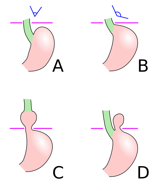
Schematic diagram of different types of hiatal hernia. A: Normal anatomy. B: Pre-stage (note the widening of the esophagogastric angle). C: Sliding hiatal hernia. D: Paraesophageal hiatal hernia
A 3–4 cm smooth muscle structure at the gastroesophageal junctionGastroesophageal junctionThe area covering the terminal portion of esophagus and the beginning of stomach at the cardiac orifice.Esophagus: Anatomy
Maintains a high-pressure zone between the esophagusEsophagusThe esophagus is a muscular tube-shaped organ of around 25 centimeters in length that connects the pharynx to the stomach. The organ extends from approximately the 6th cervical vertebra to the 11th thoracic vertebra and can be divided grossly into 3 parts: the cervical part, the thoracic part, and the abdominal part. Esophagus: Anatomy and the stomachStomachThe stomach is a muscular sac in the upper left portion of the abdomen that plays a critical role in digestion. The stomach develops from the foregut and connects the esophagus with the duodenum. Structurally, the stomach is C-shaped and forms a greater and lesser curvature and is divided grossly into regions: the cardia, fundus, body, and pylorus. Stomach: Anatomy
Relaxes transiently in response to meals
Some reflux of stomachStomachThe stomach is a muscular sac in the upper left portion of the abdomen that plays a critical role in digestion. The stomach develops from the foregut and connects the esophagus with the duodenum. Structurally, the stomach is C-shaped and forms a greater and lesser curvature and is divided grossly into regions: the cardia, fundus, body, and pylorus. Stomach: Anatomy contents is normal, but cleared by esophageal contractions.
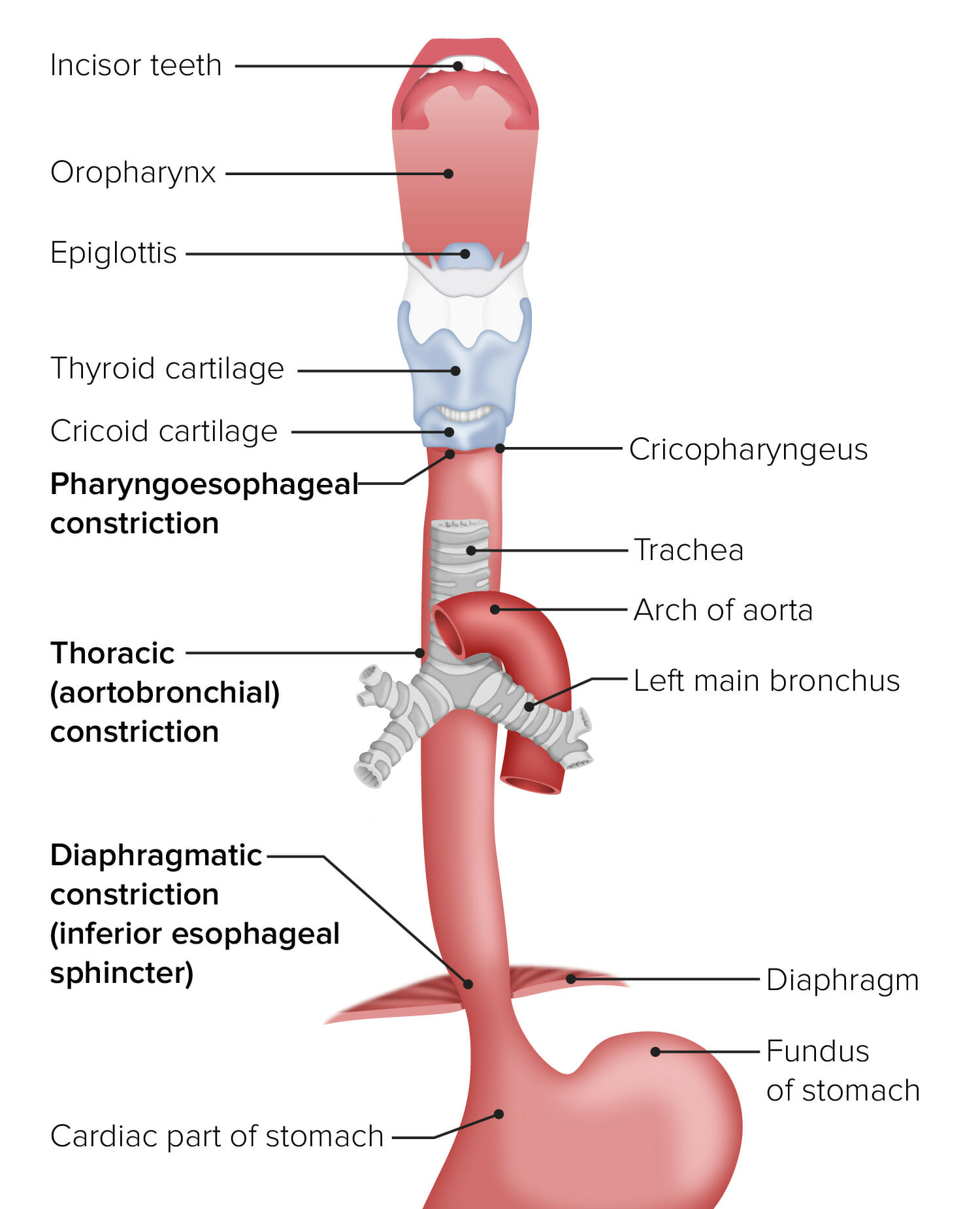
The esophagus in relation to other organs from the mouth to the stomach
Image by Lecturio.
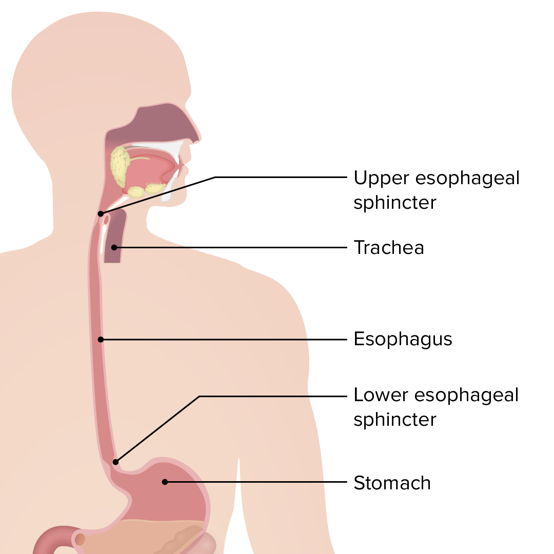
Esophagus: the upper esophageal sphincter controls the movement of food from the pharynx to the esophagus. The lower esophageal sphincter controls the movement of food from the esophagus to the stomach.
Image by Lecturio.
Pathophysiology[2,4]
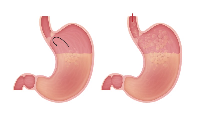
Factors leading to increased exposure of esophageal mucosaEsophageal MucosaCircular innermost layer of the esophagus wall that mediates esophageal peristalsis which pushes ingested food bolus toward the stomach.Esophagus: Anatomy to gastric acidGastric acidHydrochloric acid present in gastric juice.Gastroesophageal Reflux Disease (GERD)/contents:
Increased frequency and duration of reflux episodes
Incompetent LES (lower baseline pressure)
Increased frequency of TLESRs
Hiatal herniaHiatal herniaStomach herniation located at or near the diaphragmatic opening for the esophagus, the esophageal hiatus.Congenital Diaphragmatic Hernias (shorter and weaker LES)
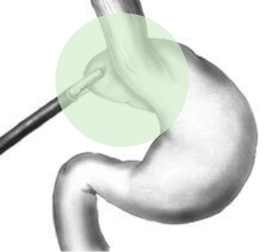
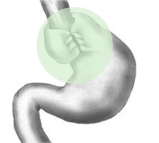
Pathophysiology of gastroesophageal reflux (GERD): Left image (normal): The LES, a structure at the gastroesophageal junction, maintains a high-pressure zone between the esophagus and the stomach. This prevents the reflux of gastric contents. The LES relaxes transiently in response to meals. Right image (GERD): An incompetent LES (lower baseline pressure) and increased frequency of TLESRs are among the factors causing GERD.
DysphagiaDysphagiaDysphagia is the subjective sensation of difficulty swallowing. Symptoms can range from a complete inability to swallow, to the sensation of solids or liquids becoming “stuck.” Dysphagia is classified as either oropharyngeal or esophageal, with esophageal dysphagia having 2 sub-types: functional and mechanical. Dysphagia/odynophagiaOdynophagiaEpiglottitis (secondary to mucosal irritation/damage)
Belching, nauseaNauseaAn unpleasant sensation in the stomach usually accompanied by the urge to vomit. Common causes are early pregnancy, sea and motion sickness, emotional stress, intense pain, food poisoning, and various enteroviruses.Antiemetics
Chest painPainAn unpleasant sensation induced by noxious stimuli which are detected by nerve endings of nociceptive neurons.Pain: Types and Pathways
Globus sensationGlobus sensationA feeling of a lump in the throat that occurs between meals in the absence of other gastrointestinal and motility disorders (e.g., dysphagia; gastroesophageal reflux).Esophagitis (“lump in the throatThroatThe pharynx is a component of the digestive system that lies posterior to the nasal cavity, oral cavity, and larynx. The pharynx can be divided into the oropharynx, nasopharynx, and laryngopharynx. Pharyngeal muscles play an integral role in vital processes such as breathing, swallowing, and speaking.Pharynx: Anatomy”)
Extraesophageal symptoms (reflux into the larynxLarynxThe larynx, also commonly called the voice box, is a cylindrical space located in the neck at the level of the C3-C6 vertebrae. The major structures forming the framework of the larynx are the thyroid cartilage, cricoid cartilage, and epiglottis. The larynx serves to produce sound (phonation), conducts air to the trachea, and prevents large molecules from reaching the lungs.Larynx: Anatomy, mouth, and respiratory tract)
LaryngitisLaryngitisLaryngitis is an inflammation of the larynx most commonly due to infection or trauma that can be either acute or chronic. In this condition, the 2 folds of mucous membranes that make up the vocal cords become inflamed and irritated. The inflammation results in a distortion of the voice produced, resulting in a hoarse sound or aphonia.Laryngitis
PharyngitisPharyngitisPharyngitis is an inflammation of the back of the throat (pharynx). Pharyngitis is usually caused by an upper respiratory tract infection, which is viral in most cases. It typically results in a sore throat and fever. Other symptoms may include a runny nose, cough, headache, and hoarseness. Pharyngitis
AsthmaAsthmaAsthma is a chronic inflammatory respiratory condition characterized by bronchial hyperresponsiveness and airflow obstruction. The disease is believed to result from the complex interaction of host and environmental factors that increase disease predisposition, with inflammation causing symptoms and structural changes. Patients typically present with wheezing, cough, and dyspnea. Asthma exacerbation
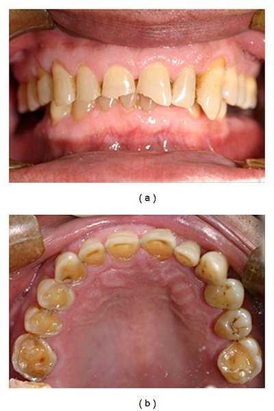
Frontal and maxillary occlusal views of severe tooth erosion caused by endogenous acid in a patient with GERD
Image: “Tooth erosion due to GERD” by Dr. A. Dickson. License: CC BY 2.5
DysphagiaDysphagiaDysphagia is the subjective sensation of difficulty swallowing. Symptoms can range from a complete inability to swallow, to the sensation of solids or liquids becoming “stuck.” Dysphagia is classified as either oropharyngeal or esophageal, with esophageal dysphagia having 2 sub-types: functional and mechanical. Dysphagia
Bleeding/anemiaAnemiaAnemia is a condition in which individuals have low Hb levels, which can arise from various causes. Anemia is accompanied by a reduced number of RBCs and may manifest with fatigue, shortness of breath, pallor, and weakness. Subtypes are classified by the size of RBCs, chronicity, and etiology. Anemia: Overview and Types
VomitingVomitingThe forcible expulsion of the contents of the stomach through the mouth.Hypokalemia
Diagnostics may vary depending on practice location. The following information is based on US recommendations for adults. For UK guidelines, refer to the National Institute for Health and Care Excellence.
Generally, once daily PPI therapy is given for 8 weeks.
If extraesophageal symptoms are present, may augment PPI trial to twice daily dosing for 8–12 weeks.
If symptoms resolve, taper to the lowest effective dose or switch to on-demand therapy.
However, this finding alone is not necessarily diagnostic. as PPI responsiveness in these cases is variableVariableVariables represent information about something that can change. The design of the measurement scales, or of the methods for obtaining information, will determine the data gathered and the characteristics of that data. As a result, a variable can be qualitative or quantitative, and may be further classified into subgroups.Types of Variables.
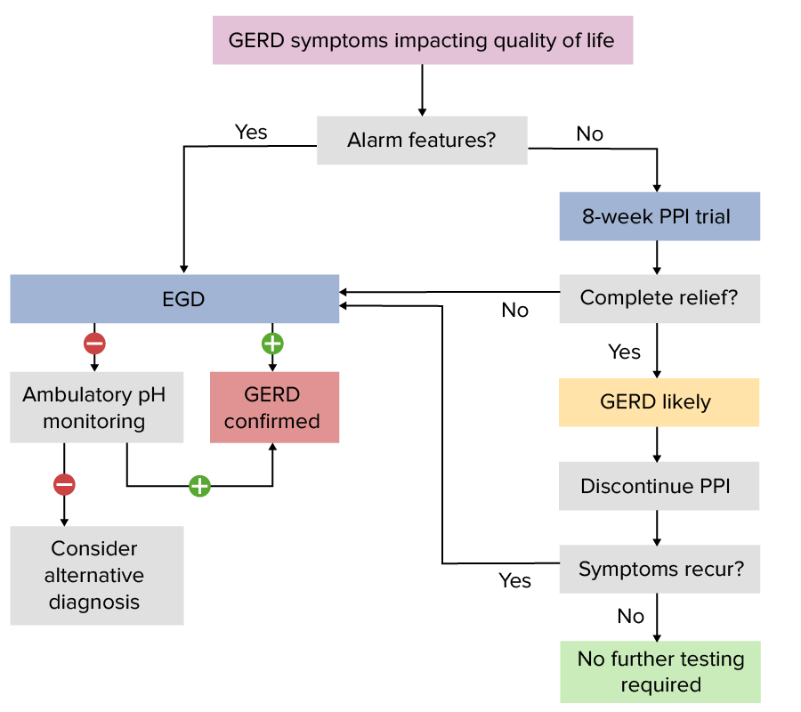
Additional workup is necessary if:
Alarm features
Extraesophageal symptoms, particularly if:
PPI trial fails
Typical GERDGERDGastroesophageal reflux disease (GERD) occurs when the stomach acid frequently flows back into the esophagus. This backwash (acid reflux) can irritate the lining of the esophagus, causing symptoms such as retrosternal burning pain (heartburn). Gastroesophageal Reflux Disease (GERD) symptoms are absent.
Incomplete resolution or recurrence of symptoms
Simplified algorithm for the evaluation of GERD based on recommendations by the American College of Gastroenterology
EGD: esophagogastroduodenoscopy; PPI: proton pump inhibitor
Image by Lecturio.
Esophagogastroduodenoscopy (EGD)[1,4,7]
Not necessary for a typical GERDGERDGastroesophageal reflux disease (GERD) occurs when the stomach acid frequently flows back into the esophagus. This backwash (acid reflux) can irritate the lining of the esophagus, causing symptoms such as retrosternal burning pain (heartburn). Gastroesophageal Reflux Disease (GERD) presentation
May be used to evaluate histology (via biopsyBiopsyRemoval and pathologic examination of specimens from the living body.Ewing Sarcoma) in conjunction with visualization
Often used to assess patientsPatientsIndividuals participating in the health care system for the purpose of receiving therapeutic, diagnostic, or preventive procedures.Clinician–Patient Relationship who have:
Incomplete resolution or recurrence of symptoms after PPI trial → should discontinue PPI for 2–4 weeks before EGD
Alarm features
Atypical symptoms
Alarm features that may warrant endoscopyEndoscopyProcedures of applying endoscopes for disease diagnosis and treatment. Endoscopy involves passing an optical instrument through a small incision in the skin i.e., percutaneous; or through a natural orifice and along natural body pathways such as the digestive tract; and/or through an incision in the wall of a tubular structure or organ, i.e. Transluminal, to examine or perform surgery on the interior parts of the body.Gastroesophageal Reflux Disease (GERD):
DysphagiaDysphagiaDysphagia is the subjective sensation of difficulty swallowing. Symptoms can range from a complete inability to swallow, to the sensation of solids or liquids becoming “stuck.” Dysphagia is classified as either oropharyngeal or esophageal, with esophageal dysphagia having 2 sub-types: functional and mechanical. Dysphagia/odynophagiaOdynophagiaEpiglottitis
Chest painPainAn unpleasant sensation induced by noxious stimuli which are detected by nerve endings of nociceptive neurons.Pain: Types and Pathways
Age > 50
AnemiaAnemiaAnemia is a condition in which individuals have low Hb levels, which can arise from various causes. Anemia is accompanied by a reduced number of RBCs and may manifest with fatigue, shortness of breath, pallor, and weakness. Subtypes are classified by the size of RBCs, chronicity, and etiology. Anemia: Overview and Types/melenaMelenaThe black, tarry, foul-smelling feces that contain degraded blood.Gastrointestinal Bleeding/hematemesisHematemesisVomiting of blood that is either fresh bright red, or older ‘coffee-ground’ in character. It generally indicates bleeding of the upper gastrointestinal tract.Mallory-Weiss Syndrome (Mallory-Weiss Tear)
Persistent vomitingVomitingThe forcible expulsion of the contents of the stomach through the mouth.Hypokalemia
Gastrointestinal cancer in 1st-degree relative
Long-standing symptoms (> 5 years), especially with at least 1 risk factor for Barrett’s esophagusEsophagusThe esophagus is a muscular tube-shaped organ of around 25 centimeters in length that connects the pharynx to the stomach. The organ extends from approximately the 6th cervical vertebra to the 11th thoracic vertebra and can be divided grossly into 3 parts: the cervical part, the thoracic part, and the abdominal part. Esophagus: Anatomy (male, age >50, white, central obesityCentral ObesityCushing Syndrome, smoker, or family historyFamily HistoryAdult Health Maintenance)
Findings:
Visual:
Upper endoscopyEndoscopyProcedures of applying endoscopes for disease diagnosis and treatment. Endoscopy involves passing an optical instrument through a small incision in the skin i.e., percutaneous; or through a natural orifice and along natural body pathways such as the digestive tract; and/or through an incision in the wall of a tubular structure or organ, i.e. Transluminal, to examine or perform surgery on the interior parts of the body.Gastroesophageal Reflux Disease (GERD) findings may be normal.
EsophagitisEsophagitisEsophagitis is the inflammation or irritation of the esophagus. The major types of esophagitis are medication-induced, infectious, eosinophilic, corrosive, and acid reflux. Patients typically present with odynophagia, dysphagia, and retrosternal chest pain. Esophagitis is common.
Strictures
Can identify areas concerning for metaplasiaMetaplasiaA condition in which there is a change of one adult cell type to another similar adult cell type.Cellular Adaptation and carcinoma
Histologic:
Histologic changes may be present in areas without visual changes, but they are not specific to GERDGERDGastroesophageal reflux disease (GERD) occurs when the stomach acid frequently flows back into the esophagus. This backwash (acid reflux) can irritate the lining of the esophagus, causing symptoms such as retrosternal burning pain (heartburn). Gastroesophageal Reflux Disease (GERD).
PenetrationPenetrationX-rays of inflammatory cells (eosinophilsEosinophilsGranular leukocytes with a nucleus that usually has two lobes connected by a slender thread of chromatin, and cytoplasm containing coarse, round granules that are uniform in size and stainable by eosin.Innate Immunity: Phagocytes and Antigen Presentation, neutrophilsNeutrophilsGranular leukocytes having a nucleus with three to five lobes connected by slender threads of chromatin, and cytoplasm containing fine inconspicuous granules and stainable by neutral dyes.Innate Immunity: Phagocytes and Antigen Presentation)
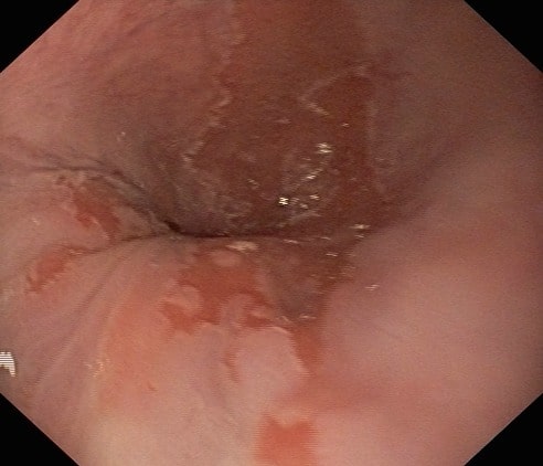
Esophagogastroduodenoscopy of a patient with persistent GERD: The image shows replacement of the squamous epithelium with columnar epithelium (Barrett esophagus).
Image: “Barretts esophagus” by US National Library of Medicine. License: CC BY 2.0
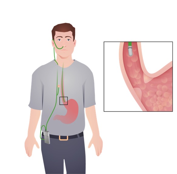
Ambulatory pHpHThe quantitative measurement of the acidity or basicity of a solution.Acid-Base Balance monitoring
24-hour ambulatory pHpHThe quantitative measurement of the acidity or basicity of a solution.Acid-Base Balance monitoring is considered the gold standard for GERDGERDGastroesophageal reflux disease (GERD) occurs when the stomach acid frequently flows back into the esophagus. This backwash (acid reflux) can irritate the lining of the esophagus, causing symptoms such as retrosternal burning pain (heartburn). Gastroesophageal Reflux Disease (GERD) diagnosis.
Used to confirm the diagnosis and check the adequacy of treatment
pHpHThe quantitative measurement of the acidity or basicity of a solution.Acid-Base Balance monitoring doneon PPI therapy to evaluate refractory GERDGERDGastroesophageal reflux disease (GERD) occurs when the stomach acid frequently flows back into the esophagus. This backwash (acid reflux) can irritate the lining of the esophagus, causing symptoms such as retrosternal burning pain (heartburn). Gastroesophageal Reflux Disease (GERD) (assessing acid suppressionSuppressionDefense Mechanisms)
Done off PPIs when establishing a de novo diagnosis of GERDGERDGastroesophageal reflux disease (GERD) occurs when the stomach acid frequently flows back into the esophagus. This backwash (acid reflux) can irritate the lining of the esophagus, causing symptoms such as retrosternal burning pain (heartburn). Gastroesophageal Reflux Disease (GERD)
Performed for 24 or 48–96 hours:
Catheter-based pHpHThe quantitative measurement of the acidity or basicity of a solution.Acid-Base Balance monitoring:
Catheter with a sensor is inserted through the noseNoseThe nose is the human body’s primary organ of smell and functions as part of the upper respiratory system. The nose may be best known for inhaling oxygen and exhaling carbon dioxide, but it also contributes to other important functions, such as tasting. The anatomy of the nose can be divided into the external nose and the nasal cavity. Nose Anatomy (External & Internal), going to the esophagusEsophagusThe esophagus is a muscular tube-shaped organ of around 25 centimeters in length that connects the pharynx to the stomach. The organ extends from approximately the 6th cervical vertebra to the 11th thoracic vertebra and can be divided grossly into 3 parts: the cervical part, the thoracic part, and the abdominal part. Esophagus: Anatomy.
Typically 24 hours
Wireless Bravo system:
Wireless capsuleCapsuleAn envelope of loose gel surrounding a bacterial cell which is associated with the virulence of pathogenic bacteria. Some capsules have a well-defined border, whereas others form a slime layer that trails off into the medium. Most capsules consist of relatively simple polysaccharides but there are some bacteria whose capsules are made of polypeptides.Bacteroides attached to esophageal lining senses the pHpHThe quantitative measurement of the acidity or basicity of a solution.Acid-Base Balance.
Can extend to 48–96 hours
Esophageal pH monitoringEsophageal ph monitoringAnalysis of the hydrogen ion concentration in the lumen of the esophagus. It is used to record the pattern, frequency, and duration of gastroesophageal reflux.Dysphagia with impedance:
Evaluates non-acidic or weakly acidic reflux
Used when there are persistent GERDGERDGastroesophageal reflux disease (GERD) occurs when the stomach acid frequently flows back into the esophagus. This backwash (acid reflux) can irritate the lining of the esophagus, causing symptoms such as retrosternal burning pain (heartburn). Gastroesophageal Reflux Disease (GERD) symptoms despite PPI therapy
Preoperative assessment before antireflux surgery
Measures the frequency of the pHpHThe quantitative measurement of the acidity or basicity of a solution.Acid-Base Balance dropping below < 4.0 (tends to coincide with symptoms)
Reliably detects:
Pathologic acid exposure
Frequency of reflux episodes
CorrelationCorrelationDetermination of whether or not two variables are correlated. This means to study whether an increase or decrease in one variable corresponds to an increase or decrease in the other variable.Causality, Validity, and Reliability of symptoms with reflux episodes
For patientsPatientsIndividuals participating in the health care system for the purpose of receiving therapeutic, diagnostic, or preventive procedures.Clinician–Patient Relationship with:
Extraesophageal symptoms
GERDGERDGastroesophageal reflux disease (GERD) occurs when the stomach acid frequently flows back into the esophagus. This backwash (acid reflux) can irritate the lining of the esophagus, causing symptoms such as retrosternal burning pain (heartburn). Gastroesophageal Reflux Disease (GERD) refractory to medications
No endoscopic findings
Ambulatory esophageal pH monitoring: This test aids in the diagnosis of GERD and evaluates the adequacy of treatment in those with persistent symptoms. In this image, the device with a pH sensor is placed transnasally, and is attached to a portable data recorder.
Contrast medium is swallowed and serial X-raysX-raysX-rays are high-energy particles of electromagnetic radiation used in the medical field for the generation of anatomical images. X-rays are projected through the body of a patient and onto a film, and this technique is called conventional or projectional radiography. X-rays are taken to delineate anatomy.
Limited use in diagnosing GERDGERDGastroesophageal reflux disease (GERD) occurs when the stomach acid frequently flows back into the esophagus. This backwash (acid reflux) can irritate the lining of the esophagus, causing symptoms such as retrosternal burning pain (heartburn). Gastroesophageal Reflux Disease (GERD) itself
Can be considered in patientsPatientsIndividuals participating in the health care system for the purpose of receiving therapeutic, diagnostic, or preventive procedures.Clinician–Patient Relationship with dysphagiaDysphagiaDysphagia is the subjective sensation of difficulty swallowing. Symptoms can range from a complete inability to swallow, to the sensation of solids or liquids becoming “stuck.” Dysphagia is classified as either oropharyngeal or esophageal, with esophageal dysphagia having 2 sub-types: functional and mechanical. Dysphagia
May show strictures, tumors, hiatal hernias, and severe esophagitisEsophagitisEsophagitis is the inflammation or irritation of the esophagus. The major types of esophagitis are medication-induced, infectious, eosinophilic, corrosive, and acid reflux. Patients typically present with odynophagia, dysphagia, and retrosternal chest pain. Esophagitis
Esophageal manometryManometryMeasurement of the pressure or tension of liquids or gases with a manometer.Achalasia[4,7]
Not recommended as a sole diagnostic tool
A pressure-sensing probeProbeA device placed on the patient’s body to visualize a targetUltrasound (Sonography) is passed into the upper GI and serial measurmements are taken.
Usually done prior to anti-reflux surgery if this surgery is considered
Management
Management may vary depending on practice location. The following information is primarily based on US recommendations for adults. For UK guidelines, see the National Institute for Health and Care Excellence. See your local recommendations for further guidance.
Elevate the head of the bed (if nocturnal symptoms).
Avoid triggers, such as:
Alcohol
CoffeeCoffeeA beverage made from ground coffee beans (seeds) infused in hot water. It generally contains caffeine and theophylline unless it is decaffeinated.Constipation
Chocolate
Carbonated beverages
Spicy foods
Tobacco cessation (smokingSmokingWillful or deliberate act of inhaling and exhaling smoke from burning substances or agents held by hand.Interstitial Lung Diseases or chewing)
Medical management of GERDGERDGastroesophageal reflux disease (GERD) occurs when the stomach acid frequently flows back into the esophagus. This backwash (acid reflux) can irritate the lining of the esophagus, causing symptoms such as retrosternal burning pain (heartburn). Gastroesophageal Reflux Disease (GERD) in adults
For patientsPatientsIndividuals participating in the health care system for the purpose of receiving therapeutic, diagnostic, or preventive procedures.Clinician–Patient Relationship with persistent symptoms (> 2 episodes per week):[1,4,7]
Presence of erosive esophagitisEsophagitisEsophagitis is the inflammation or irritation of the esophagus. The major types of esophagitis are medication-induced, infectious, eosinophilic, corrosive, and acid reflux. Patients typically present with odynophagia, dysphagia, and retrosternal chest pain. Esophagitis or Barrett esophagusEsophagusThe esophagus is a muscular tube-shaped organ of around 25 centimeters in length that connects the pharynx to the stomach. The organ extends from approximately the 6th cervical vertebra to the 11th thoracic vertebra and can be divided grossly into 3 parts: the cervical part, the thoracic part, and the abdominal part. Esophagus: Anatomy
Severe symptoms that impair qualityQualityActivities and programs intended to assure or improve the quality of care in either a defined medical setting or a program. The concept includes the assessment or evaluation of the quality of care; identification of problems or shortcomings in the delivery of care; designing activities to overcome these deficiencies; and follow-up monitoring to ensure effectiveness of corrective steps.Quality Measurement and Improvement of life
Options:
OmeprazoleOmeprazoleA 4-methoxy-3, 5-dimethylpyridyl, 5-methoxybenzimidazole derivative of timoprazole that is used in the therapy of stomach ulcers and zollinger-ellison syndrome. The drug inhibits an h(+)-k(+)-exchanging ATPase which is found in gastric parietal cells.Gastric Acid Drugs
LansoprazoleLansoprazoleA 2, 2, 2-trifluoroethoxypyridyl derivative of timoprazole that is used in the therapy of stomach ulcers and zollinger-ellison syndrome. The drug inhibits h(+)-k(+)-exchanging ATPase which is found in gastric parietal cells. Lansoprazole is a racemic mixture of (r)- and (s)-isomers.Gastric Acid Drugs
Esomeprazole
PantoprazolePantoprazole2-pyridinylmethylsulfinylbenzimidazole proton pump inhibitor that is used in the treatment of gastroesophageal reflux and peptic ulcer.Gastric Acid Drugs
Instruct patientsPatientsIndividuals participating in the health care system for the purpose of receiving therapeutic, diagnostic, or preventive procedures.Clinician–Patient Relationship to take 30–60 minutes prior to meals
If symptoms:
Resolve completely → discontinue PPI
Recur within 3 months → as needed PPI or continue long-term maintenance therapy with PPI (at lowest effective dose)
Recur after 3 months → repeat 8-week course of previously effective acid-suppressive therapy
Notes:[4]
Severe erosive esophagitisEsophagitisEsophagitis is the inflammation or irritation of the esophagus. The major types of esophagitis are medication-induced, infectious, eosinophilic, corrosive, and acid reflux. Patients typically present with odynophagia, dysphagia, and retrosternal chest pain. Esophagitis and Barrett esophagusEsophagusThe esophagus is a muscular tube-shaped organ of around 25 centimeters in length that connects the pharynx to the stomach. The organ extends from approximately the 6th cervical vertebra to the 11th thoracic vertebra and can be divided grossly into 3 parts: the cervical part, the thoracic part, and the abdominal part. Esophagus: Anatomy require long-term maintenance PPI therapy.
Routinely augmenting therapy with additional medications is not recommended.
Alternative to PPIs in patientsPatientsIndividuals participating in the health care system for the purpose of receiving therapeutic, diagnostic, or preventive procedures.Clinician–Patient Relationship with persistent symptoms:[3]
Potassium-competitive acid blocker (PCAB)
Vonoprazan
Blocks potassiumPotassiumAn element in the alkali group of metals with an atomic symbol k, atomic number 19, and atomic weight 39. 10. It is the chief cation in the intracellular fluid of muscle and other cells. Potassium ion is a strong electrolyte that plays a significant role in the regulation of fluid volume and maintenance of the water-electrolyte balance.Hyperkalemia channel of the H+-K+ ATPase pumpPumpACES and RUSH: Resuscitation Ultrasound Protocols
Used in cases where symptoms persist despite adequate trial of PPIs at maximum dose
Also an agent recommended for erosive esophagitisEsophagitisEsophagitis is the inflammation or irritation of the esophagus. The major types of esophagitis are medication-induced, infectious, eosinophilic, corrosive, and acid reflux. Patients typically present with odynophagia, dysphagia, and retrosternal chest pain. Esophagitis (LA classification B to D)
Dosing: 10 mg daily for 4 to 8 weeks
Just like PPIs, recommended to be discontinued or tapered to the lowest effective dose if there is resolution of symptoms
Expensive (FDA approved drug in the U.S. in 2023)
Table: PPI therapy for GERDGERDGastroesophageal reflux disease (GERD) occurs when the stomach acid frequently flows back into the esophagus. This backwash (acid reflux) can irritate the lining of the esophagus, causing symptoms such as retrosternal burning pain (heartburn). Gastroesophageal Reflux Disease (GERD) in adults[14]
Medication
Standard adult dose (oral)
Esomeprazole
20 mg daily
LansoprazoleLansoprazoleA 2, 2, 2-trifluoroethoxypyridyl derivative of timoprazole that is used in the therapy of stomach ulcers and zollinger-ellison syndrome. The drug inhibits h(+)-k(+)-exchanging ATPase which is found in gastric parietal cells. Lansoprazole is a racemic mixture of (r)- and (s)-isomers.Gastric Acid Drugs
30 mg daily
OmeprazoleOmeprazoleA 4-methoxy-3, 5-dimethylpyridyl, 5-methoxybenzimidazole derivative of timoprazole that is used in the therapy of stomach ulcers and zollinger-ellison syndrome. The drug inhibits an h(+)-k(+)-exchanging ATPase which is found in gastric parietal cells.Gastric Acid Drugs
20 mg daily
PantoprazolePantoprazole2-pyridinylmethylsulfinylbenzimidazole proton pump inhibitor that is used in the treatment of gastroesophageal reflux and peptic ulcer.Gastric Acid Drugs
40 mg daily
For patientsPatientsIndividuals participating in the health care system for the purpose of receiving therapeutic, diagnostic, or preventive procedures.Clinician–Patient Relationship with mild and intermittent GERDGERDGastroesophageal reflux disease (GERD) occurs when the stomach acid frequently flows back into the esophagus. This backwash (acid reflux) can irritate the lining of the esophagus, causing symptoms such as retrosternal burning pain (heartburn). Gastroesophageal Reflux Disease (GERD) symptoms (< 2 episodes per week):[1,4,7]
Antacids:
Do not treat disease → balance pHpHThe quantitative measurement of the acidity or basicity of a solution.Acid-Base Balance
Used on demand, not as maintenance therapy
May include:
MagnesiumMagnesiumA metallic element that has the atomic symbol mg, atomic number 12, and atomic weight 24. 31. It is important for the activity of many enzymes, especially those involved in oxidative phosphorylation.Electrolytes salts
Aluminum hydroxide
Calcium carbonateCalcium carbonateCarbonic acid calcium salt. An odorless, tasteless powder or crystal that occurs in nature. It is used therapeutically as a phosphate buffer in hemodialysis patients and as a calcium supplement.Hypocalcemia
SodiumSodiumA member of the alkali group of metals. It has the atomic symbol na, atomic number 11, and atomic weight 23.Hyponatremia alginate:
Derived from seaweed
Forms a viscous gel to reduce esophageal reflux
Sometimes added to antacids
Histamine-2 receptorReceptorReceptors are proteins located either on the surface of or within a cell that can bind to signaling molecules known as ligands (e.g., hormones) and cause some type of response within the cell.Receptors antagonists (H2RAs, or “H2-blockers”):
FamotidineFamotidineA competitive histamine h2-receptor antagonist. Its main pharmacodynamic effect is the inhibition of gastric secretion.Antihistamines (preferred)
NizatidineNizatidineA histamine h2 receptor antagonist with low toxicity that inhibits gastric acid secretion. The drug is used for the treatment of duodenal ulcers.Antihistamines
Consider PPI if symptoms persist
Can be used as a step-down option when de-escalating from PPI therapy
Not recommended if erosive esophagitisEsophagitisEsophagitis is the inflammation or irritation of the esophagus. The major types of esophagitis are medication-induced, infectious, eosinophilic, corrosive, and acid reflux. Patients typically present with odynophagia, dysphagia, and retrosternal chest pain. Esophagitis is present
PregnancyPregnancyThe status during which female mammals carry their developing young (embryos or fetuses) in utero before birth, beginning from fertilization to birth.Pregnancy: Diagnosis, Physiology, and Care[4]
Start with lifestyle and dietary modifications
Next, try 1st-line therapies:
Antacids
Alginates
Sucralfate 1 g 3 times daily (only recommended use for GERDGERDGastroesophageal reflux disease (GERD) occurs when the stomach acid frequently flows back into the esophagus. This backwash (acid reflux) can irritate the lining of the esophagus, causing symptoms such as retrosternal burning pain (heartburn). Gastroesophageal Reflux Disease (GERD) is during pregnancyPregnancyThe status during which female mammals carry their developing young (embryos or fetuses) in utero before birth, beginning from fertilization to birth.Pregnancy: Diagnosis, Physiology, and Care)
If failure to respond, then consider H2RAs.
PPIs are given if symptoms do not resolve with H2RA intake.
Surgical management[4,9]
Decisions regarding surgical management will generally be guided by a gastroenterologist and a surgeon.
Indications:
Presence of large hiatal herniaHiatal herniaStomach herniation located at or near the diaphragmatic opening for the esophagus, the esophageal hiatus.Congenital Diaphragmatic Hernias along with the symptoms of GERDGERDGastroesophageal reflux disease (GERD) occurs when the stomach acid frequently flows back into the esophagus. This backwash (acid reflux) can irritate the lining of the esophagus, causing symptoms such as retrosternal burning pain (heartburn). Gastroesophageal Reflux Disease (GERD)
Refractory symptoms after giving maximal medical therapy
Side effects of medications
Desire to discontinue medications
Severe erosive esophagitisEsophagitisEsophagitis is the inflammation or irritation of the esophagus. The major types of esophagitis are medication-induced, infectious, eosinophilic, corrosive, and acid reflux. Patients typically present with odynophagia, dysphagia, and retrosternal chest pain. Esophagitis (LA grade C or D)
Endoscopic therapy:
Transoral incisionless fundoplication (TIF):
With or without hiatal herniaHiatal herniaStomach herniation located at or near the diaphragmatic opening for the esophagus, the esophageal hiatus.Congenital Diaphragmatic Hernias repair
Minimally invasive
Stretta procedure (radiofrequency application to LES):
Effectiveness is variableVariableVariables represent information about something that can change. The design of the measurement scales, or of the methods for obtaining information, will determine the data gathered and the characteristics of that data. As a result, a variable can be qualitative or quantitative, and may be further classified into subgroups.Types of Variables.
Currently not recommended by the American College of Gastroenterology
Surgery:
Fundoplication (laparoscopic): gastric fundusGastric fundusThe superior portion of the body of the stomach above the level of the cardiac notch.Peptic Ulcer Disease is wrapped around the lower esophagusEsophagusThe esophagus is a muscular tube-shaped organ of around 25 centimeters in length that connects the pharynx to the stomach. The organ extends from approximately the 6th cervical vertebra to the 11th thoracic vertebra and can be divided grossly into 3 parts: the cervical part, the thoracic part, and the abdominal part. Esophagus: Anatomy
Complete (Nissen, 360 degrees)
Partial (Toupet, 270 degrees; Dor, 180 degrees)
Laparoscopic Hill gastropexy: anchors the esophagusEsophagusThe esophagus is a muscular tube-shaped organ of around 25 centimeters in length that connects the pharynx to the stomach. The organ extends from approximately the 6th cervical vertebra to the 11th thoracic vertebra and can be divided grossly into 3 parts: the cervical part, the thoracic part, and the abdominal part. Esophagus: Anatomy to the prevertebral fasciaFasciaLayers of connective tissue of variable thickness. The superficial fascia is found immediately below the skin; the deep fascia invests muscles, nerves, and other organs.Cellulitis
Magnetic sphincter augmentation (MSAMSAA syndrome complex composed of three conditions which represent clinical variants of the same disease process: striatonigral degeneration; shy-drager syndrome; and the sporadic form of olivopontocerebellar atrophies. Clinical features include autonomic, cerebellar, and basal ganglia dysfunction. Pathologic examination reveals atrophy of the basal ganglia, cerebellum, pons, and medulla, with prominent loss of autonomic neurons in the brain stem and spinal cord.Atypical Parkinsonian Syndromes):
Magnetic device is implanted around the LES.
LES closure is enhanced.
Gastric bypassGastric bypassSurgical procedure in which the stomach is transected high on the body. The resulting small proximal gastric pouch is joined to any parts of the small intestine by an end-to-side surgical anastomosis, depending on the amounts of intestinal surface being bypasses. This procedure is used frequently in the treatment of morbid obesity by limiting the size of functional stomach, food intake, and food absorption.Gastroesophageal Reflux Disease (GERD) (Roux-en-Y) if obesityObesityObesity is a condition associated with excess body weight, specifically with the deposition of excessive adipose tissue. Obesity is considered a global epidemic. Major influences come from the western diet and sedentary lifestyles, but the exact mechanisms likely include a mixture of genetic and environmental factors. Obesity (BMIBMIAn indicator of body density as determined by the relationship of body weight to body height. Bmi=weight (kg)/height squared (m2). Bmi correlates with body fat (adipose tissue). Their relationship varies with age and gender. For adults, bmi falls into these categories: below 18. 5 (underweight); 18. 5-24. 9 (normal); 25. 0-29. 9 (overweight); 30. 0 and above (obese).Obesity > 35) is present
Nissen fundoplication: Gastric fundus is wrapped around the lower esophagus, causing narrowing of the gastroesophageal junction (GEJ).
Image by James P. Gray, PD.
Nissen fundoplication: Gastric fundus is wrapped around the lower esophagus, causing narrowing of the gastroesophageal junction (GEJ).
Erosive esophagitisEsophagitisEsophagitis is the inflammation or irritation of the esophagus. The major types of esophagitis are medication-induced, infectious, eosinophilic, corrosive, and acid reflux. Patients typically present with odynophagia, dysphagia, and retrosternal chest pain. Esophagitis[1,4]
30% of patientsPatientsIndividuals participating in the health care system for the purpose of receiving therapeutic, diagnostic, or preventive procedures.Clinician–Patient Relationship with untreatedGERDGERDGastroesophageal reflux disease (GERD) occurs when the stomach acid frequently flows back into the esophagus. This backwash (acid reflux) can irritate the lining of the esophagus, causing symptoms such as retrosternal burning pain (heartburn). Gastroesophageal Reflux Disease (GERD)
Irregular or linear multiple ulcerations in the distal esophagusEsophagusThe esophagus is a muscular tube-shaped organ of around 25 centimeters in length that connects the pharynx to the stomach. The organ extends from approximately the 6th cervical vertebra to the 11th thoracic vertebra and can be divided grossly into 3 parts: the cervical part, the thoracic part, and the abdominal part. Esophagus: Anatomy
Graded based on severity (LosLOSNeisseria Angeles Classification of Gastroesophageal Reflux DiseaseGastroesophageal Reflux DiseaseGastroesophageal reflux disease (GERD) occurs when the stomach acid frequently flows back into the esophagus. This backwash (acid reflux) can irritate the lining of the esophagus, causing symptoms such as retrosternal burning pain (heartburn). Gastroesophageal Reflux Disease (GERD)):
Grade A: at least 1 mucosal break mucosal breaks < 5 mm in length
Grade B: at least 1 mucosal break > 5 mm (not continuous between adjacent mucosal folds)
Grade C: at least 1 mucosal break (continuous between mucosal folds, but not circumferential; < ¾ of circumference)
Grade D: Mucosal break involves at least ¾ of luminal circumference
Results from healing of erosive esophagitisEsophagitisEsophagitis is the inflammation or irritation of the esophagus. The major types of esophagitis are medication-induced, infectious, eosinophilic, corrosive, and acid reflux. Patients typically present with odynophagia, dysphagia, and retrosternal chest pain. Esophagitis
CollagenCollagenA polypeptide substance comprising about one third of the total protein in mammalian organisms. It is the main constituent of skin; connective tissue; and the organic substance of bones (bone and bones) and teeth (tooth).Connective Tissue: Histology deposition and contraction lead to luminal narrowing.
Causes dysphagiaDysphagiaDysphagia is the subjective sensation of difficulty swallowing. Symptoms can range from a complete inability to swallow, to the sensation of solids or liquids becoming “stuck.” Dysphagia is classified as either oropharyngeal or esophageal, with esophageal dysphagia having 2 sub-types: functional and mechanical. Dysphagia to solids/food impaction
Treated with endoscopic dilation and PPIs to prevent recurrence
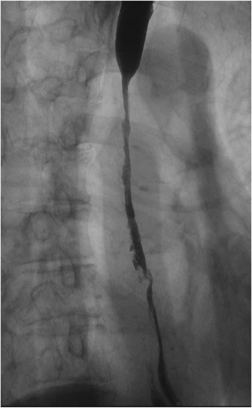
Upper gastrointestinal series: A severe stricture measuring 85 mm along the longitudinal axis was observed extending from the middle to lower thoracic esophagus caused by reflux esophagitis
Image: “Upper gastrointestinal series” by Department of Gastroenterological Surgery, Tokai University School of Medicine, 143 Shimokasuya, Isehara, Kanagawa, 259-1193, Japan. License: CC BY 4.0
Barrett esophagusEsophagusThe esophagus is a muscular tube-shaped organ of around 25 centimeters in length that connects the pharynx to the stomach. The organ extends from approximately the 6th cervical vertebra to the 11th thoracic vertebra and can be divided grossly into 3 parts: the cervical part, the thoracic part, and the abdominal part. Esophagus: Anatomy[1,2,7]
Columnar intestinal metaplasiaMetaplasiaA condition in which there is a change of one adult cell type to another similar adult cell type.Cellular Adaptation of the squamous mucosa of distal esophagusEsophagusThe esophagus is a muscular tube-shaped organ of around 25 centimeters in length that connects the pharynx to the stomach. The organ extends from approximately the 6th cervical vertebra to the 11th thoracic vertebra and can be divided grossly into 3 parts: the cervical part, the thoracic part, and the abdominal part. Esophagus: Anatomy
Diagnosed by endoscopyEndoscopyProcedures of applying endoscopes for disease diagnosis and treatment. Endoscopy involves passing an optical instrument through a small incision in the skin i.e., percutaneous; or through a natural orifice and along natural body pathways such as the digestive tract; and/or through an incision in the wall of a tubular structure or organ, i.e. Transluminal, to examine or perform surgery on the interior parts of the body.Gastroesophageal Reflux Disease (GERD) with biopsyBiopsyRemoval and pathologic examination of specimens from the living body.Ewing Sarcoma
Salmon-colored mucosa on white-light endoscopyEndoscopyProcedures of applying endoscopes for disease diagnosis and treatment. Endoscopy involves passing an optical instrument through a small incision in the skin i.e., percutaneous; or through a natural orifice and along natural body pathways such as the digestive tract; and/or through an incision in the wall of a tubular structure or organ, i.e. Transluminal, to examine or perform surgery on the interior parts of the body.Gastroesophageal Reflux Disease (GERD)
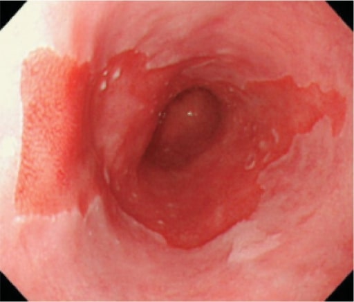
Long segment of Barrett esophagus
Image: “Long segment Barrett’s esophagus” by Japan Esophageal Society. License: CC BY 4.0
GERDGERDGastroesophageal reflux disease (GERD) occurs when the stomach acid frequently flows back into the esophagus. This backwash (acid reflux) can irritate the lining of the esophagus, causing symptoms such as retrosternal burning pain (heartburn). Gastroesophageal Reflux Disease (GERD) and/or Barrett esophagusEsophagusThe esophagus is a muscular tube-shaped organ of around 25 centimeters in length that connects the pharynx to the stomach. The organ extends from approximately the 6th cervical vertebra to the 11th thoracic vertebra and can be divided grossly into 3 parts: the cervical part, the thoracic part, and the abdominal part. Esophagus: Anatomy are well-established risk factors.
Affects the distal 3rd of the esophagusEsophagusThe esophagus is a muscular tube-shaped organ of around 25 centimeters in length that connects the pharynx to the stomach. The organ extends from approximately the 6th cervical vertebra to the 11th thoracic vertebra and can be divided grossly into 3 parts: the cervical part, the thoracic part, and the abdominal part. Esophagus: Anatomy
Appears as massMassThree-dimensional lesion that occupies a space within the breastImaging of the Breast, strictureStricturePrimary Sclerosing Cholangitis, or large ulcer on endoscopyEndoscopyProcedures of applying endoscopes for disease diagnosis and treatment. Endoscopy involves passing an optical instrument through a small incision in the skin i.e., percutaneous; or through a natural orifice and along natural body pathways such as the digestive tract; and/or through an incision in the wall of a tubular structure or organ, i.e. Transluminal, to examine or perform surgery on the interior parts of the body.Gastroesophageal Reflux Disease (GERD)
PatientsPatientsIndividuals participating in the health care system for the purpose of receiving therapeutic, diagnostic, or preventive procedures.Clinician–Patient Relationship present with dysphagiaDysphagiaDysphagia is the subjective sensation of difficulty swallowing. Symptoms can range from a complete inability to swallow, to the sensation of solids or liquids becoming “stuck.” Dysphagia is classified as either oropharyngeal or esophageal, with esophageal dysphagia having 2 sub-types: functional and mechanical. Dysphagia, weight lossWeight lossDecrease in existing body weight.Bariatric Surgery, and anemiaAnemiaAnemia is a condition in which individuals have low Hb levels, which can arise from various causes. Anemia is accompanied by a reduced number of RBCs and may manifest with fatigue, shortness of breath, pallor, and weakness. Subtypes are classified by the size of RBCs, chronicity, and etiology. Anemia: Overview and Types.
The following conditions are differential diagnoses of GERDGERDGastroesophageal reflux disease (GERD) occurs when the stomach acid frequently flows back into the esophagus. This backwash (acid reflux) can irritate the lining of the esophagus, causing symptoms such as retrosternal burning pain (heartburn). Gastroesophageal Reflux Disease (GERD)/reflux esophagitisEsophagitisEsophagitis is the inflammation or irritation of the esophagus. The major types of esophagitis are medication-induced, infectious, eosinophilic, corrosive, and acid reflux. Patients typically present with odynophagia, dysphagia, and retrosternal chest pain. Esophagitis:
Pill-induced esophagitisEsophagitisEsophagitis is the inflammation or irritation of the esophagus. The major types of esophagitis are medication-induced, infectious, eosinophilic, corrosive, and acid reflux. Patients typically present with odynophagia, dysphagia, and retrosternal chest pain. Esophagitis: also presents with retrosternal painPainAn unpleasant sensation induced by noxious stimuli which are detected by nerve endings of nociceptive neurons.Pain: Types and Pathways and dysphagiaDysphagiaDysphagia is the subjective sensation of difficulty swallowing. Symptoms can range from a complete inability to swallow, to the sensation of solids or liquids becoming “stuck.” Dysphagia is classified as either oropharyngeal or esophageal, with esophageal dysphagia having 2 sub-types: functional and mechanical. Dysphagia; however, a history of taking triggering medication and the presence of odynophagiaOdynophagiaEpiglottitis since the early stages of the disease help differentiate pill-induced esophagitisEsophagitisEsophagitis is the inflammation or irritation of the esophagus. The major types of esophagitis are medication-induced, infectious, eosinophilic, corrosive, and acid reflux. Patients typically present with odynophagia, dysphagia, and retrosternal chest pain. Esophagitis from reflux esophagitisEsophagitisEsophagitis is the inflammation or irritation of the esophagus. The major types of esophagitis are medication-induced, infectious, eosinophilic, corrosive, and acid reflux. Patients typically present with odynophagia, dysphagia, and retrosternal chest pain. Esophagitis. Frequently, symptoms resolve on discontinuation of the offending medication.
Infectious esophagitisEsophagitisEsophagitis is the inflammation or irritation of the esophagus. The major types of esophagitis are medication-induced, infectious, eosinophilic, corrosive, and acid reflux. Patients typically present with odynophagia, dysphagia, and retrosternal chest pain. Esophagitis: typically presents with retrosternal painPainAn unpleasant sensation induced by noxious stimuli which are detected by nerve endings of nociceptive neurons.Pain: Types and Pathways in immunocompromisedimmunocompromisedA human or animal whose immunologic mechanism is deficient because of an immunodeficiency disorder or other disease or as the result of the administration of immunosuppressive drugs or radiation.GastroenteritispatientsPatientsIndividuals participating in the health care system for the purpose of receiving therapeutic, diagnostic, or preventive procedures.Clinician–Patient Relationship. Decreased immunity and odynophagiaOdynophagiaEpiglottitis early in the disease help differentiate infectious esophagitisEsophagitisEsophagitis is the inflammation or irritation of the esophagus. The major types of esophagitis are medication-induced, infectious, eosinophilic, corrosive, and acid reflux. Patients typically present with odynophagia, dysphagia, and retrosternal chest pain. Esophagitis from reflux esophagitisEsophagitisEsophagitis is the inflammation or irritation of the esophagus. The major types of esophagitis are medication-induced, infectious, eosinophilic, corrosive, and acid reflux. Patients typically present with odynophagia, dysphagia, and retrosternal chest pain. Esophagitis. Diagnosis is confirmed by biopsyBiopsyRemoval and pathologic examination of specimens from the living body.Ewing Sarcoma.
Eosinophilic esophagitisEosinophilic esophagitisChronic esophagitis characterized by esophageal mucosal eosinophilia. It is diagnosed when an increase in eosinophils are present over the entire esophagus. The reflux symptoms fail to respond to proton pump inhibitors treatment, unlike in gastroesophageal reflux disease. The symptoms are associated with ige-mediated hypersensitivity to food or inhalant allergens.Esophagitis: also presents with retrosternal painPainAn unpleasant sensation induced by noxious stimuli which are detected by nerve endings of nociceptive neurons.Pain: Types and Pathways and dysphagiaDysphagiaDysphagia is the subjective sensation of difficulty swallowing. Symptoms can range from a complete inability to swallow, to the sensation of solids or liquids becoming “stuck.” Dysphagia is classified as either oropharyngeal or esophageal, with esophageal dysphagia having 2 sub-types: functional and mechanical. Dysphagia; however, the presence of concurrent atopyAtopyAtopic Dermatitis (Eczema) or asthmaAsthmaAsthma is a chronic inflammatory respiratory condition characterized by bronchial hyperresponsiveness and airflow obstruction. The disease is believed to result from the complex interaction of host and environmental factors that increase disease predisposition, with inflammation causing symptoms and structural changes. Patients typically present with wheezing, cough, and dyspnea. Asthma helps differentiate eosinophilic esophagitisEosinophilic esophagitisChronic esophagitis characterized by esophageal mucosal eosinophilia. It is diagnosed when an increase in eosinophils are present over the entire esophagus. The reflux symptoms fail to respond to proton pump inhibitors treatment, unlike in gastroesophageal reflux disease. The symptoms are associated with ige-mediated hypersensitivity to food or inhalant allergens.Esophagitis from reflux esophagitisEsophagitisEsophagitis is the inflammation or irritation of the esophagus. The major types of esophagitis are medication-induced, infectious, eosinophilic, corrosive, and acid reflux. Patients typically present with odynophagia, dysphagia, and retrosternal chest pain. Esophagitis. For confirmation, EGD with biopsyBiopsyRemoval and pathologic examination of specimens from the living body.Ewing Sarcoma is required.
Corrosive esophagitisEsophagitisEsophagitis is the inflammation or irritation of the esophagus. The major types of esophagitis are medication-induced, infectious, eosinophilic, corrosive, and acid reflux. Patients typically present with odynophagia, dysphagia, and retrosternal chest pain. Esophagitis: also presents with retrosternal painPainAn unpleasant sensation induced by noxious stimuli which are detected by nerve endings of nociceptive neurons.Pain: Types and Pathways and dysphagiaDysphagiaDysphagia is the subjective sensation of difficulty swallowing. Symptoms can range from a complete inability to swallow, to the sensation of solids or liquids becoming “stuck.” Dysphagia is classified as either oropharyngeal or esophageal, with esophageal dysphagia having 2 sub-types: functional and mechanical. Dysphagia. The disease can present with injuries to adjacent structures such as mediastinitisMediastinitisMediastinitis refers to an infection or inflammation involving the mediastinum (a region in the thoracic cavity containing the heart, thymus gland, portions of the esophagus, and trachea). Acute mediastinitis can be caused by bacterial infection due to direct contamination, hematogenous or lymphatic spread, or extension of infection from nearby structures. Mediastinitis. The history of ingesting a corrosive and the presence of odynophagiaOdynophagiaEpiglottitis differentiate corrosive esophagitisEsophagitisEsophagitis is the inflammation or irritation of the esophagus. The major types of esophagitis are medication-induced, infectious, eosinophilic, corrosive, and acid reflux. Patients typically present with odynophagia, dysphagia, and retrosternal chest pain. Esophagitis from reflux esophagitisEsophagitisEsophagitis is the inflammation or irritation of the esophagus. The major types of esophagitis are medication-induced, infectious, eosinophilic, corrosive, and acid reflux. Patients typically present with odynophagia, dysphagia, and retrosternal chest pain. Esophagitis.
Esophageal motilityEsophageal MotilityGastrointestinal Motility disorders: frequently present with dysphagiaDysphagiaDysphagia is the subjective sensation of difficulty swallowing. Symptoms can range from a complete inability to swallow, to the sensation of solids or liquids becoming “stuck.” Dysphagia is classified as either oropharyngeal or esophageal, with esophageal dysphagia having 2 sub-types: functional and mechanical. Dysphagia and chest painPainAn unpleasant sensation induced by noxious stimuli which are detected by nerve endings of nociceptive neurons.Pain: Types and Pathways. These disorders are disruptions of normal esophageal peristalsisPeristalsisA movement, caused by sequential muscle contraction, that pushes the contents of the intestines or other tubular organs in one direction.Gastrointestinal Motility. Esophageal motilityEsophageal MotilityGastrointestinal Motility disorders are diagnosed with esophageal manometryManometryMeasurement of the pressure or tension of liquids or gases with a manometer.Achalasia. The disorders may be present on their own or coexist with reflux.
Billing and Coding
Diagnosis Codes:
These codes are used to classify GERDGERDGastroesophageal reflux disease (GERD) occurs when the stomach acid frequently flows back into the esophagus. This backwash (acid reflux) can irritate the lining of the esophagus, causing symptoms such as retrosternal burning pain (heartburn). Gastroesophageal Reflux Disease (GERD), distinguishing between cases with esophagitisEsophagitisEsophagitis is the inflammation or irritation of the esophagus. The major types of esophagitis are medication-induced, infectious, eosinophilic, corrosive, and acid reflux. Patients typically present with odynophagia, dysphagia, and retrosternal chest pain. Esophagitis (inflammationInflammationInflammation is a complex set of responses to infection and injury involving leukocytes as the principal cellular mediators in the body’s defense against pathogenic organisms. Inflammation is also seen as a response to tissue injury in the process of wound healing. The 5 cardinal signs of inflammation are pain, heat, redness, swelling, and loss of function. Inflammation of the esophagusEsophagusThe esophagus is a muscular tube-shaped organ of around 25 centimeters in length that connects the pharynx to the stomach. The organ extends from approximately the 6th cervical vertebra to the 11th thoracic vertebra and can be divided grossly into 3 parts: the cervical part, the thoracic part, and the abdominal part. Esophagus: Anatomy) and those without.
Coding System
Code
Description
ICD-10-CM
K21.9
Gastro-esophageal reflux disease without esophagitisEsophagitisEsophagitis is the inflammation or irritation of the esophagus. The major types of esophagitis are medication-induced, infectious, eosinophilic, corrosive, and acid reflux. Patients typically present with odynophagia, dysphagia, and retrosternal chest pain. Esophagitis
ICD-10-CM
K21.0
Gastro-esophageal reflux disease with esophagitisEsophagitisEsophagitis is the inflammation or irritation of the esophagus. The major types of esophagitis are medication-induced, infectious, eosinophilic, corrosive, and acid reflux. Patients typically present with odynophagia, dysphagia, and retrosternal chest pain. Esophagitis
SNOMED CT
66091005
Gastroesophageal reflux diseaseGastroesophageal Reflux DiseaseGastroesophageal reflux disease (GERD) occurs when the stomach acid frequently flows back into the esophagus. This backwash (acid reflux) can irritate the lining of the esophagus, causing symptoms such as retrosternal burning pain (heartburn). Gastroesophageal Reflux Disease (GERD) (disorder)
Evaluation & Workup:
This CPT code is for an upper endoscopyEndoscopyProcedures of applying endoscopes for disease diagnosis and treatment. Endoscopy involves passing an optical instrument through a small incision in the skin i.e., percutaneous; or through a natural orifice and along natural body pathways such as the digestive tract; and/or through an incision in the wall of a tubular structure or organ, i.e. Transluminal, to examine or perform surgery on the interior parts of the body.Gastroesophageal Reflux Disease (GERD) (EGD), a procedure used in patientsPatientsIndividuals participating in the health care system for the purpose of receiving therapeutic, diagnostic, or preventive procedures.Clinician–Patient Relationship with alarm symptoms or to evaluate for complications of GERDGERDGastroesophageal reflux disease (GERD) occurs when the stomach acid frequently flows back into the esophagus. This backwash (acid reflux) can irritate the lining of the esophagus, causing symptoms such as retrosternal burning pain (heartburn). Gastroesophageal Reflux Disease (GERD) by directly visualizing the esophagusEsophagusThe esophagus is a muscular tube-shaped organ of around 25 centimeters in length that connects the pharynx to the stomach. The organ extends from approximately the 6th cervical vertebra to the 11th thoracic vertebra and can be divided grossly into 3 parts: the cervical part, the thoracic part, and the abdominal part. Esophagus: Anatomy.
Coding System
Code
Description
CPT
43235
Upper gastrointestinal endoscopyEndoscopyProcedures of applying endoscopes for disease diagnosis and treatment. Endoscopy involves passing an optical instrument through a small incision in the skin i.e., percutaneous; or through a natural orifice and along natural body pathways such as the digestive tract; and/or through an incision in the wall of a tubular structure or organ, i.e. Transluminal, to examine or perform surgery on the interior parts of the body.Gastroesophageal Reflux Disease (GERD), including esophagusEsophagusThe esophagus is a muscular tube-shaped organ of around 25 centimeters in length that connects the pharynx to the stomach. The organ extends from approximately the 6th cervical vertebra to the 11th thoracic vertebra and can be divided grossly into 3 parts: the cervical part, the thoracic part, and the abdominal part. Esophagus: Anatomy, stomachStomachThe stomach is a muscular sac in the upper left portion of the abdomen that plays a critical role in digestion. The stomach develops from the foregut and connects the esophagus with the duodenum. Structurally, the stomach is C-shaped and forms a greater and lesser curvature and is divided grossly into regions: the cardia, fundus, body, and pylorus. Stomach: Anatomy, and either the duodenumDuodenumThe shortest and widest portion of the small intestine adjacent to the pylorus of the stomach. It is named for having the length equal to about the width of 12 fingers.Small Intestine: Anatomy and/or jejunumJejunumThe middle portion of the small intestine, between duodenum and ileum. It represents about 2/5 of the remaining portion of the small intestine below duodenum.Small Intestine: Anatomy as appropriate; diagnostic
Procedures/Interventions:
This code is for a laparoscopic Nissen fundoplication, a surgical procedure to create a new valve at the bottom of the esophagusEsophagusThe esophagus is a muscular tube-shaped organ of around 25 centimeters in length that connects the pharynx to the stomach. The organ extends from approximately the 6th cervical vertebra to the 11th thoracic vertebra and can be divided grossly into 3 parts: the cervical part, the thoracic part, and the abdominal part. Esophagus: Anatomy to prevent reflux, reserved for severe or refractory GERDGERDGastroesophageal reflux disease (GERD) occurs when the stomach acid frequently flows back into the esophagus. This backwash (acid reflux) can irritate the lining of the esophagus, causing symptoms such as retrosternal burning pain (heartburn). Gastroesophageal Reflux Disease (GERD).
Coding System
Code
Description
CPT
43280
LaparoscopyLaparoscopyLaparoscopy is surgical exploration and interventions performed through small incisions with a camera and long instruments. Laparotomy and Laparoscopy, surgical, esophagogastric fundoplasty (eg, Nissen, Toupet procedures)
Medications:
These codes are for the primary medical treatments for GERDGERDGastroesophageal reflux disease (GERD) occurs when the stomach acid frequently flows back into the esophagus. This backwash (acid reflux) can irritate the lining of the esophagus, causing symptoms such as retrosternal burning pain (heartburn). Gastroesophageal Reflux Disease (GERD), including Proton PumpPumpACES and RUSH: Resuscitation Ultrasound Protocols Inhibitors (PPIs) like omeprazoleOmeprazoleA 4-methoxy-3, 5-dimethylpyridyl, 5-methoxybenzimidazole derivative of timoprazole that is used in the therapy of stomach ulcers and zollinger-ellison syndrome. The drug inhibits an h(+)-k(+)-exchanging ATPase which is found in gastric parietal cells.Gastric Acid Drugs and H2-receptor antagonists like famotidineFamotidineA competitive histamine h2-receptor antagonist. Its main pharmacodynamic effect is the inhibition of gastric secretion.Antihistamines, both of which reduce stomachStomachThe stomach is a muscular sac in the upper left portion of the abdomen that plays a critical role in digestion. The stomach develops from the foregut and connects the esophagus with the duodenum. Structurally, the stomach is C-shaped and forms a greater and lesser curvature and is divided grossly into regions: the cardia, fundus, body, and pylorus. Stomach: Anatomy acid production.
Coding System
Code
Description
RxNorm
7646
OmeprazoleOmeprazoleA 4-methoxy-3, 5-dimethylpyridyl, 5-methoxybenzimidazole derivative of timoprazole that is used in the therapy of stomach ulcers and zollinger-ellison syndrome. The drug inhibits an h(+)-k(+)-exchanging ATPase which is found in gastric parietal cells.Gastric Acid Drugs (ingredient)
RxNorm
4300
FamotidineFamotidineA competitive histamine h2-receptor antagonist. Its main pharmacodynamic effect is the inhibition of gastric secretion.Antihistamines (ingredient)
ATC
A02BC01
OmeprazoleOmeprazoleA 4-methoxy-3, 5-dimethylpyridyl, 5-methoxybenzimidazole derivative of timoprazole that is used in the therapy of stomach ulcers and zollinger-ellison syndrome. The drug inhibits an h(+)-k(+)-exchanging ATPase which is found in gastric parietal cells.Gastric Acid Drugs
Complications:
These codes document serious long-term complications of chronic, untreated GERDGERDGastroesophageal reflux disease (GERD) occurs when the stomach acid frequently flows back into the esophagus. This backwash (acid reflux) can irritate the lining of the esophagus, causing symptoms such as retrosternal burning pain (heartburn). Gastroesophageal Reflux Disease (GERD), such as the precancerousPrecancerousPathological conditions that tend eventually to become malignant.Barrett Esophagus condition Barrett’s esophagusEsophagusThe esophagus is a muscular tube-shaped organ of around 25 centimeters in length that connects the pharynx to the stomach. The organ extends from approximately the 6th cervical vertebra to the 11th thoracic vertebra and can be divided grossly into 3 parts: the cervical part, the thoracic part, and the abdominal part. Esophagus: Anatomy and the formation of an esophageal strictureStricturePrimary Sclerosing Cholangitis (narrowing).
Coding System
Code
Description
ICD-10-CM
K22.7
Barrett’s esophagusEsophagusThe esophagus is a muscular tube-shaped organ of around 25 centimeters in length that connects the pharynx to the stomach. The organ extends from approximately the 6th cervical vertebra to the 11th thoracic vertebra and can be divided grossly into 3 parts: the cervical part, the thoracic part, and the abdominal part. Esophagus: Anatomy
Katz, P. O., Dunbar, K. B., et al. (2022). ACG clinical guideline for the diagnosis and management of gastroesophageal reflux disease. American Journal of Gastroenterology, 117(1), 27–56. doi: 10.14309/ajg.0000000000001538
Yadlapati, R., Masihi, M., et al. (2021). Ambulatory reflux monitoring guides proton pump inhibitor discontinuation in patients with gastroesophageal reflux symptoms: a clinical trial. Gastroenterology, 160(1), 174.e1–182.e1. doi: 10.1053/j.gastro.2020.09.013
Mehta, R. S., Song, M., Staller, K., Chan, A. T. (2020). Association between beverage intake and incidence of gastroesophageal reflux symptoms. Clinical Gastroenterology and Hepatology, 18(10), 2226.e4–2233.e4. doi: 10.1016/j.cgh.2019.11.040
Katzka, D. A., Kahrilas, P. J. (2020). Advances in the diagnosis and management of gastroesophageal reflux disease. BMJ, 371, m3786. doi: 10.1136/bmj.m3786
Slater, B. J., Dirks, R. C., et al. (2021). SAGES guidelines for the surgical treatment of gastroesophageal reflux (GERD). Surgical Endoscopy, 35(9), 4903–4917. doi: 10.1007/s00464-021-08625-5
Pouchain, D., Bigard, M. A., Liard, F., Childs, M., Decaudin, A., McVey, D. (2012). Gaviscon® vs. omeprazole in symptomatic treatment of moderate gastroesophageal reflux: a direct comparative randomised trial. BMC Gastroenterology, 12, 18. doi: 10.1186/1471-230X-12-18
Wang, Y. H., Wintzell, V., Ludvigsson, J. F., Svanström, H., Pasternak, B. (2020). Association between proton pump inhibitor use and risk of fracture in children. JAMA Pediatrics, 174(6), 543–551. doi: 10.1001/jamapediatrics.2020.0007
Chang, A. B., Oppenheimer, J. J., et al. (2019). Chronic cough and gastroesophageal reflux in children: CHEST guideline and expert panel report. Chest, 156(1), 131–140. doi: 10.1016/j.chest.2019.03.035
Gyawali, C. P., Fass, R. (2018). Management of gastroesophageal reflux disease. Gastroenterology, 154(2), 302–318. doi: 10.1053/j.gastro.2017.07.049