Corneal anomalies are conditions in which the structure or function of the corneaCorneaThe transparent anterior portion of the fibrous coat of the eye consisting of five layers: stratified squamous corneal epithelium; bowman membrane; corneal stroma; descemet membrane; and mesenchymal corneal endothelium. It serves as the first refracting medium of the eye.Eye: Anatomy is impaired due to various congenital or acquired pathologies. Corneal anomalies are classified based on anomalies of size, clarity, ectatic anomalies, corneal dystrophies, and acquired conditions. Pathological changes can result in opacityOpacityImaging of the Lungs and Pleura or clouding of the corneaCorneaThe transparent anterior portion of the fibrous coat of the eye consisting of five layers: stratified squamous corneal epithelium; bowman membrane; corneal stroma; descemet membrane; and mesenchymal corneal endothelium. It serves as the first refracting medium of the eye.Eye: Anatomy and, hence, reduce visual acuityVisual AcuityClarity or sharpness of ocular vision or the ability of the eye to see fine details. Visual acuity depends on the functions of retina, neuronal transmission, and the interpretative ability of the brain. Normal visual acuity is expressed as 20/20 indicating that one can see at 20 feet what should normally be seen at that distance. Visual acuity can also be influenced by brightness, color, and contrast.Ophthalmic Exam. Corneal anomalies are usually diagnosed based on clinical findings. Management includes correction of refractive errorsRefractive errorsBy refraction, the light that enters the eye is focused onto a particular point of the retina. The main refractive components of the eye are the cornea and the lens. When the corneal curvature, the refractive power of the lens, does not match the size of the eye, ametropia or a refractive error occurs. Refractive Errors and treatment of the underlying conditions.
CorneaCorneaThe transparent anterior portion of the fibrous coat of the eye consisting of five layers: stratified squamous corneal epithelium; bowman membrane; corneal stroma; descemet membrane; and mesenchymal corneal endothelium. It serves as the first refracting medium of the eye.Eye: Anatomy
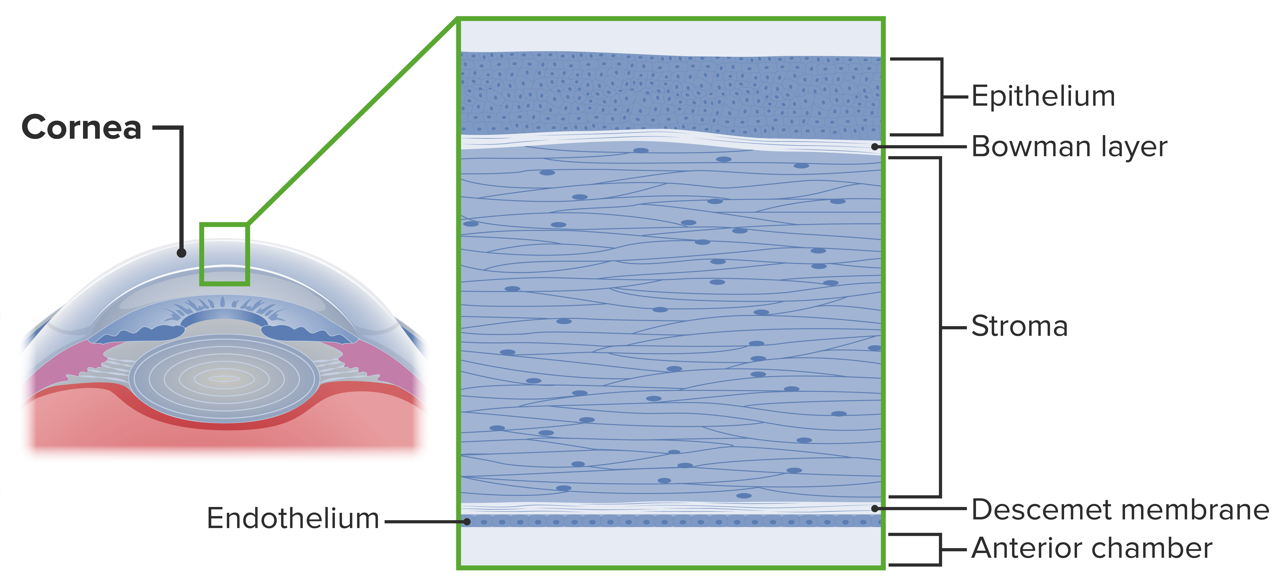
The corneaCorneaThe transparent anterior portion of the fibrous coat of the eye consisting of five layers: stratified squamous corneal epithelium; bowman membrane; corneal stroma; descemet membrane; and mesenchymal corneal endothelium. It serves as the first refracting medium of the eye.Eye: Anatomy is a transparent, avascularAvascularCorneal Abrasions, Erosion, and Ulcers, watch glass-like structure that covers the iris, anterior chamberAnterior chamberThe space in the eye, filled with aqueous humor, bounded anteriorly by the cornea and a small portion of the sclera and posteriorly by a small portion of the ciliary body, the iris, and that part of the crystalline lens which presents through the pupil.Eye: Anatomy, and pupilPupilThe pupil is the space within the eye that permits light to project onto the retina. Anatomically located in front of the lens, the pupil’s size is controlled by the surrounding iris. The pupil provides insight into the function of the central and autonomic nervous systems. Pupil: Physiology and Abnormalities.
Curvature of the corneaCorneaThe transparent anterior portion of the fibrous coat of the eye consisting of five layers: stratified squamous corneal epithelium; bowman membrane; corneal stroma; descemet membrane; and mesenchymal corneal endothelium. It serves as the first refracting medium of the eye.Eye: Anatomy is > the rest of the globe.
5 histological layers:
EpitheliumEpitheliumThe epithelium is a complex of specialized cellular organizations arranged into sheets and lining cavities and covering the surfaces of the body. The cells exhibit polarity, having an apical and a basal pole. Structures important for the epithelial integrity and function involve the basement membrane, the semipermeable sheet on which the cells rest, and interdigitations, as well as cellular junctions. Surface Epithelium: Histology
EndotheliumEndotheliumA layer of epithelium that lines the heart, blood vessels (vascular endothelium), lymph vessels (lymphatic endothelium), and the serous cavities of the body.Arteries: Histology
Classification of corneal abnormalities
Cryptophthalmos
Anomalies of size:
Microcornea
Megalocornea
Ectatic anomalies:
Keratoconus
Keratoglobus
Anomalies of clarity:
Peters anomaly
Sclerocornea
Corneal dystrophies:
Congenital stromal corneal dystrophy
Congenital endothelial corneal dystrophy
Acquired conditions:
Corneal keloids
Band-shaped keratopathy
Layers of the cornea
Image by Lecturio.
Cryptophthalmos
Cryptophthalmos is an autosomal recessiveAutosomal recessiveAutosomal inheritance, both dominant and recessive, refers to the transmission of genes from the 22 autosomal chromosomes. Autosomal recessive diseases are only expressed when 2 copies of the recessive allele are inherited.Autosomal Recessive and Autosomal Dominant Inheritance congenital anomaly associated with systemic anomalies and is characterized by an uninterrupted continuity of the skinSkinThe skin, also referred to as the integumentary system, is the largest organ of the body. The skin is primarily composed of the epidermis (outer layer) and dermis (deep layer). The epidermis is primarily composed of keratinocytes that undergo rapid turnover, while the dermis contains dense layers of connective tissue.Skin: Structure and Functions extending from the foreheadForeheadThe part of the face above the eyes.Melasma to the malar region.
Epidemiology
The incidenceIncidenceThe number of new cases of a given disease during a given period in a specified population. It also is used for the rate at which new events occur in a defined population. It is differentiated from prevalence, which refers to all cases in the population at a given time.Measures of Disease Frequency of cryptophthalmos is unknown.
May occur in isolation
Also associated with Fraser syndrome, an autosomal recessiveAutosomal recessiveAutosomal inheritance, both dominant and recessive, refers to the transmission of genes from the 22 autosomal chromosomes. Autosomal recessive diseases are only expressed when 2 copies of the recessive allele are inherited.Autosomal Recessive and Autosomal Dominant Inheritance malformation disorder presenting with:
Cryptophthalmos
SyndactylySyndactylyA congenital anomaly of the hand or foot, marked by the webbing between adjacent fingers or toes. Syndactylies are classified as complete or incomplete by the degree of joining. Syndactylies can also be simple or complex. Simple syndactyly indicates joining of only skin or soft tissue; complex syndactyly marks joining of bony elements.Development of the Limbs
Abnormalities of the respiratory and urogenital tracts
Bilateral > unilateral
Clinical presentation
Three forms:
Complete = total failure of eyelid formation
Most common type
EyelidsEyelidsEach of the upper and lower folds of skin which cover the eye when closed.Blepharitis are replaced by a layer of skinSkinThe skin, also referred to as the integumentary system, is the largest organ of the body. The skin is primarily composed of the epidermis (outer layer) and dermis (deep layer). The epidermis is primarily composed of keratinocytes that undergo rapid turnover, while the dermis contains dense layers of connective tissue.Skin: Structure and Functions extending from the foreheadForeheadThe part of the face above the eyes.Melasma to the malar region.
Absence or poor development of the eyebrow
CorneaCorneaThe transparent anterior portion of the fibrous coat of the eye consisting of five layers: stratified squamous corneal epithelium; bowman membrane; corneal stroma; descemet membrane; and mesenchymal corneal endothelium. It serves as the first refracting medium of the eye.Eye: Anatomy is absent or fused with the skinSkinThe skin, also referred to as the integumentary system, is the largest organ of the body. The skin is primarily composed of the epidermis (outer layer) and dermis (deep layer). The epidermis is primarily composed of keratinocytes that undergo rapid turnover, while the dermis contains dense layers of connective tissue.Skin: Structure and Functions.
Partial = result of the eyelid fold developing abnormally
Comprises approximately 20% of the cases
Rudimentary lids are present with a small conjunctival sac placed laterally.
The globe is small and almost completely covered by skinSkinThe skin, also referred to as the integumentary system, is the largest organ of the body. The skin is primarily composed of the epidermis (outer layer) and dermis (deep layer). The epidermis is primarily composed of keratinocytes that undergo rapid turnover, while the dermis contains dense layers of connective tissue.Skin: Structure and Functions.
Abortive = The part of the lid fold derived from the frontonasal process fails to develop, whereas the part derived from the maxillary process develops normally.
The abnormal upper lid covers and adheres to up to 75% of the upper corneaCorneaThe transparent anterior portion of the fibrous coat of the eye consisting of five layers: stratified squamous corneal epithelium; bowman membrane; corneal stroma; descemet membrane; and mesenchymal corneal endothelium. It serves as the first refracting medium of the eye.Eye: Anatomy.
Treatment is aimed at the surgical reconstruction of the eyelidsEyelidsEach of the upper and lower folds of skin which cover the eye when closed.Blepharitis to allow for visual development. There is no standard approach, and stepwise surgeries differ according to the severity of the abnormalities and orbit development.
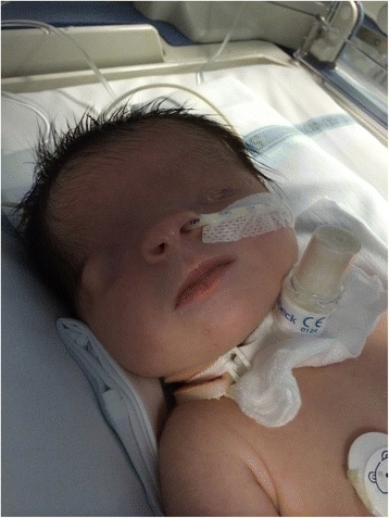
Bilateral cryptophthalmos
Image: “Bilateral cryptophthalmos with microphthalmos into left ocular globe and abnormal right ocular globe in a female infant with Fraser syndrome” by De Bernardo et al. License: CC BY 4.0
Corneal Anomalies of Size
Microcornea
The normal horizontal diameter of the corneaCorneaThe transparent anterior portion of the fibrous coat of the eye consisting of five layers: stratified squamous corneal epithelium; bowman membrane; corneal stroma; descemet membrane; and mesenchymal corneal endothelium. It serves as the first refracting medium of the eye.Eye: Anatomy at birth is 10 mm. The adult size of about 11.7 mm is normally attained by 2 years of age.
Overview:
Microcornea is characterized by a corneal diameter < 10 mm.
Can be unilateral or bilateral
Can occur as an independent anomaly
May be associated with:
Microphthalmos: abnormally small globe
Nanophthalmos: abnormally small eye with a normal-sized lensLensA transparent, biconvex structure of the eye, enclosed in a capsule and situated behind the iris and in front of the vitreous humor (vitreous body). It is slightly overlapped at its margin by the ciliary processes. Adaptation by the ciliary body is crucial for ocular accommodation.Eye: Anatomy
May present as isolated microcornea or relative anterior microphthalmos
Individuals usually develop glaucomaGlaucomaGlaucoma is an optic neuropathy characterized by typical visual field defects and optic nerve atrophy seen as optic disc cupping on examination. The acute form of glaucoma is a medical emergency. Glaucoma is often, but not always, caused by increased intraocular pressure (IOP). Glaucoma in the 4th decade of life.
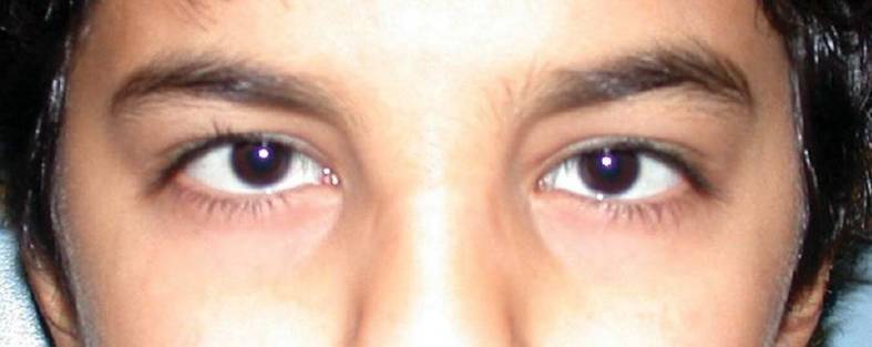
Child with microcornea (diameters < 10 mm)
Image: “At 12 years of age, the child had horizontal corneal diameters of 10 mm but otherwise unremarkable anterior segments by slit-lamp examination” by Hazin R et al. License: CC BY 2.0
Megalocornea
Megalocornea is a bilateral, nonprogressive corneal enlargement characterized by a horizontal diameter of the corneaCorneaThe transparent anterior portion of the fibrous coat of the eye consisting of five layers: stratified squamous corneal epithelium; bowman membrane; corneal stroma; descemet membrane; and mesenchymal corneal endothelium. It serves as the first refracting medium of the eye.Eye: Anatomy > 12 mm at birth and 13 mm at 2 years of age.
Overview:
Usually, a clear corneaCorneaThe transparent anterior portion of the fibrous coat of the eye consisting of five layers: stratified squamous corneal epithelium; bowman membrane; corneal stroma; descemet membrane; and mesenchymal corneal endothelium. It serves as the first refracting medium of the eye.Eye: Anatomy with normal thickness and visionVisionOphthalmic Exam
Marfan syndromeMarfan syndromeMarfan syndrome is a genetic condition with autosomal dominant inheritance. Marfan syndrome affects the elasticity of connective tissues throughout the body, most notably in the cardiovascular, ocular, and musculoskeletal systems. Marfan Syndrome
Ehlers-Danlos syndromeEhlers-Danlos syndromeEhlers-Danlos syndrome (EDS) is a heterogeneous group of inherited connective tissue disorders that are characterized by hyperextensible skin, hypermobile joints, and fragility of the skin and connective tissue. Ehlers-Danlos Syndrome
Down syndromeDown syndromeDown syndrome, or trisomy 21, is the most common chromosomal aberration and the most frequent genetic cause of developmental delay. Both boys and girls are affected and have characteristic craniofacial and musculoskeletal features, as well as multiple medical anomalies involving the cardiac, gastrointestinal, ocular, and auditory systems.Down syndrome (Trisomy 21)
Pathophysiology:
During corneal development, a defect in the formation of the optic cup allows it to overgrow.
Considered a primary overgrowth of the corneaCorneaThe transparent anterior portion of the fibrous coat of the eye consisting of five layers: stratified squamous corneal epithelium; bowman membrane; corneal stroma; descemet membrane; and mesenchymal corneal endothelium. It serves as the first refracting medium of the eye.Eye: Anatomy
Normal endothelial cell density is present.
Clinical presentation:
Presents after 12 months of age when the corneaCorneaThe transparent anterior portion of the fibrous coat of the eye consisting of five layers: stratified squamous corneal epithelium; bowman membrane; corneal stroma; descemet membrane; and mesenchymal corneal endothelium. It serves as the first refracting medium of the eye.Eye: Anatomy has fully developed
Symmetric, dome-shaped corneas > 13 mm in diameter
Reduced visual acuityVisual AcuityClarity or sharpness of ocular vision or the ability of the eye to see fine details. Visual acuity depends on the functions of retina, neuronal transmission, and the interpretative ability of the brain. Normal visual acuity is expressed as 20/20 indicating that one can see at 20 feet what should normally be seen at that distance. Visual acuity can also be influenced by brightness, color, and contrast.Ophthalmic Exam is uncommon.
AstigmatismAstigmatismUnequal curvature of the refractive surfaces of the eye. Thus a point source of light cannot be brought to a point focus on the retina but is spread over a more or less diffuse area. This results from the radius of curvature in one plane being longer or shorter than the radius at right angles to it. .Refractive Errors is common.
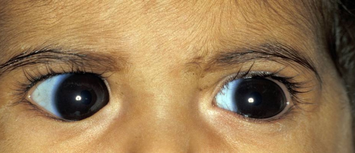
Megalocornea in a child with a corneal diameter > 14 mm
Image: “Megalocornea in a child with a corneal diameter greater than 14 mm” by Khan AO et al. License: CC BY 3.0, cropped by Lecturio.
Corneal Ectatic Anomalies
Keratoconus
Keratoconus refers to the thinning near the center of the corneaCorneaThe transparent anterior portion of the fibrous coat of the eye consisting of five layers: stratified squamous corneal epithelium; bowman membrane; corneal stroma; descemet membrane; and mesenchymal corneal endothelium. It serves as the first refracting medium of the eye.Eye: Anatomy, resulting in corneal steepening and conical shape.
Overview:
Noninflammatory
Progressive disorder
Caused by a congenital weakness of the corneaCorneaThe transparent anterior portion of the fibrous coat of the eye consisting of five layers: stratified squamous corneal epithelium; bowman membrane; corneal stroma; descemet membrane; and mesenchymal corneal endothelium. It serves as the first refracting medium of the eye.Eye: Anatomy
Presents at pubertyPubertyPuberty is a complex series of physical, psychosocial, and cognitive transitions usually experienced by adolescents (11-19 years of age). Puberty is marked by a growth in stature and the development of secondary sexual characteristics, achievement of fertility, and changes in most body systems.Puberty or in early adulthood
Usually bilateral, may often be more severe on 1 side
Difficulties in correcting visionVisionOphthalmic Exam for myopiaMyopiaRefractive Errors and astigmatismAstigmatismUnequal curvature of the refractive surfaces of the eye. Thus a point source of light cannot be brought to a point focus on the retina but is spread over a more or less diffuse area. This results from the radius of curvature in one plane being longer or shorter than the radius at right angles to it. .Refractive Errors
Findings with advanced disease:
Munson’s sign: v-shaped indentation of the lower eyelid on downgaze (see image)
Vogt striae: deep stromal stress lines that are pathognomonic for keratoconus
Acute hydropsHydropsCholecystitis: Ruptures may develop in the Descemet’s membrane causing the stroma to become suddenly edematous and opaque.
Can present with photophobiaPhotophobiaAbnormal sensitivity to light. This may occur as a manifestation of eye diseases; migraine; subarachnoid hemorrhage; meningitis; and other disorders. Photophobia may also occur in association with depression and other mental disorders.Migraine Headache and a sudden painful drop in visual acuityVisual AcuityClarity or sharpness of ocular vision or the ability of the eye to see fine details. Visual acuity depends on the functions of retina, neuronal transmission, and the interpretative ability of the brain. Normal visual acuity is expressed as 20/20 indicating that one can see at 20 feet what should normally be seen at that distance. Visual acuity can also be influenced by brightness, color, and contrast.Ophthalmic Exam
Treatments include pressure patching and bandage contact lenses.
Topical hyperosmotics, cycloplegics, and steroidsSteroidsA group of polycyclic compounds closely related biochemically to terpenes. They include cholesterol, numerous hormones, precursors of certain vitamins, bile acids, alcohols (sterols), and certain natural drugs and poisons. Steroids have a common nucleus, a fused, reduced 17-carbon atom ring system, cyclopentanoperhydrophenanthrene. Most steroids also have two methyl groups and an aliphatic side-chain attached to the nucleus.Benign Liver Tumors are used to decrease edemaEdemaEdema is a condition in which excess serous fluid accumulates in the body cavity or interstitial space of connective tissues. Edema is a symptom observed in several medical conditions. It can be categorized into 2 types, namely, peripheral (in the extremities) and internal (in an organ or body cavity). Edema, painPainAn unpleasant sensation induced by noxious stimuli which are detected by nerve endings of nociceptive neurons.Pain: Types and Pathways, and inflammationInflammationInflammation is a complex set of responses to infection and injury involving leukocytes as the principal cellular mediators in the body’s defense against pathogenic organisms. Inflammation is also seen as a response to tissue injury in the process of wound healing. The 5 cardinal signs of inflammation are pain, heat, redness, swelling, and loss of function. Inflammation.
Diagnosis:
Slit-lamp exam: CorneaCorneaThe transparent anterior portion of the fibrous coat of the eye consisting of five layers: stratified squamous corneal epithelium; bowman membrane; corneal stroma; descemet membrane; and mesenchymal corneal endothelium. It serves as the first refracting medium of the eye.Eye: Anatomy may appear normal.
Corneal collagenCollagenA polypeptide substance comprising about one third of the total protein in mammalian organisms. It is the main constituent of skin; connective tissue; and the organic substance of bones (bone and bones) and teeth (tooth).Connective Tissue: Histology crosslinking: A procedure that uses riboflavin drops, UV lightUV lightThat portion of the electromagnetic spectrum immediately below the visible range and extending into the x-ray frequencies. The longer wavelengths (near-uv or biotic or vital rays) are necessary for the endogenous synthesis of vitamin D and are also called antirachitic rays; the shorter, ionizing wavelengths (far-uv or abiotic or extravital rays) are viricidal, bactericidal, mutagenic, and carcinogenic and are used as disinfectants.Bullous Pemphigoid and Pemphigus Vulgaris, and a photosensitizer to strengthen the bonds in the corneaCorneaThe transparent anterior portion of the fibrous coat of the eye consisting of five layers: stratified squamous corneal epithelium; bowman membrane; corneal stroma; descemet membrane; and mesenchymal corneal endothelium. It serves as the first refracting medium of the eye.Eye: Anatomy and measure progression.
Glasses: to correct myopic astigmatismAstigmatismUnequal curvature of the refractive surfaces of the eye. Thus a point source of light cannot be brought to a point focus on the retina but is spread over a more or less diffuse area. This results from the radius of curvature in one plane being longer or shorter than the radius at right angles to it. .Refractive Errors as long as the patient can function
Contact lenses:
Rigid gas-permeable lenses neutralize the irregularity of the corneaCorneaThe transparent anterior portion of the fibrous coat of the eye consisting of five layers: stratified squamous corneal epithelium; bowman membrane; corneal stroma; descemet membrane; and mesenchymal corneal endothelium. It serves as the first refracting medium of the eye.Eye: Anatomy, but may become intolerable.
Scleral lenses that sit on the scleraScleraThe white, opaque, fibrous, outer tunic of the eyeball, covering it entirely excepting the segment covered anteriorly by the cornea. It is essentially avascular but contains apertures for vessels, lymphatics, and nerves.Eye: Anatomy without touching the corneaCorneaThe transparent anterior portion of the fibrous coat of the eye consisting of five layers: stratified squamous corneal epithelium; bowman membrane; corneal stroma; descemet membrane; and mesenchymal corneal endothelium. It serves as the first refracting medium of the eye.Eye: Anatomy may be used.
Surgical interventions:
Keratoplasty: required in 10%–15% of patientsPatientsIndividuals participating in the health care system for the purpose of receiving therapeutic, diagnostic, or preventive procedures.Clinician–Patient Relationship
Intrastromal corneal ring segments
Munson’s sign in a patient with keratoconus
Image: “Münson’s sign in a keratoconus patient which appears as bulging of the lower lid during downgaze” by BioMed Research International/Mariam Lotfy Khaled et al. License: CC BY 4.0
Keratoglobus
Keratoglobus is a hemispherical protrusion of the whole corneaCorneaThe transparent anterior portion of the fibrous coat of the eye consisting of five layers: stratified squamous corneal epithelium; bowman membrane; corneal stroma; descemet membrane; and mesenchymal corneal endothelium. It serves as the first refracting medium of the eye.Eye: Anatomy with limbus-to-limbus thinning of the corneaCorneaThe transparent anterior portion of the fibrous coat of the eye consisting of five layers: stratified squamous corneal epithelium; bowman membrane; corneal stroma; descemet membrane; and mesenchymal corneal endothelium. It serves as the first refracting medium of the eye.Eye: Anatomy.
Chronic marginal blepharitisBlepharitisBlepharitis is an ocular condition characterized by eyelid inflammation. Anterior blepharitis involves the eyelid skin and eyelashes, while the posterior type affects the meibomian glands. Often, these conditions overlap. Blepharitis
IdiopathicIdiopathicDermatomyositis orbital inflammationInflammationInflammation is a complex set of responses to infection and injury involving leukocytes as the principal cellular mediators in the body’s defense against pathogenic organisms. Inflammation is also seen as a response to tissue injury in the process of wound healing. The 5 cardinal signs of inflammation are pain, heat, redness, swelling, and loss of function. Inflammation
Noninflammatory in origin
Progressive in nature
Strong association with Ehlers-Danlos syndromeEhlers-Danlos syndromeEhlers-Danlos syndrome (EDS) is a heterogeneous group of inherited connective tissue disorders that are characterized by hyperextensible skin, hypermobile joints, and fragility of the skin and connective tissue. Ehlers-Danlos Syndrome type VI
Usually associated with defects in collagenCollagenA polypeptide substance comprising about one third of the total protein in mammalian organisms. It is the main constituent of skin; connective tissue; and the organic substance of bones (bone and bones) and teeth (tooth).Connective Tissue: HistologysynthesisSynthesisPolymerase Chain Reaction (PCR) or degradation
High myopiaMyopiaRefractive Errors with irregular astigmatismAstigmatismUnequal curvature of the refractive surfaces of the eye. Thus a point source of light cannot be brought to a point focus on the retina but is spread over a more or less diffuse area. This results from the radius of curvature in one plane being longer or shorter than the radius at right angles to it. .Refractive Errors
Protective eyewear reduces the incidenceIncidenceThe number of new cases of a given disease during a given period in a specified population. It also is used for the rate at which new events occur in a defined population. It is differentiated from prevalence, which refers to all cases in the population at a given time.Measures of Disease Frequency of corneal perforations.
Fitting of customized scleral lenses
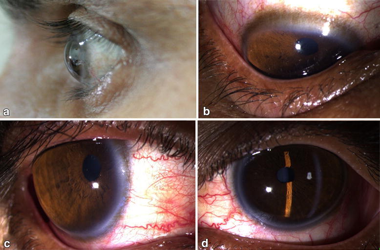
a: diffuse corneal thinning with outward protrusion of the globe b: indentation of the lower lid c & d: stretched out limbus with clear anterior chamber
Image: “Clinical picture of the patient showing diffuse thinning of cornea with outward globular protrusion” by Gupta N et al. License: CC BY 4.0
Peters anomaly is a congenital form of anterior segment dysgenesis in which abnormal cleavage of the anterior chamberAnterior chamberThe space in the eye, filled with aqueous humor, bounded anteriorly by the cornea and a small portion of the sclera and posteriorly by a small portion of the ciliary body, the iris, and that part of the crystalline lens which presents through the pupil.Eye: Anatomy results in a central defect in the corneal endotheliumEndotheliumA layer of epithelium that lines the heart, blood vessels (vascular endothelium), lymph vessels (lymphatic endothelium), and the serous cavities of the body.Arteries: Histology and causes leukoma (whitish plaquePlaquePrimary Skin Lesions).
Overview:
Multiple geneGeneA category of nucleic acid sequences that function as units of heredity and which code for the basic instructions for the development, reproduction, and maintenance of organisms.Basic Terms of Genetics loci:
PAX6
PITX2
FOXC1
CYP1B1
Can occur sporadically, but dominant and recessive inheritances are common
Systemic associations include:
TrisomyTrisomyThe possession of a third chromosome of any one type in an otherwise diploid cell.Types of Mutations 13
TrisomyTrisomyThe possession of a third chromosome of any one type in an otherwise diploid cell.Types of Mutations 15
Partial deletion of chromosomeChromosomeIn a prokaryotic cell or in the nucleus of a eukaryotic cell, a structure consisting of or containing DNA which carries the genetic information essential to the cell.Basic Terms of GeneticsarmArmThe arm, or “upper arm” in common usage, is the region of the upper limb that extends from the shoulder to the elbow joint and connects inferiorly to the forearm through the cubital fossa. It is divided into 2 fascial compartments (anterior and posterior).Arm: Anatomy 11q
IncidenceIncidenceThe number of new cases of a given disease during a given period in a specified population. It also is used for the rate at which new events occur in a defined population. It is differentiated from prevalence, which refers to all cases in the population at a given time.Measures of Disease Frequency is unknown: part of the congenital corneal opacities seen in 3–6 individuals per 100,000
Pathogenesis: due to a disruption in neural crestNeural crestThe two longitudinal ridges along the primitive streak appearing near the end of gastrulation during development of nervous system (neurulation). The ridges are formed by folding of neural plate. Between the ridges is a neural groove which deepens as the fold become elevated. When the folds meet at midline, the groove becomes a closed tube, the neural tube.Hirschsprung Disease migration or separation that occurs in the 7th week of gestation
Iris strands are often seen across the anterior chamberAnterior chamberThe space in the eye, filled with aqueous humor, bounded anteriorly by the cornea and a small portion of the sclera and posteriorly by a small portion of the ciliary body, the iris, and that part of the crystalline lens which presents through the pupil.Eye: Anatomy to the posterior surface of the corneaCorneaThe transparent anterior portion of the fibrous coat of the eye consisting of five layers: stratified squamous corneal epithelium; bowman membrane; corneal stroma; descemet membrane; and mesenchymal corneal endothelium. It serves as the first refracting medium of the eye.Eye: Anatomy.
Diagnosis: based on clinical findings and confirmed using ultrasonography
Management:
RegularRegularInsulin monitoring for glaucomaGlaucomaGlaucoma is an optic neuropathy characterized by typical visual field defects and optic nerve atrophy seen as optic disc cupping on examination. The acute form of glaucoma is a medical emergency. Glaucoma is often, but not always, caused by increased intraocular pressure (IOP). Glaucoma
Surgery: full-thickness penetrating keratoplasty plus lensectomy in patientsPatientsIndividuals participating in the health care system for the purpose of receiving therapeutic, diagnostic, or preventive procedures.Clinician–Patient Relationship with cataractCataractPartial or complete opacity on or in the lens or capsule of one or both eyes, impairing vision or causing blindness. The many kinds of cataract are classified by their morphology (size, shape, location) or etiology (cause and time of occurrence).Neurofibromatosis Type 2
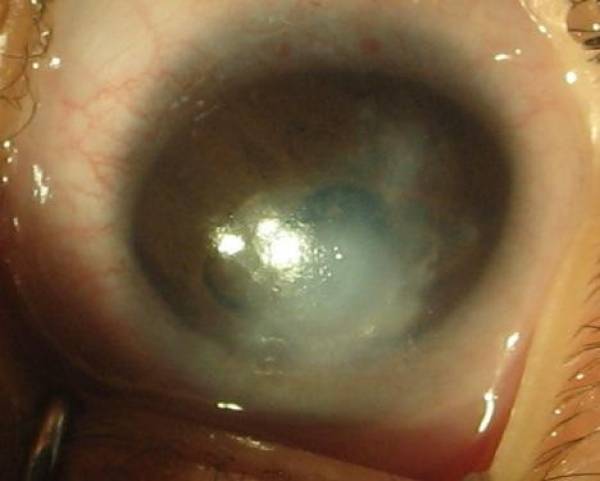
Corneal opacity in a patient with Peters anomaly
Image: “Photograph of left eye” by Tuli N et al. License: CC BY 2.0
Sclerocornea
With sclerocornea, the limits of the corneaCorneaThe transparent anterior portion of the fibrous coat of the eye consisting of five layers: stratified squamous corneal epithelium; bowman membrane; corneal stroma; descemet membrane; and mesenchymal corneal endothelium. It serves as the first refracting medium of the eye.Eye: Anatomy and scleraScleraThe white, opaque, fibrous, outer tunic of the eyeball, covering it entirely excepting the segment covered anteriorly by the cornea. It is essentially avascular but contains apertures for vessels, lymphatics, and nerves.Eye: Anatomy are indistinct, resulting in a congenital anomaly of clarity. Irregularly arranged collagenCollagenA polypeptide substance comprising about one third of the total protein in mammalian organisms. It is the main constituent of skin; connective tissue; and the organic substance of bones (bone and bones) and teeth (tooth).Connective Tissue: Histology fibrils are noted.
Overview:
Additional ocular anomalies, such as cataracts or colobomaColobomaCongenital anomaly in which some of the structures of the eye are absent due to incomplete fusion of the fetal intraocular fissure during gestation.Esophageal Atresia and Tracheoesophageal Fistula, are usually present.
Nonprogressive
Noninflammatory
May affect the entire corneaCorneaThe transparent anterior portion of the fibrous coat of the eye consisting of five layers: stratified squamous corneal epithelium; bowman membrane; corneal stroma; descemet membrane; and mesenchymal corneal endothelium. It serves as the first refracting medium of the eye.Eye: Anatomy or be limited to a part of the corneaCorneaThe transparent anterior portion of the fibrous coat of the eye consisting of five layers: stratified squamous corneal epithelium; bowman membrane; corneal stroma; descemet membrane; and mesenchymal corneal endothelium. It serves as the first refracting medium of the eye.Eye: Anatomy:
Peripheral type of sclerocornea: The affected area is vascularized with superficial scleral vessels.
Total sclerocornea: The entire corneaCorneaThe transparent anterior portion of the fibrous coat of the eye consisting of five layers: stratified squamous corneal epithelium; bowman membrane; corneal stroma; descemet membrane; and mesenchymal corneal endothelium. It serves as the first refracting medium of the eye.Eye: Anatomy is opaque and vascularized.
Etiology:
Autosomal dominantAutosomal dominantAutosomal inheritance, both dominant and recessive, refers to the transmission of genes from the 22 autosomal chromosomes. Autosomal dominant diseases are expressed when only 1 copy of the dominant allele is inherited. Autosomal Recessive and Autosomal Dominant Inheritance or autosomal recessiveAutosomal recessiveAutosomal inheritance, both dominant and recessive, refers to the transmission of genes from the 22 autosomal chromosomes. Autosomal recessive diseases are only expressed when 2 copies of the recessive allele are inherited.Autosomal Recessive and Autosomal Dominant Inheritance form (much more severe)
Commonly occurs bilaterally
Pathophysiology:
Corneal development occurs during the 7th and 8th weeks of gestation.
Failure of the mesenchymal cells to differentiate into corneal and scleral cells allows the corneal curvature to exceed that of the scleraScleraThe white, opaque, fibrous, outer tunic of the eyeball, covering it entirely excepting the segment covered anteriorly by the cornea. It is essentially avascular but contains apertures for vessels, lymphatics, and nerves.Eye: Anatomy.
Clinical presentation:
Partial sclerocornea: presence of a peripheral, white, vascularized, 1–2-mm corneal rim that blends with the scleraScleraThe white, opaque, fibrous, outer tunic of the eyeball, covering it entirely excepting the segment covered anteriorly by the cornea. It is essentially avascular but contains apertures for vessels, lymphatics, and nerves.Eye: Anatomy. The central corneaCorneaThe transparent anterior portion of the fibrous coat of the eye consisting of five layers: stratified squamous corneal epithelium; bowman membrane; corneal stroma; descemet membrane; and mesenchymal corneal endothelium. It serves as the first refracting medium of the eye.Eye: Anatomy is generally normal.
Total sclerocornea:
Entire corneaCorneaThe transparent anterior portion of the fibrous coat of the eye consisting of five layers: stratified squamous corneal epithelium; bowman membrane; corneal stroma; descemet membrane; and mesenchymal corneal endothelium. It serves as the first refracting medium of the eye.Eye: Anatomy is involved.
Center is clearer than the periphery.
Management:
Generally, no treatment is required.
Refractive errorsRefractive errorsBy refraction, the light that enters the eye is focused onto a particular point of the retina. The main refractive components of the eye are the cornea and the lens. When the corneal curvature, the refractive power of the lens, does not match the size of the eye, ametropia or a refractive error occurs. Refractive Errors are corrected if present.
Artificial tears may be used.
In generalized sclerocornea, early keratoplasty should be considered to provide and/or preserve visionVisionOphthalmic Exam.
Peripheral scleral changes seen in a patient with sclerocornea
Image: “Sclerocornea” by Mataftsi A et al. License: CC BY 3.0
Corneal Dystrophies
Congenital stromal corneal dystrophy
A noninflammatory, inherited corneal disorder not usually associated with any other ocular or systemic conditions
Overview:
Usually involves the central area of the corneaCorneaThe transparent anterior portion of the fibrous coat of the eye consisting of five layers: stratified squamous corneal epithelium; bowman membrane; corneal stroma; descemet membrane; and mesenchymal corneal endothelium. It serves as the first refracting medium of the eye.Eye: Anatomy
Discrete areas of opacification develop in the superficial layers of the stroma.
Etiology:
Caused by mutations in the DCN geneGeneA category of nucleic acid sequences that function as units of heredity and which code for the basic instructions for the development, reproduction, and maintenance of organisms.Basic Terms of Genetics
Autosomal dominantAutosomal dominantAutosomal inheritance, both dominant and recessive, refers to the transmission of genes from the 22 autosomal chromosomes. Autosomal dominant diseases are expressed when only 1 copy of the dominant allele is inherited. Autosomal Recessive and Autosomal Dominant Inheritance
Classification:
Lattice dystrophy types I and II
Granular dystrophy types I and II
Macular dystrophy
Reis-Bucklers dystrophy
Pathophysiology:
Clinical symptoms arise due to atrophyAtrophyDecrease in the size of a cell, tissue, organ, or multiple organs, associated with a variety of pathological conditions such as abnormal cellular changes, ischemia, malnutrition, or hormonal changes.Cellular Adaptation and degeneration of basal epithelial cells.
Amyloid and hyaline deposits also contribute to the development of symptoms.
There is an increase in number and density until the Bowman’s membraneBowman’S MembraneCorneal Abrasions, Erosion, and Ulcers becomes eroded and the epitheliumEpitheliumThe epithelium is a complex of specialized cellular organizations arranged into sheets and lining cavities and covering the surfaces of the body. The cells exhibit polarity, having an apical and a basal pole. Structures important for the epithelial integrity and function involve the basement membrane, the semipermeable sheet on which the cells rest, and interdigitations, as well as cellular junctions. Surface Epithelium: Histology desquamates.
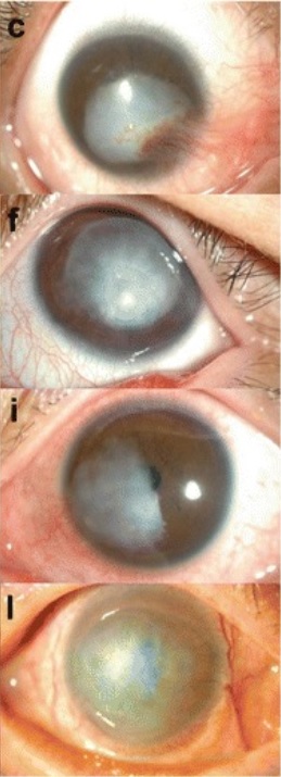
Corneal dystrophies can present as granular, lattice, or macular forms.
The corneal surface is normal or slightly irregular; small opacities are seen throughout the stroma of the entire corneaCorneaThe transparent anterior portion of the fibrous coat of the eye consisting of five layers: stratified squamous corneal epithelium; bowman membrane; corneal stroma; descemet membrane; and mesenchymal corneal endothelium. It serves as the first refracting medium of the eye.Eye: Anatomy and give it a cloudy appearance.
StrabismusStrabismusStrabismus is the misalignment of the eyes while fixating the gaze on an object. Strabismus can be idiopathic, but it may also be caused by cerebral palsy, uncorrected refractive errors, and extraocular muscle or cranial nerve dysfunction. Strabismus is common.
Slit-lamp exam: Anterior corneaCorneaThe transparent anterior portion of the fibrous coat of the eye consisting of five layers: stratified squamous corneal epithelium; bowman membrane; corneal stroma; descemet membrane; and mesenchymal corneal endothelium. It serves as the first refracting medium of the eye.Eye: Anatomy displays flaky or feathery opacities.
Management:
Glasses or contact lenses for correction of refractive errorsRefractive errorsBy refraction, the light that enters the eye is focused onto a particular point of the retina. The main refractive components of the eye are the cornea and the lens. When the corneal curvature, the refractive power of the lens, does not match the size of the eye, ametropia or a refractive error occurs. Refractive Errors
Patching or surgical correction of strabismusStrabismusStrabismus is the misalignment of the eyes while fixating the gaze on an object. Strabismus can be idiopathic, but it may also be caused by cerebral palsy, uncorrected refractive errors, and extraocular muscle or cranial nerve dysfunction. Strabismus
Keratoplasty
Congenital endothelial corneal dystrophy (CHED)
Congenital hereditary endothelial dystrophy is a corneal dystrophy characterized by bilateral diffuse clouding of corneas.
Etiology and epidemiology:
Autosomal recessiveAutosomal recessiveAutosomal inheritance, both dominant and recessive, refers to the transmission of genes from the 22 autosomal chromosomes. Autosomal recessive diseases are only expressed when 2 copies of the recessive allele are inherited.Autosomal Recessive and Autosomal Dominant Inheritance
MutationMutationGenetic mutations are errors in DNA that can cause protein misfolding and dysfunction. There are various types of mutations, including chromosomal, point, frameshift, and expansion mutations. Types of Mutations on chromosomeChromosomeIn a prokaryotic cell or in the nucleus of a eukaryotic cell, a structure consisting of or containing DNA which carries the genetic information essential to the cell.Basic Terms of Genetics 20p13
Affected geneGeneA category of nucleic acid sequences that function as units of heredity and which code for the basic instructions for the development, reproduction, and maintenance of organisms.Basic Terms of Genetics: SLC4A11
IncidenceIncidenceThe number of new cases of a given disease during a given period in a specified population. It also is used for the rate at which new events occur in a defined population. It is differentiated from prevalence, which refers to all cases in the population at a given time.Measures of Disease Frequency: 3 in 100,000 births
Children of consanguineous marriages have a comparatively greater incidenceIncidenceThe number of new cases of a given disease during a given period in a specified population. It also is used for the rate at which new events occur in a defined population. It is differentiated from prevalence, which refers to all cases in the population at a given time.Measures of Disease Frequency.
Classification:
CHED
Posterior polymorphous corneal dystrophy (PPCD)
Pathophysiology:
The SLC4A11 geneGeneA category of nucleic acid sequences that function as units of heredity and which code for the basic instructions for the development, reproduction, and maintenance of organisms.Basic Terms of Genetics codes for a transmembrane protein, which acts as a pumpPumpACES and RUSH: Resuscitation Ultrasound Protocols between the endotheliumEndotheliumA layer of epithelium that lines the heart, blood vessels (vascular endothelium), lymph vessels (lymphatic endothelium), and the serous cavities of the body.Arteries: Histology and corneal stroma.
Dysfunction of this protein leads to deposition of material in the layers of the corneaCorneaThe transparent anterior portion of the fibrous coat of the eye consisting of five layers: stratified squamous corneal epithelium; bowman membrane; corneal stroma; descemet membrane; and mesenchymal corneal endothelium. It serves as the first refracting medium of the eye.Eye: Anatomy and corneal clouding.
Clinical presentation and diagnosis:
History of congenital or perinatal clouding of the corneas bilaterally
AmblyopiaAmblyopiaA nonspecific term referring to impaired vision. Major subcategories include stimulus deprivation-induced amblyopia and toxic amblyopia. Stimulus deprivation-induced amblyopia is a developmental disorder of the visual cortex. A discrepancy between visual information received by the visual cortex from each eye results in abnormal cortical development. Strabismus and refractive errors may cause this condition. Toxic amblyopia is a disorder of the optic nerve which is associated with alcoholism, tobacco smoking, and other toxins and as an adverse effect of the use of some medications.Strabismus
NystagmusNystagmusInvoluntary movements of the eye that are divided into two types, jerk and pendular. Jerk nystagmus has a slow phase in one direction followed by a corrective fast phase in the opposite direction, and is usually caused by central or peripheral vestibular dysfunction. Pendular nystagmus features oscillations that are of equal velocity in both directions and this condition is often associated with visual loss early in life.Albinism
Management:
Surgery is the mainstay of treatment.
Penetrating keratoplasty or Descemet’s stripping automated endothelial keratoplasty are used.
Acquired Corneal Conditions
Corneal keloids
Corneal keloids are rare, benignBenignFibroadenoma, gray-white corneal lesions that result from abnormal fibrousFibrousFibrocystic Change tissue proliferation. Accumulation of collagenCollagenA polypeptide substance comprising about one third of the total protein in mammalian organisms. It is the main constituent of skin; connective tissue; and the organic substance of bones (bone and bones) and teeth (tooth).Connective Tissue: Histology and various glycoproteinsGlycoproteinsConjugated protein-carbohydrate compounds including mucins, mucoid, and amyloid glycoproteins.Basics of Carbohydrates results in hyperplasiaHyperplasiaAn increase in the number of cells in a tissue or organ without tumor formation. It differs from hypertrophy, which is an increase in bulk without an increase in the number of cells.Cellular Adaptation of the epitheliumEpitheliumThe epithelium is a complex of specialized cellular organizations arranged into sheets and lining cavities and covering the surfaces of the body. The cells exhibit polarity, having an apical and a basal pole. Structures important for the epithelial integrity and function involve the basement membrane, the semipermeable sheet on which the cells rest, and interdigitations, as well as cellular junctions. Surface Epithelium: Histology and disruption of the Bowman’s layer.
Overview:
Congenital: Bilateral cases of corneal keloids are associated with Lowe syndrome and Rubinstein-Taybi syndrome.
Reported in ages ranging from 2 months to 72 years, but rare overall
Majority of corneal keloids occur in the 1st 3 decades of life.
Pathogenesis: Corneal stromal overgrowth during the healing process results in the transformationTransformationChange brought about to an organism’s genetic composition by unidirectional transfer (transfection; transduction, genetic; conjugation, genetic, etc.) and incorporation of foreign DNA into prokaryotic or eukaryotic cells by recombination of part or all of that DNA into the cell’s genome.Bacteriology of keratocytes into fibroblastsFibroblastsConnective tissue cells which secrete an extracellular matrix rich in collagen and other macromolecules.Sarcoidosis and myofibroblastsMyofibroblastsSpindle-shaped cells with characteristic contractile proteins and structures that contribute to the wound healing process. They occur in granulation tissue and also in pathological processes such as fibrosis.Hypertrophic and Keloid Scars, leading to keloid formation.
Clinical presentation:
Most patientsPatientsIndividuals participating in the health care system for the purpose of receiving therapeutic, diagnostic, or preventive procedures.Clinician–Patient Relationship present with a slow-growing corneal lesion that appears to be raised from the corneal surface.
Failure to close eyelidsEyelidsEach of the upper and lower folds of skin which cover the eye when closed.Blepharitis in the case of large keloids
Diagnosis is made using visual acuityVisual AcuityClarity or sharpness of ocular vision or the ability of the eye to see fine details. Visual acuity depends on the functions of retina, neuronal transmission, and the interpretative ability of the brain. Normal visual acuity is expressed as 20/20 indicating that one can see at 20 feet what should normally be seen at that distance. Visual acuity can also be influenced by brightness, color, and contrast.Ophthalmic Exam and slit-lamp exams.
Management:
No treatment needed when asymptomatic
Close monitoring:
Size of the keloid
Visual acuityVisual AcuityClarity or sharpness of ocular vision or the ability of the eye to see fine details. Visual acuity depends on the functions of retina, neuronal transmission, and the interpretative ability of the brain. Normal visual acuity is expressed as 20/20 indicating that one can see at 20 feet what should normally be seen at that distance. Visual acuity can also be influenced by brightness, color, and contrast.Ophthalmic Exam
Surgery is considered when the keloid causes a decrease in visual acuityVisual AcuityClarity or sharpness of ocular vision or the ability of the eye to see fine details. Visual acuity depends on the functions of retina, neuronal transmission, and the interpretative ability of the brain. Normal visual acuity is expressed as 20/20 indicating that one can see at 20 feet what should normally be seen at that distance. Visual acuity can also be influenced by brightness, color, and contrast.Ophthalmic Exam:
Local excision
Superficial lamellar or phototherapeutic keratectomy
Keratoplasty
Corneal keloids appear as opacities that rise from the epicorneal surface.
Image: “Slit-lamp microscopic photographs of corneal keloids” by BMC Ophthalmology/Hyo Kyung Lee et al. License: CC BY 4.0, cropped by Lecturio.
Band-shaped keratopathy
A degenerative change associated with the deposition of calciumCalciumA basic element found in nearly all tissues. It is a member of the alkaline earth family of metals with the atomic symbol ca, atomic number 20, and atomic weight 40. Calcium is the most abundant mineral in the body and combines with phosphorus to form calcium phosphate in the bones and teeth. It is essential for the normal functioning of nerves and muscles and plays a role in blood coagulation (as factor IV) and in many enzymatic processes.Electrolytes salts in the Bowman’s membrane and anterior stromal lamellae of the corneal epitheliumEpitheliumThe epithelium is a complex of specialized cellular organizations arranged into sheets and lining cavities and covering the surfaces of the body. The cells exhibit polarity, having an apical and a basal pole. Structures important for the epithelial integrity and function involve the basement membrane, the semipermeable sheet on which the cells rest, and interdigitations, as well as cellular junctions. Surface Epithelium: Histology
Overview:
Deposition may be due to a multitude of factors:
Precipitation of tears
pHpHThe quantitative measurement of the acidity or basicity of a solution.Acid-Base Balance change
Compromise of endothelial function
Corneal edemaEdemaEdema is a condition in which excess serous fluid accumulates in the body cavity or interstitial space of connective tissues. Edema is a symptom observed in several medical conditions. It can be categorized into 2 types, namely, peripheral (in the extremities) and internal (in an organ or body cavity). Edema
Remainder of the corneaCorneaThe transparent anterior portion of the fibrous coat of the eye consisting of five layers: stratified squamous corneal epithelium; bowman membrane; corneal stroma; descemet membrane; and mesenchymal corneal endothelium. It serves as the first refracting medium of the eye.Eye: Anatomy is clear.
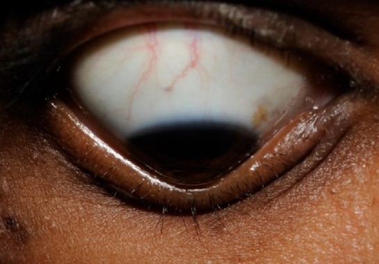
Presents as a band-shaped opacityOpacityImaging of the Lungs and Pleura in the interpalpebral zone, with a clear interval between the ends of the band and the limbusLimbusAn annular transitional zone, approximately 1 mm wide, between the cornea and the bulbar conjunctiva and sclera. It is highly vascular and is involved in the metabolism of the cornea.Eye: Anatomy
Caused by chronic ocular inflammationInflammationInflammation is a complex set of responses to infection and injury involving leukocytes as the principal cellular mediators in the body’s defense against pathogenic organisms. Inflammation is also seen as a response to tissue injury in the process of wound healing. The 5 cardinal signs of inflammation are pain, heat, redness, swelling, and loss of function. Inflammation due to:
UveitisUveitisUveitis is the inflammation of the uvea, the pigmented middle layer of the eye, which comprises the iris, ciliary body, and choroid. The condition is categorized based on the site of disease; anterior uveitis is the most common. Diseases of the Uvea
Herpes zosterHerpes ZosterVaricella-zoster virus (VZV) is a linear, double-stranded DNA virus in the Herpesviridae family. Shingles (also known as herpes zoster) is more common in adults and occurs due to the reactivation of VZV. Varicella-Zoster Virus/ChickenpoxkeratitisKeratitisInflammation of the cornea.Herpes Simplex Virus 1 and 2
May be associated with systemic disorders:
HyperparathyroidismHyperparathyroidismHyperparathyroidism is a condition associated with elevated blood levels of parathyroid hormone (PTH). Depending on the pathogenesis of this condition, hyperparathyroidism can be defined as primary, secondary or tertiary. Hyperparathyroidism
CKDCKDChronic kidney disease (CKD) is kidney impairment that lasts for ≥ 3 months, implying that it is irreversible. Hypertension and diabetes are the most common causes; however, there are a multitude of other etiologies. In the early to moderate stages, CKD is usually asymptomatic and is primarily diagnosed by laboratory abnormalities.Chronic Kidney Disease
HypophosphatasiaHypophosphatasiaA genetic metabolic disorder resulting from serum and bone alkaline phosphatase deficiency leading to hypercalcemia, ethanolamine phosphatemia, and ethanolamine phosphaturia. Clinical manifestations include severe skeletal defects resembling vitamin d-resistant rickets, failure of the calvarium to calcify, dyspnea, cyanosis, vomiting, constipation, renal calcinosis, failure to thrive, disorders of movement, beading of the costochondral junction, and rachitic bone changes.Osteomalacia and Rickets
SarcoidosisSarcoidosisSarcoidosis is a multisystem inflammatory disease that causes noncaseating granulomas. The exact etiology is unknown. Sarcoidosis usually affects the lungs and thoracic lymph nodes, but it can also affect almost every system in the body, including the skin, heart, and eyes, most commonly. Sarcoidosis
Clinical presentation:
Decreased visual acuityVisual AcuityClarity or sharpness of ocular vision or the ability of the eye to see fine details. Visual acuity depends on the functions of retina, neuronal transmission, and the interpretative ability of the brain. Normal visual acuity is expressed as 20/20 indicating that one can see at 20 feet what should normally be seen at that distance. Visual acuity can also be influenced by brightness, color, and contrast.Ophthalmic Exam is directly proportional to the amount of calciumCalciumA basic element found in nearly all tissues. It is a member of the alkaline earth family of metals with the atomic symbol ca, atomic number 20, and atomic weight 40. Calcium is the most abundant mineral in the body and combines with phosphorus to form calcium phosphate in the bones and teeth. It is essential for the normal functioning of nerves and muscles and plays a role in blood coagulation (as factor IV) and in many enzymatic processes.Electrolytes deposition in the corneaCorneaThe transparent anterior portion of the fibrous coat of the eye consisting of five layers: stratified squamous corneal epithelium; bowman membrane; corneal stroma; descemet membrane; and mesenchymal corneal endothelium. It serves as the first refracting medium of the eye.Eye: Anatomy.
PhotophobiaPhotophobiaAbnormal sensitivity to light. This may occur as a manifestation of eye diseases; migraine; subarachnoid hemorrhage; meningitis; and other disorders. Photophobia may also occur in association with depression and other mental disorders.Migraine Headache
CalciumCalciumA basic element found in nearly all tissues. It is a member of the alkaline earth family of metals with the atomic symbol ca, atomic number 20, and atomic weight 40. Calcium is the most abundant mineral in the body and combines with phosphorus to form calcium phosphate in the bones and teeth. It is essential for the normal functioning of nerves and muscles and plays a role in blood coagulation (as factor IV) and in many enzymatic processes.Electrolytes deposition begins in the periphery and progresses inwards.
Management:
Superficial debridementDebridementThe removal of foreign material and devitalized or contaminated tissue from or adjacent to a traumatic or infected lesion until surrounding healthy tissue is exposed.Stevens-Johnson Syndrome and lamellar keratectomy
Treat the underlying cause, if known, to decrease further calciumCalciumA basic element found in nearly all tissues. It is a member of the alkaline earth family of metals with the atomic symbol ca, atomic number 20, and atomic weight 40. Calcium is the most abundant mineral in the body and combines with phosphorus to form calcium phosphate in the bones and teeth. It is essential for the normal functioning of nerves and muscles and plays a role in blood coagulation (as factor IV) and in many enzymatic processes.Electrolytes deposition in the corneaCorneaThe transparent anterior portion of the fibrous coat of the eye consisting of five layers: stratified squamous corneal epithelium; bowman membrane; corneal stroma; descemet membrane; and mesenchymal corneal endothelium. It serves as the first refracting medium of the eye.Eye: Anatomy.