There are multiple different types of malignancies that can affect the vulvaVulvaThe vulva is the external genitalia of the female and includes the mons pubis, labia majora, labia minora, clitoris, vestibule, vestibular bulb, and greater vestibular glands. Vagina, Vulva, and Pelvic Floor: Anatomy. The most common histologic type is squamous cell carcinomaSquamous cell carcinomaCutaneous squamous cell carcinoma (cSCC) is caused by malignant proliferation of atypical keratinocytes. This condition is the 2nd most common skin malignancy and usually affects sun-exposed areas of fair-skinned patients. The cancer presents as a firm, erythematous, keratotic plaque or papule. Squamous Cell Carcinoma (SCC) (SCC), which accounts for approximately 75%–85% of all vulvar cancers. Other types include melanomaMelanomaMelanoma is a malignant tumor arising from melanocytes, the melanin-producing cells of the epidermis. These tumors are most common in fair-skinned individuals with a history of excessive sun exposure and sunburns. Melanoma, basal cellBasal CellErythema Multiforme carcinoma, sarcoma, malignancyMalignancyHemothorax of the Bartholin glandsBartholin glandsMucus-secreting glands situated on the posterior and lateral aspect of the vestibule of the vagina.Vagina, Vulva, and Pelvic Floor: Anatomy, and Paget disease of the vulvaVulvaThe vulva is the external genitalia of the female and includes the mons pubis, labia majora, labia minora, clitoris, vestibule, vestibular bulb, and greater vestibular glands. Vagina, Vulva, and Pelvic Floor: Anatomy (an adenocarcinoma). Squamous cell carcinomaSquamous cell carcinomaCutaneous squamous cell carcinoma (cSCC) is caused by malignant proliferation of atypical keratinocytes. This condition is the 2nd most common skin malignancy and usually affects sun-exposed areas of fair-skinned patients. The cancer presents as a firm, erythematous, keratotic plaque or papule. Squamous Cell Carcinoma (SCC) is typically associated with either high-risk HPVHPVHuman papillomavirus (HPV) is a nonenveloped, circular, double-stranded DNA virus belonging to the Papillomaviridae family. Humans are the only reservoir, and transmission occurs through close skin-to-skin or sexual contact. Human papillomaviruses infect basal epithelial cells and can affect cell-regulatory proteins to result in cell proliferation. Papillomavirus (HPV) infection or lichen sclerosusLichen SclerosusAtrophy and shriveling of the skin of the vulva that is characterized by the whitish lichen sclerosus appearance, inflammation, and pruritus.Benign Vulvar Conditions. Vulvar cancer presents as vulvar lesions that can have a variety of appearances, which may include warty or nodular masses, scaly plaques, pigmented lesions, and ulcers; pruritusPruritusAn intense itching sensation that produces the urge to rub or scratch the skin to obtain relief.Atopic Dermatitis (Eczema) is also common. Diagnosis usually requires a biopsyBiopsyRemoval and pathologic examination of specimens from the living body.Ewing Sarcoma, and management is primarily with surgical excision. Unfortunately, lymphLymphThe interstitial fluid that is in the lymphatic system.Secondary Lymphatic Organs node metastasisMetastasisThe transfer of a neoplasm from one organ or part of the body to another remote from the primary site.Grading, Staging, and Metastasis occurs early in the natural history of the disease and is associated with a poor prognosisPrognosisA prediction of the probable outcome of a disease based on a individual's condition and the usual course of the disease as seen in similar situations.Non-Hodgkin Lymphomas.
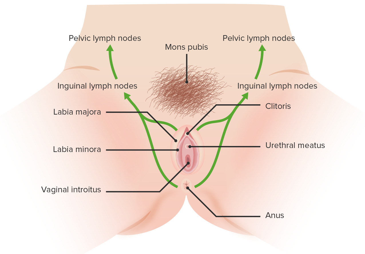
Vulvar cancer is a malignancyMalignancyHemothorax of the vulvaVulvaThe vulva is the external genitalia of the female and includes the mons pubis, labia majora, labia minora, clitoris, vestibule, vestibular bulb, and greater vestibular glands. Vagina, Vulva, and Pelvic Floor: Anatomy, which includes structures of the external female genitalia:
ClitorisClitorisAn erectile structure homologous with the penis, situated beneath the anterior labial commissure, partially hidden between the anterior ends of the labia minora.Vagina, Vulva, and Pelvic Floor: Anatomy
Vulvar cancer is a rare cancer that constitutes only 0.3% of new cancer diagnoses.
Lifetime risk: 0.3%
Squamous cell carcinomaSquamous cell carcinomaCutaneous squamous cell carcinoma (cSCC) is caused by malignant proliferation of atypical keratinocytes. This condition is the 2nd most common skin malignancy and usually affects sun-exposed areas of fair-skinned patients. The cancer presents as a firm, erythematous, keratotic plaque or papule. Squamous Cell Carcinoma (SCC) (SCC) is the most common histologic subtype: approximately 75%–85%
IncidenceIncidenceThe number of new cases of a given disease during a given period in a specified population. It also is used for the rate at which new events occur in a defined population. It is differentiated from prevalence, which refers to all cases in the population at a given time.Measures of Disease Frequency: approximately 5000–10,000 cases per year in the United States
MortalityMortalityAll deaths reported in a given population.Measures of Health Status: approximately 1000–2000 deaths per year in the United States
Age:
Typically diagnosed at 55–85 years of age
Most common age group affected: 65–75 years
Ethnicity: more common among White women
Risk factors
HPVHPVHuman papillomavirus (HPV) is a nonenveloped, circular, double-stranded DNA virus belonging to the Papillomaviridae family. Humans are the only reservoir, and transmission occurs through close skin-to-skin or sexual contact. Human papillomaviruses infect basal epithelial cells and can affect cell-regulatory proteins to result in cell proliferation. Papillomavirus (HPV) infection with high-risk subtypes:
Most important risk factor for SCC
High-risk HPVHPVHuman papillomavirus (HPV) is a nonenveloped, circular, double-stranded DNA virus belonging to the Papillomaviridae family. Humans are the only reservoir, and transmission occurs through close skin-to-skin or sexual contact. Human papillomaviruses infect basal epithelial cells and can affect cell-regulatory proteins to result in cell proliferation. Papillomavirus (HPV) subtypes: 16, 18, 31, 33, and 45
Approximately 70%–75% of HPV-associated SCCs are HPV-16.
Increasing age
SmokingSmokingWillful or deliberate act of inhaling and exhaling smoke from burning substances or agents held by hand.Interstitial Lung Diseases
Lichen sclerosusLichen SclerosusAtrophy and shriveling of the skin of the vulva that is characterized by the whitish lichen sclerosus appearance, inflammation, and pruritus.Benign Vulvar Conditions (key risk factor for non–HPV-associated SCC)
Squamous cell hyperplasiaHyperplasiaAn increase in the number of cells in a tissue or organ without tumor formation. It differs from hypertrophy, which is an increase in bulk without an increase in the number of cells.Cellular Adaptation
Prior pelvic radiationRadiationEmission or propagation of acoustic waves (sound), electromagnetic energy waves (such as light; radio waves; gamma rays; or x-rays), or a stream of subatomic particles (such as electrons; neutrons; protons; or alpha particles).Osteosarcoma
Squamous cell carcinomaSquamous cell carcinomaCutaneous squamous cell carcinoma (cSCC) is caused by malignant proliferation of atypical keratinocytes. This condition is the 2nd most common skin malignancy and usually affects sun-exposed areas of fair-skinned patients. The cancer presents as a firm, erythematous, keratotic plaque or papule. Squamous Cell Carcinoma (SCC) (75%–85%):
HPV-associated (warty type)
Non–HPV-associated (keratinized type)
MelanomaMelanomaMelanoma is a malignant tumor arising from melanocytes, the melanin-producing cells of the epidermis. These tumors are most common in fair-skinned individuals with a history of excessive sun exposure and sunburns. Melanoma (approximately 5%–10%)
Leiomyosarcomas: malignancies of smooth muscle tissue
Rhabdomyosarcomas: malignancies of skeletal muscle tissue
Liposarcomas: malignancies of adipose tissueAdipose tissueAdipose tissue is a specialized type of connective tissue that has both structural and highly complex metabolic functions, including energy storage, glucose homeostasis, and a multitude of endocrine capabilities. There are three types of adipose tissue, white adipose tissue, brown adipose tissue, and beige or “brite” adipose tissue, which is a transitional form.Adipose Tissue: Histology
Adenocarcinoma or carcinoma of the Bartholin glandsBartholin glandsMucus-secreting glands situated on the posterior and lateral aspect of the vestibule of the vagina.Vagina, Vulva, and Pelvic Floor: Anatomy (approximately 1%)
Paget disease of the vulvaVulvaThe vulva is the external genitalia of the female and includes the mons pubis, labia majora, labia minora, clitoris, vestibule, vestibular bulb, and greater vestibular glands. Vagina, Vulva, and Pelvic Floor: Anatomy (an adenocarcinoma originating from glandular cells, < 1%)
Vulvar cancer is staged on the basis of the size and location of the tumorTumorInflammation, regional lymphLymphThe interstitial fluid that is in the lymphatic system.Secondary Lymphatic Organs node involvement, and the presence of metastasisMetastasisThe transfer of a neoplasm from one organ or part of the body to another remote from the primary site.Grading, Staging, and Metastasis.
Stage I:
TumorTumorInflammation confined to the vulvaVulvaThe vulva is the external genitalia of the female and includes the mons pubis, labia majora, labia minora, clitoris, vestibule, vestibular bulb, and greater vestibular glands. Vagina, Vulva, and Pelvic Floor: Anatomy
Lower urethraUrethraA tube that transports urine from the urinary bladder to the outside of the body in both the sexes. It also has a reproductive function in the male by providing a passage for sperm.Urinary Tract: Anatomy
Lower vaginaVaginaThe vagina is the female genital canal, extending from the vulva externally to the cervix uteri internally. The structures have sexual, reproductive, and urinary functions and a rich blood supply, mainly arising from the internal iliac artery.Vagina, Vulva, and Pelvic Floor: Anatomy
MetastasisMetastasisThe transfer of a neoplasm from one organ or part of the body to another remote from the primary site.Grading, Staging, and Metastasis to local lymph nodesLymph NodesThey are oval or bean shaped bodies (1 – 30 mm in diameter) located along the lymphatic system.Lymphatic Drainage System: Anatomy (superficial or deep inguinal nodes)
Stage IV, which may be defined by the presence of any of the following:
Upper urethraUrethraA tube that transports urine from the urinary bladder to the outside of the body in both the sexes. It also has a reproductive function in the male by providing a passage for sperm.Urinary Tract: Anatomy
Upper vaginaVaginaThe vagina is the female genital canal, extending from the vulva externally to the cervix uteri internally. The structures have sexual, reproductive, and urinary functions and a rich blood supply, mainly arising from the internal iliac artery.Vagina, Vulva, and Pelvic Floor: Anatomy
BladderBladderA musculomembranous sac along the urinary tract. Urine flows from the kidneys into the bladder via the ureters, and is held there until urination.Pyelonephritis and Perinephric Abscess
RectumRectumThe rectum and anal canal are the most terminal parts of the lower GI tract/large intestine that form a functional unit and control defecation. Fecal continence is maintained by several important anatomic structures including rectal folds, anal valves, the sling-like puborectalis muscle, and internal and external anal sphincters. Rectum and Anal Canal: Anatomy
Pelvic bonePelvic boneBones that constitute each half of the pelvic girdle in vertebrates, formed by fusion of the ilium; ischium; and pubic bone.Pelvis: Anatomy
Vulvar cancer develops when there is uncontrolled cellular proliferation in vulvar tissue. This cancer may progress from a premalignant lesion caused by an HPVHPVHuman papillomavirus (HPV) is a nonenveloped, circular, double-stranded DNA virus belonging to the Papillomaviridae family. Humans are the only reservoir, and transmission occurs through close skin-to-skin or sexual contact. Human papillomaviruses infect basal epithelial cells and can affect cell-regulatory proteins to result in cell proliferation. Papillomavirus (HPV) infection, or it may develop from other mutations, unrelated to HPVHPVHuman papillomavirus (HPV) is a nonenveloped, circular, double-stranded DNA virus belonging to the Papillomaviridae family. Humans are the only reservoir, and transmission occurs through close skin-to-skin or sexual contact. Human papillomaviruses infect basal epithelial cells and can affect cell-regulatory proteins to result in cell proliferation. Papillomavirus (HPV).
Vulvar squamous intraepithelial lesions (SILs)
Vulvar SILs are abnormalities of the squamous epitheliumEpitheliumThe epithelium is a complex of specialized cellular organizations arranged into sheets and lining cavities and covering the surfaces of the body. The cells exhibit polarity, having an apical and a basal pole. Structures important for the epithelial integrity and function involve the basement membrane, the semipermeable sheet on which the cells rest, and interdigitations, as well as cellular junctions. Surface Epithelium: Histology. Previously referred to as vulvar intraepithelial neoplasia (VIN), the preferred term is now SIL, which can be classified as:
Low-grade SIL (LSIL):
Previously referred to as VIN1
Not considered a premalignant lesion (no significant risk of progression to vulvar cancer)
Generally associated with low-risk HPVHPVHuman papillomavirus (HPV) is a nonenveloped, circular, double-stranded DNA virus belonging to the Papillomaviridae family. Humans are the only reservoir, and transmission occurs through close skin-to-skin or sexual contact. Human papillomaviruses infect basal epithelial cells and can affect cell-regulatory proteins to result in cell proliferation. Papillomavirus (HPV) types (6, 11) that cause genital wartsWartsBenign epidermal proliferations or tumors; some are viral in origin.Female Genitourinary Examination
Lesions are typically flat condylomas.
High-grade SIL (HSIL):
Previously referred to as VIN2 and VIN3
Premalignant lesions
Significant risk of progression to SCC if untreated
Generally associated with high-risk HPVHPVHuman papillomavirus (HPV) is a nonenveloped, circular, double-stranded DNA virus belonging to the Papillomaviridae family. Humans are the only reservoir, and transmission occurs through close skin-to-skin or sexual contact. Human papillomaviruses infect basal epithelial cells and can affect cell-regulatory proteins to result in cell proliferation. Papillomavirus (HPV) types (16 (especially), 18, 31, 33, and 45)
High association with concurrent cervical intraepithelial neoplasia (CINCINAn increased tendency to acquire chromosome aberrations when various processes involved in chromosome replication, repair, or segregation are dysfunctional.Colorectal Cancer)
Differentiated VIN (dVIN):
Previously referred to as VIN simplex type
Premalignant lesions
Not associated with HPVHPVHuman papillomavirus (HPV) is a nonenveloped, circular, double-stranded DNA virus belonging to the Papillomaviridae family. Humans are the only reservoir, and transmission occurs through close skin-to-skin or sexual contact. Human papillomaviruses infect basal epithelial cells and can affect cell-regulatory proteins to result in cell proliferation. Papillomavirus (HPV)infectionsInfectionsInvasion of the host organism by microorganisms or their toxins or by parasites that can cause pathological conditions or diseases.Chronic Granulomatous Disease
Refers to vulvar changes associated with vulvar dermatoses (primarily lichen sclerosusLichen SclerosusAtrophy and shriveling of the skin of the vulva that is characterized by the whitish lichen sclerosus appearance, inflammation, and pruritus.Benign Vulvar Conditions)
Low-grade squamous intraepithelial lesion
Image: “Vulvar intra-epithelial neoplasia (VIN grade I)” by Kotsopoulos IC, et al. License: CC BY 2.0
HPV-associated squamous cell carcinomaSquamous cell carcinomaCutaneous squamous cell carcinoma (cSCC) is caused by malignant proliferation of atypical keratinocytes. This condition is the 2nd most common skin malignancy and usually affects sun-exposed areas of fair-skinned patients. The cancer presents as a firm, erythematous, keratotic plaque or papule. Squamous Cell Carcinoma (SCC) vulvar cancer
HPV-associated SCC vulvar cancer is most commonly due to HPV-16. HPVHPVHuman papillomavirus (HPV) is a nonenveloped, circular, double-stranded DNA virus belonging to the Papillomaviridae family. Humans are the only reservoir, and transmission occurs through close skin-to-skin or sexual contact. Human papillomaviruses infect basal epithelial cells and can affect cell-regulatory proteins to result in cell proliferation. Papillomavirus (HPV) has the ability to affect host cell protein expression:
HPVHPVHuman papillomavirus (HPV) is a nonenveloped, circular, double-stranded DNA virus belonging to the Papillomaviridae family. Humans are the only reservoir, and transmission occurs through close skin-to-skin or sexual contact. Human papillomaviruses infect basal epithelial cells and can affect cell-regulatory proteins to result in cell proliferation. Papillomavirus (HPV) has 2 major oncoproteins:
After cells lose tumorTumorInflammation suppressor proteinsProteinsLinear polypeptides that are synthesized on ribosomes and may be further modified, crosslinked, cleaved, or assembled into complex proteins with several subunits. The specific sequence of amino acids determines the shape the polypeptide will take, during protein folding, and the function of the protein.Energy Homeostasis → unregulated proliferation → HSIL
Invasion through the basement membraneBasement membraneA darkly stained mat-like extracellular matrix (ecm) that separates cell layers, such as epithelium from endothelium or a layer of connective tissue. The ecm layer that supports an overlying epithelium or endothelium is called basal lamina. Basement membrane (bm) can be formed by the fusion of either two adjacent basal laminae or a basal lamina with an adjacent reticular lamina of connective tissue. Bm, composed mainly of type IV collagen; glycoprotein laminin; and proteoglycan, provides barriers as well as channels between interacting cell layers.Thin Basement Membrane Nephropathy (TBMN) → SCC
Non–HPV-associated squamous cell carcinomaSquamous cell carcinomaCutaneous squamous cell carcinoma (cSCC) is caused by malignant proliferation of atypical keratinocytes. This condition is the 2nd most common skin malignancy and usually affects sun-exposed areas of fair-skinned patients. The cancer presents as a firm, erythematous, keratotic plaque or papule. Squamous Cell Carcinoma (SCC) vulvar cancer
Non–HPV-associated SCC vulvar cancer most commonly occurs in the setting of lichen sclerosusLichen SclerosusAtrophy and shriveling of the skin of the vulva that is characterized by the whitish lichen sclerosus appearance, inflammation, and pruritus.Benign Vulvar Conditions.
InflammationInflammationInflammation is a complex set of responses to infection and injury involving leukocytes as the principal cellular mediators in the body’s defense against pathogenic organisms. Inflammation is also seen as a response to tissue injury in the process of wound healing. The 5 cardinal signs of inflammation are pain, heat, redness, swelling, and loss of function. Inflammation or autoimmunityAutoimmunityAutoimmunity is a pathologic immune response toward self-antigens, resulting from a combination of factors: immunologic, genetic, and environmental. The immune system is equipped with self-tolerance, allowing immune cells such as T cells and B cells to recognize self-antigens and to not mount a reaction against them. Defects in this mechanism, along with environmental triggers (such as infections) and genetic susceptibility factors (most notable of which are the HLA genes) can lead to autoimmune diseases. Autoimmunity leads to the loss of cyclin-dependent kinase 2A (p16) → unregulated proliferation → dVIN
P53 remains intact
Invasion through basement membraneBasement membraneA darkly stained mat-like extracellular matrix (ecm) that separates cell layers, such as epithelium from endothelium or a layer of connective tissue. The ecm layer that supports an overlying epithelium or endothelium is called basal lamina. Basement membrane (bm) can be formed by the fusion of either two adjacent basal laminae or a basal lamina with an adjacent reticular lamina of connective tissue. Bm, composed mainly of type IV collagen; glycoprotein laminin; and proteoglycan, provides barriers as well as channels between interacting cell layers.Thin Basement Membrane Nephropathy (TBMN) → SCC
EmbolizationEmbolizationA method of hemostasis utilizing various agents such as gelfoam, silastic, metal, glass, or plastic pellets, autologous clot, fat, and muscle as emboli. It has been used in the treatment of spinal cord and intracranial arteriovenous malformations, renal arteriovenous fistulas, gastrointestinal bleeding, epistaxis, hypersplenism, certain highly vascular tumors, traumatic rupture of blood vessels, and control of operative hemorrhage.Gastrointestinal Bleeding into regional lymph nodesLymph NodesThey are oval or bean shaped bodies (1 – 30 mm in diameter) located along the lymphatic system.Lymphatic Drainage System: Anatomy:
Vulvar cancer is often asymptomatic in its early stages. Early symptoms tend to be mild and are easily overlooked by the woman and/or her physician.
Vulvar squamous cell carcinomaSquamous cell carcinomaCutaneous squamous cell carcinoma (cSCC) is caused by malignant proliferation of atypical keratinocytes. This condition is the 2nd most common skin malignancy and usually affects sun-exposed areas of fair-skinned patients. The cancer presents as a firm, erythematous, keratotic plaque or papule. Squamous Cell Carcinoma (SCC)
PruritusPruritusAn intense itching sensation that produces the urge to rub or scratch the skin to obtain relief.Atopic Dermatitis (Eczema)
Burning sensation
Vulvar bleeding
LymphadenopathyLymphadenopathyLymphadenopathy is lymph node enlargement (> 1 cm) and is benign and self-limited in most patients. Etiologies include malignancy, infection, and autoimmune disorders, as well as iatrogenic causes such as the use of certain medications. Generalized lymphadenopathy often indicates underlying systemic disease. Lymphadenopathy in the groinGroinThe external junctural region between the lower part of the abdomen and the thigh.Male Genitourinary Examination (may be present even with small vulvar lesions)
Symptoms suggestive of advanced disease:
Vulvar painPainAn unpleasant sensation induced by noxious stimuli which are detected by nerve endings of nociceptive neurons.Pain: Types and Pathways
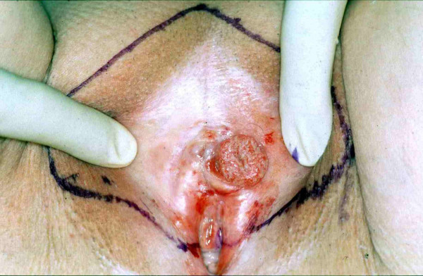
Squamous cell carcinoma of the vulva immediately prior to surgical resection: Planned surgical resection margins are marked.
Image: “Microinvasive squamous cell carcinoma of the vulva” by Sidor J, Diallo-Danebrock R, Eltze E, Lellé RJ. License: CC BY 2.0
Findings suggestive of non–squamous cell carcinomaSquamous cell carcinomaCutaneous squamous cell carcinoma (cSCC) is caused by malignant proliferation of atypical keratinocytes. This condition is the 2nd most common skin malignancy and usually affects sun-exposed areas of fair-skinned patients. The cancer presents as a firm, erythematous, keratotic plaque or papule. Squamous Cell Carcinoma (SCC) vulvar cancers
MelanomaMelanomaMelanoma is a malignant tumor arising from melanocytes, the melanin-producing cells of the epidermis. These tumors are most common in fair-skinned individuals with a history of excessive sun exposure and sunburns. Melanoma:
Usually a pigmented lesion
Characteristics similar to nonvulvar melanomaMelanomaMelanoma is a malignant tumor arising from melanocytes, the melanin-producing cells of the epidermis. These tumors are most common in fair-skinned individuals with a history of excessive sun exposure and sunburns. Melanoma (ABCDEs):
Asymmetric
Irregular Borders
Black Color
Diameter > 6 mmMMMultiple myeloma (MM) is a malignant condition of plasma cells (activated B lymphocytes) primarily seen in the elderly. Monoclonal proliferation of plasma cells results in cytokine-driven osteoclastic activity and excessive secretion of IgG antibodies.Multiple Myeloma
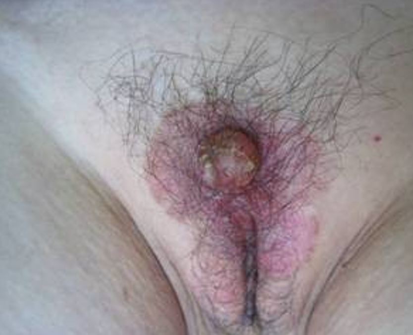
MassMassThree-dimensional lesion that occupies a space within the breastImaging of the Breast in the region of the Bartholin glandsBartholin glandsMucus-secreting glands situated on the posterior and lateral aspect of the vestibule of the vagina.Vagina, Vulva, and Pelvic Floor: Anatomy (4:00 and 8:00 when viewing the vulvaVulvaThe vulva is the external genitalia of the female and includes the mons pubis, labia majora, labia minora, clitoris, vestibule, vestibular bulb, and greater vestibular glands. Vagina, Vulva, and Pelvic Floor: Anatomy as a clock face)
MassMassThree-dimensional lesion that occupies a space within the breastImaging of the Breast may be irregular with solid components.
Paget disease of the vulva (also known as extramammary Paget disease): Note the firm red mass on an eczematoid erythema covering the mons pubis.
Image: “The clinical features of the patient with EMPD.” by Wang X, Yang W, Yang J. License: CC BY 2.0
Diagnosis
Vulvar cancer is a histologicdiagnosis. Once the diagnosis is established, stagingStagingMethods which attempt to express in replicable terms the extent of the neoplasm in the patient.Grading, Staging, and Metastasis requires a combination of clinical and surgical assessments.
Examination
Pelvic exam:
Identify any abnormal masses or lesions that should be biopsied.
Measure the size of any lesions.
Palpate lymph nodesLymph NodesThey are oval or bean shaped bodies (1 – 30 mm in diameter) located along the lymphatic system.Lymphatic Drainage System: Anatomy for lymphadenopathyLymphadenopathyLymphadenopathy is lymph node enlargement (> 1 cm) and is benign and self-limited in most patients. Etiologies include malignancy, infection, and autoimmune disorders, as well as iatrogenic causes such as the use of certain medications. Generalized lymphadenopathy often indicates underlying systemic disease. Lymphadenopathy.
Pap smearPap smearCytological preparation of cells collected from a mucosal surface and stained with Papanicolaou stain.Cervical Cancer Screening: should be up-to-date per screeningScreeningPreoperative Care guidelines owing to high rate of concurrent cervical pathology
ColposcopyColposcopyThe examination, therapy or surgery of the cervix and vagina by means of a specially designed endoscope introduced vaginally.Cervical Cancer Screening:
Performed on the vulvaVulvaThe vulva is the external genitalia of the female and includes the mons pubis, labia majora, labia minora, clitoris, vestibule, vestibular bulb, and greater vestibular glands. Vagina, Vulva, and Pelvic Floor: Anatomy, vaginaVaginaThe vagina is the female genital canal, extending from the vulva externally to the cervix uteri internally. The structures have sexual, reproductive, and urinary functions and a rich blood supply, mainly arising from the internal iliac artery.Vagina, Vulva, and Pelvic Floor: Anatomy, and cervixCervixThe uterus, cervix, and fallopian tubes are part of the internal female reproductive system. The most inferior portion of the uterus is the cervix, which connects the uterine cavity to the vagina. Externally, the cervix is lined by stratified squamous cells; however, the cervical canal is lined by columnar epithelium.Uterus, Cervix, and Fallopian Tubes: Anatomy
Soak the vulvaVulvaThe vulva is the external genitalia of the female and includes the mons pubis, labia majora, labia minora, clitoris, vestibule, vestibular bulb, and greater vestibular glands. Vagina, Vulva, and Pelvic Floor: Anatomy and perianal skinSkinThe skin, also referred to as the integumentary system, is the largest organ of the body. The skin is primarily composed of the epidermis (outer layer) and dermis (deep layer). The epidermis is primarily composed of keratinocytes that undergo rapid turnover, while the dermis contains dense layers of connective tissue.Skin: Structure and Functions, vaginaVaginaThe vagina is the female genital canal, extending from the vulva externally to the cervix uteri internally. The structures have sexual, reproductive, and urinary functions and a rich blood supply, mainly arising from the internal iliac artery.Vagina, Vulva, and Pelvic Floor: Anatomy, and cervixCervixThe uterus, cervix, and fallopian tubes are part of the internal female reproductive system. The most inferior portion of the uterus is the cervix, which connects the uterine cavity to the vagina. Externally, the cervix is lined by stratified squamous cells; however, the cervical canal is lined by columnar epithelium.Uterus, Cervix, and Fallopian Tubes: Anatomy in acetic acid and examine under magnification with a standard colposcopeColposcopeInstruments inserted into the vagina for examination of the tissues of the vagina and cervix by means of a magnifying lens.Diagnostic Procedures in Gynecology.
Cystoscopy: if there is concern for bladderBladderA musculomembranous sac along the urinary tract. Urine flows from the kidneys into the bladder via the ureters, and is held there until urination.Pyelonephritis and Perinephric Abscess involvement
Proctoscopy: if there is concern for rectal involvement
BiopsyBiopsyRemoval and pathologic examination of specimens from the living body.Ewing Sarcoma
Should be obtained for any suspicious lesions because vulvar cancer can have a wide range of appearances
Change in skinSkinThe skin, also referred to as the integumentary system, is the largest organ of the body. The skin is primarily composed of the epidermis (outer layer) and dermis (deep layer). The epidermis is primarily composed of keratinocytes that undergo rapid turnover, while the dermis contains dense layers of connective tissue.Skin: Structure and Functions color or elevation
Confirm cancer diagnosis (rule out benignBenignFibroadenoma pathology such as lichen simplex chronicusLichen Simplex ChronicusA benign vulvar skin disorder characterized by hyperkeratosis (thickening of the skin) that occurs secondary to chronic vulvar irritationBenign Vulvar Conditions)
Determine the histologic type of cancer
Assess depth of invasion
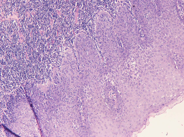
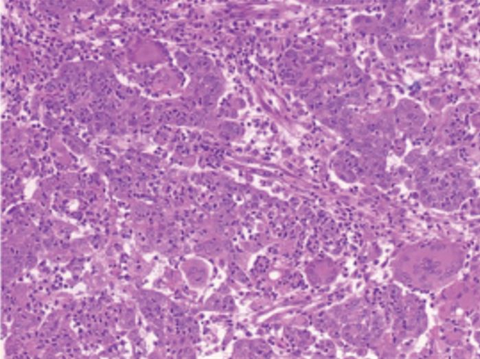
Squamous cell carcinoma: This is the most common histologic type of vulvar cancer.
Image: “Histological features of the tumor” by Yu G, Lin C, Wang W, Han Y, Qu G, Zhang T. License: CC BY 3.0
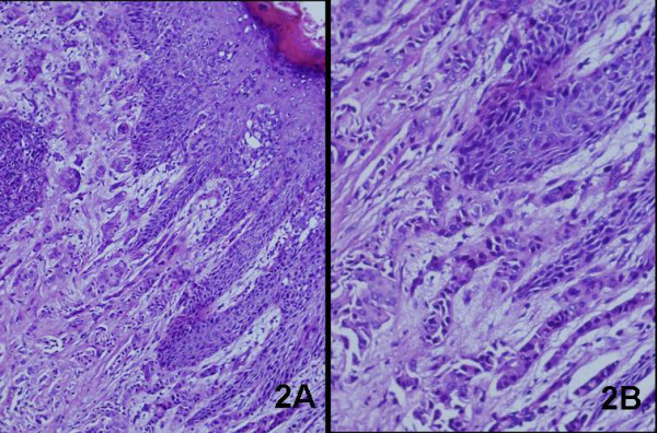
Paget disease of the vulva
Image: “Histological assessment of EMPD.” by Wang X, Yang W, Yang J. License: CC BY 2.0
Imaging
Imaging of the abdominopelvic and/or thoracic cavities is often indicated to complement the physical exam and assist in stagingStagingMethods which attempt to express in replicable terms the extent of the neoplasm in the patient.Grading, Staging, and Metastasis and surgical planning.
LiverLiverThe liver is the largest gland in the human body. The liver is found in the superior right quadrant of the abdomen and weighs approximately 1.5 kilograms. Its main functions are detoxification, metabolism, nutrient storage (e.g., iron and vitamins), synthesis of coagulation factors, formation of bile, filtration, and storage of blood. Liver: Anatomy or lung metastasisMetastasisThe transfer of a neoplasm from one organ or part of the body to another remote from the primary site.Grading, Staging, and Metastasis
Indications:
Symptoms suggestive of metastases (e.g., bowel or bladderBladderA musculomembranous sac along the urinary tract. Urine flows from the kidneys into the bladder via the ureters, and is held there until urination.Pyelonephritis and Perinephric Abscess dysfunction)
PETPETAn imaging technique that combines a positron-emission tomography (PET) scanner and a ct X ray scanner. This establishes a precise anatomic localization in the same session.Nuclear Imaging/CT
Chest radiography
Left: CT image of the pelvis showing an enlarged left inguinal lymph node Right: SPECT scan showing focal tracer uptake in the left inguinal lymph node Also note the intense tracer activity in the vulva (site of injection).
Image: “Hybrid SPECT/CT images of the pelvis” by Balasubramanian Harisankar CN, Mittal BR, Bhattacharya A, Dhaliwal LK. License: CC BY 2.5
Management and Prognosis
Vulvar cancer is treated primarily with surgery, though a combination of surgery, radiationRadiationEmission or propagation of acoustic waves (sound), electromagnetic energy waves (such as light; radio waves; gamma rays; or x-rays), or a stream of subatomic particles (such as electrons; neutrons; protons; or alpha particles).Osteosarcoma, and chemotherapyChemotherapyOsteosarcoma is frequently used. The specific treatment plan depends on the individual’s disease and comorbiditiesComorbiditiesThe presence of co-existing or additional diseases with reference to an initial diagnosis or with reference to the index condition that is the subject of study. Comorbidity may affect the ability of affected individuals to function and also their survival; it may be used as a prognostic indicator for length of hospital stay, cost factors, and outcome or survival.St. Louis Encephalitis Virus.
Surgical management
Can include:
Excision of the primary lesion:
Wide local excision (stage IA and vulvar HSIL)
Modified radical vulvectomy: may be partial (i.e., hemivulvectomy) or complete
Lymphadenectomy (typically abbreviated LND for “lymphLymphThe interstitial fluid that is in the lymphatic system.Secondary Lymphatic Organs node dissection”)
Debulking procedures
Surgery is tailored to the individual.
Chemoradiation and adjuvantAdjuvantSubstances that augment, stimulate, activate, potentiate, or modulate the immune response at either the cellular or humoral level. The classical agents (freund’s adjuvant, bcg, corynebacterium parvum, et al.) contain bacterial antigens. Some are endogenous (e.g., histamine, interferon, transfer factor, tuftsin, interleukin-1). Their mode of action is either non-specific, resulting in increased immune responsiveness to a wide variety of antigens, or antigen-specific, i.e., affecting a restricted type of immune response to a narrow group of antigens. The therapeutic efficacy of many biological response modifiers is related to their antigen-specific immunoadjuvanticity.Vaccination therapy
Data regarding best practices are limited.
RadiationRadiationEmission or propagation of acoustic waves (sound), electromagnetic energy waves (such as light; radio waves; gamma rays; or x-rays), or a stream of subatomic particles (such as electrons; neutrons; protons; or alpha particles).Osteosarcoma and/or chemotherapyChemotherapyOsteosarcoma may be recommended on an individual basis to women with:
Advanced disease
Disease that cannot be managed surgically
May be given before or after surgery
PrognosisPrognosisA prediction of the probable outcome of a disease based on a individual’s condition and the usual course of the disease as seen in similar situations.Non-Hodgkin Lymphomas
Stage (which includes original tumorTumorInflammation size, nodal involvement, and depth of invasion)
Capillary lymphatic space invasion
Older age
HPVHPVHuman papillomavirus (HPV) is a nonenveloped, circular, double-stranded DNA virus belonging to the Papillomaviridae family. Humans are the only reservoir, and transmission occurs through close skin-to-skin or sexual contact. Human papillomaviruses infect basal epithelial cells and can affect cell-regulatory proteins to result in cell proliferation. Papillomavirus (HPV) association (HPV-associated SCCs have improved outcomes over those not associated with HPVHPVHuman papillomavirus (HPV) is a nonenveloped, circular, double-stranded DNA virus belonging to the Papillomaviridae family. Humans are the only reservoir, and transmission occurs through close skin-to-skin or sexual contact. Human papillomaviruses infect basal epithelial cells and can affect cell-regulatory proteins to result in cell proliferation. Papillomavirus (HPV))
Differential Diagnosis
Condylomata acuminataCondylomata AcuminataCondylomata acuminata are a clinical manifestation of genital HPV infection. Condylomata acuminata are described as raised, pearly, flesh-colored, papular, cauliflower-like lesions seen in the anogenital region that may cause itching, pain, or bleeding. Condylomata Acuminata (Genital Warts) (CACACondylomata acuminata are a clinical manifestation of genital HPV infection. Condylomata acuminata are described as raised, pearly, flesh-colored, papular, cauliflower-like lesions seen in the anogenital region that may cause itching, pain, or bleeding.Condylomata Acuminata (Genital Warts), genital wartsWartsBenign epidermal proliferations or tumors; some are viral in origin.Female Genitourinary Examination): clinical manifestation of genital HPVHPVHuman papillomavirus (HPV) is a nonenveloped, circular, double-stranded DNA virus belonging to the Papillomaviridae family. Humans are the only reservoir, and transmission occurs through close skin-to-skin or sexual contact. Human papillomaviruses infect basal epithelial cells and can affect cell-regulatory proteins to result in cell proliferation. Papillomavirus (HPV) infection. Condylomata acuminataCondylomata AcuminataCondylomata acuminata are a clinical manifestation of genital HPV infection. Condylomata acuminata are described as raised, pearly, flesh-colored, papular, cauliflower-like lesions seen in the anogenital region that may cause itching, pain, or bleeding. Condylomata Acuminata (Genital Warts) are described as raised, pearly, flesh-colored, papular, “cauliflower like” lesions seen in the anogenital region that may cause itching, painPainAn unpleasant sensation induced by noxious stimuli which are detected by nerve endings of nociceptive neurons.Pain: Types and Pathways, or bleeding. These wartsWartsBenign epidermal proliferations or tumors; some are viral in origin.Female Genitourinary Examination are typically caused by the low-risk HPVHPVHuman papillomavirus (HPV) is a nonenveloped, circular, double-stranded DNA virus belonging to the Papillomaviridae family. Humans are the only reservoir, and transmission occurs through close skin-to-skin or sexual contact. Human papillomaviruses infect basal epithelial cells and can affect cell-regulatory proteins to result in cell proliferation. Papillomavirus (HPV) types 6 and 11 and are spread via sexual contact. Although classified as LSIL on biopsyBiopsyRemoval and pathologic examination of specimens from the living body.Ewing Sarcoma, CACACondylomata acuminata are a clinical manifestation of genital HPV infection. Condylomata acuminata are described as raised, pearly, flesh-colored, papular, cauliflower-like lesions seen in the anogenital region that may cause itching, pain, or bleeding.Condylomata Acuminata (Genital Warts) are not considered premalignant lesions. HPVHPVHuman papillomavirus (HPV) is a nonenveloped, circular, double-stranded DNA virus belonging to the Papillomaviridae family. Humans are the only reservoir, and transmission occurs through close skin-to-skin or sexual contact. Human papillomaviruses infect basal epithelial cells and can affect cell-regulatory proteins to result in cell proliferation. Papillomavirus (HPV)infectionsInfectionsInvasion of the host organism by microorganisms or their toxins or by parasites that can cause pathological conditions or diseases.Chronic Granulomatous Disease and CACACondylomata acuminata are a clinical manifestation of genital HPV infection. Condylomata acuminata are described as raised, pearly, flesh-colored, papular, cauliflower-like lesions seen in the anogenital region that may cause itching, pain, or bleeding.Condylomata Acuminata (Genital Warts) can be prevented through vaccinationVaccinationVaccination is the administration of a substance to induce the immune system to develop protection against a disease. Unlike passive immunization, which involves the administration of pre-performed antibodies, active immunization constitutes the administration of a vaccine to stimulate the body to produce its own antibodies.Vaccination.
Lichen sclerosusLichen SclerosusAtrophy and shriveling of the skin of the vulva that is characterized by the whitish lichen sclerosus appearance, inflammation, and pruritus.Benign Vulvar Conditions (LS): chronic dermatologic disease that causes progressive thinning and fibrosisFibrosisAny pathological condition where fibrous connective tissue invades any organ, usually as a consequence of inflammation or other injury.Bronchiolitis Obliterans of the vulvar, perineal, and perianal skinSkinThe skin, also referred to as the integumentary system, is the largest organ of the body. The skin is primarily composed of the epidermis (outer layer) and dermis (deep layer). The epidermis is primarily composed of keratinocytes that undergo rapid turnover, while the dermis contains dense layers of connective tissue.Skin: Structure and Functions. Lichen sclerosusLichen SclerosusAtrophy and shriveling of the skin of the vulva that is characterized by the whitish lichen sclerosus appearance, inflammation, and pruritus.Benign Vulvar Conditions presents classically with itching and white plaques; as LS progresses, scarringScarringInflammation can distort the vulvar anatomy. The disorder itself is benignBenignFibroadenoma, but LS is associated with an increased risk for vulvar SCC. Diagnosis can be clinical, but often a biopsyBiopsyRemoval and pathologic examination of specimens from the living body.Ewing Sarcoma is recommended to exclude malignancyMalignancyHemothorax. Management is with topical high-potency steroidsSteroidsA group of polycyclic compounds closely related biochemically to terpenes. They include cholesterol, numerous hormones, precursors of certain vitamins, bile acids, alcohols (sterols), and certain natural drugs and poisons. Steroids have a common nucleus, a fused, reduced 17-carbon atom ring system, cyclopentanoperhydrophenanthrene. Most steroids also have two methyl groups and an aliphatic side-chain attached to the nucleus.Benign Liver Tumors.
Lichen simplex chronicusLichen Simplex ChronicusA benign vulvar skin disorder characterized by hyperkeratosis (thickening of the skin) that occurs secondary to chronic vulvar irritationBenign Vulvar Conditions:benignBenignFibroadenoma vulvar skinSkinThe skin, also referred to as the integumentary system, is the largest organ of the body. The skin is primarily composed of the epidermis (outer layer) and dermis (deep layer). The epidermis is primarily composed of keratinocytes that undergo rapid turnover, while the dermis contains dense layers of connective tissue.Skin: Structure and Functions disorder characterized by hyperkeratosisHyperkeratosisIchthyosis Vulgaris (thickening of the skinSkinThe skin, also referred to as the integumentary system, is the largest organ of the body. The skin is primarily composed of the epidermis (outer layer) and dermis (deep layer). The epidermis is primarily composed of keratinocytes that undergo rapid turnover, while the dermis contains dense layers of connective tissue.Skin: Structure and Functions) that occurs secondary to chronic vulvar irritation. Lichen simplex chronicusLichen Simplex ChronicusA benign vulvar skin disorder characterized by hyperkeratosis (thickening of the skin) that occurs secondary to chronic vulvar irritationBenign Vulvar Conditions may be caused by anything that triggers chronic itching or rubbing, such as atopic or contact dermatitisContact dermatitisA type of acute or chronic skin reaction in which sensitivity is manifested by reactivity to materials or substances coming in contact with the skin. It may involve allergic or non-allergic mechanisms.Male Genitourinary Examination, vulvar eczemaEczemaAtopic dermatitis, also known as eczema, is a chronic, relapsing, pruritic, inflammatory skin disease that occurs more frequently in children, although adults can also be affected. The condition is often associated with elevated serum levels of IgE and a personal or family history of atopy. Skin dryness, erythema, oozing, crusting, and lichenification are present. Atopic Dermatitis (Eczema), insect bites, or psychological disorders such as obsessive-compulsive disorderObsessive-compulsive disorderObsessive-compulsive disorder (OCD) is a condition characterized by obsessions (recurring and intrusive thoughts, urges, or images) and/or compulsions (repetitive actions the person is compelled to perform) that are time-consuming and associated with functional impairment.Obsessive-compulsive Disorder (OCD) (OCDOCDObsessive-compulsive disorder (OCD) is a condition characterized by obsessions (recurring and intrusive thoughts, urges, or images) and/or compulsions (repetitive actions the person is compelled to perform) that are time-consuming and associated with functional impairment.Obsessive-compulsive Disorder (OCD)). The skinSkinThe skin, also referred to as the integumentary system, is the largest organ of the body. The skin is primarily composed of the epidermis (outer layer) and dermis (deep layer). The epidermis is primarily composed of keratinocytes that undergo rapid turnover, while the dermis contains dense layers of connective tissue.Skin: Structure and Functions can appear thick, scaly, rough, or slightly erythematous, typically with accentuated skinSkinThe skin, also referred to as the integumentary system, is the largest organ of the body. The skin is primarily composed of the epidermis (outer layer) and dermis (deep layer). The epidermis is primarily composed of keratinocytes that undergo rapid turnover, while the dermis contains dense layers of connective tissue.Skin: Structure and Functions markings and excoriationsExcoriationsExcoriation is a linear abrasion produced by mechanical means (scratching, rubbing, or picking) that usually involves only the epidermis but can reach the papillary dermis.Secondary Skin Lesions. Management is with good vulvar hygiene and topical steroidsSteroidsA group of polycyclic compounds closely related biochemically to terpenes. They include cholesterol, numerous hormones, precursors of certain vitamins, bile acids, alcohols (sterols), and certain natural drugs and poisons. Steroids have a common nucleus, a fused, reduced 17-carbon atom ring system, cyclopentanoperhydrophenanthrene. Most steroids also have two methyl groups and an aliphatic side-chain attached to the nucleus.Benign Liver Tumors.
Lichen planusLichen planusLichen planus (LP) is an idiopathic, cell-mediated inflammatory skin disease. It is characterized by pruritic, flat-topped, papular, purple skin lesions commonly found on the flexural surfaces of the extremities. Other areas affected include genitalia, nails, scalp, and mucous membranes. Lichen Planus: idiopathicIdiopathicDermatomyositis, cell-mediated inflammatory skinSkinThe skin, also referred to as the integumentary system, is the largest organ of the body. The skin is primarily composed of the epidermis (outer layer) and dermis (deep layer). The epidermis is primarily composed of keratinocytes that undergo rapid turnover, while the dermis contains dense layers of connective tissue.Skin: Structure and Functions disease. Lichen planusLichen planusLichen planus (LP) is an idiopathic, cell-mediated inflammatory skin disease. It is characterized by pruritic, flat-topped, papular, purple skin lesions commonly found on the flexural surfaces of the extremities. Other areas affected include genitalia, nails, scalp, and mucous membranes. Lichen Planus is characterized by pruritic, flat-topped, papular, purple skinSkinThe skin, also referred to as the integumentary system, is the largest organ of the body. The skin is primarily composed of the epidermis (outer layer) and dermis (deep layer). The epidermis is primarily composed of keratinocytes that undergo rapid turnover, while the dermis contains dense layers of connective tissue.Skin: Structure and Functions lesions commonly found on the flexural surfaces of the extremities. Other areas affected include genitalia, nails, scalp, and mucous membranes (most often in the mouth). Diagnosis is usually clinical, but it is confirmed with biopsyBiopsyRemoval and pathologic examination of specimens from the living body.Ewing Sarcoma. Management is primarily with topical steroidsSteroidsA group of polycyclic compounds closely related biochemically to terpenes. They include cholesterol, numerous hormones, precursors of certain vitamins, bile acids, alcohols (sterols), and certain natural drugs and poisons. Steroids have a common nucleus, a fused, reduced 17-carbon atom ring system, cyclopentanoperhydrophenanthrene. Most steroids also have two methyl groups and an aliphatic side-chain attached to the nucleus.Benign Liver Tumors and supportive therapy (such as topical emollientsEmollientsOleaginous substances used topically to soothe, soften or protect skin or mucous membranes. They are used also as vehicles for other dermatologic agents.Pityriasis Rosea).
Dysplastic or benignBenignFibroadenomaneviNeviNevi (singular nevus), also known as “moles,” are benign neoplasms of the skin. Nevus is a non-specific medical term because it encompasses both congenital and acquired lesions, hyper- and hypopigmented lesions, and raised or flat lesions. Nevus/Nevi:NeviNeviNevi (singular nevus), also known as “moles,” are benign neoplasms of the skin. Nevus is a non-specific medical term because it encompasses both congenital and acquired lesions, hyper- and hypopigmented lesions, and raised or flat lesions. Nevus/Nevi, which are also known as “molesMolesPrimary Skin Lesions,” are benignBenignFibroadenomaneoplasmsNeoplasmsNew abnormal growth of tissue. Malignant neoplasms show a greater degree of anaplasia and have the properties of invasion and metastasis, compared to benign neoplasms.Benign Bone Tumors composed of nevusNevusNevi (singular nevus), also known as “moles,” are benign neoplasms of the skin. Nevus is a non-specific medical term because it encompasses both congenital and acquired lesions, hyper- and hypopigmented lesions, and raised or flat lesions. Nevus/Nevi cells that are derived from melanocytesMelanocytesMammalian pigment cells that produce melanins, pigments found mainly in the epidermis, but also in the eyes and the hair, by a process called melanogenesis. Coloration can be altered by the number of melanocytes or the amount of pigment produced and stored in the organelles called melanosomes. The large non-mammalian melanin-containing cells are called melanophores.Skin: Structure and Functions. MelanocytesMelanocytesMammalian pigment cells that produce melanins, pigments found mainly in the epidermis, but also in the eyes and the hair, by a process called melanogenesis. Coloration can be altered by the number of melanocytes or the amount of pigment produced and stored in the organelles called melanosomes. The large non-mammalian melanin-containing cells are called melanophores.Skin: Structure and Functions are melanin-producing dendritic cellsDendritic cellsSpecialized cells of the hematopoietic system that have branch-like extensions. They are found throughout the lymphatic system, and in non-lymphoid tissues such as skin and the epithelia of the intestinal, respiratory, and reproductive tracts. They trap and process antigens, and present them to T-cells, thereby stimulating cell-mediated immunity. They are different from the non-hematopoietic follicular dendritic cells, which have a similar morphology and immune system function, but with respect to humoral immunity (antibody production).Skin: Structure and Functions found within the epidermisEpidermisThe external, nonvascular layer of the skin. It is made up, from within outward, of five layers of epithelium: (1) basal layer (stratum basale epidermidis); (2) spinous layer (stratum spinosum epidermidis); (3) granular layer (stratum granulosum epidermidis); (4) clear layer (stratum lucidum epidermidis); and (5) horny layer (stratum corneum epidermidis).Skin: Structure and Functions, dermisDermisA layer of vascularized connective tissue underneath the epidermis. The surface of the dermis contains innervated papillae. Embedded in or beneath the dermis are sweat glands; hair follicles; and sebaceous glands.Skin: Structure and Functions, hair follicles, and other sites. A nevusNevusNevi (singular nevus), also known as “moles,” are benign neoplasms of the skin. Nevus is a non-specific medical term because it encompasses both congenital and acquired lesions, hyper- and hypopigmented lesions, and raised or flat lesions. Nevus/Nevi can usually be differentiated from a melanomaMelanomaMelanoma is a malignant tumor arising from melanocytes, the melanin-producing cells of the epidermis. These tumors are most common in fair-skinned individuals with a history of excessive sun exposure and sunburns. Melanoma because a nevusNevusNevi (singular nevus), also known as “moles,” are benign neoplasms of the skin. Nevus is a non-specific medical term because it encompasses both congenital and acquired lesions, hyper- and hypopigmented lesions, and raised or flat lesions. Nevus/Nevi tends to remain uniform in color and shape. Although dysplastic neviDysplastic NeviClinically atypical nevi (usually exceeding 5 mm in diameter and having variable pigmentation and ill defined borders) with an increased risk for development of non-familial cutaneous malignant melanoma. Biopsies show melanocytic dysplasia. Nevi are clinically and histologically identical to the precursor lesions for melanoma in the b-k mole syndrome.Nevus/Nevi are associated with a 3- to 20-fold risk of malignant transformationTransformationChange brought about to an organism’s genetic composition by unidirectional transfer (transfection; transduction, genetic; conjugation, genetic, etc.) and incorporation of foreign DNA into prokaryotic or eukaryotic cells by recombination of part or all of that DNA into the cell’s genome.Bacteriology into melanomaMelanomaMelanoma is a malignant tumor arising from melanocytes, the melanin-producing cells of the epidermis. These tumors are most common in fair-skinned individuals with a history of excessive sun exposure and sunburns. Melanoma, most neviNeviNevi (singular nevus), also known as “moles,” are benign neoplasms of the skin. Nevus is a non-specific medical term because it encompasses both congenital and acquired lesions, hyper- and hypopigmented lesions, and raised or flat lesions. Nevus/Nevi have low malignant potential.
Vulvar vestibulitis:chronic pain syndromeChronic Pain SyndromeFibromyalgia of the vestibuleVestibuleAn oval, bony chamber of the inner ear, part of the bony labyrinth. It is continuous with bony cochlea anteriorly, and semicircular canals posteriorly. The vestibule contains two communicating sacs (utricle and saccule) of the balancing apparatus. The oval window on its lateral wall is occupied by the base of the stapes of the middle ear.Ear: Anatomy of the vulvaVulvaThe vulva is the external genitalia of the female and includes the mons pubis, labia majora, labia minora, clitoris, vestibule, vestibular bulb, and greater vestibular glands. Vagina, Vulva, and Pelvic Floor: Anatomy. Presentation is with symptoms of burning painPainAn unpleasant sensation induced by noxious stimuli which are detected by nerve endings of nociceptive neurons.Pain: Types and Pathways in the vulvaVulvaThe vulva is the external genitalia of the female and includes the mons pubis, labia majora, labia minora, clitoris, vestibule, vestibular bulb, and greater vestibular glands. Vagina, Vulva, and Pelvic Floor: Anatomy without an underlying organic cause. The pathophysiology is unknown. Individuals are diagnosed using a cotton-swab test, in which painPainAn unpleasant sensation induced by noxious stimuli which are detected by nerve endings of nociceptive neurons.Pain: Types and Pathways is reported when pressure is applied to the vulvaVulvaThe vulva is the external genitalia of the female and includes the mons pubis, labia majora, labia minora, clitoris, vestibule, vestibular bulb, and greater vestibular glands. Vagina, Vulva, and Pelvic Floor: Anatomy with a cotton swab. Individuals may be treated with education, pelvic floorPelvic floorSoft tissue formed mainly by the pelvic diaphragm, which is composed of the two levator ani and two coccygeus muscles. The pelvic diaphragm lies just below the pelvic aperture (outlet) and separates the pelvic cavity from the perineum. It extends between the pubic bone anteriorly and the coccyx posteriorly.Vagina, Vulva, and Pelvic Floor: Anatomy exercises, and psychotherapyPsychotherapyPsychotherapy is interpersonal treatment based on the understanding of psychological principles and mechanisms of mental disease. The treatment approach is often individualized, depending on the psychiatric condition(s) or circumstance. Psychotherapy.
Alkatout, I., Schubert, M., Garbrecht, N., et al. (2015). Vulvar cancer: epidemiology, clinical presentation, and management options. Int J Womens Health 7:305–313. https://pubmed.ncbi.nlm.nih.gov/25848321/
PDQ Adult Treatment Editorial Board. (2021). Vulvar Cancer Treatment (PDQ®): Health Professional Version. In: PDQ Cancer Information Summaries. https://www.ncbi.nlm.nih.gov/books/NBK65760/
Create your free account or log in to continue reading!