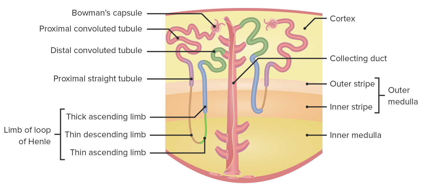
The kidneysKidneysThe kidneys are a pair of bean-shaped organs located retroperitoneally against the posterior wall of the abdomen on either side of the spine. As part of the urinary tract, the kidneys are responsible for blood filtration and excretion of water-soluble waste in the urine.Kidneys: Anatomy regulate water and solute homeostasisHomeostasisThe processes whereby the internal environment of an organism tends to remain balanced and stable.Cell Injury and Death through the processes of filtration, reabsorption, secretionSecretionCoagulation Studies, and excretion. After the filtration of blood through the glomeruli, the tubular system takes over and is responsible for adjusting the urine composition throughout the remainder of the nephronNephronThe functional units of the kidney, consisting of the glomerulus and the attached tubule.Kidneys: Anatomy. Reabsorption, secretionSecretionCoagulation Studies, and excretion occur via active and passive transportPassive transportThe passive movement of molecules exceeding the rate expected by simple diffusion. No energy is expended in the process. It is achieved by the introduction of passively diffusing molecules to an environment or path that is more favorable to the movement of those molecules. Examples of facilitated diffusion are passive transport of hydrophilic substances across a lipid membrane through hydrophilic pores that traverse the membrane, and the sliding of a DNA binding protein along a strand of DNA.The Cell: Cell Membrane mechanisms and respond dynamically to the body’s current needs to maintain homeostasisHomeostasisThe processes whereby the internal environment of an organism tends to remain balanced and stable.Cell Injury and Death of the plasmaPlasmaThe residual portion of blood that is left after removal of blood cells by centrifugation without prior blood coagulation.Transfusion Products composition and blood volume. The primary segments of the tubular system include the proximal tubule, loop of Henle, distal convoluted tubuleDistal convoluted tubuleThe portion of renal tubule that begins from the enlarged segment of the ascending limb of the loop of henle. It reenters the kidney cortex and forms the convoluted segments of the distal tubule.Gitelman Syndrome, and collecting ducts. Each segment has unique transporters and functions.
Proximal convoluted tubuleProximal convoluted tubuleThe renal tubule portion that extends from the bowman capsule in the kidney cortex into the kidney medulla. The proximal tubule consists of a convoluted proximal segment in the cortex, and a distal straight segment descending into the medulla where it forms the u-shaped loop of henle.Osmotic Diuretics
Distal convoluted tubuleDistal convoluted tubuleThe portion of renal tubule that begins from the enlarged segment of the ascending limb of the loop of henle. It reenters the kidney cortex and forms the convoluted segments of the distal tubule.Gitelman Syndrome
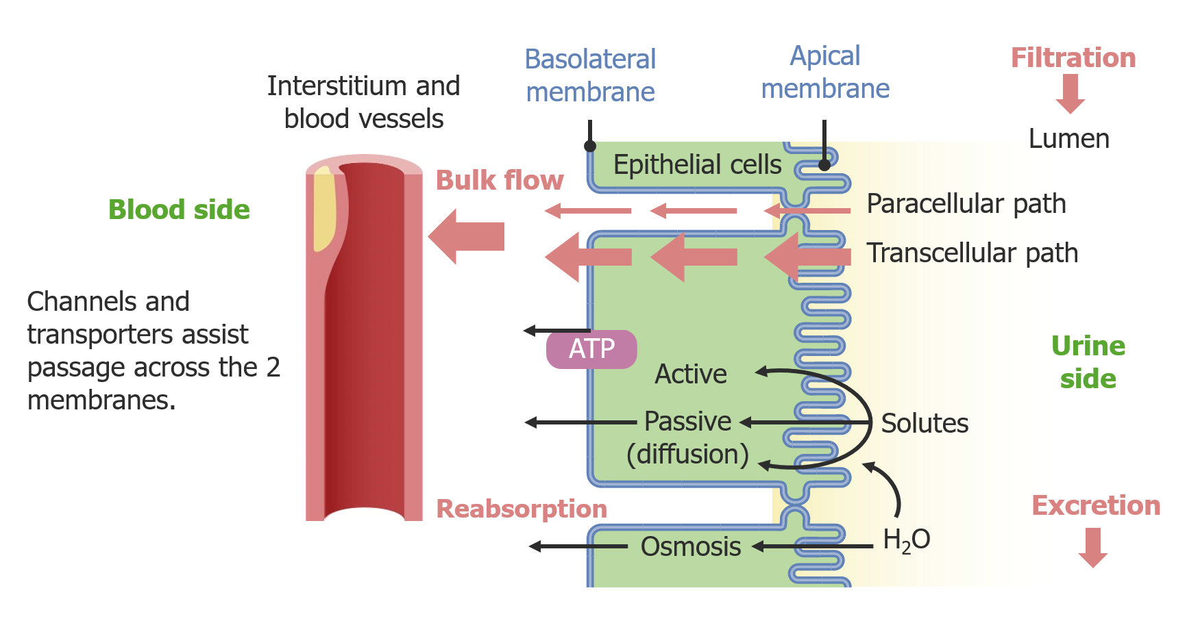
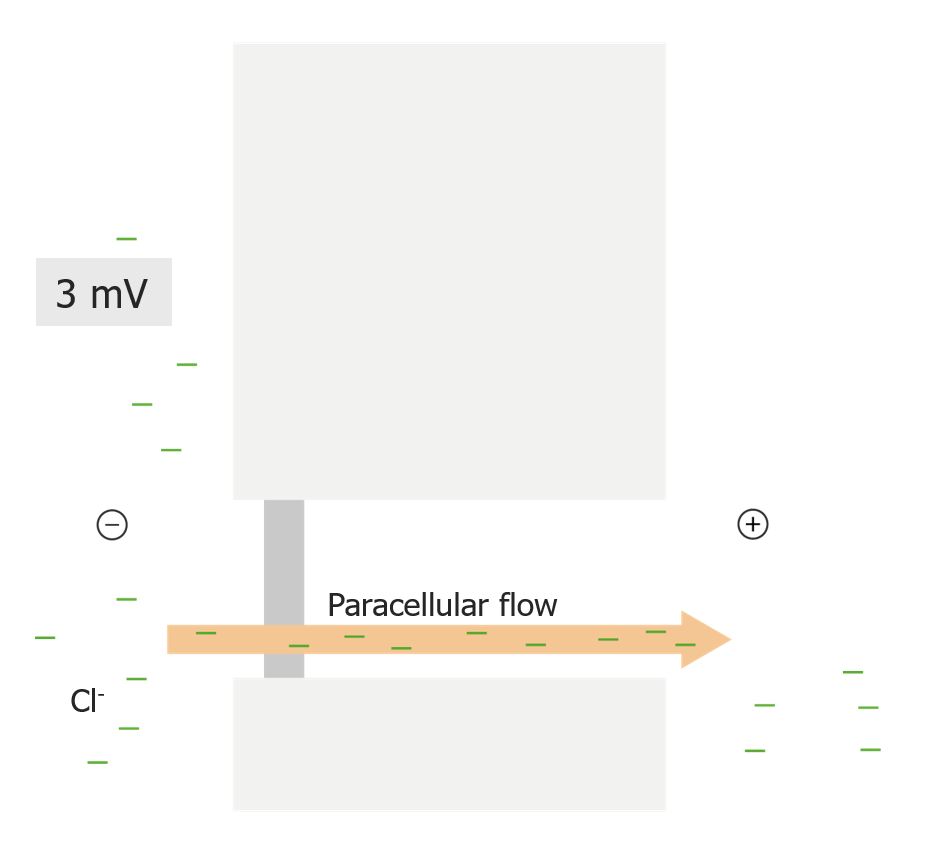
ParacellularParacellularRenal Potassium Regulation: passive transportPassive transportThe passive movement of molecules exceeding the rate expected by simple diffusion. No energy is expended in the process. It is achieved by the introduction of passively diffusing molecules to an environment or path that is more favorable to the movement of those molecules. Examples of facilitated diffusion are passive transport of hydrophilic substances across a lipid membrane through hydrophilic pores that traverse the membrane, and the sliding of a DNA binding protein along a strand of DNA.The Cell: Cell Membrane in between cells
Transcellular: transport through cells; may be active or passive
Pathways of epithelial transport of solutes from the tubular lumen
SodiumSodiumA member of the alkali group of metals. It has the atomic symbol na, atomic number 11, and atomic weight 23.Hyponatremia/PotassiumPotassiumAn element in the alkali group of metals with an atomic symbol k, atomic number 19, and atomic weight 39. 10. It is the chief cation in the intracellular fluid of muscle and other cells. Potassium ion is a strong electrolyte that plays a significant role in the regulation of fluid volume and maintenance of the water-electrolyte balance.Hyperkalemia ATPase
Located on the basolateral side of tubule cells
Transports:
3 Na+ out of the cell
2 K+ into the cell
Creates a Na+ concentration gradient and a voltage gradient:
The tubule lumen becomes electronegative in the early proximal convoluted tubuleProximal convoluted tubuleThe renal tubule portion that extends from the bowman capsule in the kidney cortex into the kidney medulla. The proximal tubule consists of a convoluted proximal segment in the cortex, and a distal straight segment descending into the medulla where it forms the u-shaped loop of henle.Osmotic Diuretics (however, the electronegativity changes as substances are absorbed throughout the nephronNephronThe functional units of the kidney, consisting of the glomerulus and the attached tubule.Kidneys: Anatomy).
Active and passive transportPassive transportThe passive movement of molecules exceeding the rate expected by simple diffusion. No energy is expended in the process. It is achieved by the introduction of passively diffusing molecules to an environment or path that is more favorable to the movement of those molecules. Examples of facilitated diffusion are passive transport of hydrophilic substances across a lipid membrane through hydrophilic pores that traverse the membrane, and the sliding of a DNA binding protein along a strand of DNA.The Cell: Cell Membrane mechanisms are dependent on these gradients.
Establishment of Na+ concentration gradient and voltage gradient by the Na+/K+-ATPase
Water moves through the epithelial tight junctionsTight junctionsCell-cell junctions that seal adjacent epithelial cells together, preventing the passage of most dissolved molecules from one side of the epithelial sheet to the other.The Cell: Cell Junctions (“leaky” subtype of tight junctionsTight junctionsCell-cell junctions that seal adjacent epithelial cells together, preventing the passage of most dissolved molecules from one side of the epithelial sheet to the other.The Cell: Cell Junctions).
Movement of water is dictated by osmolarityOsmolarityThe concentration of osmotically active particles in solution expressed in terms of osmoles of solute per liter of solution. Osmolality is expressed in terms of osmoles of solute per kilogram of solvent.Hypernatremia (osmosisOsmosisTendency of fluids (e.g., water) to move from the less concentrated to the more concentrated side of a semipermeable membrane.Body Fluid Compartments).
Collecting ductCollecting ductStraight tubes commencing in the radiate part of the kidney cortex where they receive the curved ends of the distal convoluted tubules. In the medulla the collecting tubules of each pyramid converge to join a central tube (duct of bellini) which opens on the summit of the papilla.Renal Cell Carcinoma
Solvent drag:
Some solutes are “dragged” along as water moves.
Solutes move via convective currents created by the movement of water.
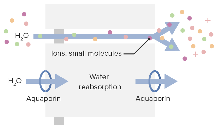
Mechanisms of water movement through the cell: Top pathway shows paracellular movement of water through tight junctions with solvent drag. Bottom pathway shows transcellular movement of water through aquaporin channels.
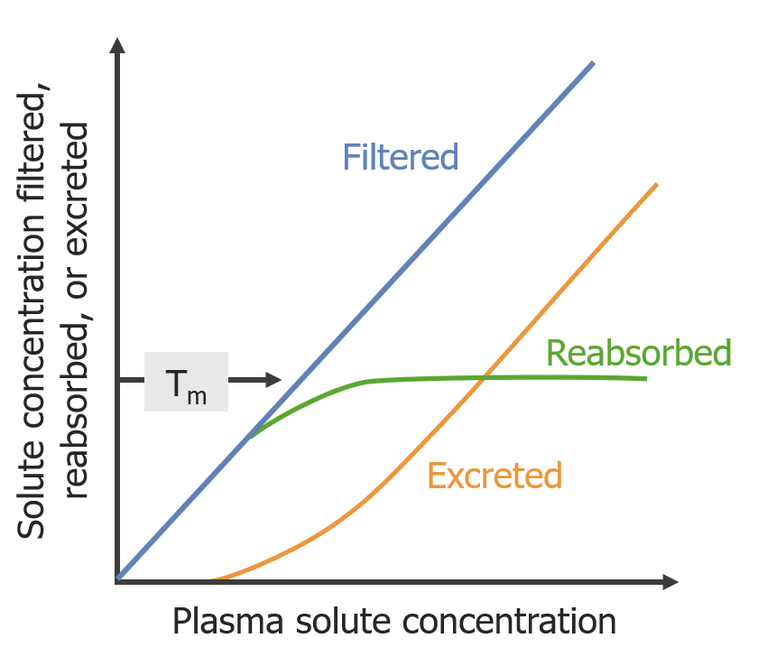
The reabsorption capacity for any given substance is limited.
Once exceeded, additional substances are lost in the urine.
Also referred to as the “renal thresholdThresholdMinimum voltage necessary to generate an action potential (an all-or-none response)Skeletal Muscle Contraction” for reabsorption
For example, glucoseGlucoseA primary source of energy for living organisms. It is naturally occurring and is found in fruits and other parts of plants in its free state. It is used therapeutically in fluid and nutrient replacement.Lactose Intolerance:
Has transport maximum (Tm) of 375 mg/min
At this rate, the kidneysKidneysThe kidneys are a pair of bean-shaped organs located retroperitoneally against the posterior wall of the abdomen on either side of the spine. As part of the urinary tract, the kidneys are responsible for blood filtration and excretion of water-soluble waste in the urine.Kidneys: Anatomy can reabsorb 100% of the filtered glucoseGlucoseA primary source of energy for living organisms. It is naturally occurring and is found in fruits and other parts of plants in its free state. It is used therapeutically in fluid and nutrient replacement.Lactose Intolerance up to a plasmaPlasmaThe residual portion of blood that is left after removal of blood cells by centrifugation without prior blood coagulation.Transfusion ProductsglucoseGlucoseA primary source of energy for living organisms. It is naturally occurring and is found in fruits and other parts of plants in its free state. It is used therapeutically in fluid and nutrient replacement.Lactose Intolerance concentration of approximately 180 mg/dL.
When plasmaPlasmaThe residual portion of blood that is left after removal of blood cells by centrifugation without prior blood coagulation.Transfusion ProductsglucoseGlucoseA primary source of energy for living organisms. It is naturally occurring and is found in fruits and other parts of plants in its free state. It is used therapeutically in fluid and nutrient replacement.Lactose Intolerance exceeds 180 mg/dL:
KidneysKidneysThe kidneys are a pair of bean-shaped organs located retroperitoneally against the posterior wall of the abdomen on either side of the spine. As part of the urinary tract, the kidneys are responsible for blood filtration and excretion of water-soluble waste in the urine.Kidneys: Anatomy are no longer capable of reabsorbing 100% of the filtered glucoseGlucoseA primary source of energy for living organisms. It is naturally occurring and is found in fruits and other parts of plants in its free state. It is used therapeutically in fluid and nutrient replacement.Lactose Intolerance.
Excess glucoseGlucoseA primary source of energy for living organisms. It is naturally occurring and is found in fruits and other parts of plants in its free state. It is used therapeutically in fluid and nutrient replacement.Lactose Intolerance is lost in urine (“glucosuriaGlucosuriaDiabetes Mellitus”).
Effect of maximum transport on excretion tm= maximum transport
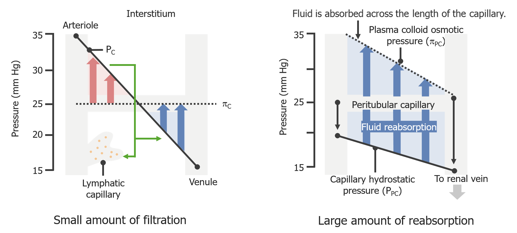
Peritubular capillary absorptionAbsorptionAbsorption involves the uptake of nutrient molecules and their transfer from the lumen of the GI tract across the enterocytes and into the interstitial space, where they can be taken up in the venous or lymphatic circulation.Digestion and Absorption
Peritubular capillary reabsorption differs from regularRegularInsulin capillary reabsorption to maximize the reabsorption of substances back into the bloodstream.
RegularRegularInsulincapillariesCapillariesCapillaries are the primary structures in the circulatory system that allow the exchange of gas, nutrients, and other materials between the blood and the extracellular fluid (ECF). Capillaries are the smallest of the blood vessels. Because a capillary diameter is so small, only 1 RBC may pass through at a time.Capillaries: Histology filter along their 1st half and reabsorb along their 2nd half:
Lower capillary hydrostatic pressureHydrostatic pressureThe pressure due to the weight of fluid.Edema and higher capillary oncotic pressureOncotic PressureEdema across their entire length
No area of filtration
Starling forces of a regular capillary (left) and a peritubular capillary (right). In both images, the dotted lines represent oncotic pressure, while the solid line represents hydrostatic pressure.
Glomerular filtrationGlomerular filtrationThe kidneys are primarily in charge of the maintenance of water and solute homeostasis through the processes of filtration, reabsorption, secretion, and excretion. Glomerular filtration is the process of converting the systemic blood supply into a filtrate, which will ultimately become the urine. Glomerular Filtration is a very nonspecific process, resulting in the filtration of large quantities of important substances that the body needs to retain (e.g., Na+, HCO3–). The primary function of the PT is to reabsorb as much of these substances as possible. Subsequently, the other nephronNephronThe functional units of the kidney, consisting of the glomerulus and the attached tubule.Kidneys: Anatomy segments fine-tune the urine composition.
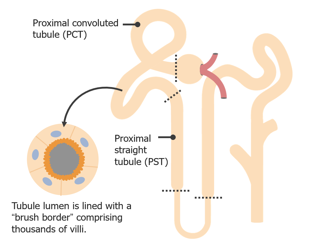
Anatomy of the PT
Divided into 2 parts: proximal convoluted tubuleProximal convoluted tubuleThe renal tubule portion that extends from the bowman capsule in the kidney cortex into the kidney medulla. The proximal tubule consists of a convoluted proximal segment in the cortex, and a distal straight segment descending into the medulla where it forms the u-shaped loop of henle.Osmotic Diuretics and PST
Proximal convoluted tubuleProximal convoluted tubuleThe renal tubule portion that extends from the bowman capsule in the kidney cortex into the kidney medulla. The proximal tubule consists of a convoluted proximal segment in the cortex, and a distal straight segment descending into the medulla where it forms the u-shaped loop of henle.Osmotic Diuretics: primary location for PT reabsorption
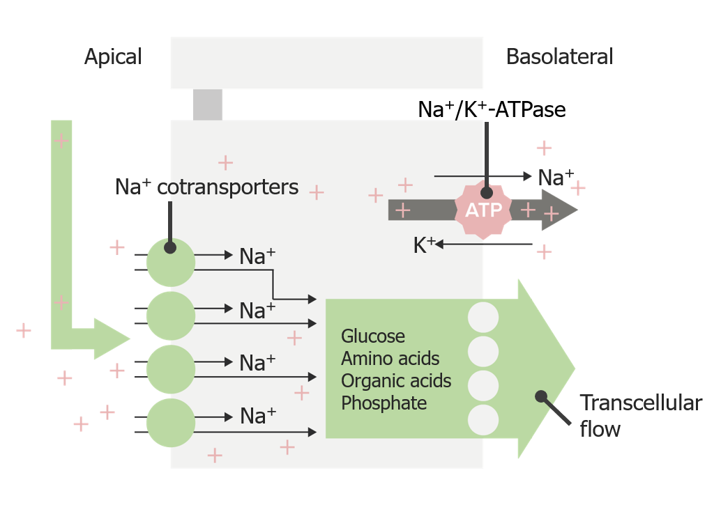
SodiumSodiumA member of the alkali group of metals. It has the atomic symbol na, atomic number 11, and atomic weight 23.Hyponatremia reabsorption in the PT
Coupled with reabsorption of other substances via cotransporters:
GlucoseGlucoseA primary source of energy for living organisms. It is naturally occurring and is found in fruits and other parts of plants in its free state. It is used therapeutically in fluid and nutrient replacement.Lactose Intolerance
Amino acidsAmino acidsOrganic compounds that generally contain an amino (-NH2) and a carboxyl (-COOH) group. Twenty alpha-amino acids are the subunits which are polymerized to form proteins.Basics of Amino Acids
Organic acidsAcidsChemical compounds which yield hydrogen ions or protons when dissolved in water, whose hydrogen can be replaced by metals or basic radicals, or which react with bases to form salts and water (neutralization). An extension of the term includes substances dissolved in media other than water.Acid-Base Balance
Powered by the Na+ gradient generated by the basolateral Na+/K+-ATPase:
Low intracellular Na+ concentration
High Na+ concentration in the tubule lumen and interstitial space on the basolateral side
Reabsorption of Na+ in the PT is isotonicIsotonicSolutions having the same osmotic pressure as blood serum, or another solution with which they are compared.Renal Sodium and Water Regulation to plasmaPlasmaThe residual portion of blood that is left after removal of blood cells by centrifugation without prior blood coagulation.Transfusion Products.
Efficiency: Approximately ⅔ of filtered water and Na+ are reabsorbed in the PT.
Sodium reabsorption via transcellular cotransport in the proximal tubule
PotassiumPotassiumAn element in the alkali group of metals with an atomic symbol k, atomic number 19, and atomic weight 39. 10. It is the chief cation in the intracellular fluid of muscle and other cells. Potassium ion is a strong electrolyte that plays a significant role in the regulation of fluid volume and maintenance of the water-electrolyte balance.Hyperkalemia, calciumCalciumA basic element found in nearly all tissues. It is a member of the alkaline earth family of metals with the atomic symbol ca, atomic number 20, and atomic weight 40. Calcium is the most abundant mineral in the body and combines with phosphorus to form calcium phosphate in the bones and teeth. It is essential for the normal functioning of nerves and muscles and plays a role in blood coagulation (as factor IV) and in many enzymatic processes.Electrolytes (CaCACondylomata acuminata are a clinical manifestation of genital HPV infection. Condylomata acuminata are described as raised, pearly, flesh-colored, papular, cauliflower-like lesions seen in the anogenital region that may cause itching, pain, or bleeding.Condylomata Acuminata (Genital Warts)2+), and magnesiumMagnesiumA metallic element that has the atomic symbol mg, atomic number 12, and atomic weight 24. 31. It is important for the activity of many enzymes, especially those involved in oxidative phosphorylation.Electrolytes (Mg2+) reabsorption in the PT
Due to upstream reabsorption of Cl– in the early PT, the polarity in the late PT flips.
In the late PT, the tubule lumen becomes more electropositive, and the basolateral interstitium becomes more electronegative.
Efficiency:
80% of the filtered K+ is reabsorbed in the PT.
65% of the filtered CaCACondylomata acuminata are a clinical manifestation of genital HPV infection. Condylomata acuminata are described as raised, pearly, flesh-colored, papular, cauliflower-like lesions seen in the anogenital region that may cause itching, pain, or bleeding.Condylomata Acuminata (Genital Warts)2+ is reabsorbed in the PT.
15% of the filtered Mg2+ is reabsorbed in the PT.
Potassium transport in the proximal tubule: In the early proximal tubule, potassium reabsorption occurs primarily via solvent drag with the reabsorption of water. In the late PT, the voltage-gradient flips (due to the upstream reabsorption of Cl–) and potassium is reabsorbed via paracellular diffusion across the tight junctions following the electrical gradient.
Reabsorption of HCO3– requires a more complex mechanism:
Sodium-hydrogen ion exchanger 3 (NHE3NHE3A sodium-hydrogen antiporter expressed primarily by epithelial cells in the kidneys, it localizes to the apical membrane of the proximal kidney tubule, where it functions in sodium and water reabsorption and possibly calcium homeostasis. It also is expressed in heart, brain, and lung tissues and is resistant to amiloride inhibition.Carbonic Anhydrase Inhibitors) reabsorbs Na+ and secretes H+.
The secreted H+ combines with the filtered HCO3– to form carbonic acid (H2CO3) in the tubular lumen.
H2CO3 is converted to H2O and CO2 by apical carbonic anhydrase-IVCarbonic anhydrase-IVA carbonic anhydrase isoenzyme found in mitochondria where it provides bicarbonate ions that are components in the urea cycle and in gluconeogenesis.Carbonic Anhydrase Inhibitors.
CO2 diffuses freely across the apical membrane back into the cell.
Intracellularcarbonic anhydrase-IICarbonic anhydrase-IIA cytosolic carbonic anhydrase isoenzyme found widely distributed in cells of almost all tissues. Deficiencies of carbonic anhydrase II produce a syndrome characterized by osteopetrosis, renal tubular acidosis and cerebral calcification.Carbonic Anhydrase Inhibitors converts CO2 and H2O back into H2CO3.
H2CO3 then dissociates into H+ and HCO3–:
H+ is recycled through the process via secretionSecretionCoagulation Studies by NHE3NHE3A sodium-hydrogen antiporter expressed primarily by epithelial cells in the kidneys, it localizes to the apical membrane of the proximal kidney tubule, where it functions in sodium and water reabsorption and possibly calcium homeostasis. It also is expressed in heart, brain, and lung tissues and is resistant to amiloride inhibition.Carbonic Anhydrase Inhibitors.
HCO3– is absorbed through the basolateral membrane via:
Na+-HCO3– cotransporter
HCO3–-Cl– exchanger
Net effects of the entire process:
Excretion of H+
AbsorptionAbsorptionAbsorption involves the uptake of nutrient molecules and their transfer from the lumen of the GI tract across the enterocytes and into the interstitial space, where they can be taken up in the venous or lymphatic circulation.Digestion and Absorption of HCO3–
Efficiency: Under normal circumstances, 80% of the filtered HCO3– is reabsorbed in the PT.
Bicarbonate reabsorption in the proximal tubule CA-IV: carbonic anhydrase IV CA-II: carbonic anhydrase II Sodium-hydrogen ion exchanger 3 (NHE3)
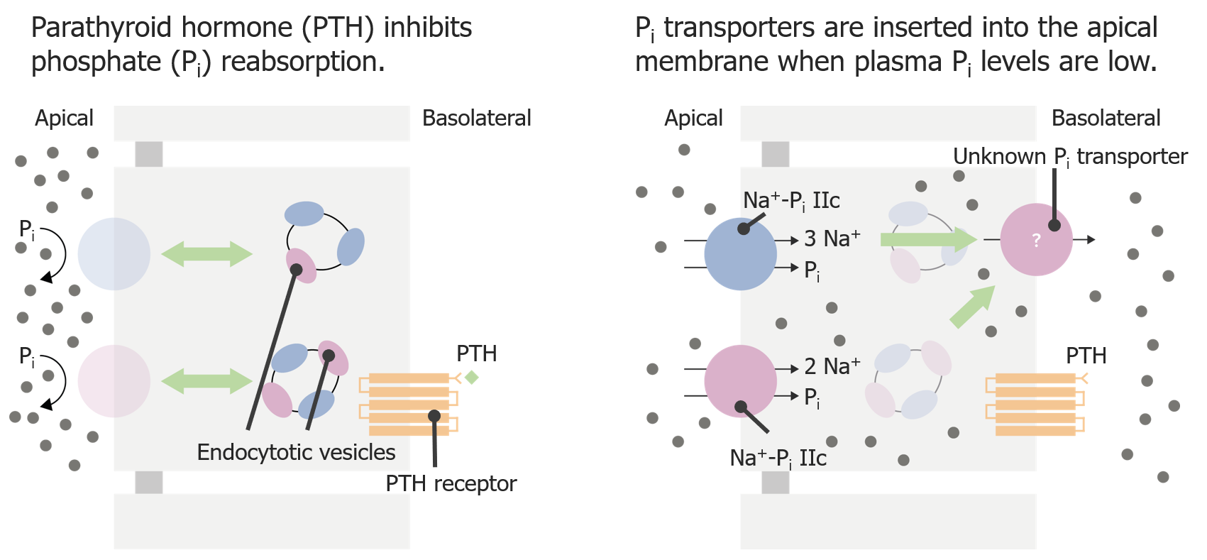
PhosphatePhosphateInorganic salts of phosphoric acid.Electrolytes (PO43-) reabsorption in the PT
PO43- reabsorption is regulated by the parathyroidParathyroidThe parathyroid glands are 2 pairs of small endocrine glands found in close proximity to the thyroid gland. The superior parathyroid glands are lodged within the parenchyma of the upper poles of the right and left thyroid lobes; the inferior parathyroid glands are close to the inferior tips or poles of the lobes.Parathyroid Glands: Anatomy hormone (PTH): PTH inhibits PO43- reabsorption.
↓ PTH → ↑ PO43- reabsorption:
In the setting of ↓ PTH, Na+/PO43- cotransporters (transporting 3 Na+ and 1 PO43-) are inserted into the apical membrane
PO43- moves across the cell and is transported across the basolateral membrane via an unknown transporter.
↑ PTH → ↓ PO43- reabsorption:
PTH binds to a basolateral PTH receptorReceptorReceptors are proteins located either on the surface of or within a cell that can bind to signaling molecules known as ligands (e.g., hormones) and cause some type of response within the cell.Receptors of the PT cell.
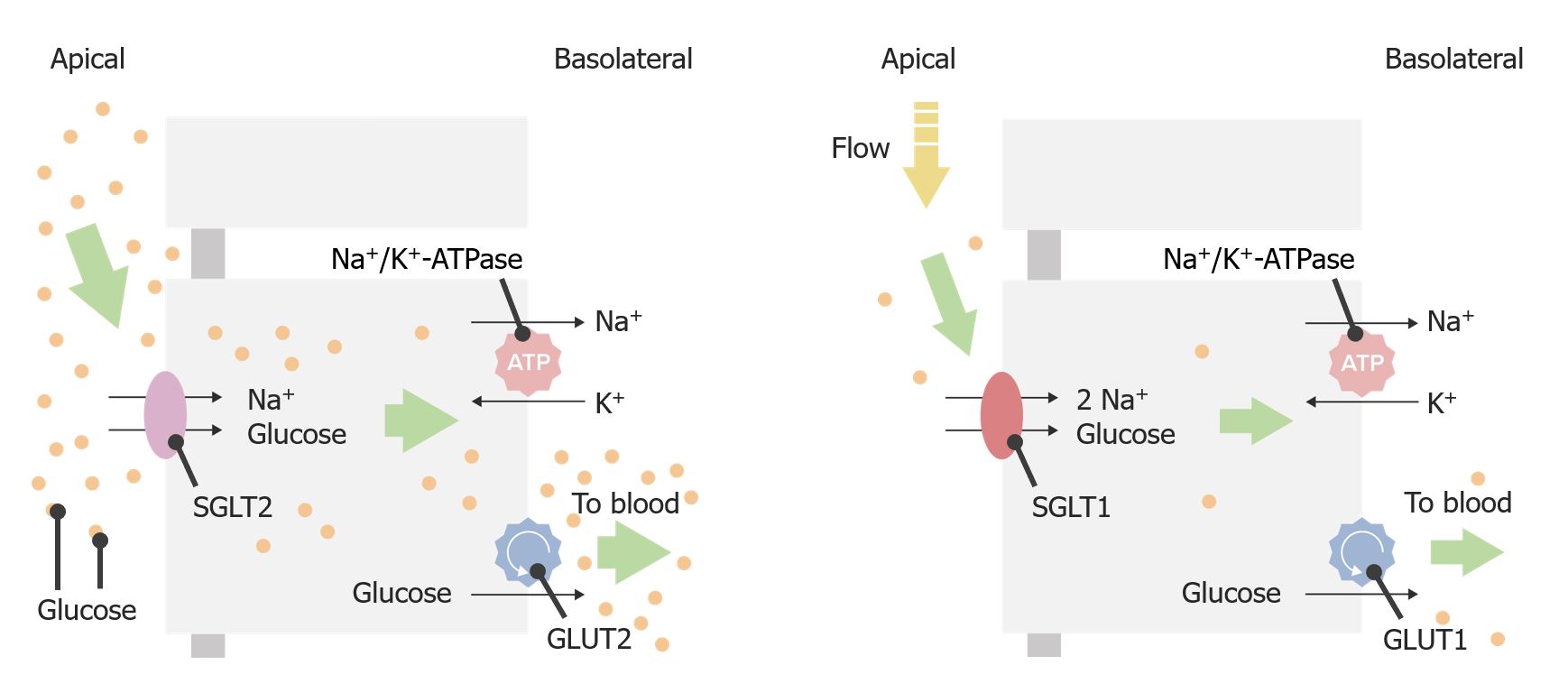
GlucoseGlucoseA primary source of energy for living organisms. It is naturally occurring and is found in fruits and other parts of plants in its free state. It is used therapeutically in fluid and nutrient replacement.Lactose Intolerance reabsorption in the PT
Apical transporters: sodium-glucose linked transporter (SGLT)2 and SGLT1
SGLT2 transporter:
1 Na+ and 1 glucoseGlucoseA primary source of energy for living organisms. It is naturally occurring and is found in fruits and other parts of plants in its free state. It is used therapeutically in fluid and nutrient replacement.Lactose Intolerance move into the cell.
Responsible for the majority of glucoseGlucoseA primary source of energy for living organisms. It is naturally occurring and is found in fruits and other parts of plants in its free state. It is used therapeutically in fluid and nutrient replacement.Lactose Intolerance reabsorption in the PT
SGLT1 transporter:
2 Na+ and 1 glucoseGlucoseA primary source of energy for living organisms. It is naturally occurring and is found in fruits and other parts of plants in its free state. It is used therapeutically in fluid and nutrient replacement.Lactose Intolerance move into the cell.
Responsible for reabsorbing glucoseGlucoseA primary source of energy for living organisms. It is naturally occurring and is found in fruits and other parts of plants in its free state. It is used therapeutically in fluid and nutrient replacement.Lactose Intolerance that is not captured by SGLT2
Lower capacity but higher affinity for glucoseGlucoseA primary source of energy for living organisms. It is naturally occurring and is found in fruits and other parts of plants in its free state. It is used therapeutically in fluid and nutrient replacement.Lactose Intolerance than SGLT2
Both are powered by the concentration gradient of Na+ created by the basolateral Na+/K+-ATPase.
Basolateral transporters: glucoseGlucoseA primary source of energy for living organisms. It is naturally occurring and is found in fruits and other parts of plants in its free state. It is used therapeutically in fluid and nutrient replacement.Lactose Intolerance transporter (GLUT)2 and GLUT1
GlucoseGlucoseA primary source of energy for living organisms. It is naturally occurring and is found in fruits and other parts of plants in its free state. It is used therapeutically in fluid and nutrient replacement.Lactose Intolerance moves out of the cell and into the interstitium via the GLUT.
GLUT2GLUT2A glucose transport facilitator that is expressed primarily in pancreatic beta cells; liver; and kidneys. It may function as a glucose sensor to regulate insulin release and glucose homeostasis.Digestion and Absorption is paired with SGLT2, and GLUT1 is paired with SGLT1.
Efficiency: Under normal circumstances, 100% of glucoseGlucoseA primary source of energy for living organisms. It is naturally occurring and is found in fruits and other parts of plants in its free state. It is used therapeutically in fluid and nutrient replacement.Lactose Intolerance is reabsorbed within the 1st 25% of the PT.
Glucose transport in the proximal tubule GLUT: glucose transporter SGLT: sodium-glucose linked transporter
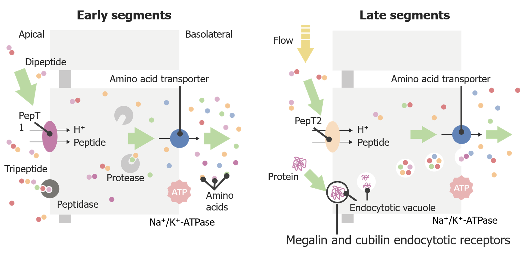
Early PT apical proteinsProteinsLinear polypeptides that are synthesized on ribosomes and may be further modified, crosslinked, cleaved, or assembled into complex proteins with several subunits. The specific sequence of amino acids determines the shape the polypeptide will take, during protein folding, and the function of the protein.Energy Homeostasis:
PepT1: H+/peptide cotransporter responsible for the majority of peptide reabsorption in the PT
Peptidase: membrane-bound enzyme
Located in the early segment of the PT
Breaks down larger tripeptides within the tubule lumen
The smaller broken-down peptides can then enter via PepT1.
Late PT apical proteinsProteinsLinear polypeptides that are synthesized on ribosomes and may be further modified, crosslinked, cleaved, or assembled into complex proteins with several subunits. The specific sequence of amino acids determines the shape the polypeptide will take, during protein folding, and the function of the protein.Energy Homeostasis:
PepT2: H+/peptide cotransporter responsible for reabsorbing peptides not captured by PepT1
Megalin and cubilin receptorsReceptorsReceptors are proteins located either on the surface of or within a cell that can bind to signaling molecules known as ligands (e.g., hormones) and cause some type of response within the cell.Receptors:
BindBINDHyperbilirubinemia of the Newborn and endocytose small proteinsProteinsLinear polypeptides that are synthesized on ribosomes and may be further modified, crosslinked, cleaved, or assembled into complex proteins with several subunits. The specific sequence of amino acids determines the shape the polypeptide will take, during protein folding, and the function of the protein.Energy Homeostasis
Peptides are digested into amino acidsAmino acidsOrganic compounds that generally contain an amino (-NH2) and a carboxyl (-COOH) group. Twenty alpha-amino acids are the subunits which are polymerized to form proteins.Basics of Amino Acids by proteasesProteasesProteins and Peptides within the cell.
Amino acidsAmino acidsOrganic compounds that generally contain an amino (-NH2) and a carboxyl (-COOH) group. Twenty alpha-amino acids are the subunits which are polymerized to form proteins.Basics of Amino Acids exit the cell via transporters on the basolateral membrane.
Amino acidAmino acidAmino acids (AAs) are composed of a central carbon atom attached to a carboxyl group, an amino group, a hydrogen atom, and a side chain (R group). Basics of Amino Acids reabsorption
Apical reabsorption:
Anionic (acidic) or cationic (basic) amino acidsAmino acidsOrganic compounds that generally contain an amino (-NH2) and a carboxyl (-COOH) group. Twenty alpha-amino acids are the subunits which are polymerized to form proteins.Basics of Amino Acids: various ion exchangers
Neutral amino acidsAmino acidsOrganic compounds that generally contain an amino (-NH2) and a carboxyl (-COOH) group. Twenty alpha-amino acids are the subunits which are polymerized to form proteins.Basics of Amino Acids: via Na+ or H+ cotransport
Basolateral reabsorption:
Aromatic amino acidsAromatic amino acidsAmino acids containing an aromatic side chain.Basics of Amino Acids: via facilitated diffusionDiffusionThe tendency of a gas or solute to pass from a point of higher pressure or concentration to a point of lower pressure or concentration and to distribute itself throughout the available space. Diffusion, especially facilitated diffusion, is a major mechanism of biological transport.Peritoneal Dialysis and Hemodialysis
Cationic (basic) and neutral amino acidsAmino acidsOrganic compounds that generally contain an amino (-NH2) and a carboxyl (-COOH) group. Twenty alpha-amino acids are the subunits which are polymerized to form proteins.Basics of Amino Acids: via Na+ cotransport
SecretionSecretionCoagulation Studies occurs primarily in the PST (i.e., late PT) and allows for the eliminationEliminationThe initial damage and destruction of tumor cells by innate and adaptive immunity. Completion of the phase means no cancer growth. Cancer Immunotherapy of endogenous and exogenous substances such as toxins and drugs.
Organic anionsAnionsNegatively charged atoms, radicals or groups of atoms which travel to the anode or positive pole during electrolysis.Electrolytes
Organic anionsAnionsNegatively charged atoms, radicals or groups of atoms which travel to the anode or positive pole during electrolysis.Electrolytes (OAOAOsteoarthritis (OA) is the most common form of arthritis, and is due to cartilage destruction and changes of the subchondral bone. The risk of developing this disorder increases with age, obesity, and repetitive joint use or trauma. Patients develop gradual joint pain, stiffness lasting < 30 minutes, and decreased range of motion.Osteoarthritis–s) are moved from the basolateral side into cells by organic anion transporters (OATs).
Transported into the tubule lumen by 2 proteinsProteinsLinear polypeptides that are synthesized on ribosomes and may be further modified, crosslinked, cleaved, or assembled into complex proteins with several subunits. The specific sequence of amino acids determines the shape the polypeptide will take, during protein folding, and the function of the protein.Energy Homeostasis:
Multidrug-resistant transporter (MRP2)
OAT4 exchanger
Examples of OAOAOsteoarthritis (OA) is the most common form of arthritis, and is due to cartilage destruction and changes of the subchondral bone. The risk of developing this disorder increases with age, obesity, and repetitive joint use or trauma. Patients develop gradual joint pain, stiffness lasting < 30 minutes, and decreased range of motion.Osteoarthritis–s secreted in the PT: bileBileAn emulsifying agent produced in the liver and secreted into the duodenum. Its composition includes bile acids and salts; cholesterol; and electrolytes. It aids digestion of fats in the duodenum.Gallbladder and Biliary Tract: Anatomy salts, urate, certain drugs (see table below)
Organic anion secretion in the late proximal tubule MRP2: multidrug-resistant transporter NaDC: Na+-dependent dicarboxylate transporter OA–: organic anion OAT: organic anion transporter
α-KG: α-ketoglutarate
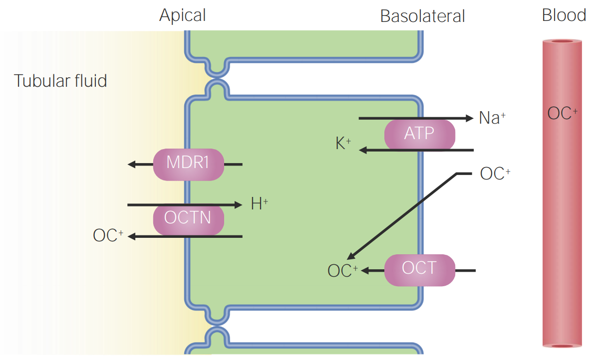
Organic cationsCationsPositively charged atoms, radicals or groups of atoms which travel to the cathode or negative pole during electrolysis.Electrolytes
Organic cationsCationsPositively charged atoms, radicals or groups of atoms which travel to the cathode or negative pole during electrolysis.Electrolytes (OC+s) are moved from the basolateral side into cells by organic cation transporters (OCTs).
Transported into the tubule lumen by 2 proteinsProteinsLinear polypeptides that are synthesized on ribosomes and may be further modified, crosslinked, cleaved, or assembled into complex proteins with several subunits. The specific sequence of amino acids determines the shape the polypeptide will take, during protein folding, and the function of the protein.Energy Homeostasis:
Multidrug-resistant transporter (MDR1)
Organic cation transporter novel (OCTN) exchanger
Examples: creatinine, dopamineDopamineOne of the catecholamine neurotransmitters in the brain. It is derived from tyrosine and is the precursor to norepinephrine and epinephrine. Dopamine is a major transmitter in the extrapyramidal system of the brain, and important in regulating movement.Receptors and Neurotransmitters of the CNS, certain drugs (see table below)
Organic cation (OC+) secretion in the proximal tubule MDR1: multidrug-resistant transporter OCT: organic cation transporter OCTN: organic cation transporter novel
Table: Organic ions secreted by the proximal tubule
Endogenous substances
Drugs
Organic anionsAnionsNegatively charged atoms, radicals or groups of atoms which travel to the anode or positive pole during electrolysis.Electrolytes
cAMPcAMPAn adenine nucleotide containing one phosphate group which is esterified to both the 3′- and 5′-positions of the sugar moiety. It is a second messenger and a key intracellular regulator, functioning as a mediator of activity for a number of hormones, including epinephrine, glucagon, and acth.Phosphodiesterase Inhibitors, cGMPcGMPGuanosine cyclic 3.Phosphodiesterase Inhibitors
BileBileAn emulsifying agent produced in the liver and secreted into the duodenum. Its composition includes bile acids and salts; cholesterol; and electrolytes. It aids digestion of fats in the duodenum.Gallbladder and Biliary Tract: Anatomy salts
Hippurates
Urate
Oxalate
ProstaglandinsProstaglandinsA group of compounds derived from unsaturated 20-carbon fatty acids, primarily arachidonic acid, via the cyclooxygenase pathway. They are extremely potent mediators of a diverse group of physiological processes.Eicosanoids: PGE2, PGF2α
Vitamins: ascorbate, folateFolateFolate and vitamin B12 are 2 of the most clinically important water-soluble vitamins. Deficiencies can present with megaloblastic anemia, GI symptoms, neuropsychiatric symptoms, and adverse pregnancy complications, including neural tube defects. Folate and Vitamin B12
AcetazolamideAcetazolamideOne of the carbonic anhydrase inhibitors that is sometimes effective against absence seizures. It is sometimes useful also as an adjunct in the treatment of tonic-clonic, myoclonic, and atonic seizures, particularly in women whose seizures occur or are exacerbated at specific times in the menstrual cycle. However, its usefulness is transient often because of rapid development of tolerance. Its antiepileptic effect may be due to its inhibitory effect on brain carbonic anhydrase, which leads to an increased transneuronal chloride gradient, increased chloride current, and increased inhibition.Carbonic Anhydrase Inhibitors
ChlorothiazideChlorothiazideA thiazide diuretic with actions and uses similar to those of hydrochlorothiazide.Hypertension Drugs
HydrochlorothiazideHydrochlorothiazideA thiazide diuretic often considered the prototypical member of this class. It reduces the reabsorption of electrolytes from the renal tubules. This results in increased excretion of water and electrolytes, including sodium, potassium, chloride, and magnesium. It is used in the treatment of several disorders including edema, hypertension, diabetes insipidus, and hypoparathyroidism.Thiazide Diuretics
FurosemideFurosemideA benzoic-sulfonamide-furan. It is a diuretic with fast onset and short duration that is used for edema and chronic renal insufficiency.Loop Diuretics
ProbenecidProbenecidThe prototypical uricosuric agent. It inhibits the renal excretion of organic anions and reduces tubular reabsorption of urate. Probenecid has also been used to treat patients with renal impairment, and, because it reduces the renal tubular excretion of other drugs, has been used as an adjunct to antibacterial therapy.Gout Drugs
Salicylates (aspirinAspirinThe prototypical analgesic used in the treatment of mild to moderate pain. It has anti-inflammatory and antipyretic properties and acts as an inhibitor of cyclooxygenase which results in the inhibition of the biosynthesis of prostaglandins. Aspirin also inhibits platelet aggregation and is used in the prevention of arterial and venous thrombosis.Nonsteroidal Antiinflammatory Drugs (NSAIDs))
Organic cationsCationsPositively charged atoms, radicals or groups of atoms which travel to the cathode or negative pole during electrolysis.Electrolytes
Creatinine
DopamineDopamineOne of the catecholamine neurotransmitters in the brain. It is derived from tyrosine and is the precursor to norepinephrine and epinephrine. Dopamine is a major transmitter in the extrapyramidal system of the brain, and important in regulating movement.Receptors and Neurotransmitters of the CNS
EpinephrineEpinephrineThe active sympathomimetic hormone from the adrenal medulla. It stimulates both the alpha- and beta- adrenergic systems, causes systemic vasoconstriction and gastrointestinal relaxation, stimulates the heart, and dilates bronchi and cerebral vessels.Sympathomimetic Drugs
NorepinephrineNorepinephrinePrecursor of epinephrine that is secreted by the adrenal medulla and is a widespread central and autonomic neurotransmitter. Norepinephrine is the principal transmitter of most postganglionic sympathetic fibers, and of the diffuse projection system in the brain that arises from the locus ceruleus.Receptors and Neurotransmitters of the CNS
AtropineAtropineAn alkaloid, originally from atropa belladonna, but found in other plants, mainly solanaceae. Hyoscyamine is the 3(s)-endo isomer of atropine.Anticholinergic Drugs
IsoproterenolIsoproterenolIsopropyl analog of epinephrine; beta-sympathomimetic that acts on the heart, bronchi, skeletal muscle, alimentary tract, etc. It is used mainly as bronchodilator and heart stimulant.Sympathomimetic Drugs
CimetidineCimetidineA histamine congener, it competitively inhibits histamine binding to histamine h2 receptors. Cimetidine has a range of pharmacological actions. It inhibits gastric acid secretion, as well as pepsin and gastrin output.Antihistamines
MorphineMorphineThe principal alkaloid in opium and the prototype opiate analgesic and narcotic. Morphine has widespread effects in the central nervous system and on smooth muscle.Opioid Analgesics
QuinineQuinineAn alkaloid derived from the bark of the cinchona tree. It is used as an antimalarial drug, and is the active ingredient in extracts of the cinchona that have been used for that purpose since before 1633. Quinine is also a mild antipyretic and analgesic and has been used in common cold preparations for that purpose. It was used commonly and as a bitter and flavoring agent, and is still useful for the treatment of babesiosis. Quinine is also useful in some muscular disorders, especially nocturnal leg cramps and myotonia congenita, because of its direct effects on muscle membrane and sodium channels. The mechanisms of its antimalarial effects are not well understood.Antimalarial Drugs
AmilorideAmilorideA pyrazine compound inhibiting sodium reabsorption through sodium channels in renal epithelial cells. This inhibition creates a negative potential in the luminal membranes of principal cells, located in the distal convoluted tubule and collecting duct. Negative potential reduces secretion of potassium and hydrogen ions. Amiloride is used in conjunction with diuretics to spare potassium loss.Liddle Syndrome
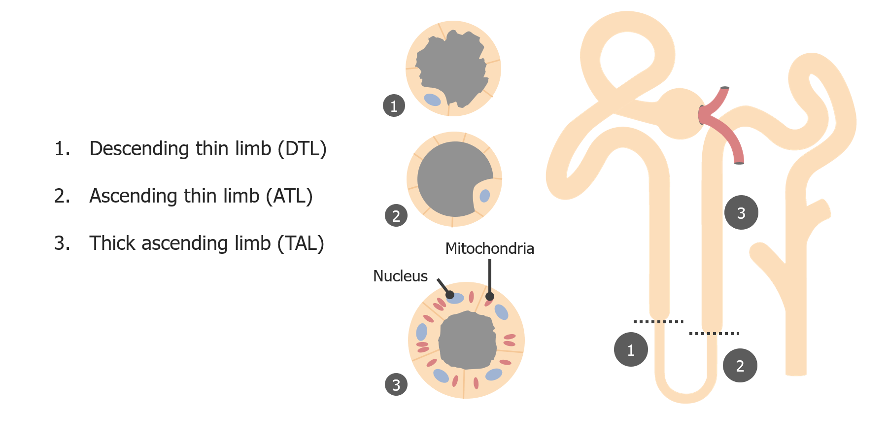
The loop of Henle is a complex segment of the nephronNephronThe functional units of the kidney, consisting of the glomerulus and the attached tubule.Kidneys: Anatomy with 2 main purposes: maintaining the corticomedullary gradient and reabsorbing moderate amounts of Na+ and water. These 2 processes are linked via the countercurrent multiplier system in the thin loops, and additional Na+absorptionAbsorptionAbsorption involves the uptake of nutrient molecules and their transfer from the lumen of the GI tract across the enterocytes and into the interstitial space, where they can be taken up in the venous or lymphatic circulation.Digestion and Absorption occurs via active transportActive transportThe movement of materials across cell membranes and epithelial layers against an electrochemical gradient, requiring the expenditure of metabolic energy.The Cell: Cell Membrane in the thick ascending limbThick ascending limbRenal Sodium and Water Regulation.
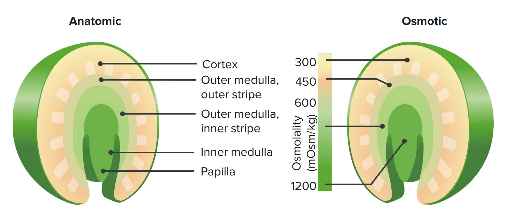
OsmolalityOsmolalityPlasma osmolality refers to the combined concentration of all solutes in the blood.Renal Sodium and Water Regulation of the renal interstitium ranges from approximately 300 mOsm/kg in the cortex to approximately 1200 mOsm/kg in the inner medulla.
This gradient is necessary for dynamic control of water reabsorption later in the collecting ductCollecting ductStraight tubes commencing in the radiate part of the kidney cortex where they receive the curved ends of the distal convoluted tubules. In the medulla the collecting tubules of each pyramid converge to join a central tube (duct of bellini) which opens on the summit of the papilla.Renal Cell Carcinoma.
The gradient is established and maintained by the passive movement of fluid and solutes according to the countercurrent multiplier theory.
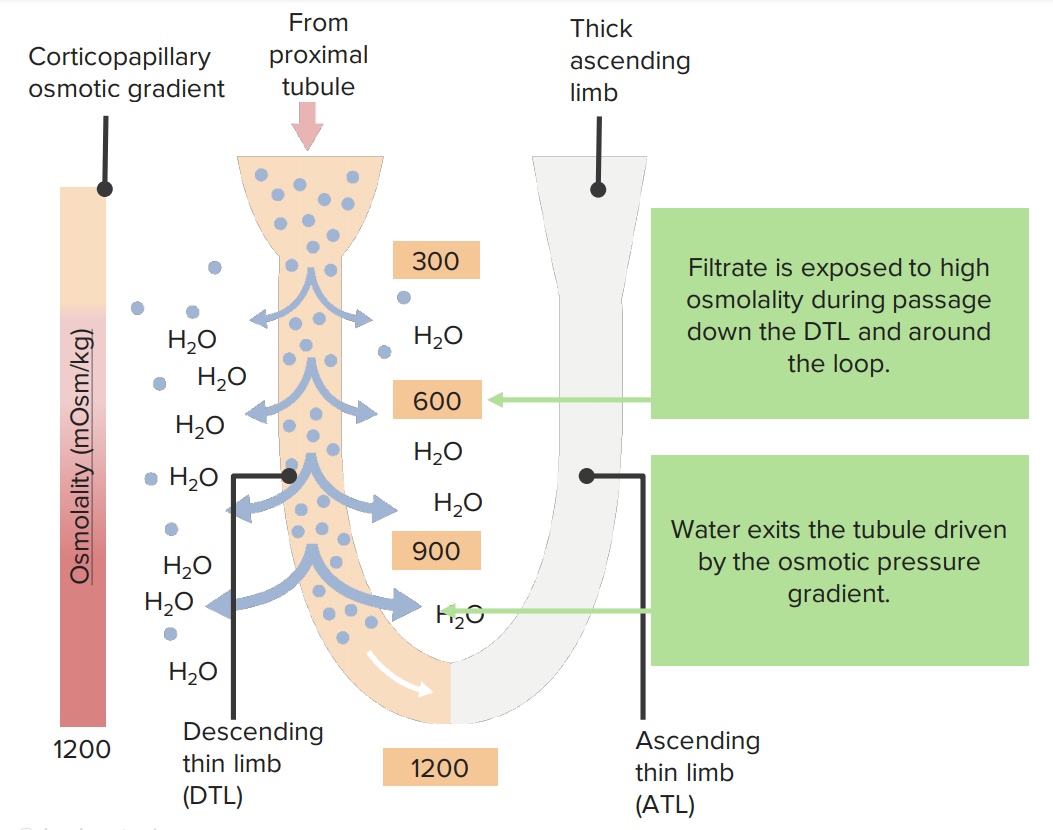
The countercurrent multiplier theory explains how the movement of fluids and solutes creates a significant corticomedullary gradient. This process occurs primarily in the thin loops of Henle and via ureaUreaA compound formed in the liver from ammonia produced by the deamination of amino acids. It is the principal end product of protein catabolism and constitutes about one half of the total urinary solids.Urea Cycle recycling.
Within the thin loops of Henle:
Thin descending limb:
Permeable only to water and not to solutes
Increasing amounts of water exit the tubules as the tubule fluid descends through areas of the interstitium with increasingly high osmolalityOsmolalityPlasma osmolality refers to the combined concentration of all solutes in the blood.Renal Sodium and Water Regulation (known as “fluid displacementDisplacementThe process by which an emotional or behavioral response that is appropriate for one situation appears in another situation for which it is inappropriate.Defense Mechanisms”).
Tubule fluid becomes concentrated.
Thin ascending limb:
Permeable only to solutes (via passive transportPassive transportThe passive movement of molecules exceeding the rate expected by simple diffusion. No energy is expended in the process. It is achieved by the introduction of passively diffusing molecules to an environment or path that is more favorable to the movement of those molecules. Examples of facilitated diffusion are passive transport of hydrophilic substances across a lipid membrane through hydrophilic pores that traverse the membrane, and the sliding of a DNA binding protein along a strand of DNA.The Cell: Cell Membrane) and not to water
Increasing amounts of solutes exit the tubule as the tubule fluid ascends through areas of the interstitium with decreasing osmolalityOsmolalityPlasma osmolality refers to the combined concentration of all solutes in the blood.Renal Sodium and Water Regulation (known as a “single effect”).
Tubule fluid becomes near isotonicIsotonicSolutions having the same osmotic pressure as blood serum, or another solution with which they are compared.Renal Sodium and Water Regulation again at the end of the thin ascending limb.
Repeating “fluid displacementDisplacementThe process by which an emotional or behavioral response that is appropriate for one situation appears in another situation for which it is inappropriate.Defense Mechanisms” followed by a “single effect” over and over again generates and maintains the corticomedullary gradient.
Effect of the corticomedullary gradient on water and sodium transport in the descending thin limb (DTL)
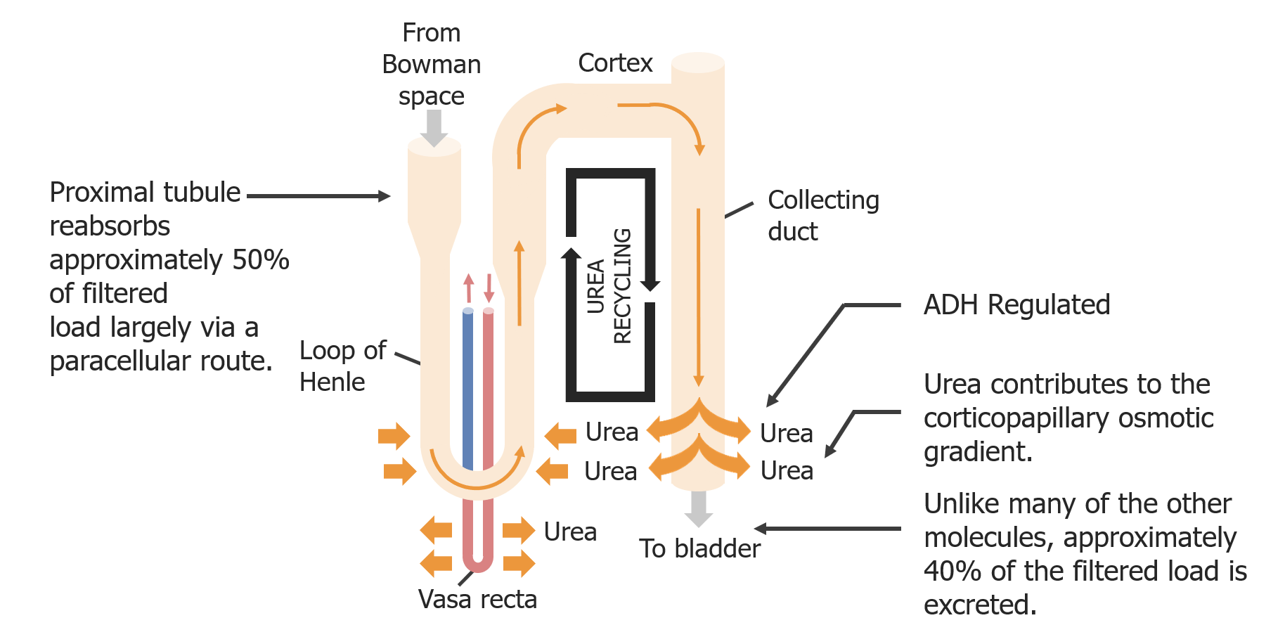
UreaUreaA compound formed in the liver from ammonia produced by the deamination of amino acids. It is the principal end product of protein catabolism and constitutes about one half of the total urinary solids.Urea Cycle recycling:
In the PT: Approximately 50% of ureaUreaA compound formed in the liver from ammonia produced by the deamination of amino acids. It is the principal end product of protein catabolism and constitutes about one half of the total urinary solids.Urea Cycle is reabsorbed via paracellularParacellularRenal Potassium Regulation transport.
In the loop of Henle:
Approximately 50% of ureaUreaA compound formed in the liver from ammonia produced by the deamination of amino acids. It is the principal end product of protein catabolism and constitutes about one half of the total urinary solids.Urea Cycle reenters the tubule (passive secretionSecretionCoagulation Studies).
Approximately 30% is reabsorbed.
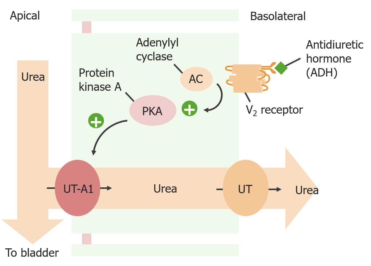
In the collecting ductCollecting ductStraight tubes commencing in the radiate part of the kidney cortex where they receive the curved ends of the distal convoluted tubules. In the medulla the collecting tubules of each pyramid converge to join a central tube (duct of bellini) which opens on the summit of the papilla.Renal Cell Carcinoma:
Approximately 50% of ureaUreaA compound formed in the liver from ammonia produced by the deamination of amino acids. It is the principal end product of protein catabolism and constitutes about one half of the total urinary solids.Urea Cycle is reabsorbed via the apical transporter UT-A1 and the basolateral transporter UT.
Antidiuretic hormoneAntidiuretic hormoneAntidiuretic hormones released by the neurohypophysis of all vertebrates (structure varies with species) to regulate water balance and osmolarity. In general, vasopressin is a nonapeptide consisting of a six-amino-acid ring with a cysteine 1 to cysteine 6 disulfide bridge or an octapeptide containing a cystine. All mammals have arginine vasopressin except the pig with a lysine at position 8. Vasopressin, a vasoconstrictor, acts on the kidney collecting ducts to increase water reabsorption, increase blood volume and blood pressure.Hypernatremia (ADH) upregulates apical UT-A1 → ↑ ureaUreaA compound formed in the liver from ammonia produced by the deamination of amino acids. It is the principal end product of protein catabolism and constitutes about one half of the total urinary solids.Urea Cycle reabsorption
Reabsorbed ureaUreaA compound formed in the liver from ammonia produced by the deamination of amino acids. It is the principal end product of protein catabolism and constitutes about one half of the total urinary solids.Urea Cycle in the interstitium contributes to the corticomedullary gradient.
Ultimately, 60% of filtered ureaUreaA compound formed in the liver from ammonia produced by the deamination of amino acids. It is the principal end product of protein catabolism and constitutes about one half of the total urinary solids.Urea Cycle is retained for this purpose and 40% is excreted.
Urea recycling as a contributor to the corticomedullary gradient
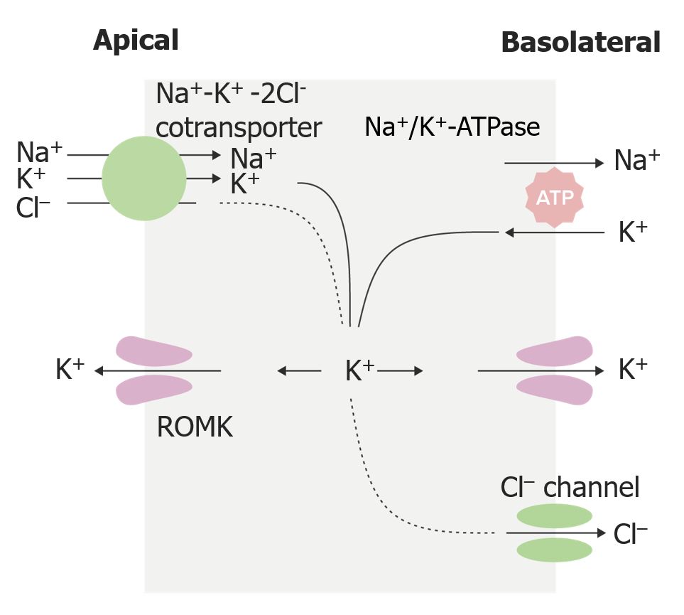
The basolateral side becomes more electronegative due to unmatched Cl–.
Important for driving the paracellularParacellularRenal Potassium Regulation transport of cationsCationsPositively charged atoms, radicals or groups of atoms which travel to the cathode or negative pole during electrolysis.Electrolytes
Contributes to the osmotic gradient between tubular fluid and interstitium:
Water does not follow solutes (Na+, K+, Cl–) into the interstitium.
Site of action for loop diureticsDiureticsAgents that promote the excretion of urine through their effects on kidney function.Heart Failure and Chronic Coronary Syndrome Medication (furosemideFurosemideA benzoic-sulfonamide-furan. It is a diuretic with fast onset and short duration that is used for edema and chronic renal insufficiency.Loop Diuretics, torsemideTorsemideA pyridine and sulfonamide derivative that acts as a sodium-potassium chloride symporter inhibitor. It is used for the treatment of edema associated with congestive heart failure; chronic renal insufficiency; and liver diseases. It is also used for the management of hypertension.Loop Diuretics, bumetanideBumetanideA sulfamyl diuretic.Loop Diuretics):
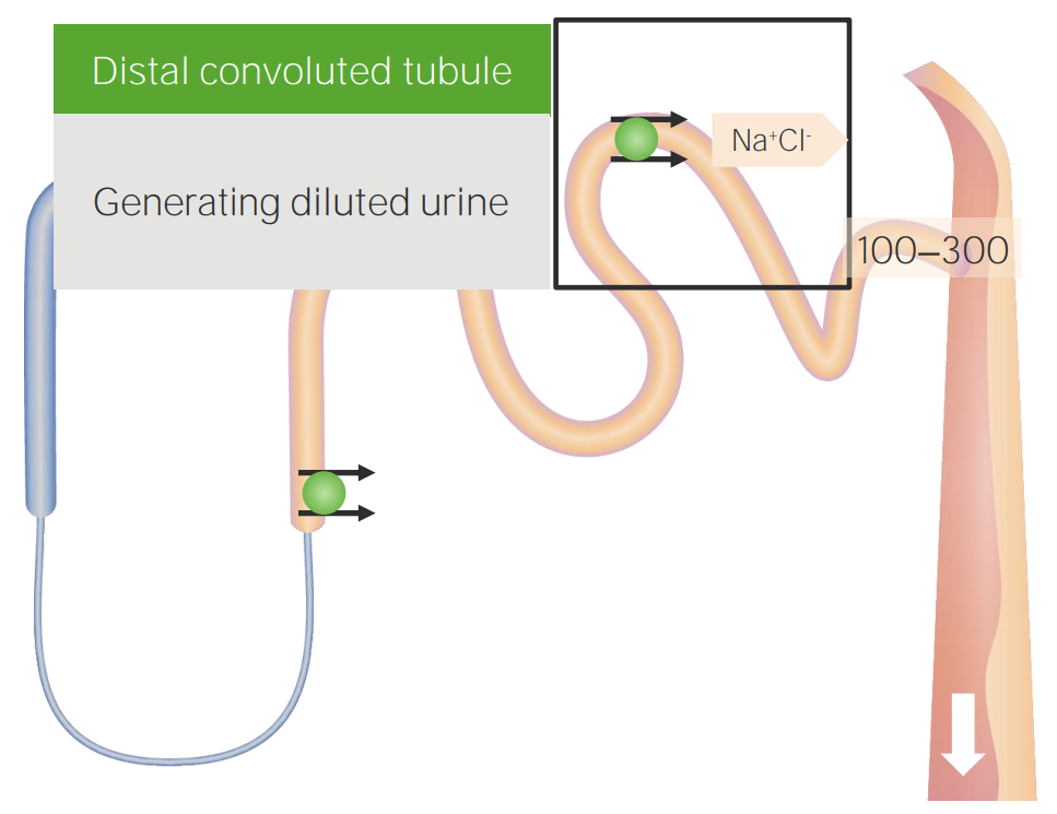
The distal convoluted tubuleDistal convoluted tubuleThe portion of renal tubule that begins from the enlarged segment of the ascending limb of the loop of henle. It reenters the kidney cortex and forms the convoluted segments of the distal tubule.Gitelman Syndrome (DCT) is another “diluting segmentDiluting segmentRenal Sodium and Water Regulation” of the nephronNephronThe functional units of the kidney, consisting of the glomerulus and the attached tubule.Kidneys: Anatomy, where the thiazide-sensitive NaClcotransporter helps generate hypotonicHypotonicSolutions that have a lesser osmotic pressure than a reference solution such as blood, plasma, or interstitial fluid.Renal Sodium and Water Regulation tubule fluid due to the DCT not being permeable to water. The transport of K+, Mg+2, and CaCACondylomata acuminata are a clinical manifestation of genital HPV infection. Condylomata acuminata are described as raised, pearly, flesh-colored, papular, cauliflower-like lesions seen in the anogenital region that may cause itching, pain, or bleeding.Condylomata Acuminata (Genital Warts)2+ also occurs in this segment.
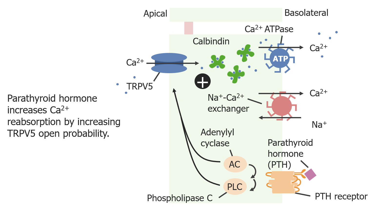
CalciumCalciumA basic element found in nearly all tissues. It is a member of the alkaline earth family of metals with the atomic symbol ca, atomic number 20, and atomic weight 40. Calcium is the most abundant mineral in the body and combines with phosphorus to form calcium phosphate in the bones and teeth. It is essential for the normal functioning of nerves and muscles and plays a role in blood coagulation (as factor IV) and in many enzymatic processes.Electrolytes reabsorption
↑ PTH → ↑ adenylyl cyclase and phospholipase CPhospholipase CA subclass of phospholipases that hydrolyze the phosphoester bond found in the third position of glycerophospholipids. Although the singular term phospholipase C specifically refers to an enzyme that catalyzes the hydrolysis of phosphatidylcholine, it is commonly used in the literature to refer to broad variety of enzymes that specifically catalyze the hydrolysis of phosphatidylinositols.Pseudomonas → ↑ phosphorylationPhosphorylationThe introduction of a phosphoryl group into a compound through the formation of an ester bond between the compound and a phosphorus moiety.Post-translational Protein Processing of TRPV5 → ↑ open probabilityProbabilityProbability is a mathematical tool used to study randomness and provide predictions about the likelihood of something happening. There are several basic rules of probability that can be used to help determine the probability of multiple events happening together, separately, or sequentially.Basics of Probability of TRPV5 channel → ↑ CaCACondylomata acuminata are a clinical manifestation of genital HPV infection. Condylomata acuminata are described as raised, pearly, flesh-colored, papular, cauliflower-like lesions seen in the anogenital region that may cause itching, pain, or bleeding.Condylomata Acuminata (Genital Warts)2+ reabsorption
Within the cell, CaCACondylomata acuminata are a clinical manifestation of genital HPV infection. Condylomata acuminata are described as raised, pearly, flesh-colored, papular, cauliflower-like lesions seen in the anogenital region that may cause itching, pain, or bleeding.Condylomata Acuminata (Genital Warts)2+ is bound to the protein calbindinCalbindinCalcium-binding proteins that are found in distal kidney tubules, intestines, brain, and other tissues where they bind, buffer and transport cytoplasmic calcium. Calbindins possess a variable number of ef-hand motifs which contain calcium-binding sites. Some isoforms are regulated by vitamin d.Calcium Hemostasis and Bone Metabolism:
Necessary due to the cytotoxicCytotoxicParvovirus B19 effects of high intracellular CaCACondylomata acuminata are a clinical manifestation of genital HPV infection. Condylomata acuminata are described as raised, pearly, flesh-colored, papular, cauliflower-like lesions seen in the anogenital region that may cause itching, pain, or bleeding.Condylomata Acuminata (Genital Warts)2+
Transports CaCACondylomata acuminata are a clinical manifestation of genital HPV infection. Condylomata acuminata are described as raised, pearly, flesh-colored, papular, cauliflower-like lesions seen in the anogenital region that may cause itching, pain, or bleeding.Condylomata Acuminata (Genital Warts)2+ to the basolateral membrane
CaCACondylomata acuminata are a clinical manifestation of genital HPV infection. Condylomata acuminata are described as raised, pearly, flesh-colored, papular, cauliflower-like lesions seen in the anogenital region that may cause itching, pain, or bleeding.Condylomata Acuminata (Genital Warts)2+ is moved into the basolateral interstitium via 2 mechanisms:
CaCACondylomata acuminata are a clinical manifestation of genital HPV infection. Condylomata acuminata are described as raised, pearly, flesh-colored, papular, cauliflower-like lesions seen in the anogenital region that may cause itching, pain, or bleeding.Condylomata Acuminata (Genital Warts)2+ ATPase
CaCACondylomata acuminata are a clinical manifestation of genital HPV infection. Condylomata acuminata are described as raised, pearly, flesh-colored, papular, cauliflower-like lesions seen in the anogenital region that may cause itching, pain, or bleeding.Condylomata Acuminata (Genital Warts)2+-Na+ exchanger
MagnesiumMagnesiumA metallic element that has the atomic symbol mg, atomic number 12, and atomic weight 24. 31. It is important for the activity of many enzymes, especially those involved in oxidative phosphorylation.Electrolytes reabsorption
Does not require a cytosolCytosolA cell’s cytoskeleton is a network of intracellular protein fibers that provides structural support, anchors organelles, and aids intra- and extracellular movement.The Cell: Cytosol and Cytoskeleton transport protein (such as calbindinCalbindinCalcium-binding proteins that are found in distal kidney tubules, intestines, brain, and other tissues where they bind, buffer and transport cytoplasmic calcium. Calbindins possess a variable number of ef-hand motifs which contain calcium-binding sites. Some isoforms are regulated by vitamin d.Calcium Hemostasis and Bone Metabolism)
Mechanism for movement into basolateral interstitium is unknown.
SodiumSodiumA member of the alkali group of metals. It has the atomic symbol na, atomic number 11, and atomic weight 23.Hyponatremia reabsorption
Occurs via 2 mechanisms:
NaCl cotransporter:
Na+ and Cl– are reabsorbed.
Site of action of thiazideThiazideHeterocyclic compounds with sulfur and nitrogen in the ring. This term commonly refers to the benzothiadiazines that inhibit sodium-potassium-chloride symporters and are used as diuretics.HyponatremiadiureticsDiureticsAgents that promote the excretion of urine through their effects on kidney function.Heart Failure and Chronic Coronary Syndrome Medication
Epithelial sodiumSodiumA member of the alkali group of metals. It has the atomic symbol na, atomic number 11, and atomic weight 23.HyponatremiachannelsChannelsThe Cell: Cell Membrane (ENaCENaCSodium channels found on salt-reabsorbing epithelial cells that line the distal nephron; the distal colon; salivary ducts; sweat glands; and the lung. They are amiloride-sensitive and play a critical role in the control of sodium balance, blood volume, and blood pressure.Liddle Syndrome):
Na+ is reabsorbed by itself.
Creates a voltage gradient because there is no matched transport of other charged ions (i.e., it is not exchanged for another cation, or cotransported with an anion)
Also found in the collecting ducts
Site of action of amilorideAmilorideA pyrazine compound inhibiting sodium reabsorption through sodium channels in renal epithelial cells. This inhibition creates a negative potential in the luminal membranes of principal cells, located in the distal convoluted tubule and collecting duct. Negative potential reduces secretion of potassium and hydrogen ions. Amiloride is used in conjunction with diuretics to spare potassium loss.Liddle Syndrome(K+-sparing diuretic)
Distal convoluted tubule (DCT) reabsorption: Sodium and chloride are reabsorbed in the DCT, but the DCT is not permeable to water, allowing for dilution of the urine.
Driven by the electrical gradientElectrical gradientRenal Potassium Regulation generated by ENaCENaCSodium channels found on salt-reabsorbing epithelial cells that line the distal nephron; the distal colon; salivary ducts; sweat glands; and the lung. They are amiloride-sensitive and play a critical role in the control of sodium balance, blood volume, and blood pressure.Liddle Syndrome activity
Cl– moves out of the electronegative lumen and toward the electropositive basolateral side.
Accounts for the majority of Cl– transport in the late DCT
PotassiumPotassiumAn element in the alkali group of metals with an atomic symbol k, atomic number 19, and atomic weight 39. 10. It is the chief cation in the intracellular fluid of muscle and other cells. Potassium ion is a strong electrolyte that plays a significant role in the regulation of fluid volume and maintenance of the water-electrolyte balance.HyperkalemiasecretionSecretionCoagulation Studies
K+ is not reabsorbed in the DCT, but it is secreted in the late DCT.
Driven by electronegative lumen from ENaCENaCSodium channels found on salt-reabsorbing epithelial cells that line the distal nephron; the distal colon; salivary ducts; sweat glands; and the lung. They are amiloride-sensitive and play a critical role in the control of sodium balance, blood volume, and blood pressure.Liddle Syndrome (same as in Cl– reabsorption)
ROMK channelsChannelsThe Cell: Cell Membrane are upregulated by aldosteroneAldosteroneA hormone secreted by the adrenal cortex that regulates electrolyte and water balance by increasing the renal retention of sodium and the excretion of potassium.Hyperkalemia.
K+secretionSecretionCoagulation Studies through ROMK will increase if ENaCENaCSodium channels found on salt-reabsorbing epithelial cells that line the distal nephron; the distal colon; salivary ducts; sweat glands; and the lung. They are amiloride-sensitive and play a critical role in the control of sodium balance, blood volume, and blood pressure.Liddle Syndrome activity increases.
Collecting ducts are the points where multiple nephrons come together during the final stages of urine formation. Intercalated cells and principal cells act to adjust the final composition and concentration of the urine, prior to eliminationEliminationThe initial damage and destruction of tumor cells by innate and adaptive immunity. Completion of the phase means no cancer growth. Cancer Immunotherapy.
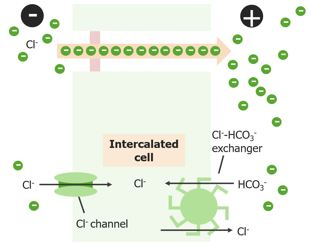
Intercalated cells
Intercalated cells are further divided into α and β subtypes, with each having a slightly different composition of transporters and other proteinsProteinsLinear polypeptides that are synthesized on ribosomes and may be further modified, crosslinked, cleaved, or assembled into complex proteins with several subunits. The specific sequence of amino acids determines the shape the polypeptide will take, during protein folding, and the function of the protein.Energy Homeostasis.
Apical proteinsProteinsLinear polypeptides that are synthesized on ribosomes and may be further modified, crosslinked, cleaved, or assembled into complex proteins with several subunits. The specific sequence of amino acids determines the shape the polypeptide will take, during protein folding, and the function of the protein.Energy Homeostasis:
Basolateral proteinsProteinsLinear polypeptides that are synthesized on ribosomes and may be further modified, crosslinked, cleaved, or assembled into complex proteins with several subunits. The specific sequence of amino acids determines the shape the polypeptide will take, during protein folding, and the function of the protein.Energy Homeostasis:
Na+/K+-ATPase
H+-ATPase (β-intercalated cells):
Paired with the apical Cl–/HCO3– exchanger
Involved with acid homeostasisHomeostasisThe processes whereby the internal environment of an organism tends to remain balanced and stable.Cell Injury and Death
Cl–/HCO3– exchanger (α-intercalated cells):
Paired with the apical Cl– channel
Involved with Cl– reabsorption
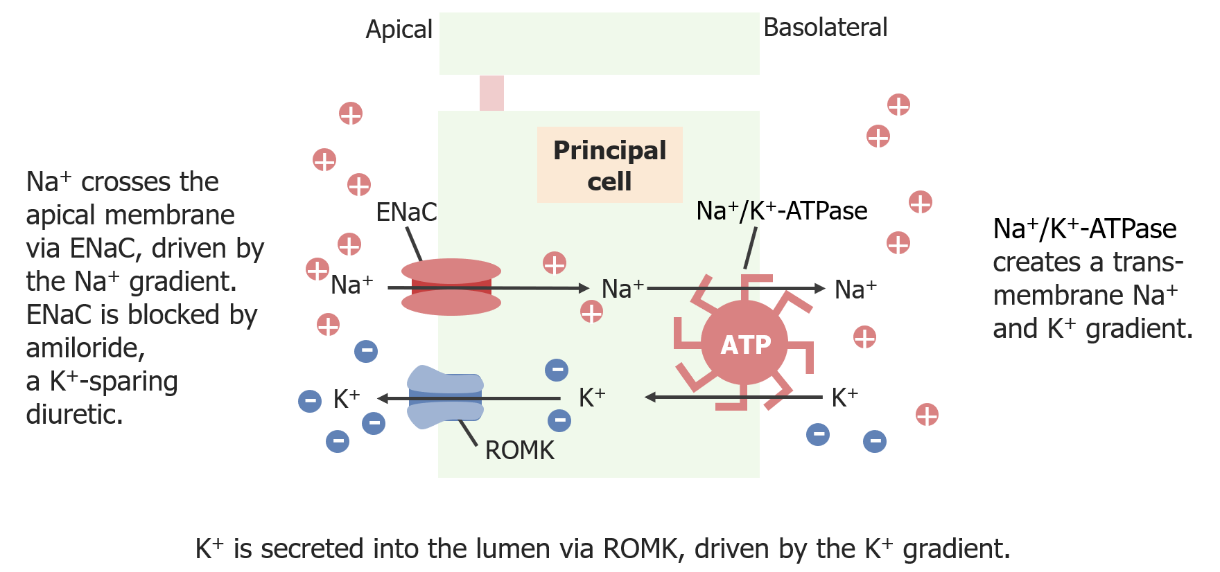
Principal cells
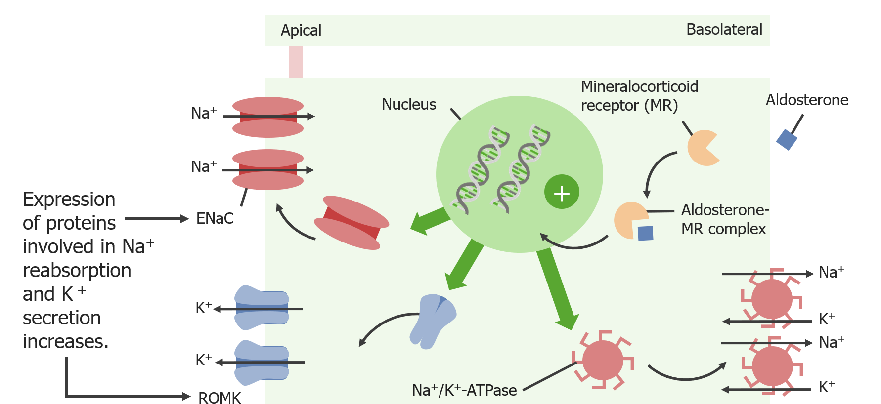
Principal cells are responsible for the fine-tuning of Na+ and K+ in the urine, which is often in response to the hormone aldosteroneAldosteroneA hormone secreted by the adrenal cortex that regulates electrolyte and water balance by increasing the renal retention of sodium and the excretion of potassium.Hyperkalemia. Principal cells are also the site of the apical aquaporin channel AQP2, which is a key component in adjusting urine concentration.
Apical proteinsProteinsLinear polypeptides that are synthesized on ribosomes and may be further modified, crosslinked, cleaved, or assembled into complex proteins with several subunits. The specific sequence of amino acids determines the shape the polypeptide will take, during protein folding, and the function of the protein.Energy Homeostasis:
ENaCENaCSodium channels found on salt-reabsorbing epithelial cells that line the distal nephron; the distal colon; salivary ducts; sweat glands; and the lung. They are amiloride-sensitive and play a critical role in the control of sodium balance, blood volume, and blood pressure.Liddle Syndrome channel: 1 Na+ moves into cell
Na+ reabsorption is powered by the Na+ gradient generated by Na+/K+-ATPase.
For each Na+ that moves into the cell, 1 Cl– is left behind in the tubular lumen.
↑ Expression/open probabilityProbabilityProbability is a mathematical tool used to study randomness and provide predictions about the likelihood of something happening. There are several basic rules of probability that can be used to help determine the probability of multiple events happening together, separately, or sequentially.Basics of Probability with aldosteroneAldosteroneA hormone secreted by the adrenal cortex that regulates electrolyte and water balance by increasing the renal retention of sodium and the excretion of potassium.Hyperkalemia
↑ Distal Na+ delivery results in ↑ ENaCENaCSodium channels found on salt-reabsorbing epithelial cells that line the distal nephron; the distal colon; salivary ducts; sweat glands; and the lung. They are amiloride-sensitive and play a critical role in the control of sodium balance, blood volume, and blood pressure.Liddle Syndrome channel activity
K+secretionSecretionCoagulation Studies is powered by K+ chemical and electrical gradients generated by the Na+/K+-ATPase and ENaCENaCSodium channels found on salt-reabsorbing epithelial cells that line the distal nephron; the distal colon; salivary ducts; sweat glands; and the lung. They are amiloride-sensitive and play a critical role in the control of sodium balance, blood volume, and blood pressure.Liddle SyndromechannelsChannelsThe Cell: Cell Membrane.
ROMK channelsChannelsThe Cell: Cell Membrane can open and close; there is an ↑ open probabilityProbabilityProbability is a mathematical tool used to study randomness and provide predictions about the likelihood of something happening. There are several basic rules of probability that can be used to help determine the probability of multiple events happening together, separately, or sequentially.Basics of Probability with:
AldosteroneAldosteroneA hormone secreted by the adrenal cortex that regulates electrolyte and water balance by increasing the renal retention of sodium and the excretion of potassium.Hyperkalemia
↓ Intracellular ATP (indicates ATP has just been used up by Na+/K+-ATPase to bring K+ into the cell)
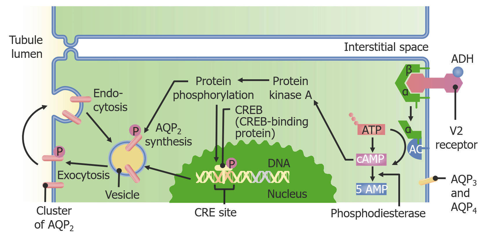
Aquaporin 2 (AQP2)channel: passive water channel
Water in the renal medulla is hypertonicHypertonicSolutions that have a greater osmotic pressure than a reference solution such as blood, plasma, or interstitial fluid.Renal Sodium and Water Regulation compared to urine due to the countercurrent multiplier system and diluting segments.
Water will leave the collecting ducts following its osmotic gradient to allow efflux if aquaporin channelsChannelsThe Cell: Cell Membrane are present.
ADH stimulates the production and insertion of aquaporins:
3 Na+ move out of cell and 2 K+ move into the cell.
Stimulated by aldosteroneAldosteroneA hormone secreted by the adrenal cortex that regulates electrolyte and water balance by increasing the renal retention of sodium and the excretion of potassium.Hyperkalemia
Aquaporin channel pathway: Antidiuretic hormone (ADH) binds to its receptor (V2), stimulating adenylate cyclase, thereby increasing cAMP and starting a protein phosphorylation cascade that ends with increased transcription and translation of the AQP2 channels.
The following table summarizes the reabsorption, secretionSecretionCoagulation Studies, and important regulatory molecules throughout the tubular system. Regulatory molecules are noted in parentheses, and “+” and “-” indicate stimulation and inhibition, respectively.
Table: Molecules reabsorbed and secreted along the nephronNephronThe functional units of the kidney, consisting of the glomerulus and the attached tubule.Kidneys: Anatomy
Segments/molecules
Proximal tubule (proximal convoluted tubuleProximal convoluted tubuleThe renal tubule portion that extends from the bowman capsule in the kidney cortex into the kidney medulla. The proximal tubule consists of a convoluted proximal segment in the cortex, and a distal straight segment descending into the medulla where it forms the u-shaped loop of henle.Osmotic Diuretics and PST)
Loop of Henle
Distal tubule
Collecting ductCollecting ductStraight tubes commencing in the radiate part of the kidney cortex where they receive the curved ends of the distal convoluted tubules. In the medulla the collecting tubules of each pyramid converge to join a central tube (duct of bellini) which opens on the summit of the papilla.Renal Cell Carcinoma
Excreted
GlucoseGlucoseA primary source of energy for living organisms. It is naturally occurring and is found in fruits and other parts of plants in its free state. It is used therapeutically in fluid and nutrient replacement.Lactose Intolerance
98% (proximal convoluted tubuleProximal convoluted tubuleThe renal tubule portion that extends from the bowman capsule in the kidney cortex into the kidney medulla. The proximal tubule consists of a convoluted proximal segment in the cortex, and a distal straight segment descending into the medulla where it forms the u-shaped loop of henle.Osmotic Diuretics); 2% (PST) reabsorbed
–
–
–
–
Amino acidsAmino acidsOrganic compounds that generally contain an amino (-NH2) and a carboxyl (-COOH) group. Twenty alpha-amino acids are the subunits which are polymerized to form proteins.Basics of Amino Acids and peptides
99% (proximal convoluted tubuleProximal convoluted tubuleThe renal tubule portion that extends from the bowman capsule in the kidney cortex into the kidney medulla. The proximal tubule consists of a convoluted proximal segment in the cortex, and a distal straight segment descending into the medulla where it forms the u-shaped loop of henle.Osmotic Diuretics); 1% (PST) reabsorbed
UreaUreaA compound formed in the liver from ammonia produced by the deamination of amino acids. It is the principal end product of protein catabolism and constitutes about one half of the total urinary solids.Urea Cycle*
50% reabsorbed
30% reabsorbed; 50% secreted
–
50% reabsorbed
40%
BicarbonateBicarbonateInorganic salts that contain the -HCO3 radical. They are an important factor in determining the ph of the blood and the concentration of bicarbonate ions is regulated by the kidney. Levels in the blood are an index of the alkali reserve or buffering capacity.Electrolytes
80% reabsorbed
10% reabsorbed
6% reabsorbed
4% reabsorbed
–
CalciumCalciumA basic element found in nearly all tissues. It is a member of the alkaline earth family of metals with the atomic symbol ca, atomic number 20, and atomic weight 40. Calcium is the most abundant mineral in the body and combines with phosphorus to form calcium phosphate in the bones and teeth. It is essential for the normal functioning of nerves and muscles and plays a role in blood coagulation (as factor IV) and in many enzymatic processes.Electrolytes
65% reabsorbed
25% reabsorbed
8% reabsorbed (+PTH)
1% reabsorbed
1%
MagnesiumMagnesiumA metallic element that has the atomic symbol mg, atomic number 12, and atomic weight 24. 31. It is important for the activity of many enzymes, especially those involved in oxidative phosphorylation.Electrolytes
15% reabsorbed
70% reabsorbed
10% reabsorbed
–
5%
PotassiumPotassiumAn element in the alkali group of metals with an atomic symbol k, atomic number 19, and atomic weight 39. 10. It is the chief cation in the intracellular fluid of muscle and other cells. Potassium ion is a strong electrolyte that plays a significant role in the regulation of fluid volume and maintenance of the water-electrolyte balance.Hyperkalemia (dietary intake)
80% reabsorbed
10% reabsorbed
Normal K intake: 10%–100% of dietary intake secreted (+Ald)
Low K diet: 2% reabsorbed
Normal K intake: 5%–50% of dietary intake reabsorbed
Low K diet: 6% reabsorbed
Normal K intake: 10%–100% of dietary intake
Low K diet: 2%
SodiumSodiumA member of the alkali group of metals. It has the atomic symbol na, atomic number 11, and atomic weight 23.Hyponatremia
67% reabsorbed (+Ang-II)
25% reabsorbed (+Ang-II)
5% reabsorbed (+Ald, -ANP)
3% reabsorbed (+Ald, -ANP)
1%
Water
67% reabsorbed
15% reabsorbed
–
18% reabsorbed (+ADH, -ANP)
1%
*Percentages add to more than 100% due to urea recycling.
PTH: parathyroid hormone
PST: proximal straight tubule
Ang-II: angiotensin-II
Ald: aldosterone
ADH: antidiuretic hormone
ANP: atrial natriuretic peptide
Renal cell carcinomaRenal cell carcinomaRenal cell carcinoma (RCC) is a tumor that arises from the lining of the renal tubular system within the renal cortex. Renal cell carcinoma is responsible for 80%-85% of all primary renal neoplasms. Most RCCs arise sporadically, but smoking, hypertension, and obesity are linked to its development. Renal Cell Carcinoma: the most common primary renal malignancyMalignancyHemothorax that originates from renal tubular cells (most commonly in the PT).
SGLT2 inhibitorsSGLT2 inhibitorsNon-insulinotropic Diabetes Drugs: a class of oral medications used in the management of type 2 diabetesDiabetesDiabetes mellitus (DM) is a metabolic disease characterized by hyperglycemia and dysfunction of the regulation of glucose metabolism by insulin. Type 1 DM is diagnosed mostly in children and young adults as the result of autoimmune destruction of β cells in the pancreas and the resulting lack of insulin. Type 2 DM has a significant association with obesity and is characterized by insulin resistance.Diabetes Mellitus mellitus. SGLT2 inhibitorsSGLT2 inhibitorsNon-insulinotropic Diabetes Drugs block glucoseGlucoseA primary source of energy for living organisms. It is naturally occurring and is found in fruits and other parts of plants in its free state. It is used therapeutically in fluid and nutrient replacement.Lactose Intolerance reabsorption via the SGLT2 transporter in the PT, causing glucoseGlucoseA primary source of energy for living organisms. It is naturally occurring and is found in fruits and other parts of plants in its free state. It is used therapeutically in fluid and nutrient replacement.Lactose Intolerance to be excreted in the urine rather than being reabsorbed. The names of SGLT2 inhibitorsSGLT2 inhibitorsNon-insulinotropic Diabetes Drugs end in -gliflozin (e.g., empagliflozinEmpagliflozinNon-insulinotropic Diabetes Drugs) and are considered 2nd-line options. An important side effect is the increased risk for genitourinary tract infectionsInfectionsInvasion of the host organism by microorganisms or their toxins or by parasites that can cause pathological conditions or diseases.Chronic Granulomatous Disease.
Loop diureticsDiureticsAgents that promote the excretion of urine through their effects on kidney function.Heart Failure and Chronic Coronary Syndrome Medication: a commonly used class of diureticsDiureticsAgents that promote the excretion of urine through their effects on kidney function.Heart Failure and Chronic Coronary Syndrome Medication (including furosemideFurosemideA benzoic-sulfonamide-furan. It is a diuretic with fast onset and short duration that is used for edema and chronic renal insufficiency.Loop Diuretics, bumetanideBumetanideA sulfamyl diuretic.Loop Diuretics, and torsemideTorsemideA pyridine and sulfonamide derivative that acts as a sodium-potassium chloride symporter inhibitor. It is used for the treatment of edema associated with congestive heart failure; chronic renal insufficiency; and liver diseases. It is also used for the management of hypertension.Loop Diuretics) that exert their effects by blocking the NKCC2NKCC2Renal Potassium Regulation cotransporter in the thick ascending limbThick ascending limbRenal Sodium and Water Regulation of the loop of Henle. SodiumSodiumA member of the alkali group of metals. It has the atomic symbol na, atomic number 11, and atomic weight 23.Hyponatremia ions remain in the tubule lumen and obligate water to remain with it, resulting in diuretic action. HypokalemiaHypokalemiaHypokalemia is defined as plasma potassium (K+) concentration < 3.5 mEq/L. Homeostatic mechanisms maintain plasma concentration between 3.5-5.2 mEq/L despite marked variation in dietary intake. Hypokalemia can be due to renal losses, GI losses, transcellular shifts, or poor dietary intake.Hypokalemia is a common side effect due to the action of increased distal delivery of Na+ on the ROMK channelsChannelsThe Cell: Cell Membrane.
ACEiACEiA class of drugs whose main indications are the treatment of hypertension and heart failure. They exert their hemodynamic effect mainly by inhibiting the renin-angiotensin system. They also modulate sympathetic nervous system activity and increase prostaglandin synthesis. They cause mainly vasodilation and mild natriuresis without affecting heart rate and contractility.Renin-Angiotensin-Aldosterone System Inhibitors: a commonly used class of antihypertensive drugs that inhibit the RAASRAASA blood pressure regulating system of interacting components that include renin; angiotensinogen; angiotensin converting enzyme; angiotensin i; angiotensin ii; and angiotensinase. Renin, an enzyme produced in the kidney, acts on angiotensinogen, an alpha-2 globulin produced by the liver, forming angiotensin I. Angiotensin-converting enzyme, contained in the lung, acts on angiotensin I in the plasma converting it to angiotensin II, an extremely powerful vasoconstrictor. Angiotensin II causes contraction of the arteriolar and renal vascular smooth muscle, leading to retention of salt and water in the kidney and increased arterial blood pressure. In addition, angiotensin II stimulates the release of aldosterone from the adrenal cortex, which in turn also increases salt and water retention in the kidney. Angiotensin-converting enzyme also breaks down bradykinin, a powerful vasodilator and component of the kallikrein-kinin system.Adrenal Hormones at the ACE level. The names of the drugs in this class end in -pril (e.g., lisinoprilLisinoprilOne of the angiotensin-converting enzyme inhibitors (ACE inhibitors), orally active, that has been used in the treatment of hypertension and congestive heart failure.Renin-Angiotensin-Aldosterone System Inhibitors, enalaprilEnalaprilAn angiotensin-converting enzyme inhibitor that is used to treat hypertension and heart failure.Hypertension Drugs) and are commonly used for the treatment of heart failureHeart FailureA heterogeneous condition in which the heart is unable to pump out sufficient blood to meet the metabolic need of the body. Heart failure can be caused by structural defects, functional abnormalities (ventricular dysfunction), or a sudden overload beyond its capacity. Chronic heart failure is more common than acute heart failure which results from sudden insult to cardiac function, such as myocardial infarction.Total Anomalous Pulmonary Venous Return (TAPVR) and proteinuriaProteinuriaThe presence of proteins in the urine, an indicator of kidney diseases.Nephrotic Syndrome in Children, in addition to hypertensionHypertensionHypertension, or high blood pressure, is a common disease that manifests as elevated systemic arterial pressures. Hypertension is most often asymptomatic and is found incidentally as part of a routine physical examination or during triage for an unrelated medical encounter. Hypertension. These drugs are clinically interchangeable with aldosteroneAldosteroneA hormone secreted by the adrenal cortex that regulates electrolyte and water balance by increasing the renal retention of sodium and the excretion of potassium.HyperkalemiareceptorReceptorReceptors are proteins located either on the surface of or within a cell that can bind to signaling molecules known as ligands (e.g., hormones) and cause some type of response within the cell.Receptors blockers.
Angiotensin IIAngiotensin IIAn octapeptide that is a potent but labile vasoconstrictor. It is produced from angiotensin I after the removal of two amino acids at the c-terminal by angiotensin converting enzyme. The amino acid in position 5 varies in different species. To block vasoconstriction and hypertension effect of angiotensin II, patients are often treated with ace inhibitors or with angiotensin II type 1 receptor blockers.Renal Sodium and Water RegulationreceptorReceptorReceptors are proteins located either on the surface of or within a cell that can bind to signaling molecules known as ligands (e.g., hormones) and cause some type of response within the cell.Receptors blockers: a commonly used class of antihypertensive drugs that inhibit the RAASRAASA blood pressure regulating system of interacting components that include renin; angiotensinogen; angiotensin converting enzyme; angiotensin i; angiotensin ii; and angiotensinase. Renin, an enzyme produced in the kidney, acts on angiotensinogen, an alpha-2 globulin produced by the liver, forming angiotensin I. Angiotensin-converting enzyme, contained in the lung, acts on angiotensin I in the plasma converting it to angiotensin II, an extremely powerful vasoconstrictor. Angiotensin II causes contraction of the arteriolar and renal vascular smooth muscle, leading to retention of salt and water in the kidney and increased arterial blood pressure. In addition, angiotensin II stimulates the release of aldosterone from the adrenal cortex, which in turn also increases salt and water retention in the kidney. Angiotensin-converting enzyme also breaks down bradykinin, a powerful vasodilator and component of the kallikrein-kinin system.Adrenal Hormones at the receptorReceptorReceptors are proteins located either on the surface of or within a cell that can bind to signaling molecules known as ligands (e.g., hormones) and cause some type of response within the cell.Receptors level. The names of the drugs in this class end in -artan (e.g., losartanLosartanAn antagonist of angiotensin type 1 receptor with antihypertensive activity due to the reduced pressor effect of angiotensin II.Hypertension Drugs, candesartan) and are commonly used for the treatment of heart failureHeart FailureA heterogeneous condition in which the heart is unable to pump out sufficient blood to meet the metabolic need of the body. Heart failure can be caused by structural defects, functional abnormalities (ventricular dysfunction), or a sudden overload beyond its capacity. Chronic heart failure is more common than acute heart failure which results from sudden insult to cardiac function, such as myocardial infarction.Total Anomalous Pulmonary Venous Return (TAPVR) and proteinuriaProteinuriaThe presence of proteins in the urine, an indicator of kidney diseases.Nephrotic Syndrome in Children, in addition to hypertensionHypertensionHypertension, or high blood pressure, is a common disease that manifests as elevated systemic arterial pressures. Hypertension is most often asymptomatic and is found incidentally as part of a routine physical examination or during triage for an unrelated medical encounter. Hypertension. These drugs are clinically interchangeable with ACE inhibitorsACE inhibitorsTruncus Arteriosus and are often used when ACE inhibitorsACE inhibitorsTruncus Arteriosus are not tolerated owing to the relatively common side effect of coughing (which is not a feature of angiotensin IIAngiotensin IIAn octapeptide that is a potent but labile vasoconstrictor. It is produced from angiotensin I after the removal of two amino acids at the c-terminal by angiotensin converting enzyme. The amino acid in position 5 varies in different species. To block vasoconstriction and hypertension effect of angiotensin II, patients are often treated with ace inhibitors or with angiotensin II type 1 receptor blockers.Renal Sodium and Water RegulationreceptorReceptorReceptors are proteins located either on the surface of or within a cell that can bind to signaling molecules known as ligands (e.g., hormones) and cause some type of response within the cell.Receptors blockers).
DiabetesDiabetesDiabetes mellitus (DM) is a metabolic disease characterized by hyperglycemia and dysfunction of the regulation of glucose metabolism by insulin. Type 1 DM is diagnosed mostly in children and young adults as the result of autoimmune destruction of β cells in the pancreas and the resulting lack of insulin. Type 2 DM has a significant association with obesity and is characterized by insulin resistance.Diabetes Mellitus insipidus (DIDIDiabetes insipidus (DI) is a condition in which the kidneys are unable to concentrate urine. There are 2 subforms of di: central di (CDI) and nephrogenic di (NDI). Both conditions result in the kidneys being unable to concentrate urine, leading to polyuria, nocturia, and polydipsia.Arginine Vasopressin Disorders (Diabetes Insipidus)): a disease resulting from either the lack of ADH secretionSecretionCoagulation Studies (central DIDIDiabetes insipidus (DI) is a condition in which the kidneys are unable to concentrate urine. There are 2 subforms of di: central di (CDI) and nephrogenic di (NDI). Both conditions result in the kidneys being unable to concentrate urine, leading to polyuria, nocturia, and polydipsia.Arginine Vasopressin Disorders (Diabetes Insipidus)) or the resistanceResistancePhysiologically, the opposition to flow of air caused by the forces of friction. As a part of pulmonary function testing, it is the ratio of driving pressure to the rate of air flow.Ventilation: Mechanics of Breathing to ADH (nephrogenic DIDIDiabetes insipidus (DI) is a condition in which the kidneys are unable to concentrate urine. There are 2 subforms of di: central di (CDI) and nephrogenic di (NDI). Both conditions result in the kidneys being unable to concentrate urine, leading to polyuria, nocturia, and polydipsia.Arginine Vasopressin Disorders (Diabetes Insipidus)). The lack of ADH stimulation in the tubular cells results in decreased aquaporin channelsChannelsThe Cell: Cell Membrane in the collecting ducts, which, in turn, leads to decreased water reabsorption, inappropriately dilute urine, and polyuriaPolyuriaUrination of a large volume of urine with an increase in urinary frequency, commonly seen in diabetes.Renal Potassium Regulation. Treatment includes the administration of desmopressinDesmopressinHemophilia (ddAVPDDAVPHemophilia), an ADH analog.
V2 receptorReceptorReceptors are proteins located either on the surface of or within a cell that can bind to signaling molecules known as ligands (e.g., hormones) and cause some type of response within the cell.Receptors blockers: also known as “vaptans,” this class of drugs inhibits the action of ADH at the receptorReceptorReceptors are proteins located either on the surface of or within a cell that can bind to signaling molecules known as ligands (e.g., hormones) and cause some type of response within the cell.Receptors level. V2 receptorReceptorReceptors are proteins located either on the surface of or within a cell that can bind to signaling molecules known as ligands (e.g., hormones) and cause some type of response within the cell.Receptors blockers are used in the treatment of SIADHSIADHSyndrome of inappropriate antidiuretic hormone secretion (SIADH) is a disorder of impaired water excretion due to the inability to suppress the secretion of antidiuretic hormone (ADH). SIADH is characterized by impaired water excretion leading to dilutional hyponatremia, which is mainly asymptomatic but may cause neurologic symptoms. SSyndrome of Inappropriate Antidiuretic Hormone Secretion (SIADH), which causes hyponatremiaHyponatremiaHyponatremia is defined as a decreased serum sodium (sNa+) concentration less than 135 mmol/L. Serum sodium is the greatest contributor to plasma osmolality, which is very tightly controlled via antidiuretic hormone (ADH) release from the hypothalamus and by the thirst mechanism.Hyponatremia from inappropriately high water reabsorption from the aquaporin channelsChannelsThe Cell: Cell Membrane in the collecting ductCollecting ductStraight tubes commencing in the radiate part of the kidney cortex where they receive the curved ends of the distal convoluted tubules. In the medulla the collecting tubules of each pyramid converge to join a central tube (duct of bellini) which opens on the summit of the papilla.Renal Cell Carcinoma. TolvaptanTolvaptanA benzazepine derivative and selective vasopressin v2 receptor antagonist that is used to treat euvolemic and hypervolemic hyponatremia. It is also used in the treatment of rapidly progressing autosomal dominant polycystic kidney disease to slow the rate of cyst development and renal insufficiency.Syndrome of Inappropriate Antidiuretic Hormone Secretion (SIADH) is the most commonly used oral drug in this class.