Injuries due to cold weather are common among children and athletes who are involved in sports played in cold conditions. There are multiple cold-related injuries, with frostbite being the most common. Frostbite is a direct freezing injury to the peripheral tissues and occurs when the skin temperature drops below 0℃ (32°F). Common sites of frostbite include the nose, ears, fingers, and toes. Clinical signs include skin pallor, anesthesia, blistering, and tissue necrosis. Initial treatment is rapid rewarming. Further interventions (including thrombolysis) will depend on the timing of presentation, severity of injury, and presence of other comorbidities.
FrostbiteFrostbiteInjuries due to cold weather are common among children and athletes who are involved in sports played in cold conditions. Frostbite is a direct freezing injury to the peripheral tissues and occurs when the skin temperature drops below 0°C (32°F). Common sites of frostbite include the nose, ears, fingers, and toes. Frostbite is injury to tissue resulting from cold exposure at temperatures below 0°C (32°F). FrostbiteFrostbiteInjuries due to cold weather are common among children and athletes who are involved in sports played in cold conditions. Frostbite is a direct freezing injury to the peripheral tissues and occurs when the skin temperature drops below 0°C (32°F). Common sites of frostbite include the nose, ears, fingers, and toes. Frostbite exists on the severe end of a spectrum, with frostnipFrostnipFrostbite and pernioPernioRecurrent localized itching, swelling and painful erythema on the fingers, toes or ears, produced by exposure to cold.Frostbite at the milder end.
Epidemiology[1,2,4]
Poor population statistics (no formal reporting)
Vulnerable populations:
Homeless
Military personnel
Children
Elderly
Risk factors:
Low ambient temperature
High-velocity wind
Exposure to water or snow
Clothing that is wet, constrictive, or inadequately insulating
Impaired judgmentJudgmentThe process of discovering or asserting an objective or intrinsic relation between two objects or concepts; a faculty or power that enables a person to make judgments; the process of bringing to light and asserting the implicit meaning of a concept; a critical evaluation of a person or situation.Psychiatric Assessment (ethyl alcohol, drugs, fear/panic)
History of peripheral vascular disease or Raynaud’s phenomenon
NoseNoseThe nose is the human body’s primary organ of smell and functions as part of the upper respiratory system. The nose may be best known for inhaling oxygen and exhaling carbon dioxide, but it also contributes to other important functions, such as tasting. The anatomy of the nose can be divided into the external nose and the nasal cavity. Nose Anatomy (External & Internal)
Ears
PenisPenisThe penis is the male organ of copulation and micturition. The organ is composed of a root, body, and glans. The root is attached to the pubic bone by the crura penis. The body consists of the 2 parallel corpora cavernosa and the corpus spongiosum. The glans is ensheathed by the prepuce or foreskin. Penis: Anatomy
Pathophysiology[1,2,4]
FrostbiteFrostbiteInjuries due to cold weather are common among children and athletes who are involved in sports played in cold conditions. Frostbite is a direct freezing injury to the peripheral tissues and occurs when the skin temperature drops below 0°C (32°F). Common sites of frostbite include the nose, ears, fingers, and toes. Frostbite injury results from:
Tissue cools below subfreezing → extra and intracellular ice crystals form
Fluid and electrolyte fluxes → lysis of cell membranes with subsequent cell deathCell deathInjurious stimuli trigger the process of cellular adaptation, whereby cells respond to withstand the harmful changes in their environment. Overwhelmed adaptive mechanisms lead to cell injury. Mild stimuli produce reversible injury. If the stimulus is severe or persistent, injury becomes irreversible. Apoptosis is programmed cell death, a mechanism with both physiologic and pathologic effects.Cell Injury and Death
Inflammatory process mediated by thromboxane A2Thromboxane A2An unstable intermediate between the prostaglandin endoperoxides and thromboxane B2. The compound has a bicyclic oxaneoxetane structure. It is a potent inducer of platelet aggregation and causes vasoconstriction. It is the principal component of rabbit aorta contracting substance (RCS).Arterial Pressure Regulation, prostaglandin F2-alpha, bradykinins, and histamine
Leads to tissue ischemiaIschemiaA hypoperfusion of the blood through an organ or tissue caused by a pathologic constriction or obstruction of its blood vessels, or an absence of blood circulation.Ischemic Cell Damage and necrosisNecrosisThe death of cells in an organ or tissue due to disease, injury or failure of the blood supply.Ischemic Cell Damage
Worsened in setting of thawing followed by refreezing
Tissue ischemiaIschemiaA hypoperfusion of the blood through an organ or tissue caused by a pathologic constriction or obstruction of its blood vessels, or an absence of blood circulation.Ischemic Cell Damage
Decreased temperatures increase blood viscosityBlood viscosityThe internal resistance of the blood to shear forces. The in vitro measure of whole blood viscosity is of limited clinical utility because it bears little relationship to the actual viscosity within the circulation, but an increase in the viscosity of circulating blood can contribute to morbidity in patients suffering from disorders such as sickle cell anemia and polycythemia.Vascular Resistance, Flow, and Mean Arterial Pressure → microthrombi
VasodilationVasodilationThe physiological widening of blood vessels by relaxing the underlying vascular smooth muscle.Pulmonary Hypertension Drugs and stasis result in tissue hypoperfusion and ischemiaIschemiaA hypoperfusion of the blood through an organ or tissue caused by a pathologic constriction or obstruction of its blood vessels, or an absence of blood circulation.Ischemic Cell Damage.
WBCs flood to perfused area, releasing inflammatory mediators.
Leads to cell deathCell deathInjurious stimuli trigger the process of cellular adaptation, whereby cells respond to withstand the harmful changes in their environment. Overwhelmed adaptive mechanisms lead to cell injury. Mild stimuli produce reversible injury. If the stimulus is severe or persistent, injury becomes irreversible. Apoptosis is programmed cell death, a mechanism with both physiologic and pathologic effects.Cell Injury and Death
IschemiaIschemiaA hypoperfusion of the blood through an organ or tissue caused by a pathologic constriction or obstruction of its blood vessels, or an absence of blood circulation.Ischemic Cell Damage
Late progressive ischemiaIschemiaA hypoperfusion of the blood through an organ or tissue caused by a pathologic constriction or obstruction of its blood vessels, or an absence of blood circulation.Ischemic Cell Damage:
Hypoperfusion
InflammationInflammationInflammation is a complex set of responses to infection and injury involving leukocytes as the principal cellular mediators in the body’s defense against pathogenic organisms. Inflammation is also seen as a response to tissue injury in the process of wound healing. The 5 cardinal signs of inflammation are pain, heat, redness, swelling, and loss of function. Inflammation
Tissue necrosisNecrosisThe death of cells in an organ or tissue due to disease, injury or failure of the blood supply.Ischemic Cell Damage
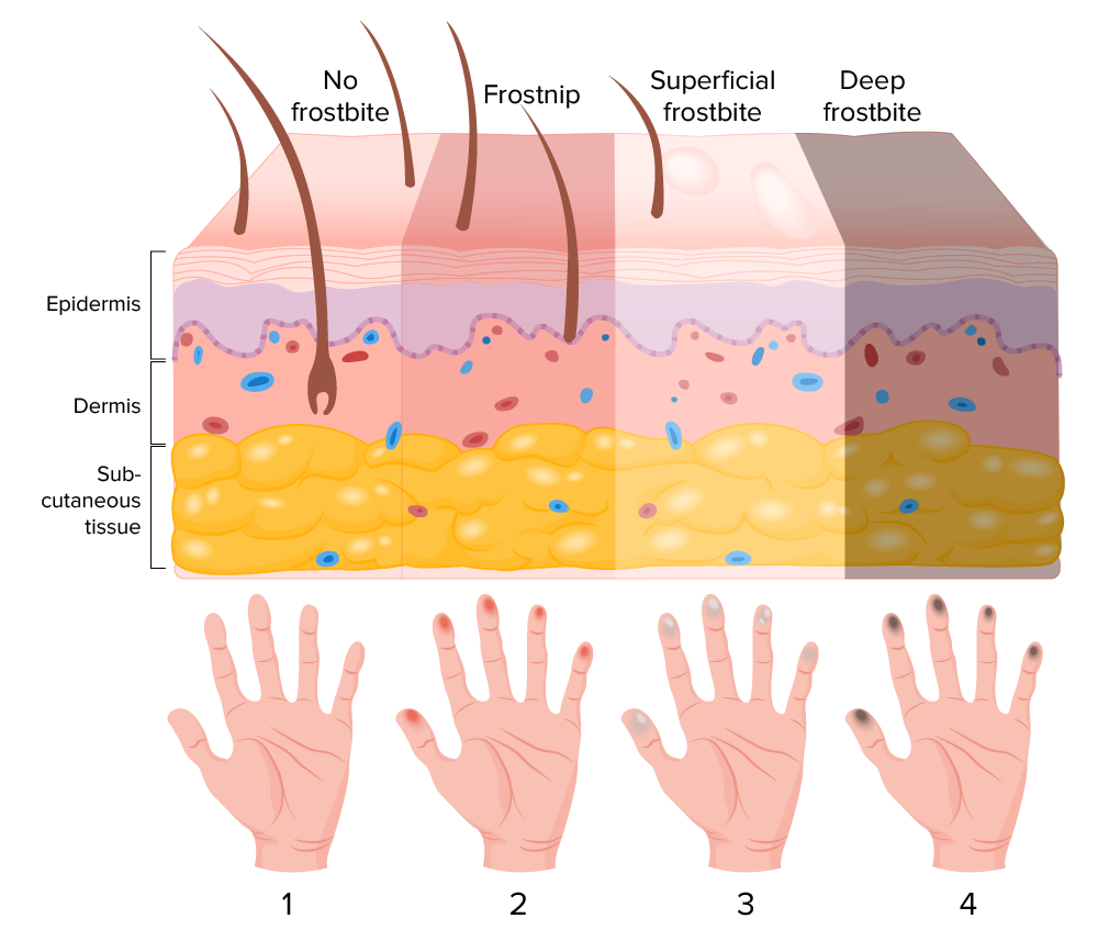
Stages of frostbite
Image by Lecturio.
Clinical Presentation and Diagnosis
The diagnosis of frostbiteFrostbiteInjuries due to cold weather are common among children and athletes who are involved in sports played in cold conditions. Frostbite is a direct freezing injury to the peripheral tissues and occurs when the skin temperature drops below 0°C (32°F). Common sites of frostbite include the nose, ears, fingers, and toes. Frostbite is clinical and should be distinguished from less severe forms of cold injuryCold injuryA physical injury caused by exposure of the body to extremely low ambient temperatures that may lead to loss of body parts, or in extreme cases, death. Examples of cold injury are frostbite and chilblains.Frostbite (frostnipFrostnipFrostbite).
Forms of cold injuryCold injuryA physical injury caused by exposure of the body to extremely low ambient temperatures that may lead to loss of body parts, or in extreme cases, death. Examples of cold injury are frostbite and chilblains.Frostbite and classification
Chilblain (pernioPernioRecurrent localized itching, swelling and painful erythema on the fingers, toes or ears, produced by exposure to cold.Frostbite):[6]
Local inflammatory injury associated with extended or repeated exposure (1–5 hours) to cold (above freezing temperatures), wet conditions
SkinSkinThe skin, also referred to as the integumentary system, is the largest organ of the body. The skin is primarily composed of the epidermis (outer layer) and dermis (deep layer). The epidermis is primarily composed of keratinocytes that undergo rapid turnover, while the dermis contains dense layers of connective tissue.Skin: Structure and Functions blood vessels constrict → hypoxemiaHypoxemiaNeonatal Respiratory Distress Syndrome, vessel wall inflammationInflammationInflammation is a complex set of responses to infection and injury involving leukocytes as the principal cellular mediators in the body’s defense against pathogenic organisms. Inflammation is also seen as a response to tissue injury in the process of wound healing. The 5 cardinal signs of inflammation are pain, heat, redness, swelling, and loss of function. Inflammation, and possibly edemaEdemaEdema is a condition in which excess serous fluid accumulates in the body cavity or interstitial space of connective tissues. Edema is a symptom observed in several medical conditions. It can be categorized into 2 types, namely, peripheral (in the extremities) and internal (in an organ or body cavity). Edema in the dermisDermisA layer of vascularized connective tissue underneath the epidermis. The surface of the dermis contains innervated papillae. Embedded in or beneath the dermis are sweat glands; hair follicles; and sebaceous glands.Skin: Structure and Functions
Purplish or reddish lesions, which can be pruritic or painful
Commonly affects the hands and feet
Trench (immersion) footFootThe foot is the terminal portion of the lower limb, whose primary function is to bear weight and facilitate locomotion. The foot comprises 26 bones, including the tarsal bones, metatarsal bones, and phalanges. The bones of the foot form longitudinal and transverse arches and are supported by various muscles, ligaments, and tendons.Foot: Anatomy:[6]
Non-freezingcold injuryCold injuryA physical injury caused by exposure of the body to extremely low ambient temperatures that may lead to loss of body parts, or in extreme cases, death. Examples of cold injury are frostbite and chilblains.Frostbite
Prolonged (> 12 hours) exposure of the feet to cold, wet conditions
Tight shoes/boots make the condition worse.
↑ Extracellular fluidExtracellular fluidThe fluid of the body that is outside of cells. It is the external environment for the cells.Body Fluid Compartments → edemaEdemaEdema is a condition in which excess serous fluid accumulates in the body cavity or interstitial space of connective tissues. Edema is a symptom observed in several medical conditions. It can be categorized into 2 types, namely, peripheral (in the extremities) and internal (in an organ or body cavity). Edema affects the soft tissues (including the blood vessels and nerves) → painPainAn unpleasant sensation induced by noxious stimuli which are detected by nerve endings of nociceptive neurons.Pain: Types and Pathways, numbness, bullaeBullaeErythema Multiforme
Most common freezing cold injuryCold injuryA physical injury caused by exposure of the body to extremely low ambient temperatures that may lead to loss of body parts, or in extreme cases, death. Examples of cold injury are frostbite and chilblains.Frostbite
No ice formation, no tissue loss
Cannot clinically distinguish between frostnipFrostnipFrostbite and frostbiteFrostbiteInjuries due to cold weather are common among children and athletes who are involved in sports played in cold conditions. Frostbite is a direct freezing injury to the peripheral tissues and occurs when the skin temperature drops below 0°C (32°F). Common sites of frostbite include the nose, ears, fingers, and toes. Frostbite initially
Numbness and pallor resolve with passive rewarming.
FrostbiteFrostbiteInjuries due to cold weather are common among children and athletes who are involved in sports played in cold conditions. Frostbite is a direct freezing injury to the peripheral tissues and occurs when the skin temperature drops below 0°C (32°F). Common sites of frostbite include the nose, ears, fingers, and toes. Frostbite:
Classic 4-tier stagingStagingMethods which attempt to express in replicable terms the extent of the neoplasm in the patient.Grading, Staging, and Metastasis (derived from burn classification):[6]
Based on acute physical examination findings
Assessed after rewarming, but before imaging
Graded from 1st-degree to 4th-degree
Alternative: 2-tier classification
Easier to use in the field setting
Assessment of severity (after rewarming) can be done without imaging.
Superficial: no or minimal expected tissue loss (1st- or 2nd-degree)
Deep: expected tissue loss (3rd- or 4th-degree)
Once in a field hospital or clinic, use Cauchy classification (grade based on anatomical extent of frostbiteFrostbiteInjuries due to cold weather are common among children and athletes who are involved in sports played in cold conditions. Frostbite is a direct freezing injury to the peripheral tissues and occurs when the skin temperature drops below 0°C (32°F). Common sites of frostbite include the nose, ears, fingers, and toes. Frostbite after rewarming, with higher grades indicating higher risk of amputationAmputationAn amputation is the separation of a portion of the limb or the entire limb from the body, along with the bone. Amputations are generally indicated for conditions that compromise the viability of the limb or promote the spread of a local process that could manifest systemically. Amputation):[9,11]
Grade 1: no lesion → no amputationAmputationAn amputation is the separation of a portion of the limb or the entire limb from the body, along with the bone. Amputations are generally indicated for conditions that compromise the viability of the limb or promote the spread of a local process that could manifest systemically. Amputation of boneBoneBone is a compact type of hardened connective tissue composed of bone cells, membranes, an extracellular mineralized matrix, and central bone marrow. The 2 primary types of bone are compact and spongy. Bones: Structure and Types
Grade 2: lesion on distal phalanx → tissue amputationAmputationAn amputation is the separation of a portion of the limb or the entire limb from the body, along with the bone. Amputations are generally indicated for conditions that compromise the viability of the limb or promote the spread of a local process that could manifest systemically. Amputation
Grade 3: lesion on the intermediary and proximal phalanx (beyond the distal phalanx) → amputationAmputationAn amputation is the separation of a portion of the limb or the entire limb from the body, along with the bone. Amputations are generally indicated for conditions that compromise the viability of the limb or promote the spread of a local process that could manifest systemically. Amputation of the digit
Grade 4: lesion on the carpal/tarsal → amputationAmputationAn amputation is the separation of a portion of the limb or the entire limb from the body, along with the bone. Amputations are generally indicated for conditions that compromise the viability of the limb or promote the spread of a local process that could manifest systemically. Amputation of the limb
Classifying frostbiteFrostbiteInjuries due to cold weather are common among children and athletes who are involved in sports played in cold conditions. Frostbite is a direct freezing injury to the peripheral tissues and occurs when the skin temperature drops below 0°C (32°F). Common sites of frostbite include the nose, ears, fingers, and toes. Frostbite is helpful to healthcare workers, as risk of tissue loss and amputationAmputationAn amputation is the separation of a portion of the limb or the entire limb from the body, along with the bone. Amputations are generally indicated for conditions that compromise the viability of the limb or promote the spread of a local process that could manifest systemically. Amputation is an important factor in evaluation decisions.
Table: 2-tier and 4-tier classification systems of frostbiteFrostbiteInjuries due to cold weather are common among children and athletes who are involved in sports played in cold conditions. Frostbite is a direct freezing injury to the peripheral tissues and occurs when the skin temperature drops below 0°C (32°F). Common sites of frostbite include the nose, ears, fingers, and toes. Frostbite[5,12]
2-tier classification
4-tier classification
Depth of damage
Signs
Superficial
1st-degree
Partial-thickness skinSkinThe skin, also referred to as the integumentary system, is the largest organ of the body. The skin is primarily composed of the epidermis (outer layer) and dermis (deep layer). The epidermis is primarily composed of keratinocytes that undergo rapid turnover, while the dermis contains dense layers of connective tissue.Skin: Structure and Functions freezing
ErythemaErythemaRedness of the skin produced by congestion of the capillaries. This condition may result from a variety of disease processes.Chalazion
Mild edemaEdemaEdema is a condition in which excess serous fluid accumulates in the body cavity or interstitial space of connective tissues. Edema is a symptom observed in several medical conditions. It can be categorized into 2 types, namely, peripheral (in the extremities) and internal (in an organ or body cavity). Edema
Slight epidermal sloughing
2nd-degree
Full-thickness skinSkinThe skin, also referred to as the integumentary system, is the largest organ of the body. The skin is primarily composed of the epidermis (outer layer) and dermis (deep layer). The epidermis is primarily composed of keratinocytes that undergo rapid turnover, while the dermis contains dense layers of connective tissue.Skin: Structure and Functions freezing
ErythemaErythemaRedness of the skin produced by congestion of the capillaries. This condition may result from a variety of disease processes.Chalazion
EdemaEdemaEdema is a condition in which excess serous fluid accumulates in the body cavity or interstitial space of connective tissues. Edema is a symptom observed in several medical conditions. It can be categorized into 2 types, namely, peripheral (in the extremities) and internal (in an organ or body cavity). Edema
Superficial skinSkinThe skin, also referred to as the integumentary system, is the largest organ of the body. The skin is primarily composed of the epidermis (outer layer) and dermis (deep layer). The epidermis is primarily composed of keratinocytes that undergo rapid turnover, while the dermis contains dense layers of connective tissue.Skin: Structure and Functions blisters (with clear or milky fluid)
Deep
3rd-degree
SkinSkinThe skin, also referred to as the integumentary system, is the largest organ of the body. The skin is primarily composed of the epidermis (outer layer) and dermis (deep layer). The epidermis is primarily composed of keratinocytes that undergo rapid turnover, while the dermis contains dense layers of connective tissue.Skin: Structure and Functions and subcutaneous tissueSubcutaneous tissueLoose connective tissue lying under the dermis, which binds skin loosely to subjacent tissues. It may contain a pad of adipocytes, which vary in number according to the area of the body and vary in size according to the nutritional state.Soft Tissue Abscess freezing
Deep, hemorrhagic blisters
Substantial edemaEdemaEdema is a condition in which excess serous fluid accumulates in the body cavity or interstitial space of connective tissues. Edema is a symptom observed in several medical conditions. It can be categorized into 2 types, namely, peripheral (in the extremities) and internal (in an organ or body cavity). Edema
May have blue/black appearance
4th-degree
SkinSkinThe skin, also referred to as the integumentary system, is the largest organ of the body. The skin is primarily composed of the epidermis (outer layer) and dermis (deep layer). The epidermis is primarily composed of keratinocytes that undergo rapid turnover, while the dermis contains dense layers of connective tissue.Skin: Structure and Functions, subcutaneous tissueSubcutaneous tissueLoose connective tissue lying under the dermis, which binds skin loosely to subjacent tissues. It may contain a pad of adipocytes, which vary in number according to the area of the body and vary in size according to the nutritional state.Soft Tissue Abscess freezing which extends to deeper structures (e.g., muscle, tendon, boneBoneBone is a compact type of hardened connective tissue composed of bone cells, membranes, an extracellular mineralized matrix, and central bone marrow. The 2 primary types of bone are compact and spongy. Bones: Structure and Types)
NecrosisNecrosisThe death of cells in an organ or tissue due to disease, injury or failure of the blood supply.Ischemic Cell Damage
Table: Cauchy classification for the severity of frostbiteFrostbiteInjuries due to cold weather are common among children and athletes who are involved in sports played in cold conditions. Frostbite is a direct freezing injury to the peripheral tissues and occurs when the skin temperature drops below 0°C (32°F). Common sites of frostbite include the nose, ears, fingers, and toes. Frostbite injury[9.11]
Grade
Initial lesion (day 0)
Blisters
AmputationAmputationAn amputation is the separation of a portion of the limb or the entire limb from the body, along with the bone. Amputations are generally indicated for conditions that compromise the viability of the limb or promote the spread of a local process that could manifest systemically. Amputation
1
No lesion
None
No amputationAmputationAn amputation is the separation of a portion of the limb or the entire limb from the body, along with the bone. Amputations are generally indicated for conditions that compromise the viability of the limb or promote the spread of a local process that could manifest systemically. Amputation
2
Lesion/cyanosisCyanosisA bluish or purplish discoloration of the skin and mucous membranes due to an increase in the amount of deoxygenated hemoglobin in the blood or a structural defect in the hemoglobin molecule.Pulmonary Examination in distal phalanx
Clear blisters
Tissue amputationAmputationAn amputation is the separation of a portion of the limb or the entire limb from the body, along with the bone. Amputations are generally indicated for conditions that compromise the viability of the limb or promote the spread of a local process that could manifest systemically. Amputation
3
Lesion/cyanosisCyanosisA bluish or purplish discoloration of the skin and mucous membranes due to an increase in the amount of deoxygenated hemoglobin in the blood or a structural defect in the hemoglobin molecule.Pulmonary Examination beyond distal phalanx (to the proximal phalanx)
Hemorrhagic blisters on the digit
AmputationAmputationAn amputation is the separation of a portion of the limb or the entire limb from the body, along with the bone. Amputations are generally indicated for conditions that compromise the viability of the limb or promote the spread of a local process that could manifest systemically. Amputation of the digit
4
Lesion on the carpal/tarsal
Hemorrhagic blisters on the carpal/tarsal region
AmputationAmputationAn amputation is the separation of a portion of the limb or the entire limb from the body, along with the bone. Amputations are generally indicated for conditions that compromise the viability of the limb or promote the spread of a local process that could manifest systemically. Amputation of the limb
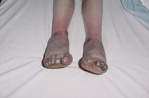
Initial presentation of frostbite on day 2
Image: “Grade IV frostbite requiring bilateral below knee amputations: a case report” by Ramdass MJ. License: CC BY 3.0
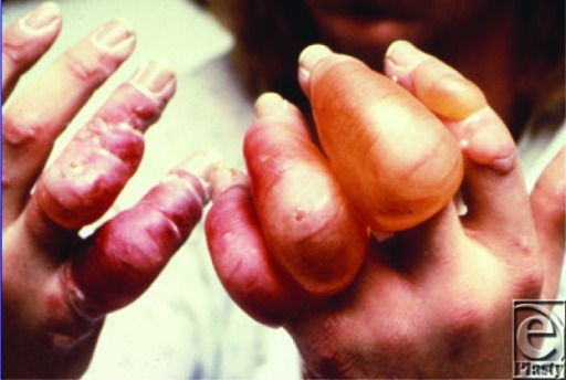
Frostbite in a 12-year-old girl who presented to the emergency department after waiting at a bus stop in winter
Image: “Urban blisters” by Chang N, Nunn R, Milner SM, Price LA. License: CC BY 2.0
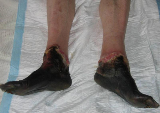
Appearance of grade IV frostbite with completely mummified feet 3 weeks post-injury
Image: “Grade IV frostbite requiring bilateral below knee amputations: a case report” by Ramdass MJ. License: CC BY 3.0
In both the field and the hospital, assess perfusion after rewarming.[9]
Examine the following:
SkinSkinThe skin, also referred to as the integumentary system, is the largest organ of the body. The skin is primarily composed of the epidermis (outer layer) and dermis (deep layer). The epidermis is primarily composed of keratinocytes that undergo rapid turnover, while the dermis contains dense layers of connective tissue.Skin: Structure and Functions color and temperature
Sensation
Pulses
Capillary refill
Equipment (found in expedition camps or field clinics) to help in assessment includes:
Fast-response infrared thermometers
Pulse oximeters
DopplerDopplerUltrasonography applying the doppler effect, with frequency-shifted ultrasound reflections produced by moving targets (usually red blood cells) in the bloodstream along the ultrasound axis in direct proportion to the velocity of movement of the targets, to determine both direction and velocity of blood flow.Ultrasound (Sonography) devices
In the hospital setting, imaging helps with evaluating the extent of injury and other comorbiditiesComorbiditiesThe presence of co-existing or additional diseases with reference to an initial diagnosis or with reference to the index condition that is the subject of study. Comorbidity may affect the ability of affected individuals to function and also their survival; it may be used as a prognostic indicator for length of hospital stay, cost factors, and outcome or survival.St. Louis Encephalitis Virus:[12]
Radiography:
Initial survey of the limb
Can show trauma-related fractures and boneBoneBone is a compact type of hardened connective tissue composed of bone cells, membranes, an extracellular mineralized matrix, and central bone marrow. The 2 primary types of bone are compact and spongy. Bones: Structure and Types destruction
Standard or digital subtraction angiographyAngiographyRadiography of blood vessels after injection of a contrast medium.Cardiac Surgery:
Imaging method of choice for those presenting within 24 hours after deep (3rd- or 4th-degree) frostbiteFrostbiteInjuries due to cold weather are common among children and athletes who are involved in sports played in cold conditions. Frostbite is a direct freezing injury to the peripheral tissues and occurs when the skin temperature drops below 0°C (32°F). Common sites of frostbite include the nose, ears, fingers, and toes. Frostbite injury
Used to assess perfusion deficits in frostbiteFrostbiteInjuries due to cold weather are common among children and athletes who are involved in sports played in cold conditions. Frostbite is a direct freezing injury to the peripheral tissues and occurs when the skin temperature drops below 0°C (32°F). Common sites of frostbite include the nose, ears, fingers, and toes. Frostbite injuries; identifies candidates for thrombolytic therapy
Indicated in those presenting with 2nd-, 3rd-, or 4th-degree frostbiteFrostbiteInjuries due to cold weather are common among children and athletes who are involved in sports played in cold conditions. Frostbite is a direct freezing injury to the peripheral tissues and occurs when the skin temperature drops below 0°C (32°F). Common sites of frostbite include the nose, ears, fingers, and toes. Frostbite injuries
In candidates for thrombolysis, the test is performed within 24 hours after thawing.
For those not undergoing thrombolysis, the test is generally performed 2 days after the original injury.
Also used after angiographyAngiographyRadiography of blood vessels after injection of a contrast medium.Cardiac Surgery and treatment with intra-arterial thrombolysis
Assesses perfusion and tissue viability
Accurately predicts level of amputationAmputationAn amputation is the separation of a portion of the limb or the entire limb from the body, along with the bone. Amputations are generally indicated for conditions that compromise the viability of the limb or promote the spread of a local process that could manifest systemically. Amputation in majority of cases (when performed on day 2)
Single-photon emission computed tomographySingle-photon emission computed tomographyAn imaging technique using a device which combines tomography, emission-computed, single-photon and tomography, x-ray computed in the same session.Nuclear Imaging (SPECTSPECTAn imaging technique using a device which combines tomography, emission-computed, single-photon and tomography, x-ray computed in the same session.Nuclear Imaging)/computed tomography (CT) combined with boneBoneBone is a compact type of hardened connective tissue composed of bone cells, membranes, an extracellular mineralized matrix, and central bone marrow. The 2 primary types of bone are compact and spongy. Bones: Structure and Types scanning can identify areas of tissue necrosisNecrosisThe death of cells in an organ or tissue due to disease, injury or failure of the blood supply.Ischemic Cell Damage even before physical manifestations are observed.
Magnetic resonance angiographyAngiographyRadiography of blood vessels after injection of a contrast medium.Cardiac Surgery:
Visualizes both blood vessels and tissues
An alternative to digital subtraction angiographyAngiographyRadiography of blood vessels after injection of a contrast medium.Cardiac Surgery to assess vascular patency but has no therapeutic potential
Limited availability and less studied than above methods
Management
Individual protocols may vary based on location. The following recommendations are based on US and UK guidelines.
Prevention[5,6,11]
Educate individuals who participate in activities involving cold exposure about injuries from the cold.
Measures:
Use of proper clothing:
Internal layer: allows evaporation of sweat with minimal absorptionAbsorptionAbsorption involves the uptake of nutrient molecules and their transfer from the lumen of the GI tract across the enterocytes and into the interstitial space, where they can be taken up in the venous or lymphatic circulation.Digestion and Absorption
Middle layer: for insulation (e.g., fleece)
Removable outer layer: wind and water-resistant; allows for evaporation of moisture
Protect toes, fingers, ears, and skinSkinThe skin, also referred to as the integumentary system, is the largest organ of the body. The skin is primarily composed of the epidermis (outer layer) and dermis (deep layer). The epidermis is primarily composed of keratinocytes that undergo rapid turnover, while the dermis contains dense layers of connective tissue.Skin: Structure and Functions (when wind-chill temperatures can cause frostbiteFrostbiteInjuries due to cold weather are common among children and athletes who are involved in sports played in cold conditions. Frostbite is a direct freezing injury to the peripheral tissues and occurs when the skin temperature drops below 0°C (32°F). Common sites of frostbite include the nose, ears, fingers, and toes. Frostbite within ≤ 30 minutes).
Remove wet clothing as soon as possible.
Pack blankets, external warmers, and additional clothing.
Bring patient to a warm environment and try to warm the area/limb with body heatHeatInflammation (e.g., place in axillaAxillaThe axilla is a pyramid-shaped space located between the upper thorax and the arm. The axilla has a base, an apex, and 4 walls (anterior, medial, lateral, posterior). The base of the pyramid is made up of the axillary skin. The apex is the axillary inlet, located between the 1st rib, superior border of the scapula, and clavicle. Axilla and Brachial Plexus: Anatomy).
Do not put pressure on frostbitten extremities (e.g., rubbing frostbitten extremities or walking on feet affected by frostbiteFrostbiteInjuries due to cold weather are common among children and athletes who are involved in sports played in cold conditions. Frostbite is a direct freezing injury to the peripheral tissues and occurs when the skin temperature drops below 0°C (32°F). Common sites of frostbite include the nose, ears, fingers, and toes. Frostbite).
Remove any constricting material or jewelry from the body part.
Avoid rubbing or applying friction (to warm the part), as this worsens the injury.
Do not rewarm if there is a chance that freezing will recur:
More extensive injury occurs with thawed tissue that refreezes.[11]
↑ Prostaglandin and thromboxane release in the freeze–thaw cycle leads to vasoconstrictionVasoconstrictionThe physiological narrowing of blood vessels by contraction of the vascular smooth muscle.Vascular Resistance, Flow, and Mean Arterial Pressure, platelet aggregationPlatelet aggregationThe attachment of platelets to one another. This clumping together can be induced by a number of agents (e.g., thrombin; collagen) and is part of the mechanism leading to the formation of a thrombus.Hemostasis, thrombosisThrombosisFormation and development of a thrombus or blood clot in the blood vessel.Epidemic Typhus → more injury
AVOID refreezing if thawing occurs in the field.
Oral, and possibly IV, hydration
Supplemental O2, if SPO2 < 88% or at altitudes > 4000 m[11]
Rewarming[2,5,6,8]
In the hospital, first treat hypothermiaHypothermiaHypothermia can be defined as a drop in the core body temperature below 35°C (95°F) and is classified into mild, moderate, severe, and profound forms based on the degree of temperature decrease. Hypothermia (decrease in core temperature to < 95℉ (35℃)), if present, and/or other life-threatening conditions. Manage the associated cold injuryCold injuryA physical injury caused by exposure of the body to extremely low ambient temperatures that may lead to loss of body parts, or in extreme cases, death. Examples of cold injury are frostbite and chilblains.Frostbite as follows:
Rewarm in warm water (37°C–39°C (98.6°F–102.2°F)) bath.
IV opioidsOpioidsOpiates are drugs that are derived from the sap of the opium poppy. Opiates have been used since antiquity for the relief of acute severe pain. Opioids are synthetic opiates with properties that are substantially similar to those of opiates. Opioid Analgesics as needed
Thawing completed when tissue is red or purple and soft to the touch:
Generally takes 20–40 minutes
Air dry → do not towel dry (to avoid friction)
Avoid ambulation on lower extremity that is thawed.
Application of bulky dressing (using non-adherent gauze as the 1st layer).
Topical aloe vera applied every 6 hours with dressing changes.
Elevation of the limb to reduce edemaEdemaEdema is a condition in which excess serous fluid accumulates in the body cavity or interstitial space of connective tissues. Edema is a symptom observed in several medical conditions. It can be categorized into 2 types, namely, peripheral (in the extremities) and internal (in an organ or body cavity). Edema
AspirinAspirinThe prototypical analgesic used in the treatment of mild to moderate pain. It has anti-inflammatory and antipyretic properties and acts as an inhibitor of cyclooxygenase which results in the inhibition of the biosynthesis of prostaglandins. Aspirin also inhibits platelet aggregation and is used in the prevention of arterial and venous thrombosis.Nonsteroidal Antiinflammatory Drugs (NSAIDs) and nonsteroidal antiinflammatory drugsNonsteroidal Antiinflammatory DrugsNonsteroidal antiinflammatory drugs (NSAIDs) are a class of medications consisting of aspirin, reversible NSAIDs, and selective NSAIDs. NSAIDs are used as antiplatelet, analgesic, antipyretic, and antiinflammatory agents. Nonsteroidal Antiinflammatory Drugs (NSAIDs) (NSAIDsNSAIDSPrimary vs Secondary Headaches) to help reduce inflammationInflammationInflammation is a complex set of responses to infection and injury involving leukocytes as the principal cellular mediators in the body’s defense against pathogenic organisms. Inflammation is also seen as a response to tissue injury in the process of wound healing. The 5 cardinal signs of inflammation are pain, heat, redness, swelling, and loss of function. Inflammation
Blisters:
Do not debride in the field.
No clear consensus on managing clear tense (non-hemorrhagic) blisters, but may be drained at the provider’s discretion (especially if movement is impaired).
Hemorrhagic blisters are a sign of deeper tissue damage and should be left intact (not debrided).
FrostbiteFrostbiteInjuries due to cold weather are common among children and athletes who are involved in sports played in cold conditions. Frostbite is a direct freezing injury to the peripheral tissues and occurs when the skin temperature drops below 0°C (32°F). Common sites of frostbite include the nose, ears, fingers, and toes. Frostbite is not inherently prone to infection.
Administer only if there is significant trauma, or signs or symptoms of cellulitisCellulitisCellulitis is a common infection caused by bacteria that affects the dermis and subcutaneous tissue of the skin. It is frequently caused by Staphylococcus aureus and Streptococcus pyogenes. The skin infection presents as an erythematous and edematous area with warmth and tenderness. Cellulitis or sepsisSepsisSystemic inflammatory response syndrome with a proven or suspected infectious etiology. When sepsis is associated with organ dysfunction distant from the site of infection, it is called severe sepsis. When sepsis is accompanied by hypotension despite adequate fluid infusion, it is called septic shock.Sepsis and Septic Shock.
Coverage should include:
StaphylococcusStaphylococcusStaphylococcus is a medically important genera of Gram-positive, aerobic cocci. These bacteria form clusters resembling grapes on culture plates. Staphylococci are ubiquitous for humans, and many strains compose the normal skin flora.Staphylococcus spp.
StreptococcusStreptococcusStreptococcus is one of the two medically important genera of gram-positive cocci, the other being Staphylococcus. Streptococci are identified as different species on blood agar on the basis of their hemolytic pattern and sensitivity to optochin and bacitracin. There are many pathogenic species of streptococci, including S. pyogenes, S. agalactiae, S. pneumoniae, and the viridans streptococci.Streptococcus spp.
PseudomonasPseudomonasPseudomonas is a non-lactose-fermenting, gram-negative bacillus that produces pyocyanin, which gives it a characteristic blue-green color. Pseudomonas is found ubiquitously in the environment, as well as in moist reservoirs, such as hospital sinks and respiratory equipment. Pseudomonas spp.
Thrombolysis[10]
Vascular thrombosisThrombosisFormation and development of a thrombus or blood clot in the blood vessel.Epidemic Typhus is associated with frostbiteFrostbiteInjuries due to cold weather are common among children and athletes who are involved in sports played in cold conditions. Frostbite is a direct freezing injury to the peripheral tissues and occurs when the skin temperature drops below 0°C (32°F). Common sites of frostbite include the nose, ears, fingers, and toes. Frostbite injury; in those at risk for amputationAmputationAn amputation is the separation of a portion of the limb or the entire limb from the body, along with the bone. Amputations are generally indicated for conditions that compromise the viability of the limb or promote the spread of a local process that could manifest systemically. Amputation, thrombolytic therapy is considered.
Used in severe injury presenting within 24 hours:
Treatment option for grades 3 or 4
Patient should be hemodynamically stable, with absent blood flowBlood flowBlood flow refers to the movement of a certain volume of blood through the vasculature over a given unit of time (e.g., mL per minute).Vascular Resistance, Flow, and Mean Arterial Pressure in affected limb after rewarming.
Perform subtraction angiographyAngiographyRadiography of blood vessels after injection of a contrast medium.Cardiac Surgery within the 1st 24 hours (or technetium-99m scanning):[5,11]
Best outcomes if thrombolysis performed within 12 hours
If in a remote area, transfer patient to a capable facility if therapy can be given within 24 hours.
Options include intravenous or intra-arterial tissue plasminogen activatorTissue plasminogen activatorA proteolytic enzyme in the serine protease family found in many tissues which converts plasminogen to fibrinolysin. It has fibrin-binding activity and is immunologically different from urokinase-type plasminogen activator. The primary sequence, composed of 527 amino acids, is identical in both the naturally occurring and synthetic proteases.Hemostasis (tPAtPAIschemic Stroke), or intravenous or intra-arterial heparin or enoxaparinEnoxaparinLow-molecular-weight fragment of heparin, having a 4-enopyranosuronate sodium structure at the non-reducing end of the chain. It is prepared by depolymerization of the benzylic ester of porcine mucosal heparin. Therapeutically, it is used as an antithrombotic agent.Anticoagulants:
tPAtPAIschemic Stroke 0.15 mg/kg IV bolus over 15 minutes, followed by infusion of 0.15 mg/kg/hr for 6 hours; total maximum dose, 100 mg, plus
Heparin 500–1000 units/hr IV (with goal of 2x the control PTT) for 6 hours, or
EnoxaparinEnoxaparinLow-molecular-weight fragment of heparin, having a 4-enopyranosuronate sodium structure at the non-reducing end of the chain. It is prepared by depolymerization of the benzylic ester of porcine mucosal heparin. Therapeutically, it is used as an antithrombotic agent.Anticoagulants 1 mg/kg SC for 1 dose
tPAtPAIschemic Stroke 3 mg intra-arterial bolus, followed an intra-arterial infusion of 0.5–1 mg/hr (via femoral or brachial arteryBrachial ArteryThe continuation of the axillary artery; it branches into the radial and ulnar arteries.Cubital Fossa: Anatomy) plus heparin 500 units/hr intra-arterial
Uncontrolled blood pressure (systolic >180 mm Hg, diastolic > 110 mm Hg)
PregnancyPregnancyThe status during which female mammals carry their developing young (embryos or fetuses) in utero before birth, beginning from fertilization to birth.Pregnancy: Diagnosis, Physiology, and Care
Treatment should be discontinued:
When perfusion is restored (angiographyAngiographyRadiography of blood vessels after injection of a contrast medium.Cardiac Surgery or technetium-99m scanning every 12–24 hours), or
When 72 hours is reached
Potential risks and complications:
Systemic and catheter-site bleeding
Compartment syndromeCompartment SyndromeCompartment syndrome is a surgical emergency usually occurring secondary to trauma. The condition is marked by increased pressure within a compartment that compromises the circulation and function of the tissues within that space.Compartment Syndrome
IV prostacyclins (e.g., iloprostIloprostAn eicosanoid, derived from the cyclooxygenase pathway of arachidonic acid metabolism. It is a stable and synthetic analog of epoprostenol, but with a longer half-life than the parent compound. Its actions are similar to prostacyclin. Iloprost produces vasodilation and inhibits platelet aggregation.Pulmonary Hypertension Drugs):[5,11]
Effects:
Potent vasodilator
↓ Platelet aggregationPlatelet aggregationThe attachment of platelets to one another. This clumping together can be induced by a number of agents (e.g., thrombin; collagen) and is part of the mechanism leading to the formation of a thrombus.Hemostasis, lymphocyte adhesionAdhesionThe process whereby platelets adhere to something other than platelets, e.g., collagen; basement membrane; microfibrils; or other ‘foreign’ surfaces.Coagulation Studies to endothelial cells
Considered in grade 3 or 4 injuries (< 72 hours after the injury)
0.5 ng/kg/min, increasing by 0.5 ng/kg/min every 30 minutes, up to 2 ng/kg/min
If headacheHeadacheThe symptom of pain in the cranial region. It may be an isolated benign occurrence or manifestation of a wide variety of headache disorders.Brain Abscess or hypotensionHypotensionHypotension is defined as low blood pressure, specifically < 90/60 mm Hg, and is most commonly a physiologic response. Hypotension may be mild, serious, or life threatening, depending on the cause. Hypotension occurs, reduce to highest rate without side effects
Infusion is a 5- to 8-day treatment for 6 hours per day.
Monotherapy with low-molecular-weight heparin or unfractionated heparinUnfractionated heparinA highly acidic mucopolysaccharide formed of equal parts of sulfated d-glucosamine and d-glucuronic acid with sulfaminic bridges. The molecular weight ranges from six to twenty thousand. Heparin occurs in and is obtained from liver, lung, mast cells, etc. , of vertebrates. Its function is unknown, but it is used to prevent blood clotting in vivo and vitro, in the form of many different salts.Anticoagulants is not recommended (insufficient data).[5]
Surgical management[1,5,7,8,10,11]
Initial assessment might overestimate real extent of tissue injury
No surgical treatment until demarcation occurs (unless overwhelming infection or sepsisSepsisSystemic inflammatory response syndrome with a proven or suspected infectious etiology. When sepsis is associated with organ dysfunction distant from the site of infection, it is called severe sepsis. When sepsis is accompanied by hypotension despite adequate fluid infusion, it is called septic shock.Sepsis and Septic Shock is present).
For many years, the axiom “FrostbiteFrostbiteInjuries due to cold weather are common among children and athletes who are involved in sports played in cold conditions. Frostbite is a direct freezing injury to the peripheral tissues and occurs when the skin temperature drops below 0°C (32°F). Common sites of frostbite include the nose, ears, fingers, and toes. Frostbite in January, amputationAmputationAn amputation is the separation of a portion of the limb or the entire limb from the body, along with the bone. Amputations are generally indicated for conditions that compromise the viability of the limb or promote the spread of a local process that could manifest systemically. Amputation in July” applied to practice:
Indicates that a waiting period is needed to determine viability, and early amputationAmputationAn amputation is the separation of a portion of the limb or the entire limb from the body, along with the bone. Amputations are generally indicated for conditions that compromise the viability of the limb or promote the spread of a local process that could manifest systemically. Amputation increases morbidityMorbidityThe proportion of patients with a particular disease during a given year per given unit of population.Measures of Health Status.
In general, it takes up to 3 months to fully determine whether tissue is viable.
Most amputations can be done 6–12 weeks after injury.
Indications for early surgical procedures:
Necrotic tissue and/or blisters → debridementDebridementThe removal of foreign material and devitalized or contaminated tissue from or adjacent to a traumatic or infected lesion until surrounding healthy tissue is exposed.Stevens-Johnson Syndrome
Compartment syndromeCompartment SyndromeCompartment syndrome is a surgical emergency usually occurring secondary to trauma. The condition is marked by increased pressure within a compartment that compromises the circulation and function of the tissues within that space.Compartment Syndrome → fasciotomyFasciotomySurgical incision on the fascia. It is used to decompress compartment pressure (e.g. in compartment syndromes; circumferential burns and extremity injuries) or to release contractures (e.g. in dupuytren’s contracture).Compartment Syndrome
Post-thaw hydrotherapy, once to twice daily at 37℃–39℃ (98.6℉–102.2℉):
Increases circulationCirculationThe movement of the blood as it is pumped through the cardiovascular system.ABCDE Assessment
Removes superficial bacteriaBacteriaBacteria are prokaryotic single-celled microorganisms that are metabolically active and divide by binary fission. Some of these organisms play a significant role in the pathogenesis of diseases. Bacteriology
Debrides devitalized tissue
Hyperbaric oxygenHyperbaric oxygenThe therapeutic intermittent administration of oxygen in a chamber at greater than sea-level atmospheric pressures (three atmospheres). It is considered effective treatment for air and gas embolisms, smoke inhalation, acute carbon monoxide poisoning, caisson disease, clostridial gangrene, etc. The list of treatment modalities includes stroke.Decompression Sickness therapy (HBOT):
Mixed results
Theoretically, HBOT increases tissue oxygenation, but this requires effective blood supply.
If performed immediately, sympathectomy actually increases edemaEdemaEdema is a condition in which excess serous fluid accumulates in the body cavity or interstitial space of connective tissues. Edema is a symptom observed in several medical conditions. It can be categorized into 2 types, namely, peripheral (in the extremities) and internal (in an organ or body cavity). Edema and tissue loss.
If performed 24–48 hours after thawing, sympathectomy has been observed to reduce edemaEdemaEdema is a condition in which excess serous fluid accumulates in the body cavity or interstitial space of connective tissues. Edema is a symptom observed in several medical conditions. It can be categorized into 2 types, namely, peripheral (in the extremities) and internal (in an organ or body cavity). Edema and tissue loss.
May have benefits in decreasing long-term painPainAn unpleasant sensation induced by noxious stimuli which are detected by nerve endings of nociceptive neurons.Pain: Types and Pathways, hyperhidrosisHyperhidrosisExcessive sweating. In the localized type, the most frequent sites are the palms, soles, axillae, inguinal folds, and the perineal area. Its chief cause is thought to be emotional. Generalized hyperhidrosis may be induced by a hot, humid environment, by fever, or by vigorous exercise.Malassezia Fungi, and paresthesiasParesthesiasSubjective cutaneous sensations (e.g., cold, warmth, tingling, pressure, etc.) that are experienced spontaneously in the absence of stimulation.Posterior Cord Syndrome.
Results are still conflicting, and no solid recommendation has been given.
Treatment of other cold injuries[6,8]
ChilblainsChilblainsRecurrent localized itching, swelling and painful erythema on the fingers, toes or ears, produced by exposure to cold.Frostbite:
Remove wet clothing.
Dry the area and massage.
Permanent damage is not common; symptoms clear up in 2 weeks.
Trench footFootThe foot is the terminal portion of the lower limb, whose primary function is to bear weight and facilitate locomotion. The foot comprises 26 bones, including the tarsal bones, metatarsal bones, and phalanges. The bones of the foot form longitudinal and transverse arches and are supported by various muscles, ligaments, and tendons.Foot: Anatomy:
Proper footFootThe foot is the terminal portion of the lower limb, whose primary function is to bear weight and facilitate locomotion. The foot comprises 26 bones, including the tarsal bones, metatarsal bones, and phalanges. The bones of the foot form longitudinal and transverse arches and are supported by various muscles, ligaments, and tendons.Foot: Anatomy hygiene (replace wet socks; use moisture-wicking socks)
Raynaud’s phenomenon: exaggerated vascular response to cold temperatures or emotional stress causing sharply demarcated color changes of digits. Blood vessels constrict, which leads to digits turning blue. Digits return to normal color 10–20 minutes after cold is removed.
HypothermiaHypothermiaHypothermia can be defined as a drop in the core body temperature below 35°C (95°F) and is classified into mild, moderate, severe, and profound forms based on the degree of temperature decrease. Hypothermia: drop in core body temperatureBody TemperatureThe measure of the level of heat of a human or animal.Heatstroke < 35°C (95°F). Classified into mild, moderate, and severe forms. Management involves rewarming of patient by external or internal methods, depending on severity.
Billing and Coding
Diagnosis Codes:
FrostbiteFrostbiteInjuries due to cold weather are common among children and athletes who are involved in sports played in cold conditions. Frostbite is a direct freezing injury to the peripheral tissues and occurs when the skin temperature drops below 0°C (32°F). Common sites of frostbite include the nose, ears, fingers, and toes. Frostbite is coded based on the location and the depth of injury, distinguishing between superficial frostbiteFrostbiteInjuries due to cold weather are common among children and athletes who are involved in sports played in cold conditions. Frostbite is a direct freezing injury to the peripheral tissues and occurs when the skin temperature drops below 0°C (32°F). Common sites of frostbite include the nose, ears, fingers, and toes. Frostbite (T33 codes) and frostbiteFrostbiteInjuries due to cold weather are common among children and athletes who are involved in sports played in cold conditions. Frostbite is a direct freezing injury to the peripheral tissues and occurs when the skin temperature drops below 0°C (32°F). Common sites of frostbite include the nose, ears, fingers, and toes. Frostbite with tissue necrosisNecrosisThe death of cells in an organ or tissue due to disease, injury or failure of the blood supply.Ischemic Cell Damage (T34 codes).
Coding System
Code
Description
ICD-10-CM
T33.522A
Superficial frostbiteFrostbiteInjuries due to cold weather are common among children and athletes who are involved in sports played in cold conditions. Frostbite is a direct freezing injury to the peripheral tissues and occurs when the skin temperature drops below 0°C (32°F). Common sites of frostbite include the nose, ears, fingers, and toes. Frostbite on left handHandThe hand constitutes the distal part of the upper limb and provides the fine, precise movements needed in activities of daily living. It consists of 5 metacarpal bones and 14 phalanges, as well as numerous muscles innervated by the median and ulnar nerves. Hand: Anatomy, initial encounter
ICD-10-CM
T34.41XA
FrostbiteFrostbiteInjuries due to cold weather are common among children and athletes who are involved in sports played in cold conditions. Frostbite is a direct freezing injury to the peripheral tissues and occurs when the skin temperature drops below 0°C (32°F). Common sites of frostbite include the nose, ears, fingers, and toes. Frostbite with tissue necrosisNecrosisThe death of cells in an organ or tissue due to disease, injury or failure of the blood supply.Ischemic Cell Damage of right footFootThe foot is the terminal portion of the lower limb, whose primary function is to bear weight and facilitate locomotion. The foot comprises 26 bones, including the tarsal bones, metatarsal bones, and phalanges. The bones of the foot form longitudinal and transverse arches and are supported by various muscles, ligaments, and tendons.Foot: Anatomy, initial encounter
SNOMED CT
241592009
FrostbiteFrostbiteInjuries due to cold weather are common among children and athletes who are involved in sports played in cold conditions. Frostbite is a direct freezing injury to the peripheral tissues and occurs when the skin temperature drops below 0°C (32°F). Common sites of frostbite include the nose, ears, fingers, and toes. Frostbite (disorder)
Procedures/Interventions:
These codes are for procedures required in severe frostbiteFrostbiteInjuries due to cold weather are common among children and athletes who are involved in sports played in cold conditions. Frostbite is a direct freezing injury to the peripheral tissues and occurs when the skin temperature drops below 0°C (32°F). Common sites of frostbite include the nose, ears, fingers, and toes. Frostbite cases. DebridementDebridementThe removal of foreign material and devitalized or contaminated tissue from or adjacent to a traumatic or infected lesion until surrounding healthy tissue is exposed.Stevens-Johnson Syndrome is the removal of dead tissue, and amputationAmputationAn amputation is the separation of a portion of the limb or the entire limb from the body, along with the bone. Amputations are generally indicated for conditions that compromise the viability of the limb or promote the spread of a local process that could manifest systemically. Amputation may be necessary if a limb is no longer viable due to extensive necrosisNecrosisThe death of cells in an organ or tissue due to disease, injury or failure of the blood supply.Ischemic Cell Damage.
Coding System
Code
Description
CPT
11043
DebridementDebridementThe removal of foreign material and devitalized or contaminated tissue from or adjacent to a traumatic or infected lesion until surrounding healthy tissue is exposed.Stevens-Johnson Syndrome, muscle and/or fasciaFasciaLayers of connective tissue of variable thickness. The superficial fascia is found immediately below the skin; the deep fascia invests muscles, nerves, and other organs.Cellulitis; first 20 sq cm or less
CPT
28820
AmputationAmputationAn amputation is the separation of a portion of the limb or the entire limb from the body, along with the bone. Amputations are generally indicated for conditions that compromise the viability of the limb or promote the spread of a local process that could manifest systemically. Amputation, toe; metatarsophalangeal jointMetatarsophalangeal JointFoot: Anatomy
Complications:
This code for gangreneGangreneDeath and putrefaction of tissue usually due to a loss of blood supply.Small Bowel Obstruction is used to document the death of body tissue due to a lack of blood flowBlood flowBlood flow refers to the movement of a certain volume of blood through the vasculature over a given unit of time (e.g., mL per minute).Vascular Resistance, Flow, and Mean Arterial Pressure, which is the most severe outcome of deep frostbiteFrostbiteInjuries due to cold weather are common among children and athletes who are involved in sports played in cold conditions. Frostbite is a direct freezing injury to the peripheral tissues and occurs when the skin temperature drops below 0°C (32°F). Common sites of frostbite include the nose, ears, fingers, and toes. Frostbite.
Coding System
Code
Description
ICD-10-CM
I96
GangreneGangreneDeath and putrefaction of tissue usually due to a loss of blood supply.Small Bowel Obstruction, not elsewhere classified
References
Petrone, P., Kuncir, E. J., Asensio, J. A. (2003). Surgical management and strategies in the treatment of hypothermia and cold injury. Emergency Medicine Clinics of North America, 21(4), 1165–1178. https://doi.org/10.1016/S0733-8627(03)00074-9
Murphy, J. V., Banwell, P. E., Roberts, A. H., McGrouther, D. A. (2000). Frostbite: pathogenesis and treatment. Journal of Trauma and Acute Care Surgery, 48(1), 171. https://doi.org/10.1097/00005373-200001000-00036
Imray, C. H., Oakley, E. H. (2005). Cold still kills: cold-related illnesses in military practice freezing and non-freezing cold injury. Journal of the Royal Army Medical Corps, 151(4), 218–222. https://doi.org/10.1136/jramc-151-04-02
McIntosh, S. E., Freer, L., Grissom, C. K., Auerbach, P. S., Rodway, G. W., Cochran, A., Giesbrecht, G. G., McDevitt, M., Imray, C. H., Johnson, E. L., Pandey, P., Dow, J., Hackett, P. H. (2019). Wilderness Medical Society clinical practice guidelines for the prevention and treatment of frostbite: 2019 update. Wilderness & Environmental Medicine, 30(4), S19–S32. https://doi.org/10.1016/j.wem.2019.05.002
Cappaert, T. A., Stone, J. A., Castellani, J. W., Krause, B. A., Smith, D., Stephens, B. A. (2008). National athletic trainers’ association position statement: environmental cold injuries. Journal of Athletic Training, 43(6), 640–658. https://doi.org/10.4085/1062-6050-43.6.640
Cauchy, E., Davis, C. B., Pasquier, M., Meyer, E. F., Hackett, P. H. (2016). A new proposal for management of severe frostbite in the austere environment. Wilderness & Environmental Medicine, 27(1), 92–99. https://doi.org/10.1016/j.wem.2015.11.014
Hickey, S., Whitson, A., Jones, L., Wibbenmeyer, L., Ryan, C., Fey, R., Litt, J., Fabia, R., Cancio, L., Mohr, W., Twomey, J., Wagner, A., Cochran, A., Bailey, J. K. (2020). Guidelines for thrombolytic therapy for frostbite. Journal of Burn Care & Research, 41(1), 176–183. https://doi.org/10.1093/jbcr/irz148
Handford, C., Buxton, P., Russell, K., Imray, C. E., McIntosh, S. E., Freer, L., Cochran, A., Imray, C. H. (2014). Frostbite: a practical approach to hospital management. Extreme Physiology & Medicine, 3(1), 7. https://doi.org/10.1186/2046-7648-3-7
Millet, J. D., Brown, R. K., Levi, B., Kraft, C. T., Jacobson, J. A., Gross, M. D., Wong, K. K. (2016). Frostbite: spectrum of imaging findings and guidelines for management. RadioGraphics, 36(7), 2154–2169. https://doi.org/10.1148/rg.2016160045
Create your free account or log in to continue reading!