Acute disseminated encephalomyelitis (ADEM) is an immune-mediated, inflammatory, monophasic, demyelinating condition that affects the white matter White Matter The region of central nervous system that appears lighter in color than the other type, gray matter. It mainly consists of myelinated nerve fibers and contains few neuronal cell bodies or dendrites. Brown-Séquard Syndrome of the brain Brain The part of central nervous system that is contained within the skull (cranium). Arising from the neural tube, the embryonic brain is comprised of three major parts including prosencephalon (the forebrain); mesencephalon (the midbrain); and rhombencephalon (the hindbrain). The developed brain consists of cerebrum; cerebellum; and other structures in the brain stem. Nervous System: Anatomy, Structure, and Classification and spinal cord Spinal cord The spinal cord is the major conduction pathway connecting the brain to the body; it is part of the CNS. In cross section, the spinal cord is divided into an H-shaped area of gray matter (consisting of synapsing neuronal cell bodies) and a surrounding area of white matter (consisting of ascending and descending tracts of myelinated axons). Spinal Cord: Anatomy. As a rapidly progressive post-infectious encephalomyelitis, ADEM is characterized by demyelination Demyelination Multiple Sclerosis in the brain Brain The part of central nervous system that is contained within the skull (cranium). Arising from the neural tube, the embryonic brain is comprised of three major parts including prosencephalon (the forebrain); mesencephalon (the midbrain); and rhombencephalon (the hindbrain). The developed brain consists of cerebrum; cerebellum; and other structures in the brain stem. Nervous System: Anatomy, Structure, and Classification and spinal cord Spinal cord The spinal cord is the major conduction pathway connecting the brain to the body; it is part of the CNS. In cross section, the spinal cord is divided into an H-shaped area of gray matter (consisting of synapsing neuronal cell bodies) and a surrounding area of white matter (consisting of ascending and descending tracts of myelinated axons). Spinal Cord: Anatomy as a result of inflammation Inflammation Inflammation is a complex set of responses to infection and injury involving leukocytes as the principal cellular mediators in the body's defense against pathogenic organisms. Inflammation is also seen as a response to tissue injury in the process of wound healing. The 5 cardinal signs of inflammation are pain, heat, redness, swelling, and loss of function. Inflammation following infection or immunization.
Last updated: Dec 15, 2025
Acute disseminated encephalomyelitis (ADEM) is an acute neurologic deficit caused by an autoimmune attack on the brain Brain The part of central nervous system that is contained within the skull (cranium). Arising from the neural tube, the embryonic brain is comprised of three major parts including prosencephalon (the forebrain); mesencephalon (the midbrain); and rhombencephalon (the hindbrain). The developed brain consists of cerebrum; cerebellum; and other structures in the brain stem. Nervous System: Anatomy, Structure, and Classification and spinal cord Spinal cord The spinal cord is the major conduction pathway connecting the brain to the body; it is part of the CNS. In cross section, the spinal cord is divided into an H-shaped area of gray matter (consisting of synapsing neuronal cell bodies) and a surrounding area of white matter (consisting of ascending and descending tracts of myelinated axons). Spinal Cord: Anatomy that leads to multifocal Multifocal Retinoblastoma demyelination Demyelination Multiple Sclerosis.
All of the following criteria must be met MET Preoperative Care for a diagnosis of pediatric ADEM:
Although no formal diagnostic criteria exist for adult ADEM, diagnosis is typically based on encephalopathy Encephalopathy Hyper-IgM Syndrome, polyfocal neurologic deficits Neurologic Deficits High-Risk Headaches, and characteristic MRI findings.
The etiology of ADEM is not fully understood, but the majority of cases follow
infections
Infections
Invasion of the host organism by microorganisms or their toxins or by parasites that can cause pathological conditions or diseases.
Chronic Granulomatous Disease, and a
minority follow
vaccination
Vaccination
Vaccination is the administration of a substance to induce the immune system to develop protection against a disease. Unlike passive immunization, which involves the administration of pre-performed antibodies, active immunization constitutes the administration of a vaccine to stimulate the body to produce its own antibodies.
Vaccination. MOG antibody-associated disease (MOGAD) has emerged as a frequent overlapping diagnosis, especially in children.
The pathophysiology of ADEM is unclear; however, 2 primary theories are proposed:
History:
Signs and symptoms:
Physical examination findings:
Acute hemorrhagic leukoencephalitis (AHL):

Meningismus
Brudzinski and Kernig signs are positive in the context of any condition that causes inflammation of the meninges (also known as meningismus). These conditions include meningitis as well as acute hemorrhagic leukoencephalitis (AHL).
Acute disseminated encephalomyelitis with peripheral nervous system Peripheral nervous system The nervous system outside of the brain and spinal cord. The peripheral nervous system has autonomic and somatic divisions. The autonomic nervous system includes the enteric, parasympathetic, and sympathetic subdivisions. The somatic nervous system includes the cranial and spinal nerves and their ganglia and the peripheral sensory receptors. Nervous System: Anatomy, Structure, and Classification involvement:
Clinical diagnosis can be confirmed using CNS imaging and lab testing.
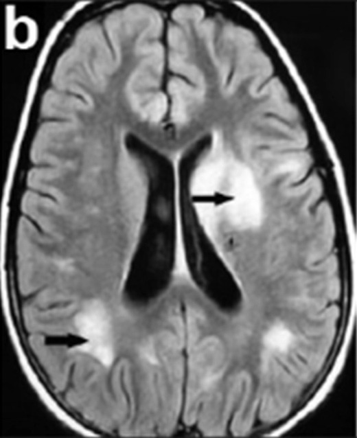
A typical MRI of an individual with acute disseminated encephalomyelitis (ADEM): Note the spotty pattern of inflammation of the cerebral parenchyma (black arrows).
Image: “MRI scan of a patient with acute disseminated encephalomyelitis (ADEM)” by Kamate M, Chetal V, Tonape V, Mahantshetti N, Hattiholi V. License: CC BY 2.0, cropped by Lecturio.