Vitiligo is the most common depigmenting disorder and is caused by the destruction of melanocytes. The etiology is unknown; however, genetic and autoimmune factors may play a role. Patients present with hypo- or depigmented macules or patches which often occur on the face, hands, knees, and/or genitalia. The diagnosis is clinical. Management depends on the severity and can include sun protection, topical or oral steroids, topical calcineurin inhibitors, immunosuppressants, and phototherapy.
VitiligoVitiligoVitiligo is the most common depigmenting disorder and is caused by the destruction of melanocytes. Patients present with hypo- or depigmented macules or patches which often occur on the face, hands, knees, and/or genitalia. Vitiligo is a progressive skinSkinThe skin, also referred to as the integumentary system, is the largest organ of the body. The skin is primarily composed of the epidermis (outer layer) and dermis (deep layer). The epidermis is primarily composed of keratinocytes that undergo rapid turnover, while the dermis contains dense layers of connective tissue.Skin: Structure and Functions condition in which there is destruction of melanocytesMelanocytesMammalian pigment cells that produce melanins, pigments found mainly in the epidermis, but also in the eyes and the hair, by a process called melanogenesis. Coloration can be altered by the number of melanocytes or the amount of pigment produced and stored in the organelles called melanosomes. The large non-mammalian melanin-containing cells are called melanophores.Skin: Structure and Functions resulting in the loss of skinSkinThe skin, also referred to as the integumentary system, is the largest organ of the body. The skin is primarily composed of the epidermis (outer layer) and dermis (deep layer). The epidermis is primarily composed of keratinocytes that undergo rapid turnover, while the dermis contains dense layers of connective tissue.Skin: Structure and Functions pigmentation.
PrevalencePrevalenceThe total number of cases of a given disease in a specified population at a designated time. It is differentiated from incidence, which refers to the number of new cases in the population at a given time.Measures of Disease Frequency: 0.1%–2% of the general population
Occurs in children and adults
Equal incidenceIncidenceThe number of new cases of a given disease during a given period in a specified population. It also is used for the rate at which new events occur in a defined population. It is differentiated from prevalence, which refers to all cases in the population at a given time.Measures of Disease Frequency in males and females
No racial or ethnic predilection
Age:
Onset generally occurs before 30 years of age.
Peak incidenceIncidenceThe number of new cases of a given disease during a given period in a specified population. It also is used for the rate at which new events occur in a defined population. It is differentiated from prevalence, which refers to all cases in the population at a given time.Measures of Disease Frequency: 10–30 years of age
The cause of vitiligoVitiligoVitiligo is the most common depigmenting disorder and is caused by the destruction of melanocytes. Patients present with hypo- or depigmented macules or patches which often occur on the face, hands, knees, and/or genitalia. Vitiligo is unknown but is postulated to be a result of multiple factors.
Approximately 36 susceptibility loci have been identified for nonsegmental vitiligoVitiligoVitiligo is the most common depigmenting disorder and is caused by the destruction of melanocytes. Patients present with hypo- or depigmented macules or patches which often occur on the face, hands, knees, and/or genitalia. Vitiligo.
May be autoimmune-mediated:
Approximately 20% of affected individuals have an autoimmune condition.
Associated conditions:
Hashimoto’s thyroiditisThyroiditisThyroiditis is a catchall term used to describe a variety of conditions that have inflammation of the thyroid gland in common. It includes pathologies that cause an acute illness with severe thyroid pain (e.g., subacute thyroiditis and infectious thyroiditis) as well as conditions in which there is no clinically evident inflammation and the manifestations primarily reflect thyroid dysfunction or a goiter (e.g., painless thyroiditis and fibrous Riedel’s thyroiditis). Thyroiditis
Graves’ disease
Type 1Type 1Spinal Muscular AtrophydiabetesDiabetesDiabetes mellitus (DM) is a metabolic disease characterized by hyperglycemia and dysfunction of the regulation of glucose metabolism by insulin. Type 1 DM is diagnosed mostly in children and young adults as the result of autoimmune destruction of β cells in the pancreas and the resulting lack of insulin. Type 2 DM has a significant association with obesity and is characterized by insulin resistance.Diabetes Mellitus mellitus
Addison disease
Pernicious anemiaPernicious anemiaA megaloblastic anemia occurring in children but more commonly in later life, characterized by histamine-fast achlorhydria, in which the laboratory and clinical manifestations are based on malabsorption of vitamin B12 due to a failure of the gastric mucosa to secrete adequate and potent intrinsic factor.Megaloblastic Anemia
Alopecia areataAlopecia AreataLoss of scalp and body hair involving microscopically inflammatory patchy areas.Alopecia
PsoriasisPsoriasisPsoriasis is a common T-cell-mediated inflammatory skin condition. The etiology is unknown, but is thought to be due to genetic inheritance and environmental triggers. There are 4 major subtypes, with the most common form being chronic plaque psoriasis. Psoriasis
Inflammatory bowel disease
The presence of antibodiesAntibodiesImmunoglobulins (Igs), also known as antibodies, are glycoprotein molecules produced by plasma cells that act in immune responses by recognizing and binding particular antigens. The various Ig classes are IgG (the most abundant), IgM, IgE, IgD, and IgA, which differ in their biologic features, structure, target specificity, and distribution.Immunoglobulins: Types and Functions to melaninMelaninInsoluble polymers of tyrosine derivatives found in and causing darkness in skin (skin pigmentation), hair, and feathers providing protection against sunburn induced by sunlight. Carotenes contribute yellow and red coloration.Seborrheic Keratosis has been noted.
Oxidative stressOxidative stressA disturbance in the prooxidant-antioxidant balance in favor of the former, leading to potential damage. Indicators of oxidative stress include damaged DNA bases, protein oxidation products, and lipid peroxidation products.Cell Injury and Death: Melanocyte self-destruction may arise from toxic phenolic compounds formed during melaninMelaninInsoluble polymers of tyrosine derivatives found in and causing darkness in skin (skin pigmentation), hair, and feathers providing protection against sunburn induced by sunlight. Carotenes contribute yellow and red coloration.Seborrheic KeratosissynthesisSynthesisPolymerase Chain Reaction (PCR).
Intrinsic defects of melanocytesMelanocytesMammalian pigment cells that produce melanins, pigments found mainly in the epidermis, but also in the eyes and the hair, by a process called melanogenesis. Coloration can be altered by the number of melanocytes or the amount of pigment produced and stored in the organelles called melanosomes. The large non-mammalian melanin-containing cells are called melanophores.Skin: Structure and Functions
Neural hypothesisHypothesisA hypothesis is a preliminary answer to a research question (i.e., a “guess” about what the results will be). There are 2 types of hypotheses: the null hypothesis and the alternative hypothesis.Statistical Tests and Data Representation: Nerve endings near pigment cells may secrete a neurochemical mediator toxic to melanocytesMelanocytesMammalian pigment cells that produce melanins, pigments found mainly in the epidermis, but also in the eyes and the hair, by a process called melanogenesis. Coloration can be altered by the number of melanocytes or the amount of pigment produced and stored in the organelles called melanosomes. The large non-mammalian melanin-containing cells are called melanophores.Skin: Structure and Functions.
Pathophysiology[1,4,8]
Destruction, or disappearance, of melanocytesMelanocytesMammalian pigment cells that produce melanins, pigments found mainly in the epidermis, but also in the eyes and the hair, by a process called melanogenesis. Coloration can be altered by the number of melanocytes or the amount of pigment produced and stored in the organelles called melanosomes. The large non-mammalian melanin-containing cells are called melanophores.Skin: Structure and Functions → loss of pigmentation in the affected area of the skinSkinThe skin, also referred to as the integumentary system, is the largest organ of the body. The skin is primarily composed of the epidermis (outer layer) and dermis (deep layer). The epidermis is primarily composed of keratinocytes that undergo rapid turnover, while the dermis contains dense layers of connective tissue.Skin: Structure and Functions
This process gives the appearance of white patchesPatchesVitiligo on the skinSkinThe skin, also referred to as the integumentary system, is the largest organ of the body. The skin is primarily composed of the epidermis (outer layer) and dermis (deep layer). The epidermis is primarily composed of keratinocytes that undergo rapid turnover, while the dermis contains dense layers of connective tissue.Skin: Structure and Functions.
Clinical Presentation and Diagnosis
Clinical presentation
VitiligoVitiligoVitiligo is the most common depigmenting disorder and is caused by the destruction of melanocytes. Patients present with hypo- or depigmented macules or patches which often occur on the face, hands, knees, and/or genitalia. Vitiligo results in hypopigmented or depigmented areas.
History[8,9,11]
Factors that preceded onset of skinSkinThe skin, also referred to as the integumentary system, is the largest organ of the body. The skin is primarily composed of the epidermis (outer layer) and dermis (deep layer). The epidermis is primarily composed of keratinocytes that undergo rapid turnover, while the dermis contains dense layers of connective tissue.Skin: Structure and Functions changes; recent cutaneous trauma (Koebner phenomenonKoebner PhenomenonLichen Planus)
Rate of onset
Associated symptoms
Extent, duration, and spread of skinSkinThe skin, also referred to as the integumentary system, is the largest organ of the body. The skin is primarily composed of the epidermis (outer layer) and dermis (deep layer). The epidermis is primarily composed of keratinocytes that undergo rapid turnover, while the dermis contains dense layers of connective tissue.Skin: Structure and Functions lesions
Current medications
Concomitant diseases
Occupational exposures
Family historyFamily HistoryAdult Health Maintenance of vitiligoVitiligoVitiligo is the most common depigmenting disorder and is caused by the destruction of melanocytes. Patients present with hypo- or depigmented macules or patches which often occur on the face, hands, knees, and/or genitalia. Vitiligo and autoimmune disease
Document psychological impact on qualityQualityActivities and programs intended to assure or improve the quality of care in either a defined medical setting or a program. The concept includes the assessment or evaluation of the quality of care; identification of problems or shortcomings in the delivery of care; designing activities to overcome these deficiencies; and follow-up monitoring to ensure effectiveness of corrective steps.Quality Measurement and Improvement of life
Hair in affected areas may also be depigmented (poliosisPoliosisBlepharitis).
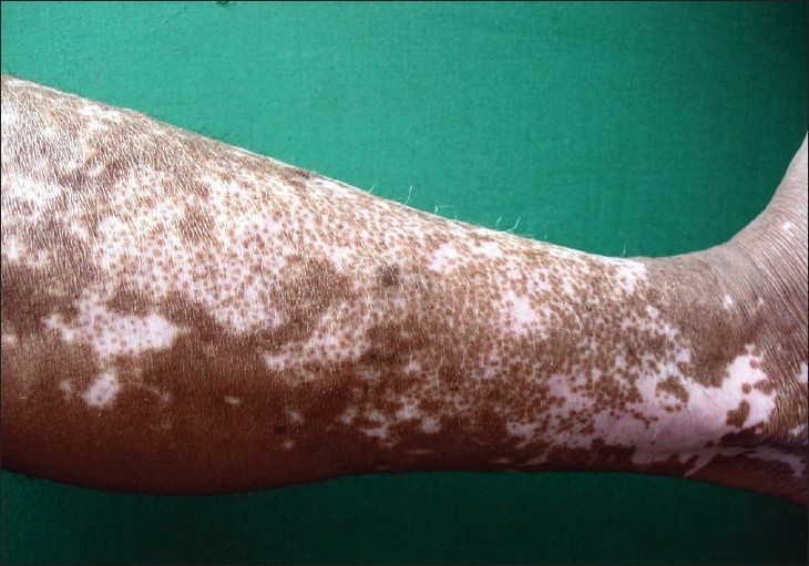
Large patches of hypopigmentation in a patient with vitiligo
Image: “Split thickness skin grafting in patients with stable vitiligo” by Sameem F, Sultan SJ, Ahmad QM. License: CC BY 2.0
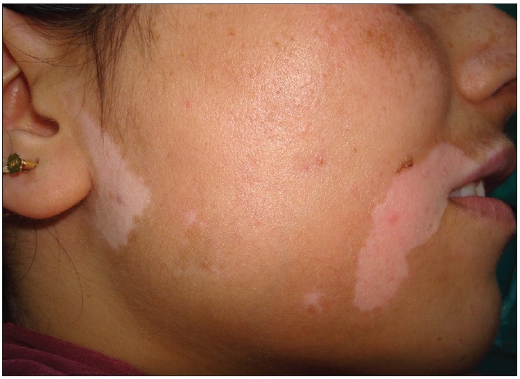
Depigmentation seen in a patient with vitiligo
Image: “Vitiligo and the melanocyte reservoir” by Falabella R. License: CC BY 2.0
Classification
VitiligoVitiligoVitiligo is the most common depigmenting disorder and is caused by the destruction of melanocytes. Patients present with hypo- or depigmented macules or patches which often occur on the face, hands, knees, and/or genitalia. Vitiligo can be classified based on affected sites and distribution:[1,11]
Nonsegmental vitiligoVitiligoVitiligo is the most common depigmenting disorder and is caused by the destruction of melanocytes. Patients present with hypo- or depigmented macules or patches which often occur on the face, hands, knees, and/or genitalia. Vitiligo (NSV; most common)
Segmental vitiligoVitiligoVitiligo is the most common depigmenting disorder and is caused by the destruction of melanocytes. Patients present with hypo- or depigmented macules or patches which often occur on the face, hands, knees, and/or genitalia. Vitiligo (SV)
Mixed
Unclassified
Additionally, clinicians will want to assess disease stability:[9]
Established areas of depigmentationDepigmentationVitiligo did not progress within the past 12 months.
Progressive:
New areas developed within the past 12 months, OR
Established areas progressed within the past 12 months.
Rapidly progressive:
There is no international consensus definition.
May consider when there is:
Rapid development of new lesions
Rapid increase in size of established lesions
Regressive: Spontaneous repigmentation occurs
Table: Classification of VitiligoVitiligoVitiligo is the most common depigmenting disorder and is caused by the destruction of melanocytes. Patients present with hypo- or depigmented macules or patches which often occur on the face, hands, knees, and/or genitalia. Vitiligo[1,9,11]
Can progress to universal vitiligoVitiligoVitiligo is the most common depigmenting disorder and is caused by the destruction of melanocytes. Patients present with hypo- or depigmented macules or patches which often occur on the face, hands, knees, and/or genitalia. Vitiligo
Includes follicular, vitiligoVitiligoVitiligo is the most common depigmenting disorder and is caused by the destruction of melanocytes. Patients present with hypo- or depigmented macules or patches which often occur on the face, hands, knees, and/or genitalia. Vitiligo minor, and vitiligoVitiligoVitiligo is the most common depigmenting disorder and is caused by the destruction of melanocytes. Patients present with hypo- or depigmented macules or patches which often occur on the face, hands, knees, and/or genitalia. Vitiligo punctata
SV
Unisegmental
Bisegmental
Plurisegmental
≥ 1 depigmented macules on 1 side of the body
Mixed
Includes characteristics of NSV + SV
Unclassified/ undetermined
Focal
Small, isolated site
Has not progressed to NSV after ≥ 2 years
Does not fit characteristics of SV
Mucosal
1 Isolated mucosal site
Adapted from the International Vitiligo Task Force[11]
BSA: body surface area; NSV: nonsegmental vitiligo; SV: segmental vitiligo
Diagnosis[1,3,8]
The diagnosis is usually clinical. However, the following examinations may be used if the diagnosis is unclear:
Exam with a Wood lamp:
Handheld device that emits ultraviolet (UV) A light
Useful in patientsPatientsIndividuals participating in the health care system for the purpose of receiving therapeutic, diagnostic, or preventive procedures.Clinician–Patient Relationship with pale skinSkinThe skin, also referred to as the integumentary system, is the largest organ of the body. The skin is primarily composed of the epidermis (outer layer) and dermis (deep layer). The epidermis is primarily composed of keratinocytes that undergo rapid turnover, while the dermis contains dense layers of connective tissue.Skin: Structure and Functions where vitiligoVitiligoVitiligo is the most common depigmenting disorder and is caused by the destruction of melanocytes. Patients present with hypo- or depigmented macules or patches which often occur on the face, hands, knees, and/or genitalia. Vitiligo lesions are more subtle
Accentuates hypo- or depigmented areas (appear white)
BiopsyBiopsyRemoval and pathologic examination of specimens from the living body.Ewing Sarcoma:
Can be used to differentiate from other hypopigmented disorders
Findings:
Absence of melanocytesMelanocytesMammalian pigment cells that produce melanins, pigments found mainly in the epidermis, but also in the eyes and the hair, by a process called melanogenesis. Coloration can be altered by the number of melanocytes or the amount of pigment produced and stored in the organelles called melanosomes. The large non-mammalian melanin-containing cells are called melanophores.Skin: Structure and Functions
Loss of epidermal pigmentation
Perifollicular lymphocytic infiltrate may be seen in some patientsPatientsIndividuals participating in the health care system for the purpose of receiving therapeutic, diagnostic, or preventive procedures.Clinician–Patient Relationship.
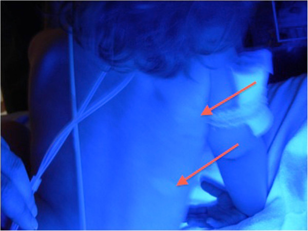
The use of a Wood’s lamp shows accentuated areas of depigmented skin (red arrows) in a patient with vitiligo.
Image: “A child with autoimmune polyendocrinopathy candidiasis and ectodermal dysplasia treated with immunosuppression: a case report” by O’Gorman CS et al. License: CC BY 2.0
Management and Prognosis
General management and considerations
No cure is currently available. Management generally aims to slow the progression of the disease and address cosmetic issues. The following is based on US, UK, and European guidelines.
Goals of treatment:
Rapid stabilization of progressive and progressive disease
All patientsPatientsIndividuals participating in the health care system for the purpose of receiving therapeutic, diagnostic, or preventive procedures.Clinician–Patient Relationship should practice sun avoidance and protection:
Sun protection factor (SPF) 50 sunscreenSunscreenChemical or physical agents that protect the skin from sunburn and erythema by absorbing or blocking ultraviolet radiation.Melanoma for UVA and UVB
Consider measuring vitamin DVitamin DA vitamin that includes both cholecalciferols and ergocalciferols, which have the common effect of preventing or curing rickets in animals. It can also be viewed as a hormone since it can be formed in skin by action of ultraviolet rays upon the precursors, 7-dehydrocholesterol and ergosterol, and acts on vitamin D receptors to regulate calcium in opposition to parathyroid hormone.Fat-soluble Vitamins and their Deficiencies levels in individuals who avoid sun exposure.
Avoid cutaneous trauma, when possible.
ThyroidThyroidThe thyroid gland is one of the largest endocrine glands in the human body. The thyroid gland is a highly vascular, brownish-red gland located in the visceral compartment of the anterior region of the neck.Thyroid Gland: Anatomy function testing should be performed due to the strong association with thyroidThyroidThe thyroid gland is one of the largest endocrine glands in the human body. The thyroid gland is a highly vascular, brownish-red gland located in the visceral compartment of the anterior region of the neck.Thyroid Gland: Anatomy disease.
British Association of Dermatology guidelines recommend screeningScreeningPreoperative Care for thyroidThyroidThe thyroid gland is one of the largest endocrine glands in the human body. The thyroid gland is a highly vascular, brownish-red gland located in the visceral compartment of the anterior region of the neck.Thyroid Gland: AnatomyantibodiesAntibodiesImmunoglobulins (Igs), also known as antibodies, are glycoprotein molecules produced by plasma cells that act in immune responses by recognizing and binding particular antigens. The various Ig classes are IgG (the most abundant), IgM, IgE, IgD, and IgA, which differ in their biologic features, structure, target specificity, and distribution.Immunoglobulins: Types and Functions and thyroidThyroidThe thyroid gland is one of the largest endocrine glands in the human body. The thyroid gland is a highly vascular, brownish-red gland located in the visceral compartment of the anterior region of the neck.Thyroid Gland: Anatomy function.[9]
US “Choosing Wisely” guidelines recommend against ordering laboratory tests for associated autoimmune diseasesAutoimmune diseasesDisorders that are characterized by the production of antibodies that react with host tissues or immune effector cells that are autoreactive to endogenous peptides.Selective IgA Deficiency in patientsPatientsIndividuals participating in the health care system for the purpose of receiving therapeutic, diagnostic, or preventive procedures.Clinician–Patient Relationship with vitiligoVitiligoVitiligo is the most common depigmenting disorder and is caused by the destruction of melanocytes. Patients present with hypo- or depigmented macules or patches which often occur on the face, hands, knees, and/or genitalia. Vitiligo who do not have signs or symptoms of other autoimmune diseasesAutoimmune diseasesDisorders that are characterized by the production of antibodies that react with host tissues or immune effector cells that are autoreactive to endogenous peptides.Selective IgA Deficiency.[10]
If patient and/or family historyFamily HistoryAdult Health Maintenance and/or laboratory parameters point to an additional autoimmune disease, other autoantibodiesAutoantibodiesAntibodies that react with self-antigens (autoantigens) of the organism that produced them.Blotting Techniques can be checked.[11]
Counseling and other psychological intervention should be considered due to the effects of the disease on patientsPatientsIndividuals participating in the health care system for the purpose of receiving therapeutic, diagnostic, or preventive procedures.Clinician–Patient Relationship‘ mental health and self-esteem.
Cosmetic camouflage:
Includes:
Cosmetics (foundation-based)
Self-tanning products containing dihydroxyacetone
Dyes
TattoosTattoosThe indelible marking of tissues, primarily skin, by pricking it with needles to imbed various coloring agents. Tattooing of the cornea is done to colorize leukoma spots.Cellular Accumulations (consider with caution owing to unpredictable nature of the disease)[12]
May be an adequate treatment choice for some individuals
Choice of therapy depends on:[8]
Type/subtype
Severity
Site location:
Face, trunk, mid-extremities → tend to respond better to therapy
Distal extremities, lipsLipsThe lips are the soft and movable most external parts of the oral cavity. The blood supply of the lips originates from the external carotid artery, and the innervation is through cranial nerves.Lips and Tongue: Anatomy → tend to be less responsive to therapy
Percentage of body surface area (BSA) affected
Whether lesions are stable or progressive
Patient preference
When to refer to a specialist:[9]
Rapidly progressive disease
Uncertainty in the diagnosis
Disease causes significant distress to the patient.
Assess and monitor qualityQualityActivities and programs intended to assure or improve the quality of care in either a defined medical setting or a program. The concept includes the assessment or evaluation of the quality of care; identification of problems or shortcomings in the delivery of care; designing activities to overcome these deficiencies; and follow-up monitoring to ensure effectiveness of corrective steps.Quality Measurement and Improvement of life and psychological distress → may use standard questionnaires for depression (Patient Health Questionnaire-9 (PHQ-9)), anxietyAnxietyFeelings or emotions of dread, apprehension, and impending disaster but not disabling as with anxiety disorders.Generalized Anxiety Disorder (Generalized AnxietyAnxietyFeelings or emotions of dread, apprehension, and impending disaster but not disabling as with anxiety disorders.Generalized Anxiety Disorder Disorder-7 (GAD7)), and others for qualityQualityActivities and programs intended to assure or improve the quality of care in either a defined medical setting or a program. The concept includes the assessment or evaluation of the quality of care; identification of problems or shortcomings in the delivery of care; designing activities to overcome these deficiencies; and follow-up monitoring to ensure effectiveness of corrective steps.Quality Measurement and Improvement of life
Keep in mind that 2–3 months of a chosen therapy may be needed before efficacy can be assessed.
Superpotent corticosteroid: clobetasolClobetasolA derivative of prednisolone with high glucocorticoid activity and low mineralocorticoid activity. Absorbed through the skin faster than fluocinonide, it is used topically in treatment of psoriasis but may cause marked adrenocortical suppression.Glucocorticoids 0.05% cream, gel, lotion, or ointment
Dosing:
No guidelines on the optimal dosing or duration
Potential options:
Continuous dosing: daily application for no longer than 2–3 months
Discontinuous dosing: application for 15 days/month over 6 months
Dark skinSkinThe skin, also referred to as the integumentary system, is the largest organ of the body. The skin is primarily composed of the epidermis (outer layer) and dermis (deep layer). The epidermis is primarily composed of keratinocytes that undergo rapid turnover, while the dermis contains dense layers of connective tissue.Skin: Structure and Functions
Recent lesions
Monitor for:
SkinSkinThe skin, also referred to as the integumentary system, is the largest organ of the body. The skin is primarily composed of the epidermis (outer layer) and dermis (deep layer). The epidermis is primarily composed of keratinocytes that undergo rapid turnover, while the dermis contains dense layers of connective tissue.Skin: Structure and FunctionsatrophyAtrophyDecrease in the size of a cell, tissue, organ, or multiple organs, associated with a variety of pathological conditions such as abnormal cellular changes, ischemia, malnutrition, or hormonal changes.Cellular Adaptation
An alternative to topical steroidsSteroidsA group of polycyclic compounds closely related biochemically to terpenes. They include cholesterol, numerous hormones, precursors of certain vitamins, bile acids, alcohols (sterols), and certain natural drugs and poisons. Steroids have a common nucleus, a fused, reduced 17-carbon atom ring system, cyclopentanoperhydrophenanthrene. Most steroids also have two methyl groups and an aliphatic side-chain attached to the nucleus.Benign Liver Tumors
Preferred in areas at high risk of skinSkinThe skin, also referred to as the integumentary system, is the largest organ of the body. The skin is primarily composed of the epidermis (outer layer) and dermis (deep layer). The epidermis is primarily composed of keratinocytes that undergo rapid turnover, while the dermis contains dense layers of connective tissue.Skin: Structure and FunctionsatrophyAtrophyDecrease in the size of a cell, tissue, organ, or multiple organs, associated with a variety of pathological conditions such as abnormal cellular changes, ischemia, malnutrition, or hormonal changes.Cellular Adaptation (e.g., head/neckNeckThe part of a human or animal body connecting the head to the rest of the body.Peritonsillar Abscess, genitals, intertriginous regions)
Optimal for new or actively progressing lesions
Options:
Topical tacrolimusTacrolimusA macrolide isolated from the culture broth of a strain of streptomyces tsukubaensis that has strong immunosuppressive activity in vivo and prevents the activation of T-lymphocytes in response to antigenic or mitogenic stimulation in vitro.Immunosuppressants 0.1% ointment
PruritusPruritusAn intense itching sensation that produces the urge to rub or scratch the skin to obtain relief.Atopic Dermatitis (Eczema)
SkinSkinThe skin, also referred to as the integumentary system, is the largest organ of the body. The skin is primarily composed of the epidermis (outer layer) and dermis (deep layer). The epidermis is primarily composed of keratinocytes that undergo rapid turnover, while the dermis contains dense layers of connective tissue.Skin: Structure and FunctionshyperpigmentationHyperpigmentationExcessive pigmentation of the skin, usually as a result of increased epidermal or dermal melanin pigmentation, hypermelanosis. Hyperpigmentation can be localized or generalized. The condition may arise from exposure to light, chemicals or other substances, or from a primary metabolic imbalance.Malassezia Fungi
Black box warning: associated with increased risk of malignancyMalignancyHemothorax
Used for nonsegmental vitiligoVitiligoVitiligo is the most common depigmenting disorder and is caused by the destruction of melanocytes. Patients present with hypo- or depigmented macules or patches which often occur on the face, hands, knees, and/or genitalia. Vitiligo
Dosing:
1.5% cream applied in a thin layer twice daily
Up to maximum of 60 g/week or 100 g/2 weeks
Treatment area should not exceed 10% BSA
Safety concerns related to systemic absorptionAbsorptionAbsorption involves the uptake of nutrient molecules and their transfer from the lumen of the GI tract across the enterocytes and into the interstitial space, where they can be taken up in the venous or lymphatic circulation.Digestion and AbsorptionlimitLimitA value (e.g., pressure or time) that should not be exceeded and which is specified by the operator to protect the lungInvasive Mechanical Ventilation the use of this drug.
Monitor for:
ErythemaErythemaRedness of the skin produced by congestion of the capillaries. This condition may result from a variety of disease processes.Chalazion
PruritusPruritusAn intense itching sensation that produces the urge to rub or scratch the skin to obtain relief.Atopic Dermatitis (Eczema)
Acne
InflammationInflammationInflammation is a complex set of responses to infection and injury involving leukocytes as the principal cellular mediators in the body’s defense against pathogenic organisms. Inflammation is also seen as a response to tissue injury in the process of wound healing. The 5 cardinal signs of inflammation are pain, heat, redness, swelling, and loss of function. Inflammation of the nasal passages and throatThroatThe pharynx is a component of the digestive system that lies posterior to the nasal cavity, oral cavity, and larynx. The pharynx can be divided into the oropharynx, nasopharynx, and laryngopharynx. Pharyngeal muscles play an integral role in vital processes such as breathing, swallowing, and speaking.Pharynx: Anatomy
HeadacheHeadacheThe symptom of pain in the cranial region. It may be an isolated benign occurrence or manifestation of a wide variety of headache disorders.Brain Abscess
UTIUTIUrinary tract infections (UTIs) represent a wide spectrum of diseases, from self-limiting simple cystitis to severe pyelonephritis that can result in sepsis and death. Urinary tract infections are most commonly caused by Escherichia coli, but may also be caused by other bacteria and fungi. Urinary Tract Infections (UTIs)
FeverFeverFever is defined as a measured body temperature of at least 38°C (100.4°F). Fever is caused by circulating endogenous and/or exogenous pyrogens that increase levels of prostaglandin E2 in the hypothalamus. Fever is commonly associated with chills, rigors, sweating, and flushing of the skin. Fever
Black box warning: associated with an increased risk of:
Serious infectionsInfectionsInvasion of the host organism by microorganisms or their toxins or by parasites that can cause pathological conditions or diseases.Chronic Granulomatous Disease
1st-line therapy for stabilization of rapidly progressive disease
Low dose systemic coroticosteroids (e.g. prednisonePrednisoneA synthetic anti-inflammatory glucocorticoid derived from cortisone. It is biologically inert and converted to prednisolone in the liver.Immunosuppressants) for no more than 2 weeks then reassess after 4 to 6 weeks (course can be repeated), OR
Minipulse therapy (in adults) then reassess after 4 to 6 weeks; examples:
DexamethasoneDexamethasoneAn anti-inflammatory 9-fluoro-glucocorticoid.Antiemetics (oral) 2.5 mg per day for 2 consecutive days/week for up to 12 weeks
TriamcinoloneTriamcinoloneA glucocorticoid given, as the free alcohol or in esterified form, orally, intramuscularly, by local injection, by inhalation, or applied topically in the management of various disorders in which corticosteroids are indicated.Glucocorticoids (IM) 40 mg every 4 to 6 weeks (up to 3 doses)
UK:
Oral betamethasoneBetamethasoneA glucocorticoid given orally, parenterally, by local injection, by inhalation, or applied topically in the management of various disorders in which corticosteroids are indicated. Its lack of mineralocorticoid properties makes betamethasone particularly suitable for treating cerebral edema and congenital adrenal hyperplasia.Glucocorticoids:
0.1 mg/kg twice weekly for 3 months
Dosing may need to be altered based on response.
Oral dexamethasoneDexamethasoneAn anti-inflammatory 9-fluoro-glucocorticoid.Antiemetics is a potential alternative.
Rarely used as monotherapy
Often combined with other therapies, such as narrow-band UVB
Systemic immunosuppressantsImmunosuppressantsImmunosuppressants are a class of drugs widely used in the management of autoimmune conditions and organ transplant rejection. The general effect is dampening of the immune response.Immunosuppressants:[12]
Require specialty referral
Options:
MethotrexateMethotrexateAn antineoplastic antimetabolite with immunosuppressant properties. It is an inhibitor of tetrahydrofolate dehydrogenase and prevents the formation of tetrahydrofolate, necessary for synthesis of thymidylate, an essential component of DNA.Antimetabolite Chemotherapy
CyclophosphamideCyclophosphamidePrecursor of an alkylating nitrogen mustard antineoplastic and immunosuppressive agent that must be activated in the liver to form the active aldophosphamide. It has been used in the treatment of lymphoma and leukemia. Its side effect, alopecia, has been used for defleecing sheep. Cyclophosphamide may also cause sterility, birth defects, mutations, and cancer.Immunosuppressants
An alternative to systemic steroidsSteroidsA group of polycyclic compounds closely related biochemically to terpenes. They include cholesterol, numerous hormones, precursors of certain vitamins, bile acids, alcohols (sterols), and certain natural drugs and poisons. Steroids have a common nucleus, a fused, reduced 17-carbon atom ring system, cyclopentanoperhydrophenanthrene. Most steroids also have two methyl groups and an aliphatic side-chain attached to the nucleus.Benign Liver Tumors
Used alone for stabilization of rapidly progressive disease if steroidsSteroidsA group of polycyclic compounds closely related biochemically to terpenes. They include cholesterol, numerous hormones, precursors of certain vitamins, bile acids, alcohols (sterols), and certain natural drugs and poisons. Steroids have a common nucleus, a fused, reduced 17-carbon atom ring system, cyclopentanoperhydrophenanthrene. Most steroids also have two methyl groups and an aliphatic side-chain attached to the nucleus.Benign Liver Tumors are contraindicated
Narrow-band (NB) UVB:
1st-line therapy for widespread vitiligoVitiligoVitiligo is the most common depigmenting disorder and is caused by the destruction of melanocytes. Patients present with hypo- or depigmented macules or patches which often occur on the face, hands, knees, and/or genitalia. Vitiligo
Treatments are 2‒3 times weekly for several months (can take 1 year to reach stabilization).
No significant association found with basal cellBasal CellErythema Multiforme carcinoma (BCC), squamous cell carcinomaSquamous cell carcinomaCutaneous squamous cell carcinoma (cSCC) is caused by malignant proliferation of atypical keratinocytes. This condition is the 2nd most common skin malignancy and usually affects sun-exposed areas of fair-skinned patients. The cancer presents as a firm, erythematous, keratotic plaque or papule. Squamous Cell Carcinoma (SCC) (SCC) and/or melanomaMelanomaMelanoma is a malignant tumor arising from melanocytes, the melanin-producing cells of the epidermis. These tumors are most common in fair-skinned individuals with a history of excessive sun exposure and sunburns. Melanoma in vitiligoVitiligoVitiligo is the most common depigmenting disorder and is caused by the destruction of melanocytes. Patients present with hypo- or depigmented macules or patches which often occur on the face, hands, knees, and/or genitalia. Vitiligo trials
For recalcitrant disease involving < 5%–10% BSA that does not respond to topical therapies:
Targeted phototherapyPhototherapyTreatment of disease by exposure to light, especially by variously concentrated light rays or specific wavelengths.Hyperbilirubinemia of the Newborn with excimer laser
For refractory and extensive disease involving > 40% of BSA
Used on areas of unaffected skinSkinThe skin, also referred to as the integumentary system, is the largest organ of the body. The skin is primarily composed of the epidermis (outer layer) and dermis (deep layer). The epidermis is primarily composed of keratinocytes that undergo rapid turnover, while the dermis contains dense layers of connective tissue.Skin: Structure and Functions
Goal is to match the diseased area for cosmetic satisfaction.
Agent: monobenzyl ether of hydroquinoneHydroquinoneDerivatives of hydroquinone (1, 4-dihydrobenzene) made by reduction of benzoquinones.Melasma (monobenzone)
PrognosisPrognosisA prediction of the probable outcome of a disease based on a individual’s condition and the usual course of the disease as seen in similar situations.Non-Hodgkin Lymphomas[2,8]
VitiligoVitiligoVitiligo is the most common depigmenting disorder and is caused by the destruction of melanocytes. Patients present with hypo- or depigmented macules or patches which often occur on the face, hands, knees, and/or genitalia. Vitiligo is a chronic skinSkinThe skin, also referred to as the integumentary system, is the largest organ of the body. The skin is primarily composed of the epidermis (outer layer) and dermis (deep layer). The epidermis is primarily composed of keratinocytes that undergo rapid turnover, while the dermis contains dense layers of connective tissue.Skin: Structure and Functions condition.
The clinical course is unpredictable.
Less favorable outcomes occur with long-standing or segmental disease.
50% repigmentation may not be satisfactory to the patient
Often no response on hands, feet, and penisPenisThe penis is the male organ of copulation and micturition. The organ is composed of a root, body, and glans. The root is attached to the pubic bone by the crura penis. The body consists of the 2 parallel corpora cavernosa and the corpus spongiosum. The glans is ensheathed by the prepuce or foreskin. Penis: Anatomy
Partial repigmentation in sun-exposed patchesPatchesVitiligo may predict response:
Most patientsPatientsIndividuals participating in the health care system for the purpose of receiving therapeutic, diagnostic, or preventive procedures.Clinician–Patient Relationship have > 75% repigmentation with 1st-line therapies.
Expected goals:
25% improvement after 3 months
50% after 6 months
75% after 9 months
Lesions may stabilize or continue to progress and expand over the years, despite treatment.
Complications[8,9,12]
Psychological dysfunction and substantial interpersonal burdens
NevusNevusNevi (singular nevus), also known as “moles,” are benign neoplasms of the skin. Nevus is a non-specific medical term because it encompasses both congenital and acquired lesions, hyper- and hypopigmented lesions, and raised or flat lesions. Nevus/Nevi depigmentosus: typicallya benignBenignFibroadenoma, solitary, localized hypopigmented maculeMaculeNonpalpable lesion < 1 cm in diameterGeneralized and Localized Rashes or patchPatchNonpalpable lesion > 1 cm in diameterGeneralized and Localized Rashes that is often noted at birth or in the early years of life. These lesions are stable and not progressive, although they may appear to enlarge as the body grows. NevusNevusNevi (singular nevus), also known as “moles,” are benign neoplasms of the skin. Nevus is a non-specific medical term because it encompasses both congenital and acquired lesions, hyper- and hypopigmented lesions, and raised or flat lesions. Nevus/Nevi depigmentosus is caused by defective melanocytesMelanocytesMammalian pigment cells that produce melanins, pigments found mainly in the epidermis, but also in the eyes and the hair, by a process called melanogenesis. Coloration can be altered by the number of melanocytes or the amount of pigment produced and stored in the organelles called melanosomes. The large non-mammalian melanin-containing cells are called melanophores.Skin: Structure and Functions that cannot produce pigment or transfer it to keratinocytesKeratinocytesEpidermal cells which synthesize keratin and undergo characteristic changes as they move upward from the basal layers of the epidermis to the cornified (horny) layer of the skin. Successive stages of differentiation of the keratinocytes forming the epidermal layers are basal cell, spinous or prickle cell, and the granular cell.Skin: Structure and Functions. Management is not necessary and, when attempted, has varied results.
Pityriasis alba: a common skinSkinThe skin, also referred to as the integumentary system, is the largest organ of the body. The skin is primarily composed of the epidermis (outer layer) and dermis (deep layer). The epidermis is primarily composed of keratinocytes that undergo rapid turnover, while the dermis contains dense layers of connective tissue.Skin: Structure and Functions disorder typically affecting children and adolescents. The condition is often considered a manifestation of atopic dermatitisDermatitisAny inflammation of the skin.Atopic Dermatitis (Eczema). Typically, patientsPatientsIndividuals participating in the health care system for the purpose of receiving therapeutic, diagnostic, or preventive procedures.Clinician–Patient Relationship present with erythemaErythemaRedness of the skin produced by congestion of the capillaries. This condition may result from a variety of disease processes.Chalazion and scaleScaleDermatologic Examination followed by round, hypopigmented macules and patchesPatchesVitiligo, which commonly occur on the face, upper trunk, or upper limbs. The diagnosis is clinical. Pityriasis alba is considered self-limitingSelf-LimitingMeningitis in Children but may take months to years to resolve. Topical steroidsSteroidsA group of polycyclic compounds closely related biochemically to terpenes. They include cholesterol, numerous hormones, precursors of certain vitamins, bile acids, alcohols (sterols), and certain natural drugs and poisons. Steroids have a common nucleus, a fused, reduced 17-carbon atom ring system, cyclopentanoperhydrophenanthrene. Most steroids also have two methyl groups and an aliphatic side-chain attached to the nucleus.Benign Liver Tumors, emollientsEmollientsOleaginous substances used topically to soothe, soften or protect skin or mucous membranes. They are used also as vehicles for other dermatologic agents.Pityriasis Rosea, and calcineurin inhibitorsCalcineurin InhibitorsCompounds that inhibit or block the phosphatase activity of calcineurin.Immunosuppressants may be used to speed up resolution.
Tinea (pityriasis) versicolor: a common superficial fungal infection caused by MalasseziaMalasseziaMalassezia is a lipophilic yeast commonly found on the skin surfaces of many animals, including humans. In the presence of certain environments or triggers, this fungus can cause pathologic diseases ranging from superficial skin conditions (tinea versicolor and dermatitis) to invasive disease (e.g., Malassezia folliculitis, catheter-associated fungemia, meningitis, and urinary tract infections). Malassezia Fungi furfur. Tinea versicolorTinea versicolorA common chronic, noninflammatory and usually symptomless disorder, characterized by the occurrence of multiple macular patches of all sizes and shapes, and varying in pigmentation from fawn-colored to brown. It is seen most frequently in hot, humid, tropical regions and is mostly caused by Malassezia furfur (formerly Pityrosporum orbiculare).Malassezia Fungi may present as hypopigmented, hyperpigmented, or erythematous macules and patchesPatchesVitiligo, most often on the trunk. Diagnosis is often clinical but may be confirmed with visualization of hyphaeHyphaeMicroscopic threadlike filaments in fungi that are filled with a layer of protoplasm. Collectively, the hyphae make up the mycelium.Mycology and buddingBuddingMycology cells on potassiumPotassiumAn element in the alkali group of metals with an atomic symbol k, atomic number 19, and atomic weight 39. 10. It is the chief cation in the intracellular fluid of muscle and other cells. Potassium ion is a strong electrolyte that plays a significant role in the regulation of fluid volume and maintenance of the water-electrolyte balance.Hyperkalemia hydroxide wet mount. Management is with topical or oral antifungals.
Halo nevusNevusNevi (singular nevus), also known as “moles,” are benign neoplasms of the skin. Nevus is a non-specific medical term because it encompasses both congenital and acquired lesions, hyper- and hypopigmented lesions, and raised or flat lesions. Nevus/Nevi: a moleMoleNevi (singular nevus), also known as “moles,” are benign neoplasms of the skin. Nevus is a non-specific medical term because it encompasses both congenital and acquired lesions, hyper- and hypopigmented lesions, and raised or flat lesions.Nevus/Nevi surrounded by a halo of hypopigmentationHypopigmentationA condition caused by a deficiency or a loss of melanin pigmentation in the epidermis, also known as hypomelanosis. Hypopigmentation can be localized or generalized, and may result from genetic defects, trauma, inflammation, or infections.Malassezia Fungi. Multiple lesions may be present, and the back is most often affected. Loss of pigment often precedes spontaneous resolution of the central nevusNevusNevi (singular nevus), also known as “moles,” are benign neoplasms of the skin. Nevus is a non-specific medical term because it encompasses both congenital and acquired lesions, hyper- and hypopigmented lesions, and raised or flat lesions. Nevus/Nevi. Repigmentation of the skinSkinThe skin, also referred to as the integumentary system, is the largest organ of the body. The skin is primarily composed of the epidermis (outer layer) and dermis (deep layer). The epidermis is primarily composed of keratinocytes that undergo rapid turnover, while the dermis contains dense layers of connective tissue.Skin: Structure and Functions may occur after the nevusNevusNevi (singular nevus), also known as “moles,” are benign neoplasms of the skin. Nevus is a non-specific medical term because it encompasses both congenital and acquired lesions, hyper- and hypopigmented lesions, and raised or flat lesions. Nevus/Nevi disappears. The diagnosis is generally clinical. No treatment is necessary.
Chemical leukoderma: also known as occupational vitiligoVitiligoVitiligo is the most common depigmenting disorder and is caused by the destruction of melanocytes. Patients present with hypo- or depigmented macules or patches which often occur on the face, hands, knees, and/or genitalia. Vitiligo, hypopigmentationHypopigmentationA condition caused by a deficiency or a loss of melanin pigmentation in the epidermis, also known as hypomelanosis. Hypopigmentation can be localized or generalized, and may result from genetic defects, trauma, inflammation, or infections.Malassezia Fungi of the skinSkinThe skin, also referred to as the integumentary system, is the largest organ of the body. The skin is primarily composed of the epidermis (outer layer) and dermis (deep layer). The epidermis is primarily composed of keratinocytes that undergo rapid turnover, while the dermis contains dense layers of connective tissue.Skin: Structure and Functions resulting from contact with certain chemicals that cause melanocytotoxicity. HypopigmentationHypopigmentationA condition caused by a deficiency or a loss of melanin pigmentation in the epidermis, also known as hypomelanosis. Hypopigmentation can be localized or generalized, and may result from genetic defects, trauma, inflammation, or infections.Malassezia Fungi initially occurs in the area of contact but may spread to additional areas. The diagnosis is clinical. Management involves removal/avoidance of the offending agent and the use of topical steroidsSteroidsA group of polycyclic compounds closely related biochemically to terpenes. They include cholesterol, numerous hormones, precursors of certain vitamins, bile acids, alcohols (sterols), and certain natural drugs and poisons. Steroids have a common nucleus, a fused, reduced 17-carbon atom ring system, cyclopentanoperhydrophenanthrene. Most steroids also have two methyl groups and an aliphatic side-chain attached to the nucleus.Benign Liver Tumors.
IdiopathicIdiopathicDermatomyositis guttate hypomelanosis: benignBenignFibroadenomaskinSkinThe skin, also referred to as the integumentary system, is the largest organ of the body. The skin is primarily composed of the epidermis (outer layer) and dermis (deep layer). The epidermis is primarily composed of keratinocytes that undergo rapid turnover, while the dermis contains dense layers of connective tissue.Skin: Structure and Functions condition that is thought to be a part of the natural aging process and is associated with a decrease in the number of melanocytesMelanocytesMammalian pigment cells that produce melanins, pigments found mainly in the epidermis, but also in the eyes and the hair, by a process called melanogenesis. Coloration can be altered by the number of melanocytes or the amount of pigment produced and stored in the organelles called melanosomes. The large non-mammalian melanin-containing cells are called melanophores.Skin: Structure and Functions. PatientsPatientsIndividuals participating in the health care system for the purpose of receiving therapeutic, diagnostic, or preventive procedures.Clinician–Patient Relationship will have diffuse, small, round or oval hypopigmented macules on sun-exposed areas, most often on the extremities. Once present, these lesions are stable and do not change. The diagnosis is clinical. Treatment is not required.
Billing and Coding
Diagnosis Codes:
This code is used to diagnose vitiligoVitiligoVitiligo is the most common depigmenting disorder and is caused by the destruction of melanocytes. Patients present with hypo- or depigmented macules or patches which often occur on the face, hands, knees, and/or genitalia. Vitiligo, an autoimmune disease that causes loss of skinSkinThe skin, also referred to as the integumentary system, is the largest organ of the body. The skin is primarily composed of the epidermis (outer layer) and dermis (deep layer). The epidermis is primarily composed of keratinocytes that undergo rapid turnover, while the dermis contains dense layers of connective tissue.Skin: Structure and Functions color in patchesPatchesVitiligo due to the destruction of melanocytesMelanocytesMammalian pigment cells that produce melanins, pigments found mainly in the epidermis, but also in the eyes and the hair, by a process called melanogenesis. Coloration can be altered by the number of melanocytes or the amount of pigment produced and stored in the organelles called melanosomes. The large non-mammalian melanin-containing cells are called melanophores.Skin: Structure and Functions.
Coding System
Code
Description
ICD-10-CM
L80
VitiligoVitiligoVitiligo is the most common depigmenting disorder and is caused by the destruction of melanocytes. Patients present with hypo- or depigmented macules or patches which often occur on the face, hands, knees, and/or genitalia. Vitiligo
SNOMED CT
5831003
VitiligoVitiligoVitiligo is the most common depigmenting disorder and is caused by the destruction of melanocytes. Patients present with hypo- or depigmented macules or patches which often occur on the face, hands, knees, and/or genitalia. Vitiligo (disorder)
Medications:
These codes are for first-line treatments aimed at repigmentation, including potent topical corticosteroidsCorticosteroidsChorioretinitis and non-steroidal topical calcineurin inhibitorsCalcineurin InhibitorsCompounds that inhibit or block the phosphatase activity of calcineurin.Immunosuppressants like tacrolimusTacrolimusA macrolide isolated from the culture broth of a strain of streptomyces tsukubaensis that has strong immunosuppressive activity in vivo and prevents the activation of T-lymphocytes in response to antigenic or mitogenic stimulation in vitro.Immunosuppressants.
Coding System
Code
Description
RxNorm
2599
ClobetasolClobetasolA derivative of prednisolone with high glucocorticoid activity and low mineralocorticoid activity. Absorbed through the skin faster than fluocinonide, it is used topically in treatment of psoriasis but may cause marked adrenocortical suppression.Glucocorticoids (ingredient)
RxNorm
35927
TacrolimusTacrolimusA macrolide isolated from the culture broth of a strain of streptomyces tsukubaensis that has strong immunosuppressive activity in vivo and prevents the activation of T-lymphocytes in response to antigenic or mitogenic stimulation in vitro.Immunosuppressants (ingredient)
Procedures/Interventions:
This code is for phototherapyPhototherapyTreatment of disease by exposure to light, especially by variously concentrated light rays or specific wavelengths.Hyperbilirubinemia of the Newborn with narrowband ultraviolet B (NB-UVB) light, a common and effective treatment for widespread vitiligoVitiligoVitiligo is the most common depigmenting disorder and is caused by the destruction of melanocytes. Patients present with hypo- or depigmented macules or patches which often occur on the face, hands, knees, and/or genitalia. Vitiligo to stimulate melanocyte activity.
Coding System
Code
Description
CPT
96910
Photochemotherapy; psoralens and ultraviolet A (PUVA) or ultraviolet B (UVB)
Eleftheriadou, V., et al. (2022). British Association of Dermatologists guidelines for the management of people with vitiligo 2021. British Journal of Dermatology, 186(1), 18–29. https://doi.org/10.1111/bjd.20596
Choosing Wisely: an initiative of the ABIM Foundation. (2021). American Academy of Pediatrics–Section on Dermatology. Do not routinely order laboratory tests for associated autoimmune diseases in patients with vitiligo in the absence of signs and/or symptoms of the diseases in question. https://www.choosingwisely.org/clinician-lists/aapd2
van Geel, N., Speeckaert, R., Taïeb, A., Ezzedine, K., Lim, H. W., Pandya, A. G., Passeron, T., Wolkerstorfer, A., Abdallah, M., Alomar, A., Bae, J. M., Bekkenk, M., Benzekri, L., Böhm, M., Eleftheriadou, V., Esmat, S., Ghia, D., Goh, B. K., Grimes, P., Gupta, S., … Seneschal, J. (2023). Worldwide expert recommendations for the diagnosis and management of vitiligo: Position statement from the International Vitiligo Task Force Part 1: towards a new management algorithm. Journal of the European Academy of Dermatology and Venereology : JEADV, 37(11), 2173–2184. https://doi.org/10.1111/jdv.19451
Seneschal, J., Speeckaert, R., Taïeb, A., Wolkerstorfer, A., Passeron, T., Pandya, A. G., Lim, H. W., Ezzedine, K., Zhou, Y., Xiang, F., Thng, S., Tanemura, A., Suzuki, T., Rosmarin, D., Rodrigues, M., Raboobee, N., Pliszewski, G., Parsad, D., Oiso, N., Monteiro, P., … van Geel, N. (2023). Worldwide expert recommendations for the diagnosis and management of vitiligo: Position statement from the international Vitiligo Task Force-Part 2: Specific treatment recommendations. Journal of the European Academy of Dermatology and Venereology : JEADV, 37(11), 2185–2195. https://doi.org/10.1111/jdv.19450
Sheikh, A., et al. (2022). FDA approves ruxolitinib (Opzelura) for vitiligo therapy: a breakthrough in the field of dermatology. Annals of Medicine & Surgery, 81, 104499. https://www.ncbi.nlm.nih.gov/pmc/articles/PMC9486756/
Create your free account or log in to continue reading!