The abdominal cavity has a complex and intricate anatomy. A physician must know in which area of the abdomen every major structure is located to understand the clinical presentation of abdominal pathologies and/or in trauma situations to estimate which organs are most likely injured. The general surgeon, especially in emergency situations, uses this knowledge to execute the most advantageous surgical approach for a particular situation.
Costal cartilages of the 7th–10th ribsRibsA set of twelve curved bones which connect to the vertebral column posteriorly, and terminate anteriorly as costal cartilage. Together, they form a protective cage around the internal thoracic organs.Chest Wall: Anatomy
Inferior:
Pubic boneBoneBone is a compact type of hardened connective tissue composed of bone cells, membranes, an extracellular mineralized matrix, and central bone marrow. The 2 primary types of bone are compact and spongy. Bones: Structure and Types and pubic symphysisPubic SymphysisA slightly movable cartilaginous joint which occurs between the pubic bones.Vagina, Vulva, and Pelvic Floor: Anatomy
Inguinal ligaments
Lateral:
Superior: inferior aspect of the 10th rib
Inferior: iliac crest
Landmarks
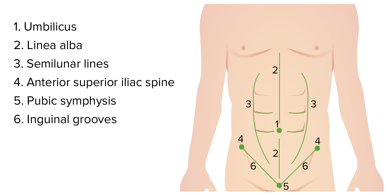
Umbilicus
Linea alba: fibrousFibrousFibrocystic Change junction of the right and left rectus muscles that runs down the midline of the abdomen
Semilunar lines: lateral borders of the rectus abdominisRectus AbdominisA long flat muscle that extends along the whole length of both sides of the abdomen. It flexes the vertebral column, particularly the lumbar portion; it also tenses the anterior abdominal wall and assists in compressing the abdominal contents. It is frequently the site of hematomas. In reconstructive surgery it is often used for the creation of myocutaneous flaps.Anterior Abdominal Wall: Anatomy muscles
SkinSkinThe skin, also referred to as the integumentary system, is the largest organ of the body. The skin is primarily composed of the epidermis (outer layer) and dermis (deep layer). The epidermis is primarily composed of keratinocytes that undergo rapid turnover, while the dermis contains dense layers of connective tissue.Skin: Structure and Functions
Superficial fasciaFasciaLayers of connective tissue of variable thickness. The superficial fascia is found immediately below the skin; the deep fascia invests muscles, nerves, and other organs.Cellulitis (subcutaneous tissueSubcutaneous tissueLoose connective tissue lying under the dermis, which binds skin loosely to subjacent tissues. It may contain a pad of adipocytes, which vary in number according to the area of the body and vary in size according to the nutritional state.Soft Tissue Abscess):
Superficial fatty layer (Camper’s fasciaFasciaLayers of connective tissue of variable thickness. The superficial fascia is found immediately below the skin; the deep fascia invests muscles, nerves, and other organs.Cellulitis)
Deeper membranous layer (Scarpa’s fasciaFasciaLayers of connective tissue of variable thickness. The superficial fascia is found immediately below the skin; the deep fascia invests muscles, nerves, and other organs.Cellulitis)
Anterior fascial layer:
Anterior rectus sheath (medial)
External abdominal oblique fasciaFasciaLayers of connective tissue of variable thickness. The superficial fascia is found immediately below the skin; the deep fascia invests muscles, nerves, and other organs.Cellulitis (lateral)
Abdominal muscles:
Rectus abdominisRectus AbdominisA long flat muscle that extends along the whole length of both sides of the abdomen. It flexes the vertebral column, particularly the lumbar portion; it also tenses the anterior abdominal wall and assists in compressing the abdominal contents. It is frequently the site of hematomas. In reconstructive surgery it is often used for the creation of myocutaneous flaps.Anterior Abdominal Wall: Anatomy
Posterior rectus sheath (medial; ends at the arcuate line midway between umbilicus and pubic symphysisPubic SymphysisA slightly movable cartilaginous joint which occurs between the pubic bones.Vagina, Vulva, and Pelvic Floor: Anatomy)
Transversalis fasciaFasciaLayers of connective tissue of variable thickness. The superficial fascia is found immediately below the skin; the deep fascia invests muscles, nerves, and other organs.Cellulitis (lateral)
PeritoneumPeritoneumThe peritoneum is a serous membrane lining the abdominopelvic cavity. This lining is formed by connective tissue and originates from the mesoderm. The membrane lines both the abdominal walls (as parietal peritoneum) and all of the visceral organs (as visceral peritoneum).Peritoneum: Anatomy
Layers of abdominal wall
Image: “Gray399” by Henry Gray. License: Public Domain, edited by Lecturio.
Relevant procedure
Ventral herniorrhaphy: surgical repair of hernias of abdominal wall
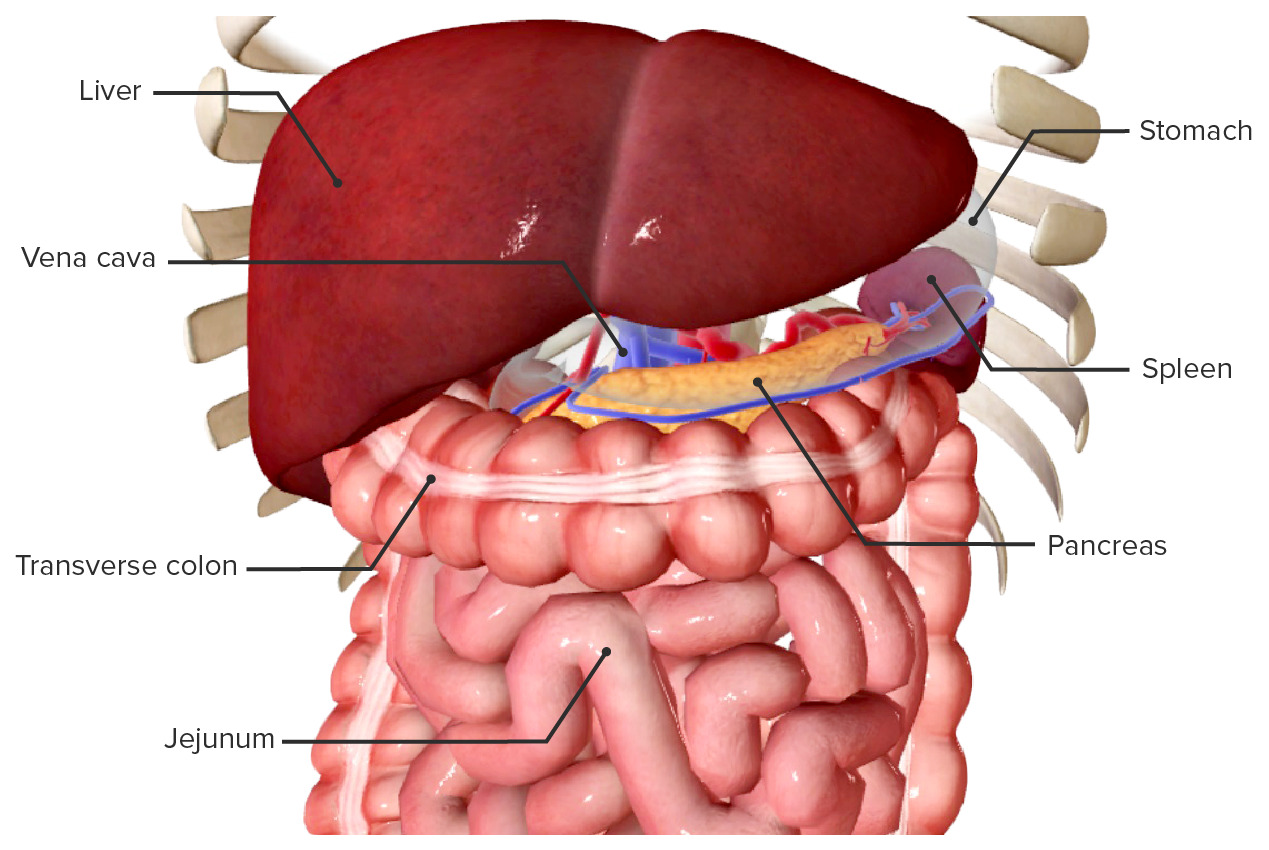
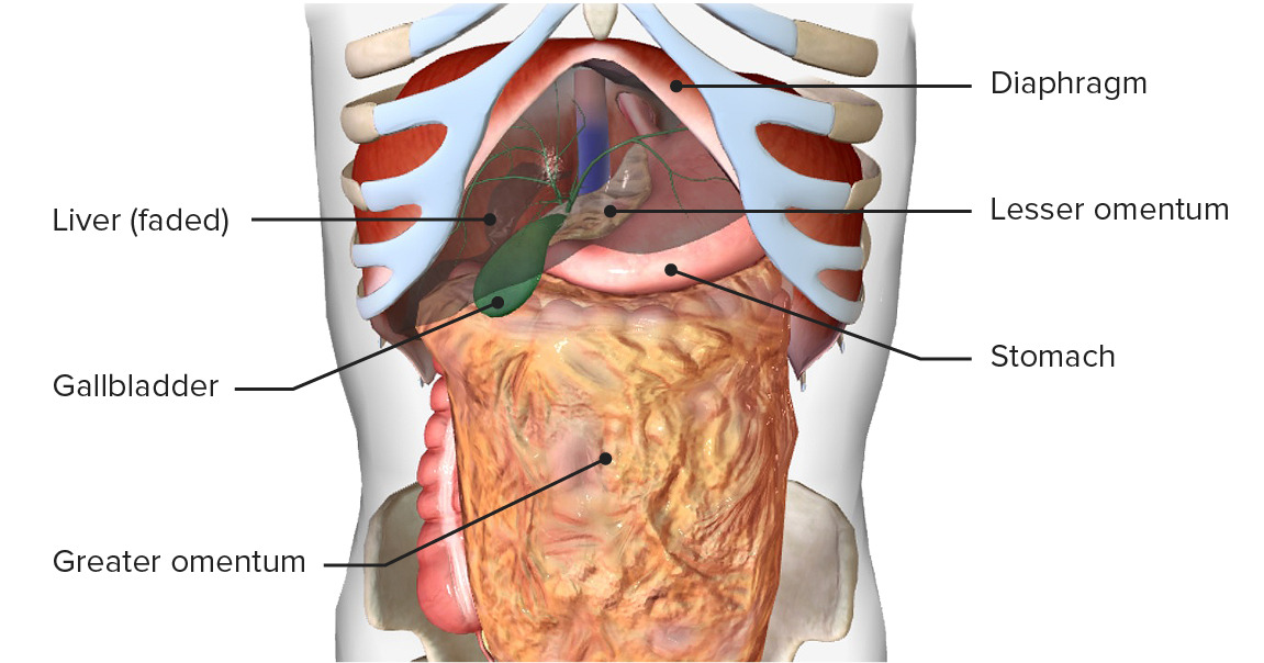
The surgically relevant structures of the right hypochondriumHypochondriumAnterior Abdominal Wall: Anatomy include the liverLiverThe liver is the largest gland in the human body. The liver is found in the superior right quadrant of the abdomen and weighs approximately 1.5 kilograms. Its main functions are detoxification, metabolism, nutrient storage (e.g., iron and vitamins), synthesis of coagulation factors, formation of bile, filtration, and storage of blood. Liver: Anatomy and the biliary treeBiliary treeThe bile ducts and the gallbladder.Gallbladder and Biliary Tract: Anatomy.
LiverLiverThe liver is the largest gland in the human body. The liver is found in the superior right quadrant of the abdomen and weighs approximately 1.5 kilograms. Its main functions are detoxification, metabolism, nutrient storage (e.g., iron and vitamins), synthesis of coagulation factors, formation of bile, filtration, and storage of blood. Liver: Anatomy
Location:
¾ in the RUQ
Adjacent to the inferior surface of diaphragmDiaphragmThe diaphragm is a large, dome-shaped muscle that separates the thoracic cavity from the abdominal cavity. The diaphragm consists of muscle fibers and a large central tendon, which is divided into right and left parts. As the primary muscle of inspiration, the diaphragm contributes 75% of the total inspiratory muscle force.Diaphragm: Anatomy
Location is breath-dependent (rises during exhalation, lowers during inhalation).
Location of the liver in the right hypochondrium and epigastrium
LiverLiverThe liver is the largest gland in the human body. The liver is found in the superior right quadrant of the abdomen and weighs approximately 1.5 kilograms. Its main functions are detoxification, metabolism, nutrient storage (e.g., iron and vitamins), synthesis of coagulation factors, formation of bile, filtration, and storage of blood. Liver: Anatomy is intraperitonealIntraperitonealPeritoneum: Anatomy except for bare area, porta hepatisPorta hepatisLiver: Anatomy, and gallbladderGallbladderThe gallbladder is a pear-shaped sac, located directly beneath the liver, that sits on top of the superior part of the duodenum. The primary functions of the gallbladder include concentrating and storing up to 50 mL of bile. Gallbladder and Biliary Tract: Anatomy fossa.
Enclosed in the Glisson capsuleCapsuleAn envelope of loose gel surrounding a bacterial cell which is associated with the virulence of pathogenic bacteria. Some capsules have a well-defined border, whereas others form a slime layer that trails off into the medium. Most capsules consist of relatively simple polysaccharides but there are some bacteria whose capsules are made of polypeptides.Bacteroides (external layer of fibrousFibrousFibrocystic Changeconnective tissueConnective tissueConnective tissues originate from embryonic mesenchyme and are present throughout the body except inside the brain and spinal cord. The main function of connective tissues is to provide structural support to organs. Connective tissues consist of cells and an extracellular matrix.Connective Tissue: Histology)
Anterior view of the diaphragmatic surface of the liver, featuring the falciform, triangular, round, and coronary ligaments:
Note that the round ligament extends from the free edge of the falciform ligament.
Inferior view of the visceral surface of the liver:
Note the uneven structure that results from impressions of the neighboring organs. The colic impression is caused by the colon’s hepatic flexure. The descending portion of the duodenum forms the duodenal impression.
Anterior view of the liver: In this image, the liver has been lifted to show the lesser omentum, which consists of the hepatogastric and hepatoduodenal ligaments. This double layer of peritoneum connects the liver with the lesser curvature of the stomach and the duodenum.
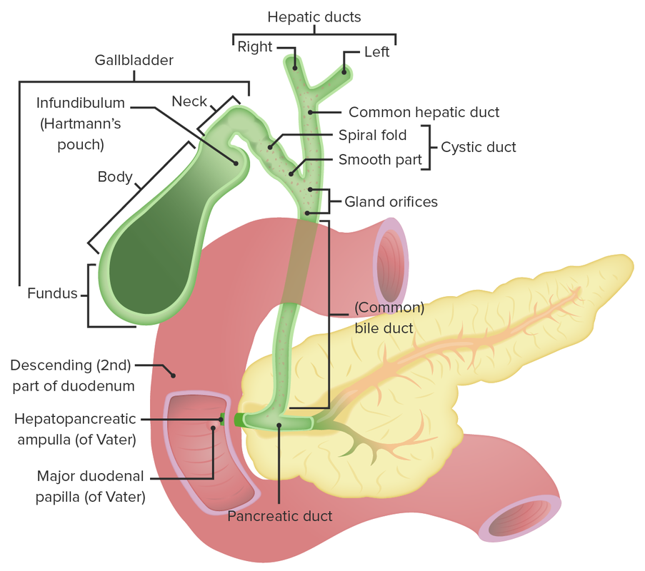
GallbladderGallbladderThe gallbladder is a pear-shaped sac, located directly beneath the liver, that sits on top of the superior part of the duodenum. The primary functions of the gallbladder include concentrating and storing up to 50 mL of bile. Gallbladder and Biliary Tract: Anatomy:
Bile-filled sac located in a fossa on the inferior aspect of the liverLiverThe liver is the largest gland in the human body. The liver is found in the superior right quadrant of the abdomen and weighs approximately 1.5 kilograms. Its main functions are detoxification, metabolism, nutrient storage (e.g., iron and vitamins), synthesis of coagulation factors, formation of bile, filtration, and storage of blood. Liver: Anatomy beneath the junction of hepatic segments Ⅳb and Ⅴ
7–10 cm in length, average capacity 30–50 mL
Anatomic divisions:
FundusFundusThe superior portion of the body of the stomach above the level of the cardiac notch.Stomach: Anatomy (superiormost aspect)
Corpus (body)
InfundibulumInfundibulumUterus, Cervix, and Fallopian Tubes: Anatomy (round, blind end extending below the liverLiverThe liver is the largest gland in the human body. The liver is found in the superior right quadrant of the abdomen and weighs approximately 1.5 kilograms. Its main functions are detoxification, metabolism, nutrient storage (e.g., iron and vitamins), synthesis of coagulation factors, formation of bile, filtration, and storage of blood. Liver: Anatomy margin)
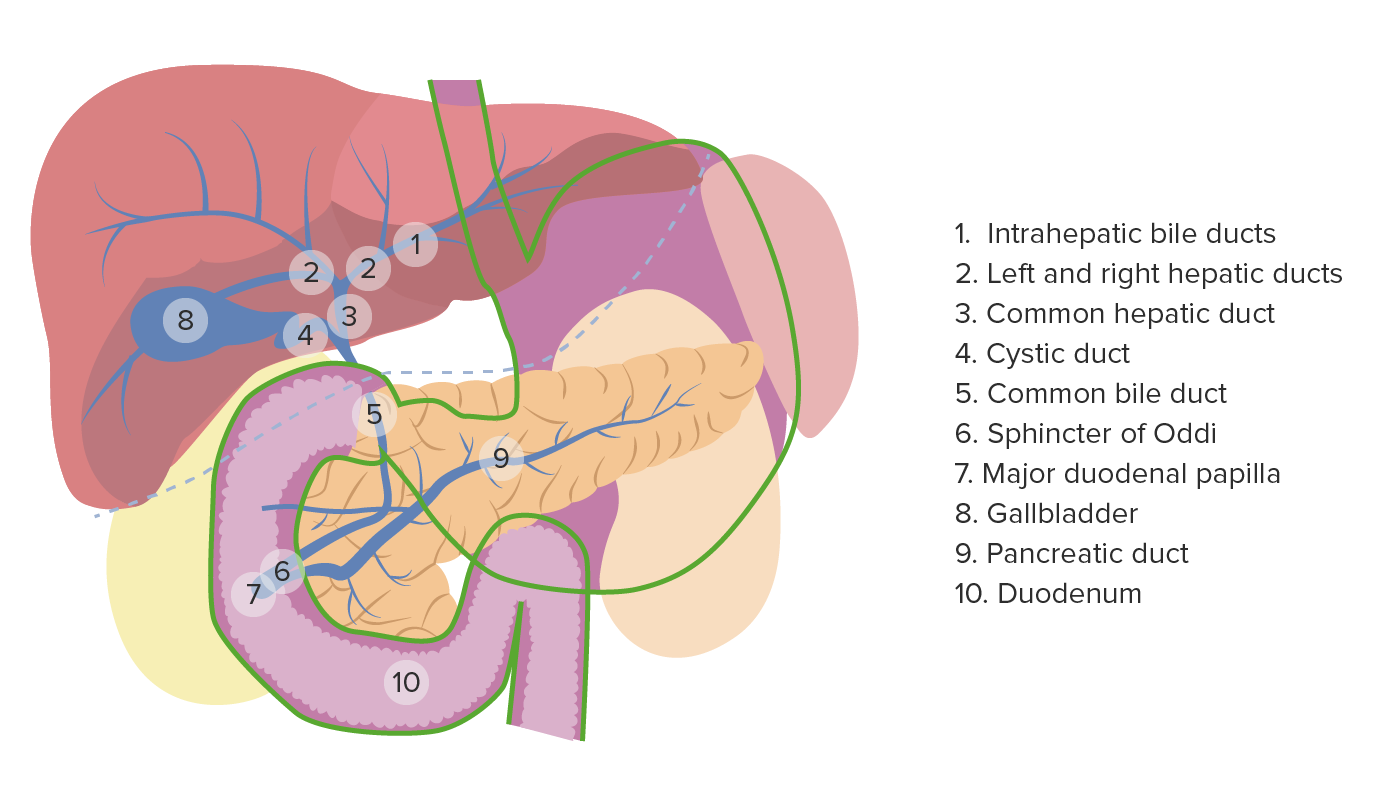
The right and left hepatic ducts join to form the common hepatic duct.
The cysticCysticFibrocystic Change duct joins the common hepatic duct to form the common bileBileAn emulsifying agent produced in the liver and secreted into the duodenum. Its composition includes bile acids and salts; cholesterol; and electrolytes. It aids digestion of fats in the duodenum.Gallbladder and Biliary Tract: Anatomy duct.
The cysticCysticFibrocystic Change duct length is highly variableVariableVariables represent information about something that can change. The design of the measurement scales, or of the methods for obtaining information, will determine the data gathered and the characteristics of that data. As a result, a variable can be qualitative or quantitative, and may be further classified into subgroups.Types of Variables.
CholecystectomyCholecystectomyCholecystectomy is a surgical procedure performed with the goal of resecting and extracting the gallbladder. It is one of the most common abdominal surgeries performed in the Western world. Cholecystectomy is performed for symptomatic cholelithiasis, cholecystitis, gallbladder polyps > 0.5 cm, porcelain gallbladder, choledocholithiasis and gallstone pancreatitis, and rarely, for gallbladder cancer. Cholecystectomy (open and laparoscopic): surgical removal of the gallbladderGallbladderThe gallbladder is a pear-shaped sac, located directly beneath the liver, that sits on top of the superior part of the duodenum. The primary functions of the gallbladder include concentrating and storing up to 50 mL of bile. Gallbladder and Biliary Tract: Anatomy, usually because of cholelithiasisCholelithiasisCholelithiasis (gallstones) is the presence of stones in the gallbladder. Most gallstones are cholesterol stones, while the rest are composed of bilirubin (pigment stones) and other mixed components. Patients are commonly asymptomatic but may present with biliary colic (intermittent pain in the right upper quadrant). Cholelithiasis (gallstone disease) with or without cholecystitisCholecystitisCholecystitis is the inflammation of the gallbladder (GB) usually caused by the obstruction of the cystic duct (acute cholecystitis). Mechanical irritation by gallstones can also produce chronic GB inflammation. Cholecystitis is one of the most common complications of cholelithiasis but inflammation without gallstones can occur in a minority of patients. Cholecystitis
Choledochostomy: surgical incisionSurgical IncisionSurgical Site Infections on the common bileBileAn emulsifying agent produced in the liver and secreted into the duodenum. Its composition includes bile acids and salts; cholesterol; and electrolytes. It aids digestion of fats in the duodenum.Gallbladder and Biliary Tract: Anatomy duct
Choledochojejunostomy: anastomosis of a common bileBileAn emulsifying agent produced in the liver and secreted into the duodenum. Its composition includes bile acids and salts; cholesterol; and electrolytes. It aids digestion of fats in the duodenum.Gallbladder and Biliary Tract: Anatomy duct to the jejunumJejunumThe middle portion of the small intestine, between duodenum and ileum. It represents about 2/5 of the remaining portion of the small intestine below duodenum.Small Intestine: Anatomy (for oncologic resections or treatment of common bileBileAn emulsifying agent produced in the liver and secreted into the duodenum. Its composition includes bile acids and salts; cholesterol; and electrolytes. It aids digestion of fats in the duodenum.Gallbladder and Biliary Tract: Anatomy duct injuries)
Hepatic lobectomy: surgical removal of a lobe of the liverLiverThe liver is the largest gland in the human body. The liver is found in the superior right quadrant of the abdomen and weighs approximately 1.5 kilograms. Its main functions are detoxification, metabolism, nutrient storage (e.g., iron and vitamins), synthesis of coagulation factors, formation of bile, filtration, and storage of blood. Liver: Anatomy
Hepatic segmentectomy: surgical removal of a segment of the liverLiverThe liver is the largest gland in the human body. The liver is found in the superior right quadrant of the abdomen and weighs approximately 1.5 kilograms. Its main functions are detoxification, metabolism, nutrient storage (e.g., iron and vitamins), synthesis of coagulation factors, formation of bile, filtration, and storage of blood. Liver: Anatomy
Needle biopsyBiopsyRemoval and pathologic examination of specimens from the living body.Ewing Sarcoma of the liverLiverThe liver is the largest gland in the human body. The liver is found in the superior right quadrant of the abdomen and weighs approximately 1.5 kilograms. Its main functions are detoxification, metabolism, nutrient storage (e.g., iron and vitamins), synthesis of coagulation factors, formation of bile, filtration, and storage of blood. Liver: Anatomy
Hepatic abscessAbscessAccumulation of purulent material in tissues, organs, or circumscribed spaces, usually associated with signs of infection.Chronic Granulomatous Disease drainage
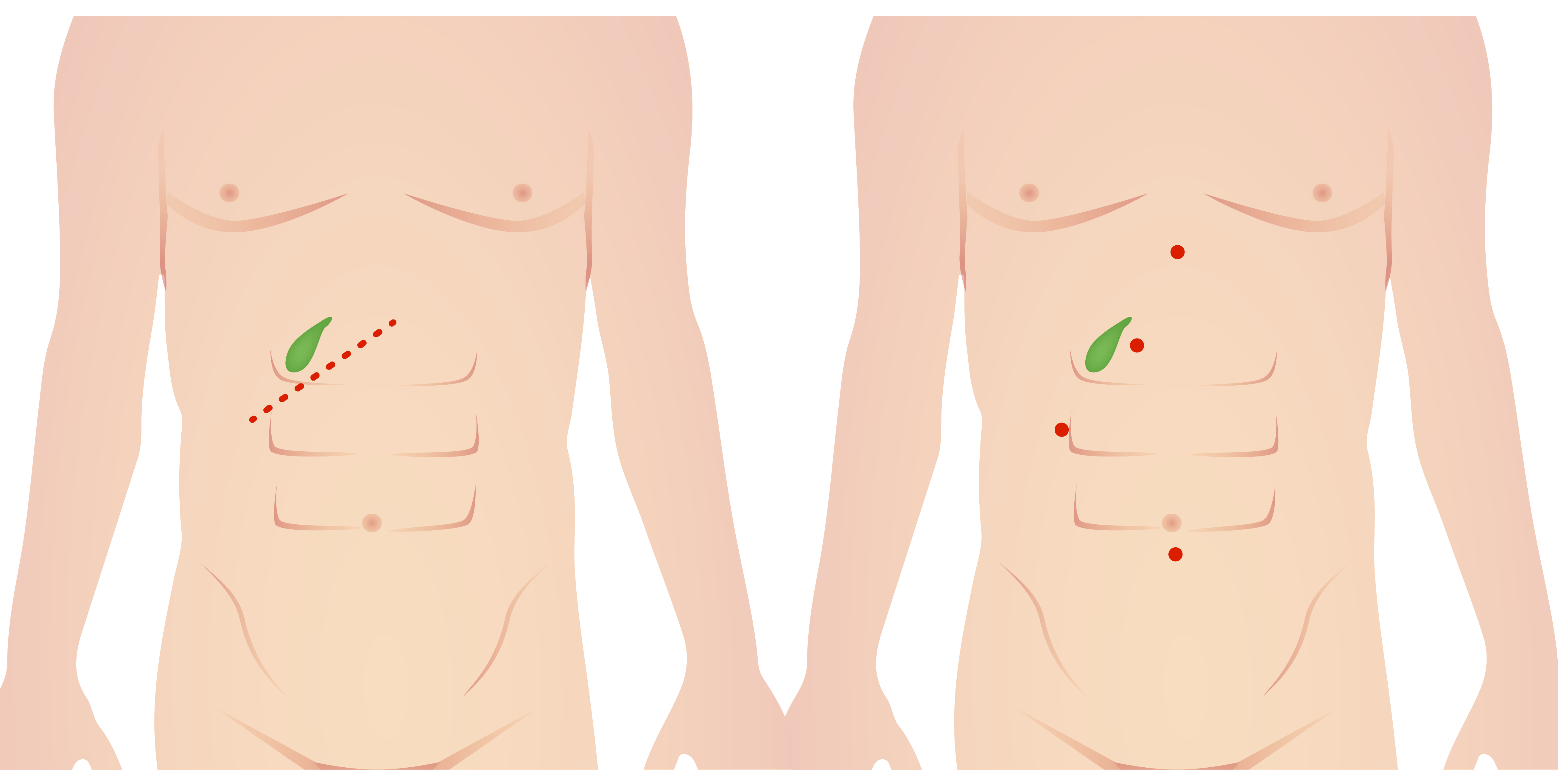
Incisions for open (left) and laparoscopic (right) cholecystectomy
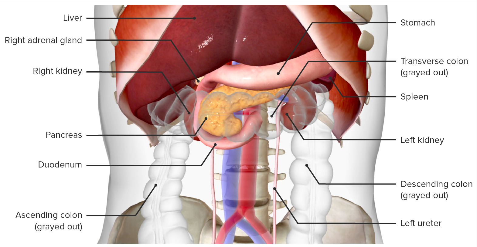
The epigastrium contains the transverse colonTransverse colonThe segment of large intestine between ascending colon and descending colon. It passes from the right colic flexure across the abdomen, then turns sharply at the left colonic flexure into the descending colon.Colon, Cecum, and Appendix: Anatomy, the duodenumDuodenumThe shortest and widest portion of the small intestine adjacent to the pylorus of the stomach. It is named for having the length equal to about the width of 12 fingers.Small Intestine: Anatomy, and the pancreasPancreasThe pancreas lies mostly posterior to the stomach and extends across the posterior abdominal wall from the duodenum on the right to the spleen on the left. This organ has both exocrine and endocrine tissue. Pancreas: Anatomy.
Transverse colonTransverse colonThe segment of large intestine between ascending colon and descending colon. It passes from the right colic flexure across the abdomen, then turns sharply at the left colonic flexure into the descending colon.Colon, Cecum, and Appendix: Anatomy
Part of the colonColonThe large intestines constitute the last portion of the digestive system. The large intestine consists of the cecum, appendix, colon (with ascending, transverse, descending, and sigmoid segments), rectum, and anal canal. The primary function of the colon is to remove water and compact the stool prior to expulsion from the body via the rectum and anal canal. Colon, Cecum, and Appendix: Anatomy distal to the ascending colonAscending colonThe segment of large intestine between the cecum and the transverse colon. It passes cephalad from the cecum to the caudal surface of the right lobe of the liver where it bends sharply to the left, forming the right colic flexure.Colon, Cecum, and Appendix: Anatomy and proximal to the descending colonDescending colonThe segment of large intestine between transverse colon and the sigmoid colon.Colon, Cecum, and Appendix: Anatomy that travels transversely across the upper abdomen
Suspended by mesenteryMesenteryA layer of the peritoneum which attaches the abdominal viscera to the abdominal wall and conveys their blood vessels and nerves.Peritoneum: Anatomy (transverse mesocolon)
Supplied by ileocolic and middle colic branches of the superior mesenteric arterySuperior mesenteric arteryA large vessel supplying the whole length of the small intestine except the superior part of the duodenum. It also supplies the cecum and the ascending part of the colon and about half the transverse part of the colon. It arises from the anterior surface of the aorta below the celiac artery at the level of the first lumbar vertebra.Small Intestine: Anatomy
Venous drainage is through the superior mesenteric vein that joins the portal veinPortal veinA short thick vein formed by union of the superior mesenteric vein and the splenic vein.Liver: Anatomy.
DuodenumDuodenumThe shortest and widest portion of the small intestine adjacent to the pylorus of the stomach. It is named for having the length equal to about the width of 12 fingers.Small Intestine: Anatomy
First C-shaped segment of the small intestineSmall intestineThe small intestine is the longest part of the GI tract, extending from the pyloric orifice of the stomach to the ileocecal junction. The small intestine is the major organ responsible for chemical digestion and absorption of nutrients. It is divided into 3 segments: the duodenum, the jejunum, and the ileum. Small Intestine: Anatomy
Surrounds the head of the pancreasPancreasThe pancreas lies mostly posterior to the stomach and extends across the posterior abdominal wall from the duodenum on the right to the spleen on the left. This organ has both exocrine and endocrine tissue. Pancreas: Anatomy
Consists of 4 segments
The 2nd, or descending, segment contains the ampulla of Vater (opening of the common bileBileAn emulsifying agent produced in the liver and secreted into the duodenum. Its composition includes bile acids and salts; cholesterol; and electrolytes. It aids digestion of fats in the duodenum.Gallbladder and Biliary Tract: Anatomy duct).
Ends at duodenojejunal flexure: fixed to the posterior abdominal wallPosterior abdominal wallThe posterior abdominal wall is a complex musculoskeletal structure that houses the abdominal aorta, the inferior vena cava, as well as important retroperitoneal organs, like the kidneys, renal glands, pancreas, and duodenum.Posterior Abdominal Wall: Anatomy by the ligament of TreitzLigament of treitzGastrointestinal Bleeding
Duodenum and its relation with the pancreas and biliary ducts
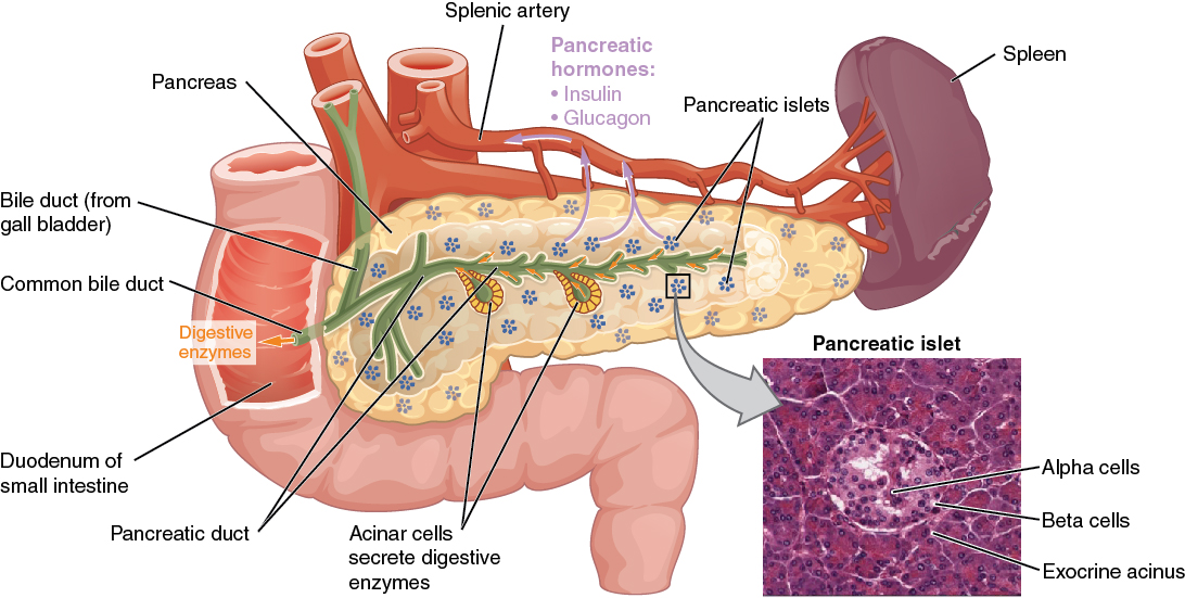
PancreasPancreasThe pancreas lies mostly posterior to the stomach and extends across the posterior abdominal wall from the duodenum on the right to the spleen on the left. This organ has both exocrine and endocrine tissue. Pancreas: Anatomy
Segments:
Caput pancreatis (head)
Uncinate process
Collum pancreatis (neckNeckThe part of a human or animal body connecting the head to the rest of the body.Peritonsillar Abscess)
Corpus pancreatis (body)
Cauda pancreatis (tail): the only intraperitonealIntraperitonealPeritoneum: Anatomy segment of the pancreasPancreasThe pancreas lies mostly posterior to the stomach and extends across the posterior abdominal wall from the duodenum on the right to the spleen on the left. This organ has both exocrine and endocrine tissue. Pancreas: Anatomy
Ducts:
Pancreatic duct (Wirsung’s duct) and common bileBileAn emulsifying agent produced in the liver and secreted into the duodenum. Its composition includes bile acids and salts; cholesterol; and electrolytes. It aids digestion of fats in the duodenum.Gallbladder and Biliary Tract: Anatomy duct open into ampulla of Vater, which presents as major duodenal papilla.
Major duodenal papilla contains the hepatopancreatic sphincter (sphincter of Oddi), which regulates the secretionSecretionCoagulation Studies of bileBileAn emulsifying agent produced in the liver and secreted into the duodenum. Its composition includes bile acids and salts; cholesterol; and electrolytes. It aids digestion of fats in the duodenum.Gallbladder and Biliary Tract: Anatomy and pancreatic fluid.
Different parts of the pancreas and its surroundings
Image: “The pancreas” by OpenStax College. License: CC BY 3.0
Relevant procedures
Transverse colostomy: an opening along the length of the transverse colonTransverse colonThe segment of large intestine between ascending colon and descending colon. It passes from the right colic flexure across the abdomen, then turns sharply at the left colonic flexure into the descending colon.Colon, Cecum, and Appendix: Anatomy to the exterior skinSkinThe skin, also referred to as the integumentary system, is the largest organ of the body. The skin is primarily composed of the epidermis (outer layer) and dermis (deep layer). The epidermis is primarily composed of keratinocytes that undergo rapid turnover, while the dermis contains dense layers of connective tissue.Skin: Structure and Functions surface
Transverse hemicolectomy: surgical removal of the transverse colonTransverse colonThe segment of large intestine between ascending colon and descending colon. It passes from the right colic flexure across the abdomen, then turns sharply at the left colonic flexure into the descending colon.Colon, Cecum, and Appendix: Anatomy
Pancreaticojejunostomy (Puestow procedure): surgically made communicationCommunicationThe exchange or transmission of ideas, attitudes, or beliefs between individuals or groups.Decision-making Capacity and Legal Competence between the pancreasPancreasThe pancreas lies mostly posterior to the stomach and extends across the posterior abdominal wall from the duodenum on the right to the spleen on the left. This organ has both exocrine and endocrine tissue. Pancreas: Anatomy and the jejunumJejunumThe middle portion of the small intestine, between duodenum and ileum. It represents about 2/5 of the remaining portion of the small intestine below duodenum.Small Intestine: Anatomy (treatment of chronic pancreatitisPancreatitisInflammation of the pancreas. Pancreatitis is classified as acute unless there are computed tomographic or endoscopic retrograde cholangiopancreatographic findings of chronic pancreatitis. The two most common forms of acute pancreatitis are alcoholic pancreatitis and gallstone pancreatitis.Acute Pancreatitis)
Pancreaticoduodenectomy (Whipple procedure): surgical removal of the head of the pancreasPancreasThe pancreas lies mostly posterior to the stomach and extends across the posterior abdominal wall from the duodenum on the right to the spleen on the left. This organ has both exocrine and endocrine tissue. Pancreas: Anatomy and the duodenumDuodenumThe shortest and widest portion of the small intestine adjacent to the pylorus of the stomach. It is named for having the length equal to about the width of 12 fingers.Small Intestine: Anatomy
Distal pancreatectomy: removal of the body and tail of the pancreasPancreasThe pancreas lies mostly posterior to the stomach and extends across the posterior abdominal wall from the duodenum on the right to the spleen on the left. This organ has both exocrine and endocrine tissue. Pancreas: Anatomy
Total pancreatectomy: surgical removal of the pancreasPancreasThe pancreas lies mostly posterior to the stomach and extends across the posterior abdominal wall from the duodenum on the right to the spleen on the left. This organ has both exocrine and endocrine tissue. Pancreas: Anatomy
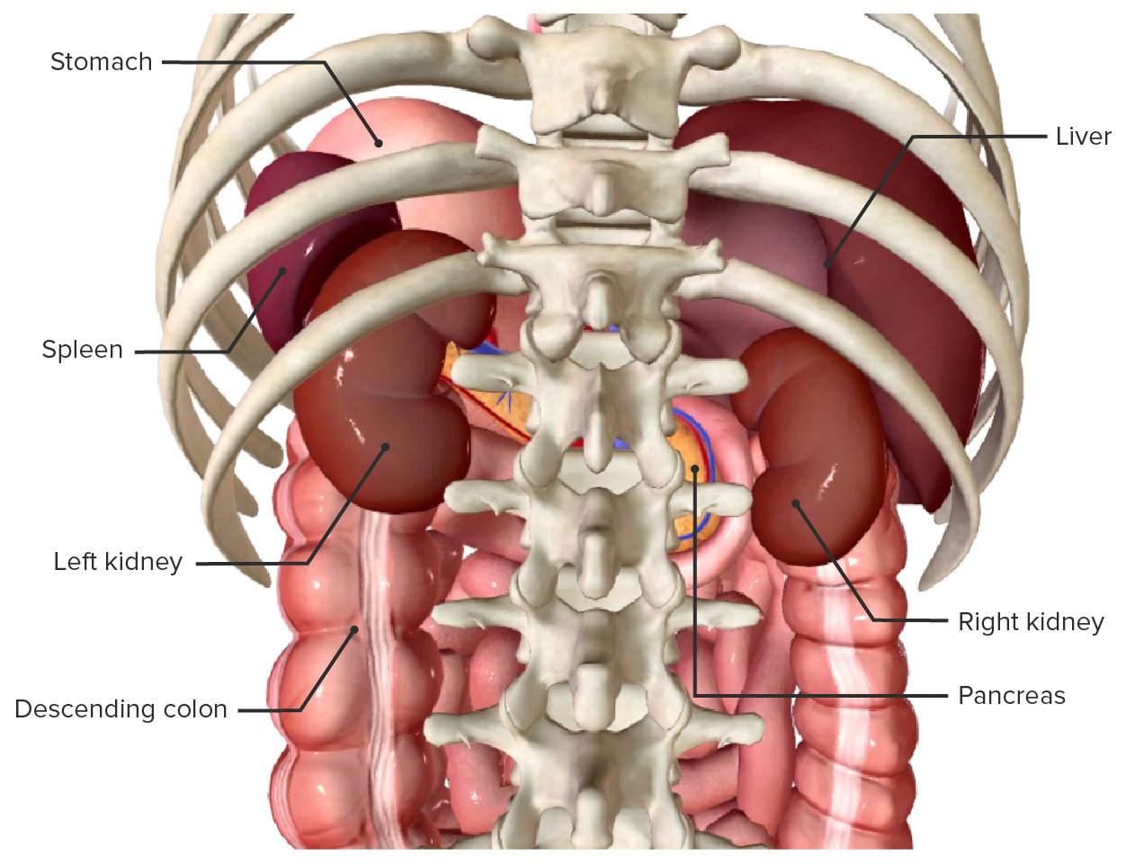
The most important structures of the left hypochondriumHypochondriumAnterior Abdominal Wall: Anatomy are the spleenSpleenThe spleen is the largest lymphoid organ in the body, located in the LUQ of the abdomen, superior to the left kidney and posterior to the stomach at the level of the 9th-11th ribs just below the diaphragm. The spleen is highly vascular and acts as an important blood filter, cleansing the blood of pathogens and damaged erythrocytes. Spleen: Anatomy and the stomachStomachThe stomach is a muscular sac in the upper left portion of the abdomen that plays a critical role in digestion. The stomach develops from the foregut and connects the esophagus with the duodenum. Structurally, the stomach is C-shaped and forms a greater and lesser curvature and is divided grossly into regions: the cardia, fundus, body, and pylorus. Stomach: Anatomy.
SpleenSpleenThe spleen is the largest lymphoid organ in the body, located in the LUQ of the abdomen, superior to the left kidney and posterior to the stomach at the level of the 9th-11th ribs just below the diaphragm. The spleen is highly vascular and acts as an important blood filter, cleansing the blood of pathogens and damaged erythrocytes. Spleen: Anatomy
Location:
Positioned against ribsRibsA set of twelve curved bones which connect to the vertebral column posteriorly, and terminate anteriorly as costal cartilage. Together, they form a protective cage around the internal thoracic organs.Chest Wall: Anatomy 9–11
Anterior: stomachStomachThe stomach is a muscular sac in the upper left portion of the abdomen that plays a critical role in digestion. The stomach develops from the foregut and connects the esophagus with the duodenum. Structurally, the stomach is C-shaped and forms a greater and lesser curvature and is divided grossly into regions: the cardia, fundus, body, and pylorus. Stomach: Anatomy
Lateral: abdominal muscles
Medial: left kidney
Posterior: diaphragmDiaphragmThe diaphragm is a large, dome-shaped muscle that separates the thoracic cavity from the abdominal cavity. The diaphragm consists of muscle fibers and a large central tendon, which is divided into right and left parts. As the primary muscle of inspiration, the diaphragm contributes 75% of the total inspiratory muscle force.Diaphragm: Anatomy
Inferior: colonColonThe large intestines constitute the last portion of the digestive system. The large intestine consists of the cecum, appendix, colon (with ascending, transverse, descending, and sigmoid segments), rectum, and anal canal. The primary function of the colon is to remove water and compact the stool prior to expulsion from the body via the rectum and anal canal. Colon, Cecum, and Appendix: Anatomy and left colic flexure
Ligaments:
Gastrosplenic
Splenorenal
Phrenicocolic
Blood supply: splenic artery and vein
Spleen in situ, anterior view (the stomach is faded): Note the spatial relations with the neighboring abdominal organs.
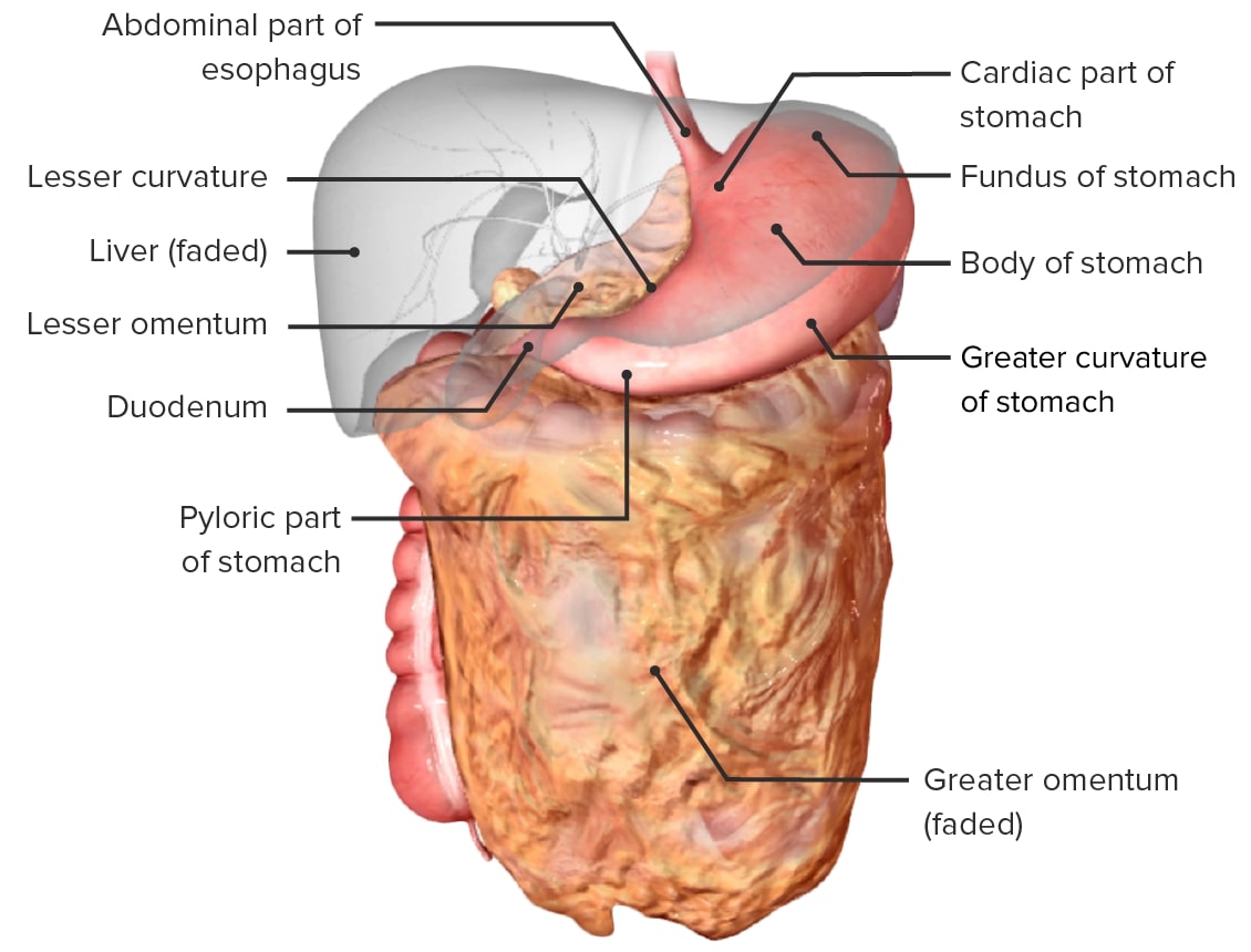
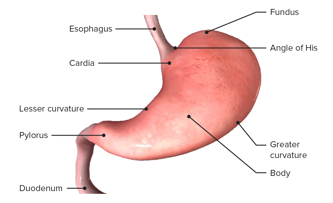
StomachStomachThe stomach is a muscular sac in the upper left portion of the abdomen that plays a critical role in digestion. The stomach develops from the foregut and connects the esophagus with the duodenum. Structurally, the stomach is C-shaped and forms a greater and lesser curvature and is divided grossly into regions: the cardia, fundus, body, and pylorus. Stomach: Anatomy
Segments:
Has 2 curvatures (lesser and greater)
Cardia:
Entrance of the stomachStomachThe stomach is a muscular sac in the upper left portion of the abdomen that plays a critical role in digestion. The stomach develops from the foregut and connects the esophagus with the duodenum. Structurally, the stomach is C-shaped and forms a greater and lesser curvature and is divided grossly into regions: the cardia, fundus, body, and pylorus. Stomach: Anatomy
Originates from the z-lineZ-lineEsophagus: Anatomy and createsthe angle of His, or angle of the cardiac orifice (the angle between the fundusFundusThe superior portion of the body of the stomach above the level of the cardiac notch.Stomach: Anatomy and abdominal esophagusEsophagusThe esophagus is a muscular tube-shaped organ of around 25 centimeters in length that connects the pharynx to the stomach. The organ extends from approximately the 6th cervical vertebra to the 11th thoracic vertebra and can be divided grossly into 3 parts: the cervical part, the thoracic part, and the abdominal part. Esophagus: Anatomy)
Crucial landmark in the construction of the gastric pouch
FundusFundusThe superior portion of the body of the stomach above the level of the cardiac notch.Stomach: Anatomy: a dome-shaped region located at the highest point of the stomachStomachThe stomach is a muscular sac in the upper left portion of the abdomen that plays a critical role in digestion. The stomach develops from the foregut and connects the esophagus with the duodenum. Structurally, the stomach is C-shaped and forms a greater and lesser curvature and is divided grossly into regions: the cardia, fundus, body, and pylorus. Stomach: Anatomy
Body:
Main section of the stomachStomachThe stomach is a muscular sac in the upper left portion of the abdomen that plays a critical role in digestion. The stomach develops from the foregut and connects the esophagus with the duodenum. Structurally, the stomach is C-shaped and forms a greater and lesser curvature and is divided grossly into regions: the cardia, fundus, body, and pylorus. Stomach: Anatomy
Extends from the fundusFundusThe superior portion of the body of the stomach above the level of the cardiac notch.Stomach: Anatomy to the pylorusPylorusThe region between the sharp indentation at the lower third of the stomach (incisura angularis) and the junction of the pylorus with the duodenum. Pyloric antral glands contain mucus-secreting cells and gastrin-secreting endocrine cells (g cells).Stomach: Anatomy
Bordered by the lesser and greater curvatures
PylorusPylorusThe region between the sharp indentation at the lower third of the stomach (incisura angularis) and the junction of the pylorus with the duodenum. Pyloric antral glands contain mucus-secreting cells and gastrin-secreting endocrine cells (g cells).Stomach: Anatomy:
Connects to the duodenumDuodenumThe shortest and widest portion of the small intestine adjacent to the pylorus of the stomach. It is named for having the length equal to about the width of 12 fingers.Small Intestine: Anatomy
Contains the pyloric sphincter
Consists of a wide pyloric antrum and narrow pyloric canal
Left gastric arteryGastric arteryAny of several branches of the splenic artery distributed to the greater curvature of the stomach.Stomach: Anatomy: main supply of the gastric pouch in a gastric bypassGastric bypassSurgical procedure in which the stomach is transected high on the body. The resulting small proximal gastric pouch is joined to any parts of the small intestine by an end-to-side surgical anastomosis, depending on the amounts of intestinal surface being bypasses. This procedure is used frequently in the treatment of morbid obesity by limiting the size of functional stomach, food intake, and food absorption.Gastroesophageal Reflux Disease (GERD)
Right gastric arteryGastric arteryAny of several branches of the splenic artery distributed to the greater curvature of the stomach.Stomach: Anatomy
Right and left gastroepiploic (gastro-omental) arteriesArteriesArteries are tubular collections of cells that transport oxygenated blood and nutrients from the heart to the tissues of the body. The blood passes through the arteries in order of decreasing luminal diameter, starting in the largest artery (the aorta) and ending in the small arterioles. Arteries are classified into 3 types: large elastic arteries, medium muscular arteries, and small arteries and arterioles. Arteries: Histology
Splenic artery
Short gastric arteriesArteriesArteries are tubular collections of cells that transport oxygenated blood and nutrients from the heart to the tissues of the body. The blood passes through the arteries in order of decreasing luminal diameter, starting in the largest artery (the aorta) and ending in the small arterioles. Arteries are classified into 3 types: large elastic arteries, medium muscular arteries, and small arteries and arterioles. Arteries: Histology
Posterior gastric arteryGastric arteryAny of several branches of the splenic artery distributed to the greater curvature of the stomach.Stomach: Anatomy
Venous drainage:
Homonymous veinsVeinsVeins are tubular collections of cells, which transport deoxygenated blood and waste from the capillary beds back to the heart. Veins are classified into 3 types: small veins/venules, medium veins, and large veins. Each type contains 3 primary layers: tunica intima, tunica media, and tunica adventitia. Veins: Histology that accompany the arteriesArteriesArteries are tubular collections of cells that transport oxygenated blood and nutrients from the heart to the tissues of the body. The blood passes through the arteries in order of decreasing luminal diameter, starting in the largest artery (the aorta) and ending in the small arterioles. Arteries are classified into 3 types: large elastic arteries, medium muscular arteries, and small arteries and arterioles. Arteries: Histology
Right and left gastric veinsVeinsVeins are tubular collections of cells, which transport deoxygenated blood and waste from the capillary beds back to the heart. Veins are classified into 3 types: small veins/venules, medium veins, and large veins. Each type contains 3 primary layers: tunica intima, tunica media, and tunica adventitia. Veins: Histology drain into the portal veinPortal veinA short thick vein formed by union of the superior mesenteric vein and the splenic vein.Liver: Anatomy.
Left gastroepiploic vein drains into the splenic vein.
Right gastroepiploic vein drains into the superior mesenteric vein.
Innervation:
Parasympathetic innervation: anterior and posterior vagal trunk
Sympathetic innervation: greater splanchnic nerve and gastric branches from celiac plexus
SplenectomySplenectomySurgical procedure involving either partial or entire removal of the spleen.Rupture of the Spleen: surgical removal of the spleenSpleenThe spleen is the largest lymphoid organ in the body, located in the LUQ of the abdomen, superior to the left kidney and posterior to the stomach at the level of the 9th-11th ribs just below the diaphragm. The spleen is highly vascular and acts as an important blood filter, cleansing the blood of pathogens and damaged erythrocytes. Spleen: Anatomy
Splenorrhaphy: surgical repair of the spleenSpleenThe spleen is the largest lymphoid organ in the body, located in the LUQ of the abdomen, superior to the left kidney and posterior to the stomach at the level of the 9th-11th ribs just below the diaphragm. The spleen is highly vascular and acts as an important blood filter, cleansing the blood of pathogens and damaged erythrocytes. Spleen: Anatomy, usually performed after trauma
Bariatric surgeryBariatric surgeryBariatric surgery refers to a group of invasive procedures used to surgically reduce the size of the stomach to produce early satiety, decrease food intake (restrictive type) and/or alter digestion, and artificially induce malabsorption of nutrients (malabsorptive type). The ultimate goal of bariatric surgery is drastic weight loss. Bariatric Surgery: group of invasive procedures that surgically reduce the size of the stomachStomachThe stomach is a muscular sac in the upper left portion of the abdomen that plays a critical role in digestion. The stomach develops from the foregut and connects the esophagus with the duodenum. Structurally, the stomach is C-shaped and forms a greater and lesser curvature and is divided grossly into regions: the cardia, fundus, body, and pylorus. Stomach: Anatomy to produce early satietyEarly SatietyBariatric Surgery and decrease food intake (restrictive type) and/or alter digestionDigestionDigestion refers to the process of the mechanical and chemical breakdown of food into smaller particles, which can then be absorbed and utilized by the body.Digestion and Absorption and artificially induce malabsorptionMalabsorptionGeneral term for a group of malnutrition syndromes caused by failure of normal intestinal absorption of nutrients.Malabsorption and Maldigestion of nutrients (malabsorptive type).
Total and subtotal gastrectomy: surgical removal of a part of or an entire stomachStomachThe stomach is a muscular sac in the upper left portion of the abdomen that plays a critical role in digestion. The stomach develops from the foregut and connects the esophagus with the duodenum. Structurally, the stomach is C-shaped and forms a greater and lesser curvature and is divided grossly into regions: the cardia, fundus, body, and pylorus. Stomach: Anatomy (for oncologic resections and gastric ulcers)
Truncal or selective vagotomy: transection of the vagal nerves or branches that can be combined with partial gastric resections to control acid secretionSecretionCoagulation Studies and prevent ulcer recurrence
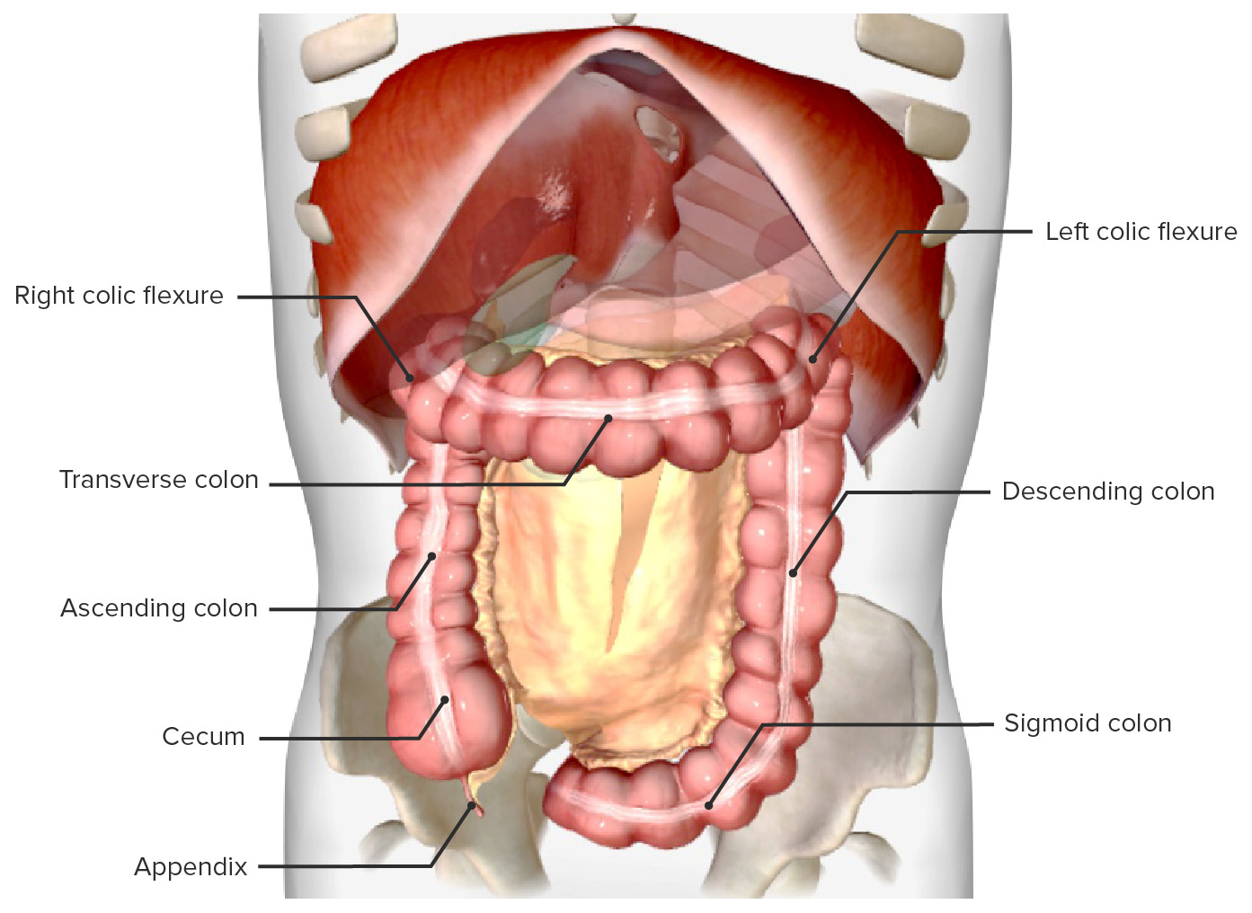
Ascending and descending portions of the large intestineLarge intestineThe large intestines constitute the last portion of the digestive system. The large intestine consists of the cecum, appendix, colon (with ascending, transverse, descending, and sigmoid segments), rectum, and anal canal. The primary function of the colon is to remove water and compact the stool prior to expulsion from the body via the rectum and anal canal. Colon, Cecum, and Appendix: Anatomy found in the right flank and the left flank, respectively
Arterial supply: superior and inferior mesenteric arteriesArteriesArteries are tubular collections of cells that transport oxygenated blood and nutrients from the heart to the tissues of the body. The blood passes through the arteries in order of decreasing luminal diameter, starting in the largest artery (the aorta) and ending in the small arterioles. Arteries are classified into 3 types: large elastic arteries, medium muscular arteries, and small arteries and arterioles. Arteries: Histology
Venous drainage:superior and inferior mesenteric veinsVeinsVeins are tubular collections of cells, which transport deoxygenated blood and waste from the capillary beds back to the heart. Veins are classified into 3 types: small veins/venules, medium veins, and large veins. Each type contains 3 primary layers: tunica intima, tunica media, and tunica adventitia. Veins: Histology that join portal circulationCirculationThe movement of the blood as it is pumped through the cardiovascular system.ABCDE Assessment
Colon in situ, anterior view, with the greater omentum and small intestines removed
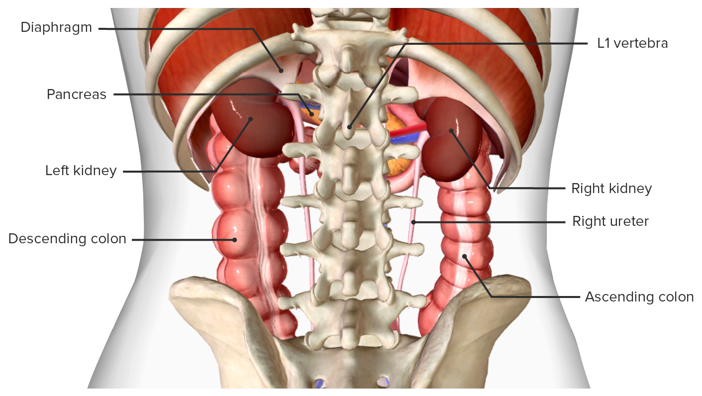
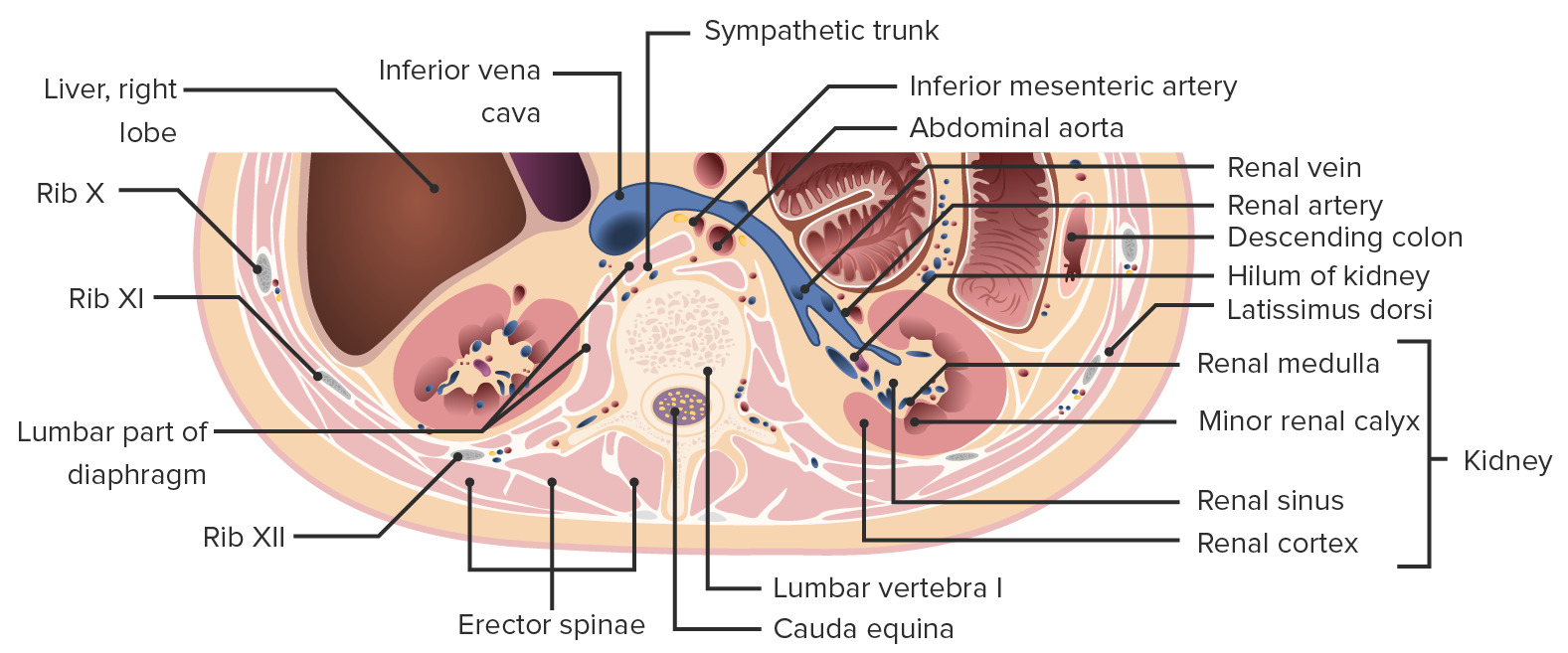
KidneysKidneysThe kidneys are a pair of bean-shaped organs located retroperitoneally against the posterior wall of the abdomen on either side of the spine. As part of the urinary tract, the kidneys are responsible for blood filtration and excretion of water-soluble waste in the urine.Kidneys: Anatomy
Location:
Between T12 and L3, in the paravertebral gutter
Their superior pole rests at the level of the 11th and 12th ribsRibsA set of twelve curved bones which connect to the vertebral column posteriorly, and terminate anteriorly as costal cartilage. Together, they form a protective cage around the internal thoracic organs.Chest Wall: Anatomy.
The parietalParietalOne of a pair of irregularly shaped quadrilateral bones situated between the frontal bone and occipital bone, which together form the sides of the cranium.Skull: AnatomyperitoneumPeritoneumThe peritoneum is a serous membrane lining the abdominopelvic cavity. This lining is formed by connective tissue and originates from the mesoderm. The membrane lines both the abdominal walls (as parietal peritoneum) and all of the visceral organs (as visceral peritoneum).Peritoneum: Anatomy anchors them to the posterior abdominal wallPosterior abdominal wallThe posterior abdominal wall is a complex musculoskeletal structure that houses the abdominal aorta, the inferior vena cava, as well as important retroperitoneal organs, like the kidneys, renal glands, pancreas, and duodenum.Posterior Abdominal Wall: Anatomy.
Holds the kidneysKidneysThe kidneys are a pair of bean-shaped organs located retroperitoneally against the posterior wall of the abdomen on either side of the spine. As part of the urinary tract, the kidneys are responsible for blood filtration and excretion of water-soluble waste in the urine.Kidneys: Anatomy in position
Consists of:
Paranephric adipose tissueAdipose tissueAdipose tissue is a specialized type of connective tissue that has both structural and highly complex metabolic functions, including energy storage, glucose homeostasis, and a multitude of endocrine capabilities. There are three types of adipose tissue, white adipose tissue, brown adipose tissue, and beige or “brite” adipose tissue, which is a transitional form.Adipose Tissue: Histology (fat)
Renal fasciaFasciaLayers of connective tissue of variable thickness. The superficial fascia is found immediately below the skin; the deep fascia invests muscles, nerves, and other organs.Cellulitis
Relations:
Superomedially: adrenal glandsAdrenal GlandsThe adrenal glands are a pair of retroperitoneal endocrine glands located above the kidneys. The outer parenchyma is called the adrenal cortex and has 3 distinct zones, each with its own secretory products. Beneath the cortex lies the adrenal medulla, which secretes catecholamines involved in the fight-or-flight response. Adrenal Glands: Anatomy
Posteriorly:
Nerves: subcostal, iliohypogastric, ilioinguinal
Muscles: diaphragmDiaphragmThe diaphragm is a large, dome-shaped muscle that separates the thoracic cavity from the abdominal cavity. The diaphragm consists of muscle fibers and a large central tendon, which is divided into right and left parts. As the primary muscle of inspiration, the diaphragm contributes 75% of the total inspiratory muscle force.Diaphragm: Anatomy, quadratus lumborumQuadratus lumborumPosterior Abdominal Wall: Anatomy
Anterior view of the kidneys and neighboring organs
Transverse cross section of the abdomen focused on the kidneys: Note how the kidney is embedded in adipose tissue within the gutters (paranephric and perinephric fat), which is continuous with the fat in the renal sinuses.
Right hemicolectomy: surgical removal of the right segment of the transverse colonTransverse colonThe segment of large intestine between ascending colon and descending colon. It passes from the right colic flexure across the abdomen, then turns sharply at the left colonic flexure into the descending colon.Colon, Cecum, and Appendix: Anatomy and ascending colonAscending colonThe segment of large intestine between the cecum and the transverse colon. It passes cephalad from the cecum to the caudal surface of the right lobe of the liver where it bends sharply to the left, forming the right colic flexure.Colon, Cecum, and Appendix: Anatomy
Left hemicolectomy: surgical removal of the left segment of the transverse colonTransverse colonThe segment of large intestine between ascending colon and descending colon. It passes from the right colic flexure across the abdomen, then turns sharply at the left colonic flexure into the descending colon.Colon, Cecum, and Appendix: Anatomy and descending colonDescending colonThe segment of large intestine between transverse colon and the sigmoid colon.Colon, Cecum, and Appendix: Anatomy
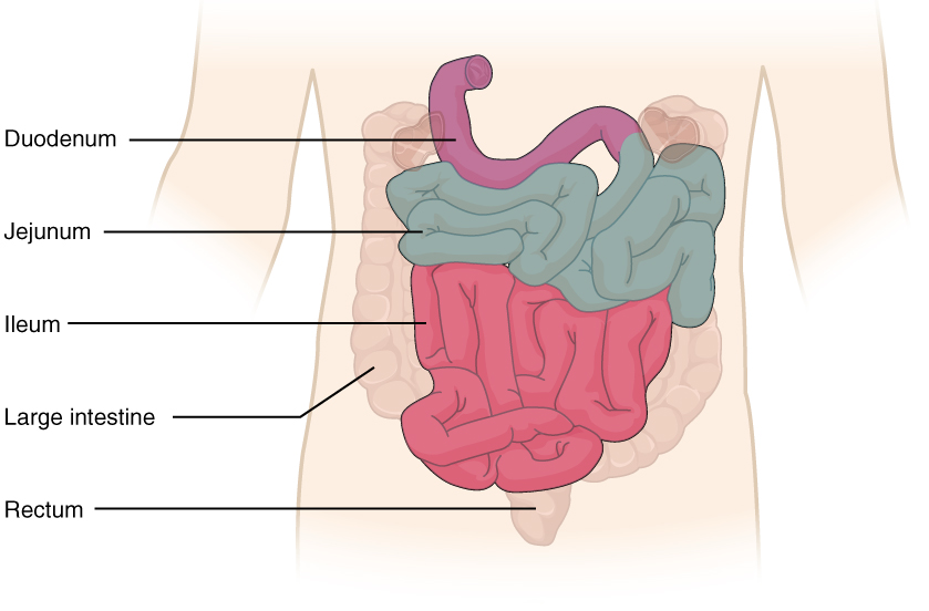
Small intestineSmall intestineThe small intestine is the longest part of the GI tract, extending from the pyloric orifice of the stomach to the ileocecal junction. The small intestine is the major organ responsible for chemical digestion and absorption of nutrients. It is divided into 3 segments: the duodenum, the jejunum, and the ileum. Small Intestine: Anatomy
Average length: 6–7 m
Segments:
DuodenumDuodenumThe shortest and widest portion of the small intestine adjacent to the pylorus of the stomach. It is named for having the length equal to about the width of 12 fingers.Small Intestine: Anatomy
JejunumJejunumThe middle portion of the small intestine, between duodenum and ileum. It represents about 2/5 of the remaining portion of the small intestine below duodenum.Small Intestine: Anatomy
IleumIleumThe distal and narrowest portion of the small intestine, between the jejunum and the ileocecal valve of the large intestine.Small Intestine: Anatomy
Blood supply:
DuodenumDuodenumThe shortest and widest portion of the small intestine adjacent to the pylorus of the stomach. It is named for having the length equal to about the width of 12 fingers.Small Intestine: Anatomy: superior and inferior pancreaticoduodenal arteriesArteriesArteries are tubular collections of cells that transport oxygenated blood and nutrients from the heart to the tissues of the body. The blood passes through the arteries in order of decreasing luminal diameter, starting in the largest artery (the aorta) and ending in the small arterioles. Arteries are classified into 3 types: large elastic arteries, medium muscular arteries, and small arteries and arterioles. Arteries: Histology
JejunumJejunumThe middle portion of the small intestine, between duodenum and ileum. It represents about 2/5 of the remaining portion of the small intestine below duodenum.Small Intestine: Anatomy and ilium: jejunal arteriesArteriesArteries are tubular collections of cells that transport oxygenated blood and nutrients from the heart to the tissues of the body. The blood passes through the arteries in order of decreasing luminal diameter, starting in the largest artery (the aorta) and ending in the small arterioles. Arteries are classified into 3 types: large elastic arteries, medium muscular arteries, and small arteries and arterioles. Arteries: Histology and ileal arteriesArteriesArteries are tubular collections of cells that transport oxygenated blood and nutrients from the heart to the tissues of the body. The blood passes through the arteries in order of decreasing luminal diameter, starting in the largest artery (the aorta) and ending in the small arterioles. Arteries are classified into 3 types: large elastic arteries, medium muscular arteries, and small arteries and arterioles. Arteries: Histology
Form anastomotic loops that run parallel to the small intestineSmall intestineThe small intestine is the longest part of the GI tract, extending from the pyloric orifice of the stomach to the ileocecal junction. The small intestine is the major organ responsible for chemical digestion and absorption of nutrients. It is divided into 3 segments: the duodenum, the jejunum, and the ileum. Small Intestine: Anatomy = arcades
Small branches leave these arcades = vasa rectaVasa rectaGlomerular Filtration (or straight arteriesArteriesArteries are tubular collections of cells that transport oxygenated blood and nutrients from the heart to the tissues of the body. The blood passes through the arteries in order of decreasing luminal diameter, starting in the largest artery (the aorta) and ending in the small arterioles. Arteries are classified into 3 types: large elastic arteries, medium muscular arteries, and small arteries and arterioles. Arteries: Histology)
Venous drainage: portal system
Small intestine and its parts
Image: “2417 Small IntestineN” by OpenStax College. License: CC BY 3.0
Extending from the aortic hiatusAortic hiatusDiaphragm: Anatomy of the diaphragmDiaphragmThe diaphragm is a large, dome-shaped muscle that separates the thoracic cavity from the abdominal cavity. The diaphragm consists of muscle fibers and a large central tendon, which is divided into right and left parts. As the primary muscle of inspiration, the diaphragm contributes 75% of the total inspiratory muscle force.Diaphragm: Anatomy (T12) to its bifurcation into the common iliac arteriesArteriesArteries are tubular collections of cells that transport oxygenated blood and nutrients from the heart to the tissues of the body. The blood passes through the arteries in order of decreasing luminal diameter, starting in the largest artery (the aorta) and ending in the small arterioles. Arteries are classified into 3 types: large elastic arteries, medium muscular arteries, and small arteries and arterioles. Arteries: Histology (L4)
Vascular branches:
Unpaired (forward branches):
Celiac trunk
Superior mesenteric
Inferior mesenteric
Paired visceral (lateral):
Renal
Gonadal
Paired parietalParietalOne of a pair of irregularly shaped quadrilateral bones situated between the frontal bone and occipital bone, which together form the sides of the cranium.Skull: Anatomy (posterolateral): inferiormost intercostal
Relevant procedures
Small intestineSmall intestineThe small intestine is the longest part of the GI tract, extending from the pyloric orifice of the stomach to the ileocecal junction. The small intestine is the major organ responsible for chemical digestion and absorption of nutrients. It is divided into 3 segments: the duodenum, the jejunum, and the ileum. Small Intestine: Anatomy resection: surgical removal of a segment of the small intestineSmall intestineThe small intestine is the longest part of the GI tract, extending from the pyloric orifice of the stomach to the ileocecal junction. The small intestine is the major organ responsible for chemical digestion and absorption of nutrients. It is divided into 3 segments: the duodenum, the jejunum, and the ileum. Small Intestine: Anatomy, usually because of adhesions, volvulusVolvulusA volvulus is the twisting or axial rotation of a portion of the bowel around its mesentery. The most common site of volvulus in adults is the colon; most frequently the sigmoid volvulus. Patients typically present with symptoms of bowel obstruction such as abdominal pain, distension, vomiting, and constipation/obstipation. Volvulus, obstruction, and regional ileitis
IleostomyIleostomySurgical creation of an external opening into the ileum for fecal diversion or drainage. This replacement for the rectum is usually created in patients with severe inflammatory bowel diseases. Loop (continent) or tube (incontinent) procedures are most often employed.Large Bowel Obstruction: surgically made communicationCommunicationThe exchange or transmission of ideas, attitudes, or beliefs between individuals or groups.Decision-making Capacity and Legal Competence between the ileumIleumThe distal and narrowest portion of the small intestine, between the jejunum and the ileocecal valve of the large intestine.Small Intestine: Anatomy and the skinSkinThe skin, also referred to as the integumentary system, is the largest organ of the body. The skin is primarily composed of the epidermis (outer layer) and dermis (deep layer). The epidermis is primarily composed of keratinocytes that undergo rapid turnover, while the dermis contains dense layers of connective tissue.Skin: Structure and Functions
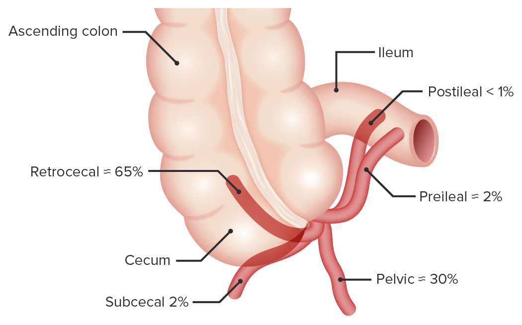
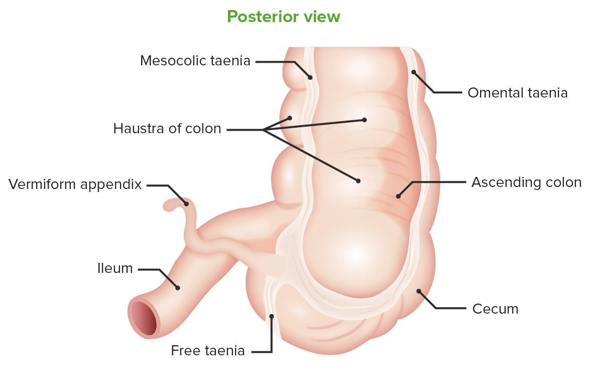
Slender, hollow, blind-ended pouch arising from the proximal cecumCecumThe blind sac or outpouching area of the large intestine that is below the entrance of the small intestine. It has a worm-like extension, the vermiform appendix.Colon, Cecum, and Appendix: Anatomy
On average, the appendixAppendixA worm-like blind tube extension from the cecum.Colon, Cecum, and Appendix: Anatomy is approximately 9 cm long, but it can vary from 2 to 22 cm.
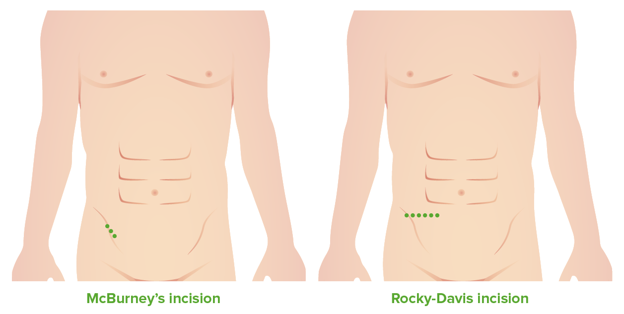
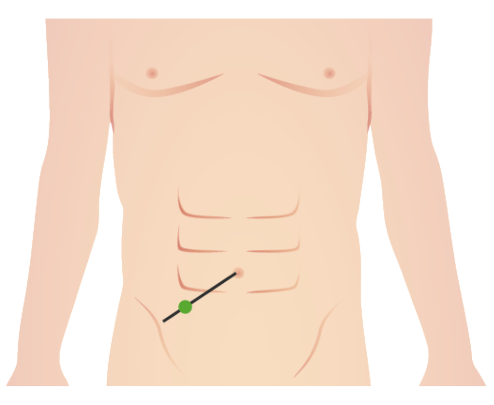
Localized tenderness at this point is a classic sign of appendicitisAppendicitisAppendicitis is the acute inflammation of the vermiform appendix and the most common abdominal surgical emergency globally. The condition has a lifetime risk of 8%. Characteristic features include periumbilical abdominal pain that migrates to the right lower quadrant, fever, anorexia, nausea, and vomiting.Appendicitis.
Both McBurney’s and Rocky-Davis incisions for open appendectomyOpen AppendectomyAppendectomy are performed through this point.
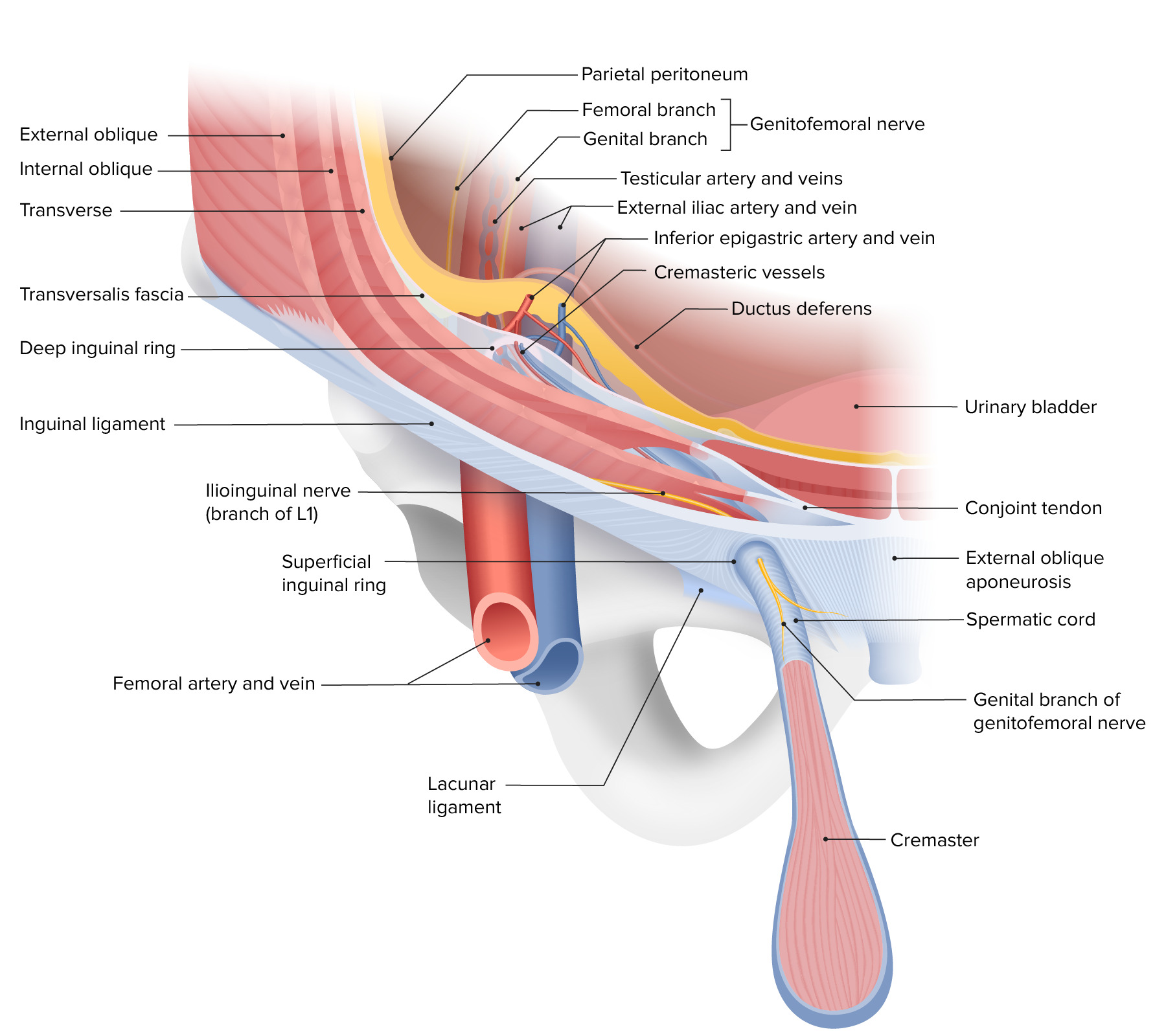
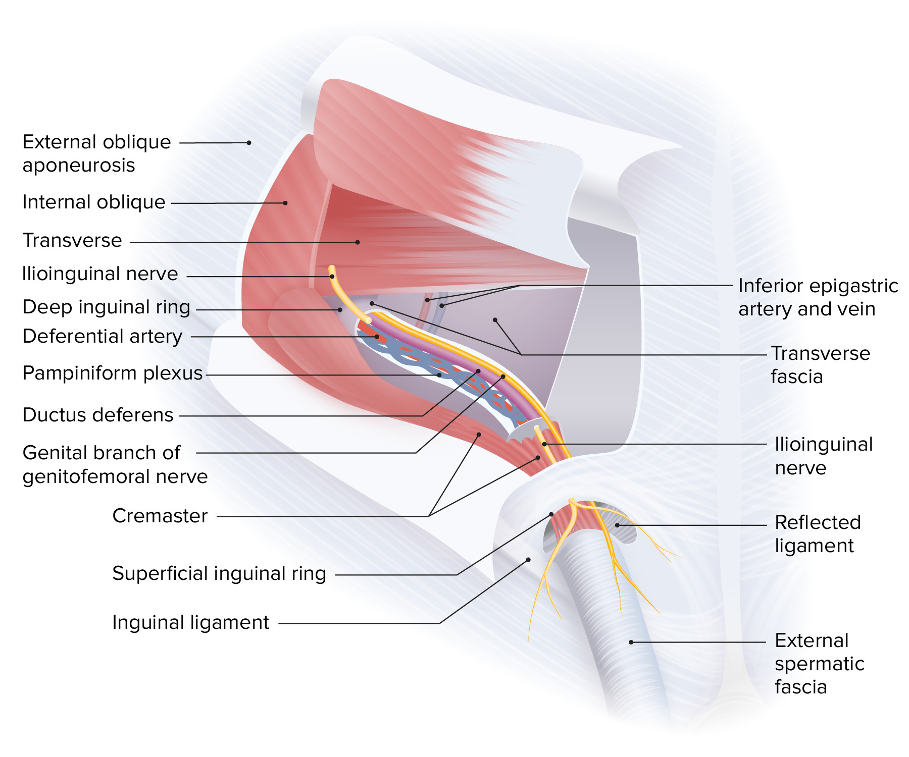
Superior: inferior edge of the internal obliqueInternal obliqueMuscles of the anterolateral abdominal wall consisting of the external oblique and the internal oblique muscles. The external abdominal oblique muscle fibers extend from lower thoracic ribs to the linea alba and the iliac crest. The internal abdominal oblique extend superomedially beneath the external oblique muscles.Anterior Abdominal Wall: Anatomy and transverse abdominal muscles
Anterior: aponeurosis of the abdominal external obliqueExternal obliqueMuscles of the anterolateral abdominal wall consisting of the external oblique and the internal oblique muscles. The external abdominal oblique muscle fibers extend from lower thoracic ribs to the linea alba and the iliac crest. The internal abdominal oblique extend superomedially beneath the external oblique muscles.Anterior Abdominal Wall: Anatomy muscle
Posterior: transversalis fasciaFasciaLayers of connective tissue of variable thickness. The superficial fascia is found immediately below the skin; the deep fascia invests muscles, nerves, and other organs.Cellulitis and interfoveolar ligament
Course:
The inguinal canalInguinal canalThe tunnel in the lower anterior abdominal wall through which the spermatic cord, in the male; round ligament, in the female; nerves; and vessels pass. Its internal end is at the deep inguinal ring and its external end is at the superficial inguinal ring.Inguinal Canal: Anatomy and Hernias is approximately 4 cm long, situated in the lower anterior abdominal wallAnterior abdominal wallThe anterior abdominal wall is anatomically delineated as a hexagonal area defined superiorly by the xiphoid process, laterally by the midaxillary lines, and inferiorly by the pubic symphysis.Anterior Abdominal Wall: Anatomy, connecting its outer and inner layers above the inguinal ligamentInguinal LigamentFemoral Region and Hernias: Anatomy (superior anterior iliac spineAnterior Iliac SpineAppendicitis to pubic tubercle).
Runs from an upper lateral to an inferior medial direction.
Inner orifice/deep inguinal ringDeep inguinal ringInguinal Canal: Anatomy and Hernias: evagination of the transversalis fasciaFasciaLayers of connective tissue of variable thickness. The superficial fascia is found immediately below the skin; the deep fascia invests muscles, nerves, and other organs.Cellulitis (surrounding the spermatic cordSpermatic CordEither of a pair of tubular structures formed by ductus deferens; arteries; veins; lymphatic vessels; and nerves. The spermatic cord extends from the deep inguinal ring through the inguinal canal to the testis in the scrotum.Testicles: Anatomy as the internal spermatic fasciaInternal spermatic fasciaInguinal Canal: Anatomy and Hernias)
External orifice/superficial inguinal ringSuperficial inguinal ringInguinal Canal: Anatomy and Hernias:fissureFissureA crack or split that extends into the dermisGeneralized and Localized Rashes in the aponeurosis of the abdominal external obliqueExternal obliqueMuscles of the anterolateral abdominal wall consisting of the external oblique and the internal oblique muscles. The external abdominal oblique muscle fibers extend from lower thoracic ribs to the linea alba and the iliac crest. The internal abdominal oblique extend superomedially beneath the external oblique muscles.Anterior Abdominal Wall: Anatomy muscle
The layers of the anterior abdominal wall, depicting the course of the inguinal canal and the composition of the deep and superficial inguinal rings
Spermatic cordSpermatic CordEither of a pair of tubular structures formed by ductus deferens; arteries; veins; lymphatic vessels; and nerves. The spermatic cord extends from the deep inguinal ring through the inguinal canal to the testis in the scrotum.Testicles: Anatomy (cremaster muscleCremaster muscleTesticles: Anatomy, internal and external spermatic fasciae surrounding the vas deferensVas DeferensThe excretory duct of the testes that carries spermatozoa. It rises from the scrotum and joins the seminal vesicles to form the ejaculatory duct.Testicles: Anatomy, pampiniform plexus, lymphatic vesselsLymphatic VesselsTubular vessels that are involved in the transport of lymph and lymphocytes.Lymphatic Drainage System: Anatomy, testicular neurovasculature, and tunica vaginalisTunica vaginalisTesticles: Anatomy)
Descending and sigmoidSigmoidA segment of the colon between the rectum and the descending colon.VolvuluscolonColonThe large intestines constitute the last portion of the digestive system. The large intestine consists of the cecum, appendix, colon (with ascending, transverse, descending, and sigmoid segments), rectum, and anal canal. The primary function of the colon is to remove water and compact the stool prior to expulsion from the body via the rectum and anal canal. Colon, Cecum, and Appendix: Anatomy
Suspended by mesenteryMesenteryA layer of the peritoneum which attaches the abdominal viscera to the abdominal wall and conveys their blood vessels and nerves.Peritoneum: Anatomy (sigmoidSigmoidA segment of the colon between the rectum and the descending colon.Volvulus mesocolon)
Arterial supply: inferior mesenteric arteryInferior mesenteric arteryThe artery supplying nearly all the left half of the transverse colon, the whole of the descending colon, the sigmoid colon, and the greater part of the rectum. It is smaller than the superior mesenteric artery and arises from the aorta above its bifurcation into the common iliac arteries.Small Intestine: Anatomy (sigmoidSigmoidA segment of the colon between the rectum and the descending colon.VolvulusarteriesArteriesArteries are tubular collections of cells that transport oxygenated blood and nutrients from the heart to the tissues of the body. The blood passes through the arteries in order of decreasing luminal diameter, starting in the largest artery (the aorta) and ending in the small arterioles. Arteries are classified into 3 types: large elastic arteries, medium muscular arteries, and small arteries and arterioles. Arteries: Histology)
Venous drainage: inferior mesenteric vein → portal veinPortal veinA short thick vein formed by union of the superior mesenteric vein and the splenic vein.Liver: Anatomy
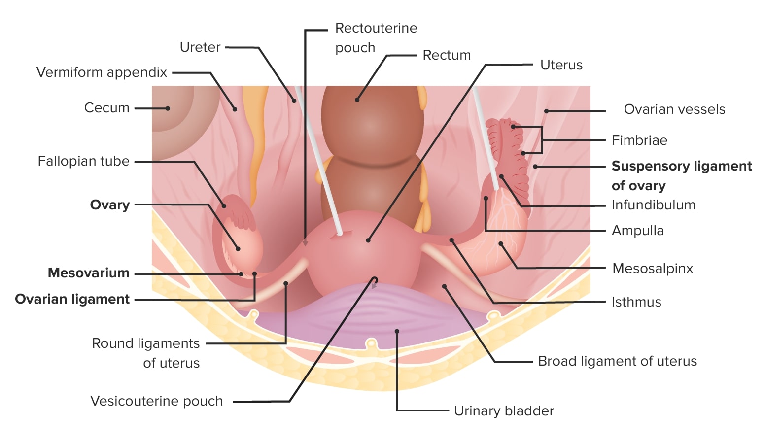
Fallopian tubesFallopian tubesThe uterus, cervix, and fallopian tubes are part of the internal female reproductive system. The fallopian tubes receive an ovum after ovulation and help move it and/or a fertilized embryo toward the uterus via ciliated cells lining the tubes and peristaltic movements of its smooth muscle. Uterus, Cervix, and Fallopian Tubes: Anatomy
InfundibulumInfundibulumUterus, Cervix, and Fallopian Tubes: Anatomy (opens into the peritoneal cavityPeritoneal CavityThe space enclosed by the peritoneum. It is divided into two portions, the greater sac and the lesser sac or omental bursa, which lies behind the stomach. The two sacs are connected by the foramen of winslow, or epiploic foramen.Peritoneum: Anatomy)
Runs from the uterusUterusThe uterus, cervix, and fallopian tubes are part of the internal female reproductive system. The uterus has a thick wall made of smooth muscle (the myometrium) and an inner mucosal layer (the endometrium). The most inferior portion of the uterus is the cervix, which connects the uterine cavity to the vagina.Uterus, Cervix, and Fallopian Tubes: Anatomy to the lateral pelvic wall
Contains the fallopian tubesFallopian tubesThe uterus, cervix, and fallopian tubes are part of the internal female reproductive system. The fallopian tubes receive an ovum after ovulation and help move it and/or a fertilized embryo toward the uterus via ciliated cells lining the tubes and peristaltic movements of its smooth muscle. Uterus, Cervix, and Fallopian Tubes: Anatomy
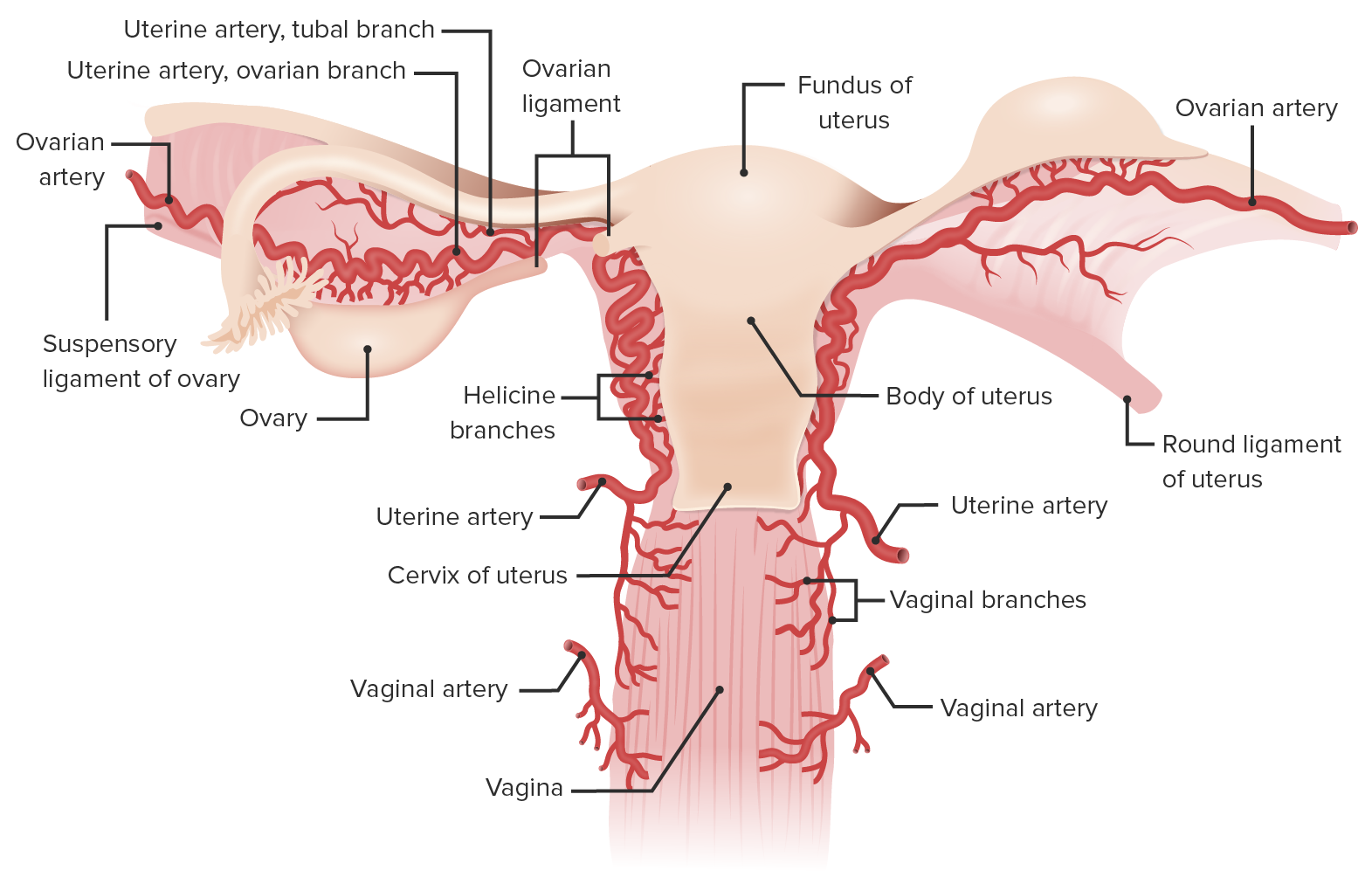
Arterial: ovarian and uterine arteriesArteriesArteries are tubular collections of cells that transport oxygenated blood and nutrients from the heart to the tissues of the body. The blood passes through the arteries in order of decreasing luminal diameter, starting in the largest artery (the aorta) and ending in the small arterioles. Arteries are classified into 3 types: large elastic arteries, medium muscular arteries, and small arteries and arterioles. Arteries: Histology
Venous supply is from ovarian veinsVeinsVeins are tubular collections of cells, which transport deoxygenated blood and waste from the capillary beds back to the heart. Veins are classified into 3 types: small veins/venules, medium veins, and large veins. Each type contains 3 primary layers: tunica intima, tunica media, and tunica adventitia. Veins: Histology:
The left ovarian vein drains into the left renal veinRenal veinShort thick veins which return blood from the kidneys to the vena cava.Glomerular Filtration.
Relevant procedures
AppendectomyAppendectomyAppendectomy is an invasive surgical procedure performed with the goal of resecting and extracting the vermiform appendix through either an open or a laparoscopic approach. The most common indication is acute appendicitis.Appendectomy: surgical removal of the vermiform appendixAppendixA worm-like blind tube extension from the cecum.Colon, Cecum, and Appendix: Anatomy, most commonly because of appendicitisAppendicitisAppendicitis is the acute inflammation of the vermiform appendix and the most common abdominal surgical emergency globally. The condition has a lifetime risk of 8%. Characteristic features include periumbilical abdominal pain that migrates to the right lower quadrant, fever, anorexia, nausea, and vomiting.Appendicitis
Inguinal herniorrhaphy: surgical repair of inguinal herniasInguinal HerniasAn abdominal hernia with an external bulge in the groin region. It can be classified by the location of herniation. Indirect inguinal hernias occur through the internal inguinal ring. Direct inguinal hernias occur through defects in the abdominal wall (transversalis fascia) in Hesselbach’s triangle. The former type is commonly seen in children and young adults; the latter in adults.Inguinal Canal: Anatomy and Hernias; can be done laparoscopically or via an open approach (Lichtenstein technique is the most common)
Salpingectomy: surgical removal of the fallopian tubeFallopian TubeA pair of highly specialized canals extending from the uterus to its corresponding ovary. They provide the means for ovum transport from the ovaries and they are the site of the ovum’s final maturation and fertilization. The fallopian tube consists of an interstitium, an isthmus, an ampulla, an infundibulum, and fimbriae. Its wall consists of three layers: serous, muscular, and an internal mucosal layer lined with both ciliated and secretory cells.Uterus, Cervix, and Fallopian Tubes: Anatomy, commonly performed to treat ectopic pregnancyEctopic pregnancyEctopic pregnancy refers to the implantation of a fertilized egg (embryo) outside the uterine cavity. The main cause is disruption of the normal anatomy of the fallopian tube. Ectopic Pregnancy
Salpingostomy: incising the fallopian tubeFallopian TubeA pair of highly specialized canals extending from the uterus to its corresponding ovary. They provide the means for ovum transport from the ovaries and they are the site of the ovum’s final maturation and fertilization. The fallopian tube consists of an interstitium, an isthmus, an ampulla, an infundibulum, and fimbriae. Its wall consists of three layers: serous, muscular, and an internal mucosal layer lined with both ciliated and secretory cells.Uterus, Cervix, and Fallopian Tubes: Anatomy open, commonly to remove an occupying lesion (ectopic pregnancyEctopic pregnancyEctopic pregnancy refers to the implantation of a fertilized egg (embryo) outside the uterine cavity. The main cause is disruption of the normal anatomy of the fallopian tube. Ectopic Pregnancy), leaving the remainder of the tube intact
SigmoidSigmoidA segment of the colon between the rectum and the descending colon.Volvulus colectomy: surgical removal of a sigmoidSigmoidA segment of the colon between the rectum and the descending colon.VolvuluscolonColonThe large intestines constitute the last portion of the digestive system. The large intestine consists of the cecum, appendix, colon (with ascending, transverse, descending, and sigmoid segments), rectum, and anal canal. The primary function of the colon is to remove water and compact the stool prior to expulsion from the body via the rectum and anal canal. Colon, Cecum, and Appendix: Anatomy, commonly performed for cancer or diverticulitisDiverticulitisInflammation of a diverticulum or diverticula.Diverticular Disease
Rectosigmoidectomy: surgical removal of the sigmoidSigmoidA segment of the colon between the rectum and the descending colon.VolvuluscolonColonThe large intestines constitute the last portion of the digestive system. The large intestine consists of the cecum, appendix, colon (with ascending, transverse, descending, and sigmoid segments), rectum, and anal canal. The primary function of the colon is to remove water and compact the stool prior to expulsion from the body via the rectum and anal canal. Colon, Cecum, and Appendix: Anatomy and rectumRectumThe rectum and anal canal are the most terminal parts of the lower GI tract/large intestine that form a functional unit and control defecation. Fecal continence is maintained by several important anatomic structures including rectal folds, anal valves, the sling-like puborectalis muscle, and internal and external anal sphincters. Rectum and Anal Canal: Anatomy
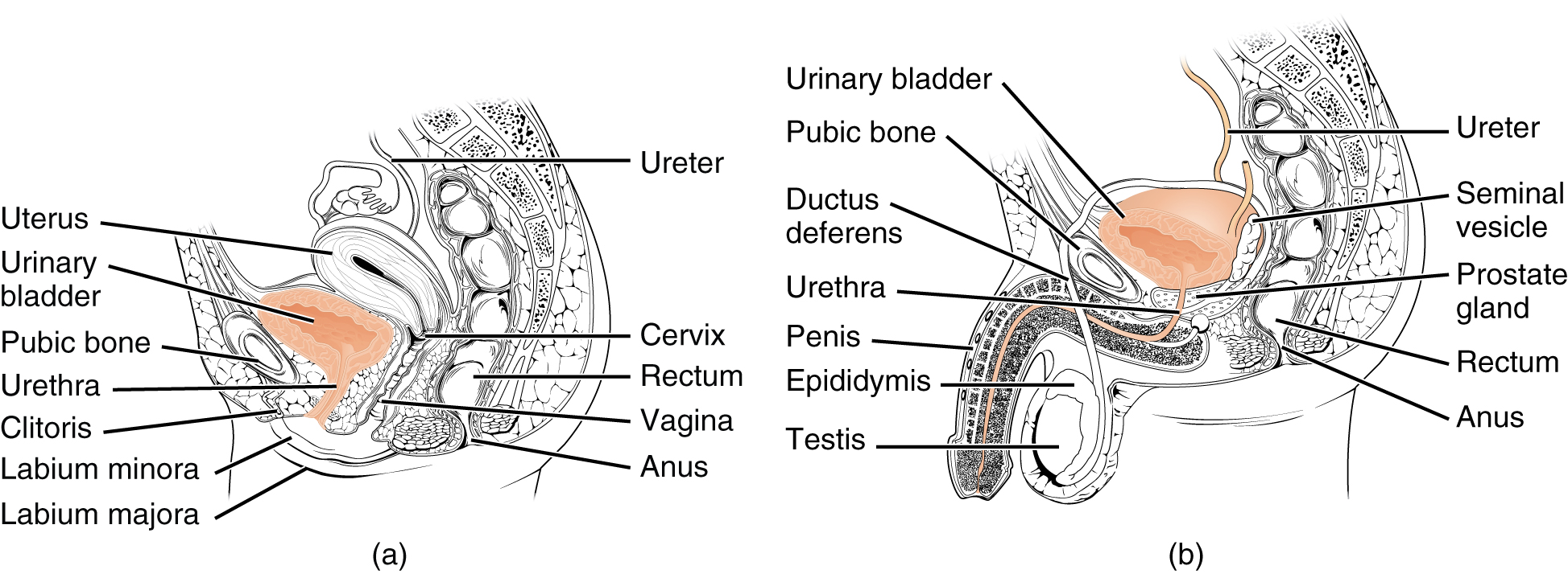
Urinary bladderUrinary BladderA musculomembranous sac along the urinary tract. Urine flows from the kidneys into the bladder via the ureters (ureter), and is held there until urination.Urinary Tract: Anatomy
In females, the urinary bladderUrinary BladderA musculomembranous sac along the urinary tract. Urine flows from the kidneys into the bladder via the ureters (ureter), and is held there until urination.Urinary Tract: Anatomy is anterior to the uterusUterusThe uterus, cervix, and fallopian tubes are part of the internal female reproductive system. The uterus has a thick wall made of smooth muscle (the myometrium) and an inner mucosal layer (the endometrium). The most inferior portion of the uterus is the cervix, which connects the uterine cavity to the vagina.Uterus, Cervix, and Fallopian Tubes: Anatomy and anterior vaginal wall.
In males, it is anterior to the rectumRectumThe rectum and anal canal are the most terminal parts of the lower GI tract/large intestine that form a functional unit and control defecation. Fecal continence is maintained by several important anatomic structures including rectal folds, anal valves, the sling-like puborectalis muscle, and internal and external anal sphincters. Rectum and Anal Canal: Anatomy.
Anchored by median umbilical ligament
Diagram of a sagittal cross section of a female (a) and male (b) pelvis: In the female pelvis, note the bladder’s close proximity to the anterior vaginal wall. In the male pelvis, note the bladder’s close proximity to the rectum.
Image: “The urethra transports urine from the bladder to the outside of the body” by OpenStax College. License: CC BY 4.0
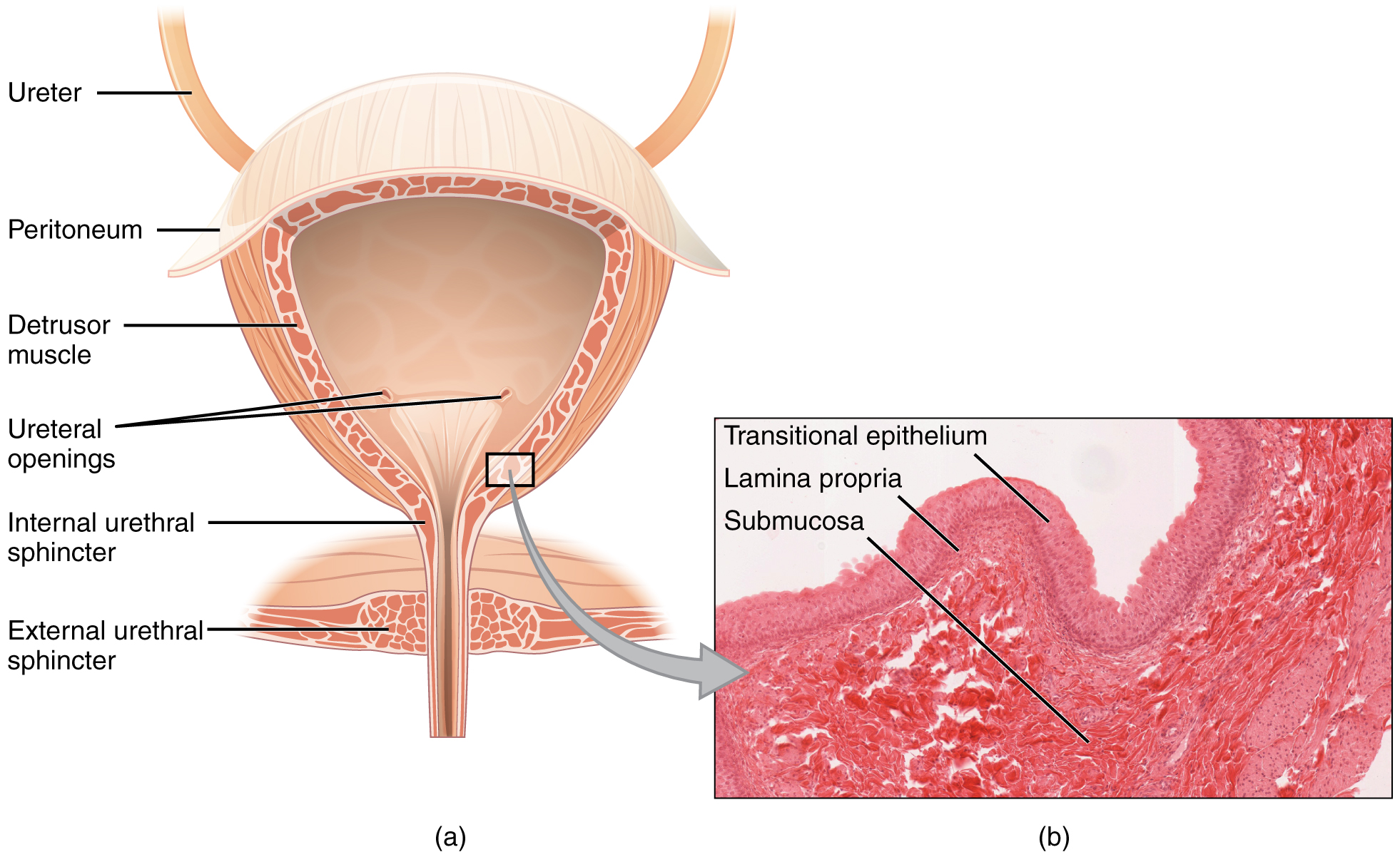
Coronal cross section of the bladder: The magnified histologic section (b) illustrates the innermost transitional epithelium, lamina propria, and submucosa.
Image: “Anterior cross section of the bladder” by OpenStax College. License: CC BY 4.0
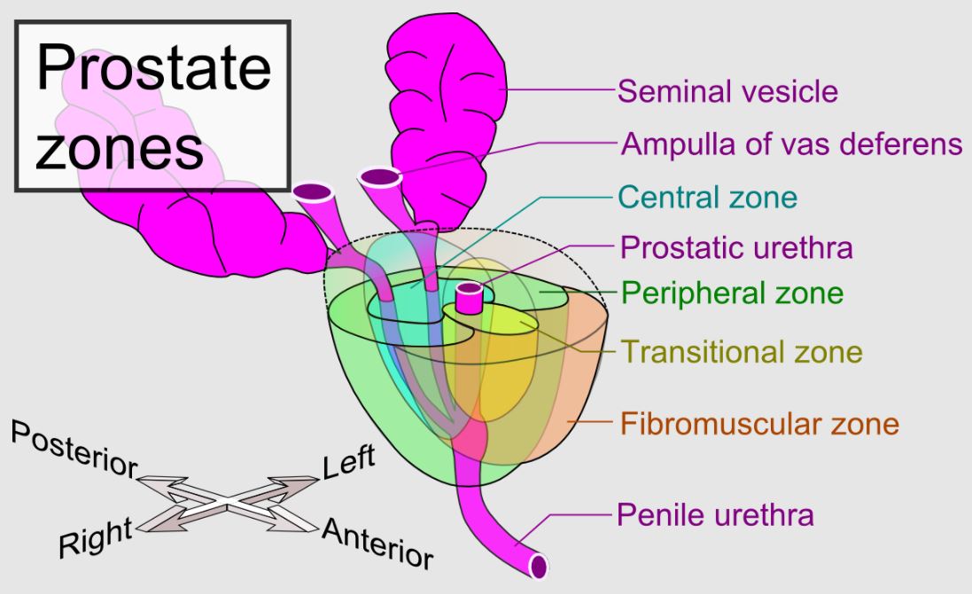
ProstateProstateThe prostate is a gland in the male reproductive system. The gland surrounds the bladder neck and a portion of the urethra. The prostate is an exocrine gland that produces a weakly acidic secretion, which accounts for roughly 20% of the seminal fluid.
Location:
Surrounding the neckNeckThe part of a human or animal body connecting the head to the rest of the body.Peritonsillar Abscess of the urinary bladderUrinary BladderA musculomembranous sac along the urinary tract. Urine flows from the kidneys into the bladder via the ureters (ureter), and is held there until urination.Urinary Tract: Anatomy
Posterior to urethraUrethraA tube that transports urine from the urinary bladder to the outside of the body in both the sexes. It also has a reproductive function in the male by providing a passage for sperm.Urinary Tract: Anatomy
Inferior to ejaculatory ductsEjaculatory DuctsPaired ducts in the human male through which semen is ejaculated into the urethra.
Lateral lobes: located on either side of the urethraUrethraA tube that transports urine from the urinary bladder to the outside of the body in both the sexes. It also has a reproductive function in the male by providing a passage for sperm.Urinary Tract: Anatomy
Median lobe: lies between the urethraUrethraA tube that transports urine from the urinary bladder to the outside of the body in both the sexes. It also has a reproductive function in the male by providing a passage for sperm.Urinary Tract: Anatomy and ejaculatory ductsEjaculatory DuctsPaired ducts in the human male through which semen is ejaculated into the urethra.
Related structures:
The urethraUrethraA tube that transports urine from the urinary bladder to the outside of the body in both the sexes. It also has a reproductive function in the male by providing a passage for sperm.Urinary Tract: Anatomy enters the prostateProstateThe prostate is a gland in the male reproductive system. The gland surrounds the bladder neck and a portion of the urethra. The prostate is an exocrine gland that produces a weakly acidic secretion, which accounts for roughly 20% of the seminal fluid. near its anterior border and usually passes between its anterior and middle thirds.
The ejaculatory ductsEjaculatory DuctsPaired ducts in the human male through which semen is ejaculated into the urethra. pass anteroinferiorly through its posterior region to open into the prostatic urethraProstatic urethra.
Bulbourethral glandsBulbourethral GlandsGlands situated on each side of the prostate that secrete a fluid component of the seminal fluid into the urethra. (Cowper’s glandsCowper’s glandsGlands situated on each side of the prostate that secrete a fluid component of the seminal fluid into the urethra.)
Prostate gland and main prostate zones: Peripheral, transitional, and central zones in relation to other structures of the male genitourinary system Note how the prostate is positioned around the prostatic urethra.
Image: “Prostate zones” by Mikael Häggström. License: CC0 1.0
UterusUterusThe uterus, cervix, and fallopian tubes are part of the internal female reproductive system. The uterus has a thick wall made of smooth muscle (the myometrium) and an inner mucosal layer (the endometrium). The most inferior portion of the uterus is the cervix, which connects the uterine cavity to the vagina.Uterus, Cervix, and Fallopian Tubes: Anatomy
Location:
7–9 cm long, found within the pelvisPelvisThe pelvis consists of the bony pelvic girdle, the muscular and ligamentous pelvic floor, and the pelvic cavity, which contains viscera, vessels, and multiple nerves and muscles. The pelvic girdle, composed of 2 “hip” bones and the sacrum, is a ring-like bony structure of the axial skeleton that links the vertebral column with the lower extremities.Pelvis: Anatomy, connected to the end of the vaginal canal via the uterine cervixCervixThe uterus, cervix, and fallopian tubes are part of the internal female reproductive system. The most inferior portion of the uterus is the cervix, which connects the uterine cavity to the vagina. Externally, the cervix is lined by stratified squamous cells; however, the cervical canal is lined by columnar epithelium.Uterus, Cervix, and Fallopian Tubes: Anatomy
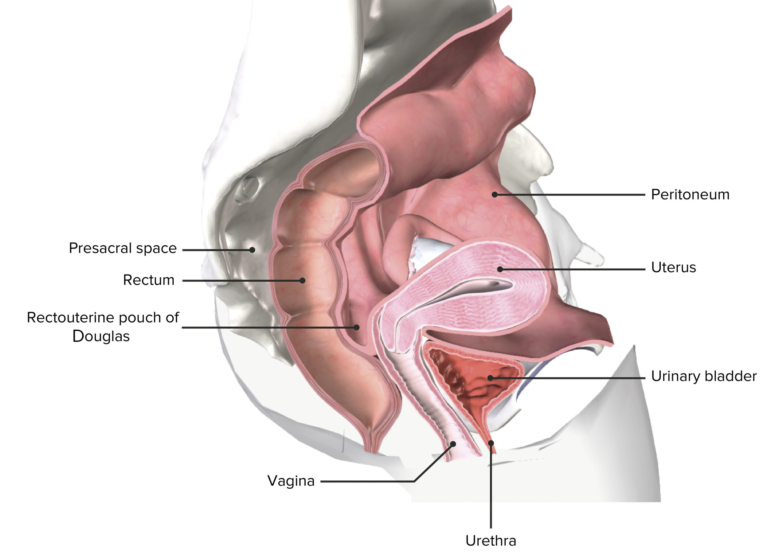
Dorsal to the urinary bladderUrinary BladderA musculomembranous sac along the urinary tract. Urine flows from the kidneys into the bladder via the ureters (ureter), and is held there until urination.Urinary Tract: Anatomy, anterior to the rectumRectumThe rectum and anal canal are the most terminal parts of the lower GI tract/large intestine that form a functional unit and control defecation. Fecal continence is maintained by several important anatomic structures including rectal folds, anal valves, the sling-like puborectalis muscle, and internal and external anal sphincters. Rectum and Anal Canal: Anatomy, in an anteverted and anteflexed position
Rectovaginal space or Douglas pouch: lowermost recess of the peritoneumPeritoneumThe peritoneum is a serous membrane lining the abdominopelvic cavity. This lining is formed by connective tissue and originates from the mesoderm. The membrane lines both the abdominal walls (as parietal peritoneum) and all of the visceral organs (as visceral peritoneum).Peritoneum: Anatomy, located between the uterusUterusThe uterus, cervix, and fallopian tubes are part of the internal female reproductive system. The uterus has a thick wall made of smooth muscle (the myometrium) and an inner mucosal layer (the endometrium). The most inferior portion of the uterus is the cervix, which connects the uterine cavity to the vagina.Uterus, Cervix, and Fallopian Tubes: Anatomy and rectumRectumThe rectum and anal canal are the most terminal parts of the lower GI tract/large intestine that form a functional unit and control defecation. Fecal continence is maintained by several important anatomic structures including rectal folds, anal valves, the sling-like puborectalis muscle, and internal and external anal sphincters. Rectum and Anal Canal: Anatomy
Vesicouterine pouch: shallower pouch between urinary bladderUrinary BladderA musculomembranous sac along the urinary tract. Urine flows from the kidneys into the bladder via the ureters (ureter), and is held there until urination.Urinary Tract: Anatomy and uterusUterusThe uterus, cervix, and fallopian tubes are part of the internal female reproductive system. The uterus has a thick wall made of smooth muscle (the myometrium) and an inner mucosal layer (the endometrium). The most inferior portion of the uterus is the cervix, which connects the uterine cavity to the vagina.Uterus, Cervix, and Fallopian Tubes: Anatomy
Structure:
FundusFundusThe superior portion of the body of the stomach above the level of the cardiac notch.Stomach: Anatomy: comprises all the uterusUterusThe uterus, cervix, and fallopian tubes are part of the internal female reproductive system. The uterus has a thick wall made of smooth muscle (the myometrium) and an inner mucosal layer (the endometrium). The most inferior portion of the uterus is the cervix, which connects the uterine cavity to the vagina.Uterus, Cervix, and Fallopian Tubes: Anatomy superior to the fallopian tubesFallopian tubesThe uterus, cervix, and fallopian tubes are part of the internal female reproductive system. The fallopian tubes receive an ovum after ovulation and help move it and/or a fertilized embryo toward the uterus via ciliated cells lining the tubes and peristaltic movements of its smooth muscle. Uterus, Cervix, and Fallopian Tubes: Anatomy
Body: region between the fundusFundusThe superior portion of the body of the stomach above the level of the cardiac notch.Stomach: Anatomy and cervixCervixThe uterus, cervix, and fallopian tubes are part of the internal female reproductive system. The most inferior portion of the uterus is the cervix, which connects the uterine cavity to the vagina. Externally, the cervix is lined by stratified squamous cells; however, the cervical canal is lined by columnar epithelium.Uterus, Cervix, and Fallopian Tubes: Anatomy
IsthmusIsthmusUterus, Cervix, and Fallopian Tubes: Anatomy: the narrowing at the cervixCervixThe uterus, cervix, and fallopian tubes are part of the internal female reproductive system. The most inferior portion of the uterus is the cervix, which connects the uterine cavity to the vagina. Externally, the cervix is lined by stratified squamous cells; however, the cervical canal is lined by columnar epithelium.Uterus, Cervix, and Fallopian Tubes: Anatomy–body transition
CervixCervixThe uterus, cervix, and fallopian tubes are part of the internal female reproductive system. The most inferior portion of the uterus is the cervix, which connects the uterine cavity to the vagina. Externally, the cervix is lined by stratified squamous cells; however, the cervical canal is lined by columnar epithelium.Uterus, Cervix, and Fallopian Tubes: Anatomy: comprised of the internal orifice (aperture to the uterine body), cervical canalCervical canalUterus, Cervix, and Fallopian Tubes: Anatomy, and the external orifice (vaginaVaginaThe vagina is the female genital canal, extending from the vulva externally to the cervix uteri internally. The structures have sexual, reproductive, and urinary functions and a rich blood supply, mainly arising from the internal iliac artery.Vagina, Vulva, and Pelvic Floor: Anatomy–cervical canalCervical canalUterus, Cervix, and Fallopian Tubes: Anatomy junction)
Superior view of the female pelvis depicting the uterus in situ, its supporting ligaments, and relation to the ovaries and neighboring organs
Round ligamentRound ligamentA fibromuscular band that attaches to the uterus and then passes along the broad ligament, out through the inguinal ring, and into the labium majus.Uterus, Cervix, and Fallopian Tubes: Anatomy (ligamentum teresLigamentum teresA cord-like remnant structure formed from the closed left fetal umbilical vein. It is located along the lower edge of the falciform ligament.Liver: Anatomy) runs from the tubouterine junction through the inguinal canalInguinal canalThe tunnel in the lower anterior abdominal wall through which the spermatic cord, in the male; round ligament, in the female; nerves; and vessels pass. Its internal end is at the deep inguinal ring and its external end is at the superficial inguinal ring.Inguinal Canal: Anatomy and Hernias to the labia majoraLabia majoraVagina, Vulva, and Pelvic Floor: Anatomy. Corresponds to the vas deferensVas DeferensThe excretory duct of the testes that carries spermatozoa. It rises from the scrotum and joins the seminal vesicles to form the ejaculatory duct.Testicles: Anatomy in men.
Cardinal ligamentCardinal ligamentUterus, Cervix, and Fallopian Tubes: Anatomy (ligamentum transversum cervicisLigamentum transversum cervicisUterus, Cervix, and Fallopian Tubes: Anatomy) consists of the transversalis fasciaFasciaLayers of connective tissue of variable thickness. The superficial fascia is found immediately below the skin; the deep fascia invests muscles, nerves, and other organs.Cellulitis connecting the cervixCervixThe uterus, cervix, and fallopian tubes are part of the internal female reproductive system. The most inferior portion of the uterus is the cervix, which connects the uterine cavity to the vagina. Externally, the cervix is lined by stratified squamous cells; however, the cervical canal is lined by columnar epithelium.Uterus, Cervix, and Fallopian Tubes: Anatomy with the pelvic wall.
Venous: Uterine veinsVeinsVeins are tubular collections of cells, which transport deoxygenated blood and waste from the capillary beds back to the heart. Veins are classified into 3 types: small veins/venules, medium veins, and large veins. Each type contains 3 primary layers: tunica intima, tunica media, and tunica adventitia. Veins: Histology drain into internal iliac veinsVeinsVeins are tubular collections of cells, which transport deoxygenated blood and waste from the capillary beds back to the heart. Veins are classified into 3 types: small veins/venules, medium veins, and large veins. Each type contains 3 primary layers: tunica intima, tunica media, and tunica adventitia. Veins: Histology.
Blood supply of the uterus and ovaries: Note the ovarian artery traveling along the ovarian suspensory ligament, supplying both the ovaries as well as the lateral ⅓ of the uterine tube. The ovarian artery continues in the mesosalpinx to anastomose with branches of the uterine artery.
Cystotomy: surgical incisionSurgical IncisionSurgical Site Infections performed on the urinary bladderUrinary BladderA musculomembranous sac along the urinary tract. Urine flows from the kidneys into the bladder via the ureters (ureter), and is held there until urination.Urinary Tract: Anatomy for repair (cystoplasty), bladderBladderA musculomembranous sac along the urinary tract. Urine flows from the kidneys into the bladder via the ureters, and is held there until urination.Pyelonephritis and Perinephric AbscesslacerationLacerationTorn, ragged, mangled wounds.Blunt Chest Trauma, or rupture due to trauma or a defect in the bladderBladderA musculomembranous sac along the urinary tract. Urine flows from the kidneys into the bladder via the ureters, and is held there until urination.Pyelonephritis and Perinephric Abscess wall
Cystectomy: surgical removal of the urinary bladderUrinary BladderA musculomembranous sac along the urinary tract. Urine flows from the kidneys into the bladder via the ureters (ureter), and is held there until urination.Urinary Tract: Anatomy, usually indicated by malignancyMalignancyHemothorax
Urostomy: surgically made communicationCommunicationThe exchange or transmission of ideas, attitudes, or beliefs between individuals or groups.Decision-making Capacity and Legal Competence between the bladderBladderA musculomembranous sac along the urinary tract. Urine flows from the kidneys into the bladder via the ureters, and is held there until urination.Pyelonephritis and Perinephric Abscess and skinSkinThe skin, also referred to as the integumentary system, is the largest organ of the body. The skin is primarily composed of the epidermis (outer layer) and dermis (deep layer). The epidermis is primarily composed of keratinocytes that undergo rapid turnover, while the dermis contains dense layers of connective tissue.Skin: Structure and Functions to facilitate evacuation of bladderBladderA musculomembranous sac along the urinary tract. Urine flows from the kidneys into the bladder via the ureters, and is held there until urination.Pyelonephritis and Perinephric Abscess contents
Abdominal hysterectomy: surgical removal of the uterusUterusThe uterus, cervix, and fallopian tubes are part of the internal female reproductive system. The uterus has a thick wall made of smooth muscle (the myometrium) and an inner mucosal layer (the endometrium). The most inferior portion of the uterus is the cervix, which connects the uterine cavity to the vagina.Uterus, Cervix, and Fallopian Tubes: Anatomy via an infraumbilical midline laparotomyMidline LaparotomyLaparotomy and Laparoscopy
Cesarean deliveryCesarean DeliveryCesarean delivery (CD) is the operative delivery of ≥ 1 infants through a surgical incision in the maternal abdomen and uterus. Cesarean deliveries may be indicated for a number of either maternal or fetal reasons, most commonly including fetal intolerance to labor, arrest of labor, a history of prior uterine surgery, fetal malpresentation, and placental abnormalities. Cesarean Delivery (CD): delivery of a fetus via a horizontal incision (Pfannenstiel or Joel-Cohen)
Suprapubic prostatectomyProstatectomyComplete or partial surgical removal of the prostate. Three primary approaches are commonly employed: suprapubic – removal through an incision above the pubis and through the urinary bladder; retropubic – as for suprapubic but without entering the urinary bladder; and transurethral (transurethral resection of prostate).Benign Prostatic Hyperplasia: surgical removal of the prostateProstateThe prostate is a gland in the male reproductive system. The gland surrounds the bladder neck and a portion of the urethra. The prostate is an exocrine gland that produces a weakly acidic secretion, which accounts for roughly 20% of the seminal fluid. via an infraumbilical midline laparotomyMidline LaparotomyLaparotomy and Laparoscopy