Mucormycosis is an angioinvasive fungal infection caused by multiple fungiFungiA kingdom of eukaryotic, heterotrophic organisms that live parasitically as saprobes, including mushrooms; yeasts; smuts, molds, etc. They reproduce either sexually or asexually, and have life cycles that range from simple to complex. Filamentous fungi, commonly known as molds, refer to those that grow as multicellular colonies.Mycology within the order, Mucorales. The fungiFungiA kingdom of eukaryotic, heterotrophic organisms that live parasitically as saprobes, including mushrooms; yeasts; smuts, molds, etc. They reproduce either sexually or asexually, and have life cycles that range from simple to complex. Filamentous fungi, commonly known as molds, refer to those that grow as multicellular colonies.Mycology are ubiquitous in the environment, but mucormycosis is very rare and almost always occurs in patientsPatientsIndividuals participating in the health care system for the purpose of receiving therapeutic, diagnostic, or preventive procedures.Clinician–Patient Relationship who are immunocompromisedimmunocompromisedA human or animal whose immunologic mechanism is deficient because of an immunodeficiency disorder or other disease or as the result of the administration of immunosuppressive drugs or radiation.Gastroenteritis. Inhalation of fungal sporesSporesThe reproductive elements of lower organisms, such as bacteria; fungi; and cryptogamic plants.Anthrax can cause rhinocerebral or pulmonary mucormycosis, direct inoculation can cause cutaneous mucormycosis, and ingestion can cause gastrointestinal mucormycosis. The clinical presentation results from fungal hyphaeHyphaeMicroscopic threadlike filaments in fungi that are filled with a layer of protoplasm. Collectively, the hyphae make up the mycelium.Mycology invading the blood vessels, causing thrombosisThrombosisFormation and development of a thrombus or blood clot in the blood vessel.Epidemic Typhus and tissue necrosisNecrosisThe death of cells in an organ or tissue due to disease, injury or failure of the blood supply.Ischemic Cell Damage. Diagnosis is confirmed with the identificationIdentificationDefense Mechanisms of the organism on histopathology of biopsyBiopsyRemoval and pathologic examination of specimens from the living body.Ewing Sarcoma specimens. PatientsPatientsIndividuals participating in the health care system for the purpose of receiving therapeutic, diagnostic, or preventive procedures.Clinician–Patient Relationship must be treated aggressively with antifungals and surgical resection of infected tissues.
Genera most commonly associated with human infection:
Rhizopus
Rhizomucor
Mucor
Cunninghamella
Lichtheimia (formerly Absidia)
Apophysomyces
Saksenaea
Morphology:
Colonies:
Fast growing
Cottony
White-to-yellow color → becomes gray
Microscopic features:
Wide hyphaeHyphaeMicroscopic threadlike filaments in fungi that are filled with a layer of protoplasm. Collectively, the hyphae make up the mycelium.Mycology
Lack of or rare septations
Branching at right angles
Sporangiophores:
Upright hyphaeHyphaeMicroscopic threadlike filaments in fungi that are filled with a layer of protoplasm. Collectively, the hyphae make up the mycelium.Mycology
Support sac-like sporangia filled with asexual sporangiospores
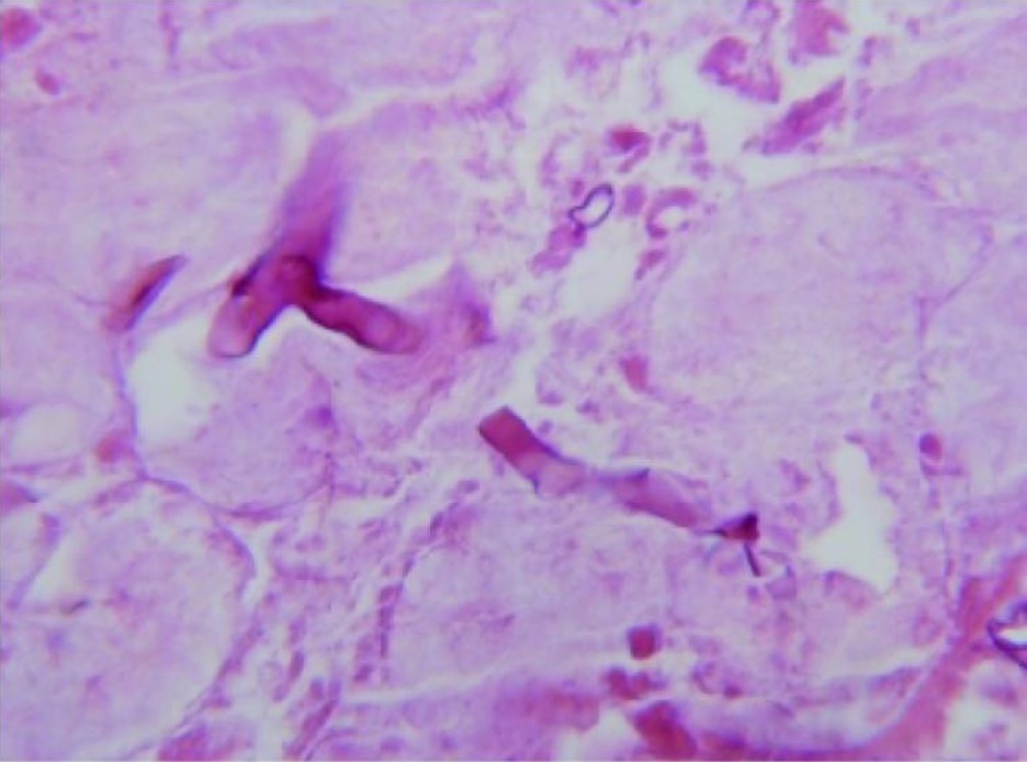
A microscopic view of the biopsy specimen shows several short, folded hyphae with nonseptate, broad and right-angle buddings, which are characteristic of mucormycosis.
Image: “Histopathological findings” by Baezzat SR et al. License: CC BY 2.5
Associated diseases
Mucormycosis is caused by many species within the Mucorales order, which can be classified based on the site of infection:
Rhino-orbital-cerebral (rhinocerebral) mucormycosis (most common in the United States)
Pulmonary mucormycosis
Cutaneous mucormycosis
Gastrointestinal mucormycosis
Epidemiology
Very rare infection; the true incidenceIncidenceThe number of new cases of a given disease during a given period in a specified population. It also is used for the rate at which new events occur in a defined population. It is differentiated from prevalence, which refers to all cases in the population at a given time.Measures of Disease Frequency is unknown.
Approximately 500 annual cases in the United States
IncidenceIncidenceThe number of new cases of a given disease during a given period in a specified population. It also is used for the rate at which new events occur in a defined population. It is differentiated from prevalence, which refers to all cases in the population at a given time.Measures of Disease Frequency is increasing due to a rising number of immunocompromisedimmunocompromisedA human or animal whose immunologic mechanism is deficient because of an immunodeficiency disorder or other disease or as the result of the administration of immunosuppressive drugs or radiation.GastroenteritispatientsPatientsIndividuals participating in the health care system for the purpose of receiving therapeutic, diagnostic, or preventive procedures.Clinician–Patient Relationship.
ReservoirReservoirAnimate or inanimate sources which normally harbor disease-causing organisms and thus serve as potential sources of disease outbreaks. Reservoirs are distinguished from vectors (disease vectors) and carriers, which are agents of disease transmission rather than continuing sources of potential disease outbreaks. Humans may serve both as disease reservoirs and carriers.Escherichia coli
Mucorales are common in the environment and are found on:
Decaying vegetation
Soil
Transmission
Inhalation of sporesSporesThe reproductive elements of lower organisms, such as bacteria; fungi; and cryptogamic plants.Anthrax (primary method)
Ingestion of contaminated food
SkinSkinThe skin, also referred to as the integumentary system, is the largest organ of the body. The skin is primarily composed of the epidermis (outer layer) and dermis (deep layer). The epidermis is primarily composed of keratinocytes that undergo rapid turnover, while the dermis contains dense layers of connective tissue.Skin: Structure and Functions inoculation
Host risk factors
Almost all infectionsInfectionsInvasion of the host organism by microorganisms or their toxins or by parasites that can cause pathological conditions or diseases.Chronic Granulomatous Disease occur in the presence of an underlying condition:
DiabetesDiabetesDiabetes mellitus (DM) is a metabolic disease characterized by hyperglycemia and dysfunction of the regulation of glucose metabolism by insulin. Type 1 DM is diagnosed mostly in children and young adults as the result of autoimmune destruction of β cells in the pancreas and the resulting lack of insulin. Type 2 DM has a significant association with obesity and is characterized by insulin resistance.Diabetes Mellitus (especially diabetic ketoacidosisKetoacidosisA life-threatening complication of diabetes mellitus, primarily of type 1 diabetes mellitus with severe insulin deficiency and extreme hyperglycemia. It is characterized by ketosis; dehydration; and depressed consciousness leading to coma.Metabolic Acidosis)
NeutropeniaNeutropeniaNeutrophils are an important component of the immune system and play a significant role in the eradication of infections. Low numbers of circulating neutrophils, referred to as neutropenia, predispose the body to recurrent infections or sepsis, though patients can also be asymptomatic. Neutropenia (neutrophilsNeutrophilsGranular leukocytes having a nucleus with three to five lobes connected by slender threads of chromatin, and cytoplasm containing fine inconspicuous granules and stainable by neutral dyes.Innate Immunity: Phagocytes and Antigen Presentation are the key host defense against the fungiFungiA kingdom of eukaryotic, heterotrophic organisms that live parasitically as saprobes, including mushrooms; yeasts; smuts, molds, etc. They reproduce either sexually or asexually, and have life cycles that range from simple to complex. Filamentous fungi, commonly known as molds, refer to those that grow as multicellular colonies.Mycology)
Iron-overload conditions (e.g., hemochromatosisHemochromatosisA disorder of iron metabolism characterized by a triad of hemosiderosis; liver cirrhosis; and diabetes mellitus. It is caused by massive iron deposits in parenchymal cells that may develop after a prolonged increase of iron absorption.Hereditary Hemochromatosis and deferoxamineDeferoxamineNatural product isolated from streptomyces pilosus. It forms iron complexes and is used as a chelating agent, particularly in the mesylate form.Hereditary Hemochromatosis therapy)
SkinSkinThe skin, also referred to as the integumentary system, is the largest organ of the body. The skin is primarily composed of the epidermis (outer layer) and dermis (deep layer). The epidermis is primarily composed of keratinocytes that undergo rapid turnover, while the dermis contains dense layers of connective tissue.Skin: Structure and Functions injury due to surgery, burnsBurnsA burn is a type of injury to the skin and deeper tissues caused by exposure to heat, electricity, chemicals, friction, or radiation. Burns are classified according to their depth as superficial (1st-degree), partial-thickness (2nd-degree), full-thickness (3rd-degree), and 4th-degree burns. Burns, or trauma
Use of injection drug
MalnutritionMalnutritionMalnutrition is a clinical state caused by an imbalance or deficiency of calories and/or micronutrients and macronutrients. The 2 main manifestations of acute severe malnutrition are marasmus (total caloric insufficiency) and kwashiorkor (protein malnutrition with characteristic edema).Malnutrition in children in resource-limited countries
Pathophysiology
Most sporesSporesThe reproductive elements of lower organisms, such as bacteria; fungi; and cryptogamic plants.Anthrax enter through the respiratory tract → adhere to mucus
Healthy individuals:
Usually clear by coughing, sneezingSneezingThe sudden, forceful, involuntary expulsion of air from the nose and mouth caused by irritation to the mucous membranes of the upper respiratory tract.Rhinovirus, or swallowingSwallowingThe act of taking solids and liquids into the gastrointestinal tract through the mouth and throat.Gastrointestinal Motility
NeutrophilsNeutrophilsGranular leukocytes having a nucleus with three to five lobes connected by slender threads of chromatin, and cytoplasm containing fine inconspicuous granules and stainable by neutral dyes.Innate Immunity: Phagocytes and Antigen Presentation phagocytize → destroy the fungus
Susceptible patientsPatientsIndividuals participating in the health care system for the purpose of receiving therapeutic, diagnostic, or preventive procedures.Clinician–Patient Relationship:
SporesSporesThe reproductive elements of lower organisms, such as bacteria; fungi; and cryptogamic plants.Anthrax transform into the hyphal form in nasal turbinatesTurbinatesThe scroll-like bony plates with curved margins on the lateral wall of the nasal cavity. Turbinates, also called nasal concha, increase the surface area of nasal cavity thus providing a mechanism for rapid warming and humidification of air as it passes to the lung.Nose Anatomy (External & Internal) or alveoliAlveoliSmall polyhedral outpouchings along the walls of the alveolar sacs, alveolar ducts and terminal bronchioles through the walls of which gas exchange between alveolar air and pulmonary capillary blood takes place.Acute Respiratory Distress Syndrome (ARDS).
HyphaeHyphaeMicroscopic threadlike filaments in fungi that are filled with a layer of protoplasm. Collectively, the hyphae make up the mycelium.Mycology invade blood vessels → tissue infarction and thrombosisThrombosisFormation and development of a thrombus or blood clot in the blood vessel.Epidemic Typhus
Other routes of infection → disease at those sites:
Traumatic inoculation or contamination → cutaneous disease
Ingestion → gastrointestinal disease
Clinical Presentation
Rhinocerebral disease
PatientsPatientsIndividuals participating in the health care system for the purpose of receiving therapeutic, diagnostic, or preventive procedures.Clinician–Patient Relationship start with symptoms of acute sinusitisSinusitisSinusitis refers to inflammation of the mucosal lining of the paranasal sinuses. The condition usually occurs concurrently with inflammation of the nasal mucosa (rhinitis), a condition known as rhinosinusitis. Acute sinusitis is due to an upper respiratory infection caused by a viral, bacterial, or fungal agent. Sinusitis. Symptoms progress due to the spread of the infection to contiguous structures.
FeverFeverFever is defined as a measured body temperature of at least 38°C (100.4°F). Fever is caused by circulating endogenous and/or exogenous pyrogens that increase levels of prostaglandin E2 in the hypothalamus. Fever is commonly associated with chills, rigors, sweating, and flushing of the skin. Fever
Nasal congestion (may have black discharge)
Unilateral retro-orbital headacheHeadacheThe symptom of pain in the cranial region. It may be an isolated benign occurrence or manifestation of a wide variety of headache disorders.Brain Abscess
Facial painFacial painPain in the facial region including orofacial pain and craniofacial pain. Associated conditions include local inflammatory and neoplastic disorders and neuralgic syndromes involving the trigeminal, facial, and glossopharyngeal nerves. Conditions which feature recurrent or persistent facial pain as the primary manifestation of disease are referred to as facial pain syndromes.Trigeminal Neuralgia
Facial cellulitisCellulitisCellulitis is a common infection caused by bacteria that affects the dermis and subcutaneous tissue of the skin. It is frequently caused by Staphylococcus aureus and Streptococcus pyogenes. The skin infection presents as an erythematous and edematous area with warmth and tenderness. Cellulitis
Eschar formation can occur on:
Nasal mucosaNasal mucosaThe mucous lining of the nasal cavity, including lining of the nostril (vestibule) and the olfactory mucosa. Nasal mucosa consists of ciliated cells, goblet cells, brush cells, small granule cells, basal cells (stem cells) and glands containing both mucous and serous cells.Nose Anatomy (External & Internal)
PalatePalateThe palate is the structure that forms the roof of the mouth and floor of the nasal cavity. This structure is divided into soft and hard palates. Palate: Anatomy
Overlying skinSkinThe skin, also referred to as the integumentary system, is the largest organ of the body. The skin is primarily composed of the epidermis (outer layer) and dermis (deep layer). The epidermis is primarily composed of keratinocytes that undergo rapid turnover, while the dermis contains dense layers of connective tissue.Skin: Structure and Functions
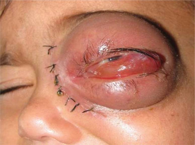
Swelling of the upper and lower lid in a patient with orbital involvement from mucormycosis
Image: “Swelling of upper and lower lid in the patient with mucormycosis” by Badiee P et al. License: CC BY 2.0
PatientsPatientsIndividuals participating in the health care system for the purpose of receiving therapeutic, diagnostic, or preventive procedures.Clinician–Patient Relationship with pulmonary diseasePulmonary diseaseDiseases involving the respiratory system.Blastomyces/Blastomycosis develop rapidly progressive pneumoniaPneumoniaPneumonia or pulmonary inflammation is an acute or chronic inflammation of lung tissue. Causes include infection with bacteria, viruses, or fungi. In more rare cases, pneumonia can also be caused through toxic triggers through inhalation of toxic substances, immunological processes, or in the course of radiotherapy.Pneumonia (often bilateral).
FeverFeverFever is defined as a measured body temperature of at least 38°C (100.4°F). Fever is caused by circulating endogenous and/or exogenous pyrogens that increase levels of prostaglandin E2 in the hypothalamus. Fever is commonly associated with chills, rigors, sweating, and flushing of the skin. Fever
DyspneaDyspneaDyspnea is the subjective sensation of breathing discomfort. Dyspnea is a normal manifestation of heavy physical or psychological exertion, but also may be caused by underlying conditions (both pulmonary and extrapulmonary). Dyspnea
Cough
HemoptysisHemoptysisHemoptysis is defined as the expectoration of blood originating in the lower respiratory tract. Hemoptysis is a consequence of another disease process and can be classified as either life threatening or non-life threatening. Hemoptysis can result in significant morbidity and mortality due to both drowning (reduced gas exchange as the lungs fill with blood) and hemorrhagic shock. Hemoptysis
Cutaneous disease
CellulitisCellulitisCellulitis is a common infection caused by bacteria that affects the dermis and subcutaneous tissue of the skin. It is frequently caused by Staphylococcus aureus and Streptococcus pyogenes. The skin infection presents as an erythematous and edematous area with warmth and tenderness. Cellulitis
Dermal necrosisNecrosisThe death of cells in an organ or tissue due to disease, injury or failure of the blood supply.Ischemic Cell Damage
Formation of black eschar
Gastrointestinal disease
The condition causes necrotic ulcers in the gastrointestinal tract (most commonly in the stomachStomachThe stomach is a muscular sac in the upper left portion of the abdomen that plays a critical role in digestion. The stomach develops from the foregut and connects the esophagus with the duodenum. Structurally, the stomach is C-shaped and forms a greater and lesser curvature and is divided grossly into regions: the cardia, fundus, body, and pylorus. Stomach: Anatomy), which can lead to perforationPerforationA pathological hole in an organ, blood vessel or other soft part of the body, occurring in the absence of external force.Esophagitis. Signs and symptoms include:
NauseaNauseaAn unpleasant sensation in the stomach usually accompanied by the urge to vomit. Common causes are early pregnancy, sea and motion sickness, emotional stress, intense pain, food poisoning, and various enteroviruses.Antiemetics
VomitingVomitingThe forcible expulsion of the contents of the stomach through the mouth.Hypokalemia
HematemesisHematemesisVomiting of blood that is either fresh bright red, or older ‘coffee-ground’ in character. It generally indicates bleeding of the upper gastrointestinal tract.Mallory-Weiss Syndrome (Mallory-Weiss Tear)
LethargyLethargyA general state of sluggishness, listless, or uninterested, with being tired, and having difficulty concentrating and doing simple tasks. It may be related to depression or drug addiction.Hyponatremia
HeadacheHeadacheThe symptom of pain in the cranial region. It may be an isolated benign occurrence or manifestation of a wide variety of headache disorders.Brain Abscess
Tissue necrosisNecrosisThe death of cells in an organ or tissue due to disease, injury or failure of the blood supply.Ischemic Cell Damage can be seen.
Imaging (e.g., head or chest CT) to assess the extent of infection and tissue damage
1,3-β-D-glucan is not useful (not a component of the Mucoralescell wallCell wallThe outermost layer of a cell in most plants; bacteria; fungi; and algae. The cell wall is usually a rigid structure that lies external to the cell membrane, and provides a protective barrier against physical or chemical agents.Cell Types: Eukaryotic versus Prokaryotic).
Management
Treat the underlying condition when possible (e.g., diabetic ketoacidosisKetoacidosisA life-threatening complication of diabetes mellitus, primarily of type 1 diabetes mellitus with severe insulin deficiency and extreme hyperglycemia. It is characterized by ketosis; dehydration; and depressed consciousness leading to coma.Metabolic Acidosis).
If possible, management should occur in a tertiary care center.
AntifungalAntifungalAzoles therapy should be started immediately:
Surgical resection of necrotic tissue is indicated to limitLimitA value (e.g., pressure or time) that should not be exceeded and which is specified by the operator to protect the lungInvasive Mechanical Ventilation further spread:
Associated with improved survival
Can lead to significant disfigurement
Comparison of Species
Table: Comparison of species
Organism
Mucorales
AspergillusAspergillusA genus of mitosporic fungi containing about 100 species and eleven different teleomorphs in the family trichocomaceae.Echinocandins
Characteristics
Wide hyphaeHyphaeMicroscopic threadlike filaments in fungi that are filled with a layer of protoplasm. Collectively, the hyphae make up the mycelium.Mycology
Lack of or rare septations
Branch at 90-degree angles
Septated hyphaeHyphaeMicroscopic threadlike filaments in fungi that are filled with a layer of protoplasm. Collectively, the hyphae make up the mycelium.Mycology
Branch at 45-degree angles
Transmission
Inhalation
Ingestion
Inoculation
Inhalation
Invasion through damaged skinSkinThe skin, also referred to as the integumentary system, is the largest organ of the body. The skin is primarily composed of the epidermis (outer layer) and dermis (deep layer). The epidermis is primarily composed of keratinocytes that undergo rapid turnover, while the dermis contains dense layers of connective tissue.Skin: Structure and Functions
ABPAABPAHypersensitivity reaction (allergic reaction) to fungus aspergillus in an individual with long-standing bronchial asthma. It is characterized by pulmonary infiltrates, eosinophilia, elevated serum immunoglobulin e, and skin reactivity to aspergillus antigen.Aspergillus/Aspergillosis
SinusitisSinusitisSinusitis refers to inflammation of the mucosal lining of the paranasal sinuses. The condition usually occurs concurrently with inflammation of the nasal mucosa (rhinitis), a condition known as rhinosinusitis. Acute sinusitis is due to an upper respiratory infection caused by a viral, bacterial, or fungal agent. Sinusitis
Invasive aspergillosisInvasive aspergillosisLung infections with the invasive forms of aspergillus, usually after surgery, transplantation, prolonged neutropenia or treatment with high-doses of corticosteroids. Invasive pulmonary aspergillosis can progress to chronic necrotizing pulmonary aspergillosis or hematogenous spread to other organs.Aspergillus/Aspergillosis
Bacterial orbital cellulitisOrbital cellulitisOrbital and preseptal cellulitis are infections differentiated by the anatomic sites affected in the orbit. Infection posterior to the septum is orbital cellulitis. Inoculation with the pathogen can occur through trauma or surgery. Cellulitis also occurs via extension from a nearby structure (such as from sinus infection or sinusitis). Orbital and Preseptal Cellulitis: infection of the orbital tissues, which can occur from hematogenousHematogenousHepatocellular Carcinoma (HCC) and Liver Metastases spread, extensionExtensionExamination of the Upper Limbs from adjacent sinuses, or traumatic inoculation. PatientsPatientsIndividuals participating in the health care system for the purpose of receiving therapeutic, diagnostic, or preventive procedures.Clinician–Patient Relationship present with swellingSwellingInflammation and rednessRednessInflammation, conjunctival erythemaErythemaRedness of the skin produced by congestion of the capillaries. This condition may result from a variety of disease processes.Chalazion, painPainAn unpleasant sensation induced by noxious stimuli which are detected by nerve endings of nociceptive neurons.Pain: Types and Pathways from eye movement, and proptosisProptosisRetinoblastoma. Diagnosis is clinical. The mainstay of treatment is antibiotic therapy and surgery is reserved for severe cases.
Cavernous sinus thrombosisCavernous sinus thrombosisFormation or presence of a blood clot (thrombus) in the cavernous sinus of the brain. Infections of the paranasal sinuses and adjacent structures, craniocerebral trauma, and thrombophilia are associated conditions. Clinical manifestations include dysfunction of cranial nerves III, IV, V, and VI, marked periorbital swelling, chemosis, fever, and visual loss.Cranial Nerve Palsies: a rare, life-threatening condition occurring from a facial infection. Cavernous sinus thrombosisCavernous sinus thrombosisFormation or presence of a blood clot (thrombus) in the cavernous sinus of the brain. Infections of the paranasal sinuses and adjacent structures, craniocerebral trauma, and thrombophilia are associated conditions. Clinical manifestations include dysfunction of cranial nerves III, IV, V, and VI, marked periorbital swelling, chemosis, fever, and visual loss.Cranial Nerve Palsies is usually bacterial in etiology. PatientsPatientsIndividuals participating in the health care system for the purpose of receiving therapeutic, diagnostic, or preventive procedures.Clinician–Patient Relationship present with feverFeverFever is defined as a measured body temperature of at least 38°C (100.4°F). Fever is caused by circulating endogenous and/or exogenous pyrogens that increase levels of prostaglandin E2 in the hypothalamus. Fever is commonly associated with chills, rigors, sweating, and flushing of the skin. Fever, headacheHeadacheThe symptom of pain in the cranial region. It may be an isolated benign occurrence or manifestation of a wide variety of headache disorders.Brain Abscess, proptosisProptosisRetinoblastoma, and ophthalmoplegiaOphthalmoplegiaParalysis of one or more of the ocular muscles due to disorders of the eye muscles, neuromuscular junction, supporting soft tissue, tendons, or innervation to the muscles.Orbital and Preseptal Cellulitis. Diagnosis is confirmed with CT or MRI. Management includes antibiotics and, occasionally, steroidsSteroidsA group of polycyclic compounds closely related biochemically to terpenes. They include cholesterol, numerous hormones, precursors of certain vitamins, bile acids, alcohols (sterols), and certain natural drugs and poisons. Steroids have a common nucleus, a fused, reduced 17-carbon atom ring system, cyclopentanoperhydrophenanthrene. Most steroids also have two methyl groups and an aliphatic side-chain attached to the nucleus.Benign Liver Tumors. AnticoagulationAnticoagulationPulmonary Hypertension Drugs is controversial.
Community-acquired pneumoniaCommunity-Acquired PneumoniaPneumonia in Children: infection of the lung parenchyma most often caused by a bacteriaBacteriaBacteria are prokaryotic single-celled microorganisms that are metabolically active and divide by binary fission. Some of these organisms play a significant role in the pathogenesis of diseases. Bacteriology or virusVirusViruses are infectious, obligate intracellular parasites composed of a nucleic acid core surrounded by a protein capsid. Viruses can be either naked (non-enveloped) or enveloped. The classification of viruses is complex and based on many factors, including type and structure of the nucleoid and capsid, the presence of an envelope, the replication cycle, and the host range. Virology. PatientsPatientsIndividuals participating in the health care system for the purpose of receiving therapeutic, diagnostic, or preventive procedures.Clinician–Patient Relationship present with feverFeverFever is defined as a measured body temperature of at least 38°C (100.4°F). Fever is caused by circulating endogenous and/or exogenous pyrogens that increase levels of prostaglandin E2 in the hypothalamus. Fever is commonly associated with chills, rigors, sweating, and flushing of the skin. Fever, dyspneaDyspneaDyspnea is the subjective sensation of breathing discomfort. Dyspnea is a normal manifestation of heavy physical or psychological exertion, but also may be caused by underlying conditions (both pulmonary and extrapulmonary). Dyspnea, and a productive cough. Chest X-rayX-rayPenetrating electromagnetic radiation emitted when the inner orbital electrons of an atom are excited and release radiant energy. X-ray wavelengths range from 1 pm to 10 nm. Hard x-rays are the higher energy, shorter wavelength x-rays. Soft x-rays or grenz rays are less energetic and longer in wavelength. The short wavelength end of the x-ray spectrum overlaps the gamma rays wavelength range. The distinction between gamma rays and x-rays is based on their radiation source.Pulmonary Function Tests typically shows lobar consolidationConsolidationPulmonary Function Tests. Management involves empiric antibiotics, which can be tailored if the causative organism is identified. Antivirals are used when a viral cause is suspected.
References
Richardson M. (2009). The ecology of the Zygomycetes and its impact on environmental exposure. Clin Microbiol Infect;15 Suppl 5:2–9. https://pubmed.ncbi.nlm.nih.gov/19754749/
Roden MM, Zaoutis TE, Buchanan WL, Knudsen TA, Sarkisova TA, Schaufele RL, et al. (2005). Epidemiology and outcome of zygomycosis: a review of 929 reported cases. Clin Infect Dis.; 41(5):634–53. https://pubmed.ncbi.nlm.nih.gov/16080086/
Kwon-Chung KJ. (2012). Taxonomy of fungi causing mucormycosis and entomophthoramycosis (zygomycosis) and nomenclature of the disease: molecular mycologic perspectives. Clin Infect Dis.; 54 Suppl 1:S8–S15. https://pubmed.ncbi.nlm.nih.gov/22247451/
Spellberg B, Edwards Jr. J, Ibrahim A. (2005). Novel perspectives on mucormycosis: pathophysiology, presentation, and management. Clin Microbiol Rev; 18(3):556–69. https://www.ncbi.nlm.nih.gov/pmc/articles/PMC1195964/
Song Y, Qiao J, Giovanni G, Liu G, Yang H, Wu J, Chen J. (2017). Mucormycosis in renal transplant recipients: a review of 174 reported cases. BMC Infect Dis.; 17(1):283. https://www.ncbi.nlm.nih.gov/pmc/articles/PMC5395857/
Andresen D, Donaldson A, Choo L, et al. (2005). Multifocal cutaneous mucormycosis complicating polymicrobial wound infections in a tsunami survivor from Sri Lanka. Lancet. 365(9462):876–8. https://pubmed.ncbi.nlm.nih.gov/15752532/
Francis JR, Villanueva P, Bryant P, Blyth CC. (2018). Mucormycosis in children: review and recommendations for management. J Pediatric Infect Dis Soc. 15;7(2):159–164. https://pubmed.ncbi.nlm.nih.gov/29294067/
Benlamkaddem, S., Zdaik, G., Doughmi, D., Bennis, A., Chraibi, F., Berdai, M. A., Abdellaoui, M., Benatiya Andaloussi, I., & Harandou, M. (2023). Rhino-orbital cerebral mucormycosis: A fatal evolution. Cureus, 15(4), e37837. https://doi.org/10.7759/cureus.37837
Create your free account or log in to continue reading!