Urticaria is raised, well-circumscribed areas (wheals) of edema (swelling) and erythema (redness) involving the dermis and epidermis with associated pruritus (itch). Urticaria is not a single disease but rather is a reaction pattern representing cutaneous mast cell degranulation resulting in the release of histamine and other vasoactive substances from mast cells and basophils in the dermis resulting in extravasation of plasma into the dermis. Urticaria can be caused by myriad inciting events, such as allergic reactions, infections, exposure, and many others. The diagnosis is made clinically. H1-antagonists are used as 1st-line treatment.
UrticariaUrticariaUrticaria is raised, well-circumscribed areas (wheals) of edema (swelling) and erythema (redness) involving the dermis and epidermis with associated pruritus (itch). Urticaria is not a single disease but rather is a reaction pattern representing cutaneous mast cell degranulation.Urticaria (Hives) is a vascular reaction of the skinSkinThe skin, also referred to as the integumentary system, is the largest organ of the body. The skin is primarily composed of the epidermis (outer layer) and dermis (deep layer). The epidermis is primarily composed of keratinocytes that undergo rapid turnover, while the dermis contains dense layers of connective tissue.Skin: Structure and Functions noted as a transient appearance of slightly elevated plaques (wheals) that are redder or paler than adjacent skinSkinThe skin, also referred to as the integumentary system, is the largest organ of the body. The skin is primarily composed of the epidermis (outer layer) and dermis (deep layer). The epidermis is primarily composed of keratinocytes that undergo rapid turnover, while the dermis contains dense layers of connective tissue.Skin: Structure and Functions and often accompanied by significant itching.
Epidemiology[1,4]
UrticariaUrticariaUrticaria is raised, well-circumscribed areas (wheals) of edema (swelling) and erythema (redness) involving the dermis and epidermis with associated pruritus (itch). Urticaria is not a single disease but rather is a reaction pattern representing cutaneous mast cell degranulation.Urticaria (Hives) is the most common dermatologic disorder seen in the ED.
Lifetime incidenceIncidenceThe number of new cases of a given disease during a given period in a specified population. It also is used for the rate at which new events occur in a defined population. It is differentiated from prevalence, which refers to all cases in the population at a given time.Measures of Disease Frequency of chronic urticariaChronic urticariaWheals and/or angioedema presented with daily symptoms lasting for more than 6 weeks. It may be classified into chronic spontaneous and chronic inducible urticaria depending on whether a specific trigger can be linked to the development of vascular reaction.Urticaria (Hives): 2%–3%.
UrticariaUrticariaUrticaria is raised, well-circumscribed areas (wheals) of edema (swelling) and erythema (redness) involving the dermis and epidermis with associated pruritus (itch). Urticaria is not a single disease but rather is a reaction pattern representing cutaneous mast cell degranulation.Urticaria (Hives) can occur in any age group.
Chronic urticariaChronic urticariaWheals and/or angioedema presented with daily symptoms lasting for more than 6 weeks. It may be classified into chronic spontaneous and chronic inducible urticaria depending on whether a specific trigger can be linked to the development of vascular reaction.Urticaria (Hives):
Signs and symptoms appearing 2–3 days per week
Duration > 6 weeks
More frequent in females (60%)
Chronic urticariaChronic urticariaWheals and/or angioedema presented with daily symptoms lasting for more than 6 weeks. It may be classified into chronic spontaneous and chronic inducible urticaria depending on whether a specific trigger can be linked to the development of vascular reaction.Urticaria (Hives) is more common between 40 and 60 years of age.
Etiology[1,7,11,13]
IgE-mediated urticarias (type I hypersensitivity, release of histamine from mast cellsMast cellsGranulated cells that are found in almost all tissues, most abundantly in the skin and the gastrointestinal tract. Like the basophils, mast cells contain large amounts of histamine and heparin. Unlike basophils, mast cells normally remain in the tissues and do not circulate in the blood. Mast cells, derived from the bone marrow stem cells, are regulated by the stem cell factor.Innate Immunity: Phagocytes and Antigen Presentation) occur within minutes to 2 hours after exposure. These are often due to exposure to certain allergens (list is not exhaustive):
Foods: fishFISHA type of in situ hybridization in which target sequences are stained with fluorescent dye so their location and size can be determined using fluorescence microscopy. This staining is sufficiently distinct that the hybridization signal can be seen both in metaphase spreads and in interphase nuclei.Chromosome Testing, shellfish, eggs, tree nuts, peanuts, milk, soy
Drugs: penicillinsPenicillinsBeta-lactam antibiotics contain a beta-lactam ring as a part of their chemical structure. Drugs in this class include penicillin G and V, penicillinase-sensitive and penicillinase-resistant penicillins, cephalosporins, carbapenems, and aztreonam. Penicillins, cephalosporinsCephalosporinsCephalosporins are a group of bactericidal beta-lactam antibiotics (similar to penicillins) that exert their effects by preventing bacteria from producing their cell walls, ultimately leading to cell death. Cephalosporins are categorized by generation and all drug names begin with “cef-” or “ceph-.” Cephalosporins
Insect venom: bee stings
Inhalants: dust mitesMitesAny arthropod of the subclass acari except the ticks. They are minute animals related to the spiders, usually having transparent or semitransparent bodies. They may be parasitic on humans and domestic animals, producing various irritations of the skin (mite infestations). Many mite species are important to human and veterinary medicine as both parasite and vector. Mites also infest plants.Scabies, pollens, molds, animal danders
Blood products
Latex
SkinSkinThe skin, also referred to as the integumentary system, is the largest organ of the body. The skin is primarily composed of the epidermis (outer layer) and dermis (deep layer). The epidermis is primarily composed of keratinocytes that undergo rapid turnover, while the dermis contains dense layers of connective tissue.Skin: Structure and Functions pressure, cold, or heatHeatInflammation
AtracuriumAtracuriumA non-depolarizing neuromuscular blocking agent with short duration of action. Its lack of significant cardiovascular effects and its lack of dependence on good kidney function for elimination provide clinical advantage over alternate non-depolarizing neuromuscular blocking agents.Neuromuscular Blockers
VecuroniumVecuroniumMonoquaternary homolog of pancuronium. A non-depolarizing neuromuscular blocking agent with shorter duration of action than pancuronium. Its lack of significant cardiovascular effects and lack of dependence on good kidney function for elimination as well as its short duration of action and easy reversibility provide advantages over, or alternatives to, other established neuromuscular blocking agents.Neuromuscular Blockers
SuccinylcholineSuccinylcholineA quaternary skeletal muscle relaxant usually used in the form of its bromide, chloride, or iodide. It is a depolarizing relaxant, acting in about 30 seconds and with a duration of effect averaging three to five minutes. Succinylcholine is used in surgical, anesthetic, and other procedures in which a brief period of muscle relaxation is called for.Cholinomimetic Drugs
Curare
VancomycinVancomycinAntibacterial obtained from streptomyces orientalis. It is a glycopeptide related to ristocetin that inhibits bacterial cell wall assembly and is toxic to kidneys and the inner ear.Glycopeptides
FluoroquinolonesFluoroquinolonesFluoroquinolones are a group of broad-spectrum, bactericidal antibiotics inhibiting bacterial DNA replication. Fluoroquinolones cover gram-negative, anaerobic, and atypical organisms, as well as some gram-positive and multidrug-resistant (MDR) organisms. Fluoroquinolones
Alcohol
Systemic disorders:
Serum sicknessSerum sicknessImmune complex disease caused by the administration of foreign serum or serum proteins and characterized by fever, lymphadenopathy, arthralgia, and urticaria. When they are complexed to protein carriers, some drugs can also cause serum sickness when they act as haptens inducing antibody responses.Type III Hypersensitivity Reaction
Cutaneous small vessel vasculitisVasculitisInflammation of any one of the blood vessels, including the arteries; veins; and rest of the vasculature system in the body.Systemic Lupus Erythematosus
Mastocytosis
Autoimmune diseasesAutoimmune diseasesDisorders that are characterized by the production of antibodies that react with host tissues or immune effector cells that are autoreactive to endogenous peptides.Selective IgA Deficiency (autoimmune thyroidThyroidThe thyroid gland is one of the largest endocrine glands in the human body. The thyroid gland is a highly vascular, brownish-red gland located in the visceral compartment of the anterior region of the neck.Thyroid Gland: Anatomy disease, rheumatoid arthritisArthritisAcute or chronic inflammation of joints.Osteoarthritis, systemic lupus erythematosusSystemic lupus erythematosusSystemic lupus erythematosus (SLE) is a chronic autoimmune, inflammatory condition that causes immune-complex deposition in organs, resulting in systemic manifestations. Women, particularly those of African American descent, are more commonly affected.Systemic Lupus Erythematosus)
Physical urticarias:
Occur as both acute and chronic forms
Wheals develop within minutes after exposure to physical factors:
Stroking/scratching skinSkinThe skin, also referred to as the integumentary system, is the largest organ of the body. The skin is primarily composed of the epidermis (outer layer) and dermis (deep layer). The epidermis is primarily composed of keratinocytes that undergo rapid turnover, while the dermis contains dense layers of connective tissue.Skin: Structure and Functions (dermographismDermographismDermatologic Examination)
IncidenceIncidenceThe number of new cases of a given disease during a given period in a specified population. It also is used for the rate at which new events occur in a defined population. It is differentiated from prevalence, which refers to all cases in the population at a given time.Measures of Disease Frequency: men = women
Wheals usually evolve and dissipate in < 24 hours.
In adults, urticariaUrticariaUrticaria is raised, well-circumscribed areas (wheals) of edema (swelling) and erythema (redness) involving the dermis and epidermis with associated pruritus (itch). Urticaria is not a single disease but rather is a reaction pattern representing cutaneous mast cell degranulation.Urticaria (Hives) is usually spontaneous/idiopathicIdiopathicDermatomyositis.
Can be spontaneous or induced by provoking stimuli (e.g., physical factors)
Chronic spontaneous urticariaUrticariaUrticaria is raised, well-circumscribed areas (wheals) of edema (swelling) and erythema (redness) involving the dermis and epidermis with associated pruritus (itch). Urticaria is not a single disease but rather is a reaction pattern representing cutaneous mast cell degranulation.Urticaria (Hives) is mainly idiopathicIdiopathicDermatomyositis but may have an underlying autoimmune cause.
Pathophysiology
Pathogenesis[9,14]
UrticariaUrticariaUrticaria is raised, well-circumscribed areas (wheals) of edema (swelling) and erythema (redness) involving the dermis and epidermis with associated pruritus (itch). Urticaria is not a single disease but rather is a reaction pattern representing cutaneous mast cell degranulation.Urticaria (Hives) is mediated by cutaneous mast cellsCutaneous Mast CellsUrticaria (Hives).
These mast cellsMast cellsGranulated cells that are found in almost all tissues, most abundantly in the skin and the gastrointestinal tract. Like the basophils, mast cells contain large amounts of histamine and heparin. Unlike basophils, mast cells normally remain in the tissues and do not circulate in the blood. Mast cells, derived from the bone marrow stem cells, are regulated by the stem cell factor.Innate Immunity: Phagocytes and Antigen Presentation release multiple mediators (histamine and other substances) in the superficial dermisDermisA layer of vascularized connective tissue underneath the epidermis. The surface of the dermis contains innervated papillae. Embedded in or beneath the dermis are sweat glands; hair follicles; and sebaceous glands.Skin: Structure and Functions.
When mast cellsMast cellsGranulated cells that are found in almost all tissues, most abundantly in the skin and the gastrointestinal tract. Like the basophils, mast cells contain large amounts of histamine and heparin. Unlike basophils, mast cells normally remain in the tissues and do not circulate in the blood. Mast cells, derived from the bone marrow stem cells, are regulated by the stem cell factor.Innate Immunity: Phagocytes and Antigen Presentation release vasoactive mediators in deeper dermisDermisA layer of vascularized connective tissue underneath the epidermis. The surface of the dermis contains innervated papillae. Embedded in or beneath the dermis are sweat glands; hair follicles; and sebaceous glands.Skin: Structure and Functions and subcutaneous tissues, angioedemaAngioedemaAngioedema is a localized, self-limited (but potentially life-threatening), nonpitting, asymmetrical edema occurring in the deep layers of the skin and mucosal tissue. The common underlying pathophysiology involves inflammatory mediators triggering significant vasodilation and increased capillary permeability. Angioedema results.
AngioedemaAngioedemaAngioedema is a localized, self-limited (but potentially life-threatening), nonpitting, asymmetrical edema occurring in the deep layers of the skin and mucosal tissue. The common underlying pathophysiology involves inflammatory mediators triggering significant vasodilation and increased capillary permeability. Angioedema that accompanies urticariaUrticariaUrticaria is raised, well-circumscribed areas (wheals) of edema (swelling) and erythema (redness) involving the dermis and epidermis with associated pruritus (itch). Urticaria is not a single disease but rather is a reaction pattern representing cutaneous mast cell degranulation.Urticaria (Hives) usually affects face, lipsLipsThe lips are the soft and movable most external parts of the oral cavity. The blood supply of the lips originates from the external carotid artery, and the innervation is through cranial nerves.Lips and Tongue: Anatomy, extremities, and genitalia.
Allergic IgE-mediated urticarias[7,9,14]
Generally related to an exogenous allergen (type I hypersensitivity reactionType I hypersensitivity reactionType I hypersensitivity reaction is an abnormal immune response triggered by exposure to specific antigens known as allergens. In this type of hypersensitivity reaction, the presentation of the antigen to the T-helper cells (Th cells) initiates a cascade of immunologic events leading to the production of antigen-specific IgE antibodies.Type I Hypersensitivity Reaction) or acute infection
Usually acute onset:
Immediate development (within minutes) of edematous, erythematous wheal-and-flare lesion
Evolve and dissipate in < 24 hours
Allergens bindBINDHyperbilirubinemia of the Newborn to IgE antibodiesIgE antibodiesAn immunoglobulin associated with mast cells. Overexpression has been associated with allergic hypersensitivity.Type I Hypersensitivity Reaction (formed in a prior sensitizing event) on mast FcFcCrystallizable fragments composed of the carboxy-terminal halves of both immunoglobulin heavy chains linked to each other by disulfide bonds. Fc fragments contain the carboxy-terminal parts of the heavy chain constant regions that are responsible for the effector functions of an immunoglobulin (complement fixation, binding to the cell membrane via fc receptors, and placental transport). This fragment can be obtained by digestion of immunoglobulins with the proteolytic enzyme papain.Immunoglobulins: Types and FunctionsreceptorsReceptorsReceptors are proteins located either on the surface of or within a cell that can bind to signaling molecules known as ligands (e.g., hormones) and cause some type of response within the cell.Receptors → release of vasoactive substances with extravasation of plasmaPlasmaThe residual portion of blood that is left after removal of blood cells by centrifugation without prior blood coagulation.Transfusion Products into the dermisDermisA layer of vascularized connective tissue underneath the epidermis. The surface of the dermis contains innervated papillae. Embedded in or beneath the dermis are sweat glands; hair follicles; and sebaceous glands.Skin: Structure and Functions
Histamine (acting predominantly on H1 and H2receptorsReceptorsReceptors are proteins located either on the surface of or within a cell that can bind to signaling molecules known as ligands (e.g., hormones) and cause some type of response within the cell.Receptors):
Smooth muscle contractionSmooth muscle contractionSmooth muscle is primarily found in the walls of hollow structures and some visceral organs, including the walls of the vasculature, GI, respiratory, and genitourinary tracts. Smooth muscle contracts more slowly and is regulated differently than skeletal muscle. Smooth muscle can be stimulated by nerve impulses, hormones, metabolic factors (like pH, CO2 or O2 levels), its own intrinsic pacemaker ability, or even mechanical stretch.Smooth Muscle Contraction → bronchospasmBronchospasmAsthma Drugs
Extravasation of blood into capillariesCapillariesCapillaries are the primary structures in the circulatory system that allow the exchange of gas, nutrients, and other materials between the blood and the extracellular fluid (ECF). Capillaries are the smallest of the blood vessels. Because a capillary diameter is so small, only 1 RBC may pass through at a time.Capillaries: Histology and vasodilationVasodilationThe physiological widening of blood vessels by relaxing the underlying vascular smooth muscle.Pulmonary Hypertension Drugs → erythemaErythemaRedness of the skin produced by congestion of the capillaries. This condition may result from a variety of disease processes.Chalazion
Increased vascular permeability and fluid shiftFluid ShiftTranslocation of body fluids from one compartment to another, such as from the vascular to the interstitial compartments. Fluid shifts are associated with profound changes in vascular permeability and water-electrolyte imbalance. The shift can also be from the lower body to the upper body as in conditions of weightlessness.Volume Depletion and Dehydration into interstitial space → edemaEdemaEdema is a condition in which excess serous fluid accumulates in the body cavity or interstitial space of connective tissues. Edema is a symptom observed in several medical conditions. It can be categorized into 2 types, namely, peripheral (in the extremities) and internal (in an organ or body cavity). Edema, wheals
VasodilationVasodilationThe physiological widening of blood vessels by relaxing the underlying vascular smooth muscle.Pulmonary Hypertension Drugs → hypotensionHypotensionHypotension is defined as low blood pressure, specifically < 90/60 mm Hg, and is most commonly a physiologic response. Hypotension may be mild, serious, or life threatening, depending on the cause. Hypotension
SensorySensoryNeurons which conduct nerve impulses to the central nervous system.Nervous System: Histology nerve activation → pruritusPruritusAn intense itching sensation that produces the urge to rub or scratch the skin to obtain relief.Atopic Dermatitis (Eczema)
Other vasoactive substances/effector molecules:
BradykininBradykininA nonapeptide messenger that is enzymatically produced from kallidin in the blood where it is a potent but short-lived agent of arteriolar dilation and increased capillary permeability. Bradykinin is also released from mast cells during asthma attacks, from gut walls as a gastrointestinal vasodilator, from damaged tissues as a pain signal, and may be a neurotransmitter.Hereditary Angioedema (C1 Esterase Inhibitor Deficiency)
Prostaglandin D2Prostaglandin D2The principal cyclooxygenase metabolite of arachidonic acid. It is released upon activation of mast cells and is also synthesized by alveolar macrophages. Among its many biological actions, the most important are its bronchoconstrictor, platelet-activating-factor-inhibitory, and cytotoxic effects.Eicosanoids
CytokinesCytokinesNon-antibody proteins secreted by inflammatory leukocytes and some non-leukocytic cells, that act as intercellular mediators. They differ from classical hormones in that they are produced by a number of tissue or cell types rather than by specialized glands. They generally act locally in a paracrine or autocrine rather than endocrine manner.Adaptive Immune Response (e.g., IL-4, IL-5, IL-6, TNF-α)
Platelet-activating factor (PAF)
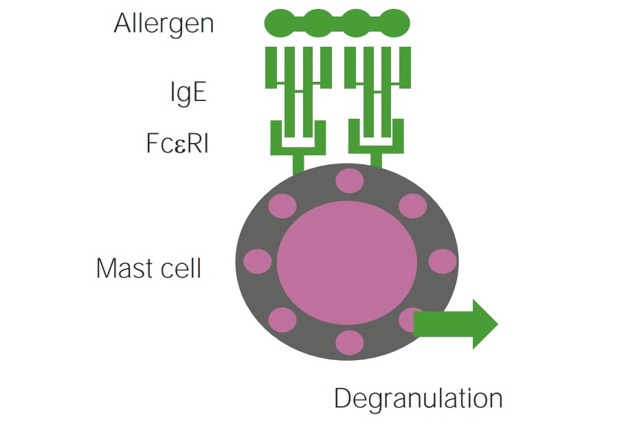
Mechanism of type 1 hypersensitivity reaction involved in IgE-mediated mast cell degranulation
Image by Lecturio.
Nonallergic urticariaUrticariaUrticaria is raised, well-circumscribed areas (wheals) of edema (swelling) and erythema (redness) involving the dermis and epidermis with associated pruritus (itch). Urticaria is not a single disease but rather is a reaction pattern representing cutaneous mast cell degranulation.Urticaria (Hives)[1,7,11,13]
No prior sensitization required
Mechanisms of non–IgE-mediated histamine release include direct mast cellMast cellGranulated cells that are found in almost all tissues, most abundantly in the skin and the gastrointestinal tract. Like the basophils, mast cells contain large amounts of histamine and heparin. Unlike basophils, mast cells normally remain in the tissues and do not circulate in the blood. Mast cells, derived from the bone marrow stem cells, are regulated by the stem cell factor.Angioedema degranulation via Mas-related G protein-coupled receptorReceptorReceptors are proteins located either on the surface of or within a cell that can bind to signaling molecules known as ligands (e.g., hormones) and cause some type of response within the cell.Receptors member X2 (MRGPRX2) receptorReceptorReceptors are proteins located either on the surface of or within a cell that can bind to signaling molecules known as ligands (e.g., hormones) and cause some type of response within the cell.Receptors (seen in drugs).
Other mechanisms include complement activationComplement ActivationThe sequential activation of serum complement proteins to create the complement membrane attack complex. Factors initiating complement activation include antigen-antibody complexes, microbial antigens, or cell surface polysaccharides.Systemic Lupus Erythematosus(in serum sicknessSerum sicknessImmune complex disease caused by the administration of foreign serum or serum proteins and characterized by fever, lymphadenopathy, arthralgia, and urticaria. When they are complexed to protein carriers, some drugs can also cause serum sickness when they act as haptens inducing antibody responses.Type III Hypersensitivity Reaction) and autoimmune disorders (seen in ~30–50% of chronic spontaneous urticariaUrticariaUrticaria is raised, well-circumscribed areas (wheals) of edema (swelling) and erythema (redness) involving the dermis and epidermis with associated pruritus (itch). Urticaria is not a single disease but rather is a reaction pattern representing cutaneous mast cell degranulation.Urticaria (Hives)).
Physical urticariaPhysical UrticariaUrticaria (Hives) is triggered by direct or local mast cellMast cellGranulated cells that are found in almost all tissues, most abundantly in the skin and the gastrointestinal tract. Like the basophils, mast cells contain large amounts of histamine and heparin. Unlike basophils, mast cells normally remain in the tissues and do not circulate in the blood. Mast cells, derived from the bone marrow stem cells, are regulated by the stem cell factor.Angioedema activation, possibly involving neuronal pathways.
EdemaEdemaEdema is a condition in which excess serous fluid accumulates in the body cavity or interstitial space of connective tissues. Edema is a symptom observed in several medical conditions. It can be categorized into 2 types, namely, peripheral (in the extremities) and internal (in an organ or body cavity). Edema
ErythemaErythemaRedness of the skin produced by congestion of the capillaries. This condition may result from a variety of disease processes.Chalazion
Well-demarcated edges
Central pallor
Surrounding red flare
Can be localized or generalized
VariableVariableVariables represent information about something that can change. The design of the measurement scales, or of the methods for obtaining information, will determine the data gathered and the characteristics of that data. As a result, a variable can be qualitative or quantitative, and may be further classified into subgroups.Types of Variables size and shape
Typically blanch with pressure
Wheals dissipate rapidly (< 24 hours).
PruritusPruritusAn intense itching sensation that produces the urge to rub or scratch the skin to obtain relief.Atopic Dermatitis (Eczema): most common associated symptom
AngioedemaAngioedemaAngioedema is a localized, self-limited (but potentially life-threatening), nonpitting, asymmetrical edema occurring in the deep layers of the skin and mucosal tissue. The common underlying pathophysiology involves inflammatory mediators triggering significant vasodilation and increased capillary permeability. Angioedema is simultaneously present in half the patientsPatientsIndividuals participating in the health care system for the purpose of receiving therapeutic, diagnostic, or preventive procedures.Clinician–Patient Relationship with urticariaUrticariaUrticaria is raised, well-circumscribed areas (wheals) of edema (swelling) and erythema (redness) involving the dermis and epidermis with associated pruritus (itch). Urticaria is not a single disease but rather is a reaction pattern representing cutaneous mast cell degranulation.Urticaria (Hives).
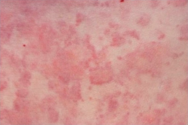
Urticarial rash
Image: “Urticarial rash at presentation” by Regional Immunology Service, Royal Hospitals, The Belfast Trust, Grosvenor Road, Belfast BT12 6BN. License: CC BY 2.0
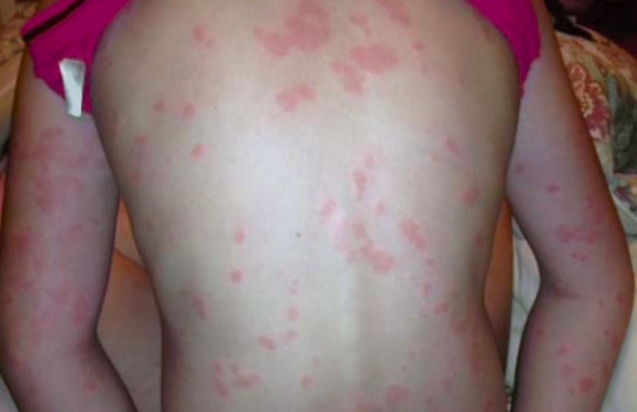
Urticarial rash
Image: “Chronic spontaneous urticaria” by University of Toronto, Medicine, Toronto, ON Canada. License: CC BY 4.0
Diagnosis
History and examination will usually lead to an accurate clinical classification.[8]
History[7,11,12]
Wheals:
Morphology, color, shape, size, pattern
Duration of onset and time to dissipation
Timing in relation to medication and other exposures
Cutaneous findings are often transient; therefore, photographsPhotographsUltrasound (Sonography) and patient documentationDocumentationSystematic organization, storage, retrieval, and dissemination of specialized information, especially of a scientific or technical nature. It often involves authenticating or validating information.Advance Directives may be helpful.
Associated symptoms:
PainPainAn unpleasant sensation induced by noxious stimuli which are detected by nerve endings of nociceptive neurons.Pain: Types and Pathways
PruritusPruritusAn intense itching sensation that produces the urge to rub or scratch the skin to obtain relief.Atopic Dermatitis (Eczema)
Systemic symptoms; headacheHeadacheThe symptom of pain in the cranial region. It may be an isolated benign occurrence or manifestation of a wide variety of headache disorders.Brain Abscess, joint painPainAn unpleasant sensation induced by noxious stimuli which are detected by nerve endings of nociceptive neurons.Pain: Types and Pathways, GI symptoms
Exposure:
Precipitating factors (see above)
Medication history
ComorbiditiesComorbiditiesThe presence of co-existing or additional diseases with reference to an initial diagnosis or with reference to the index condition that is the subject of study. Comorbidity may affect the ability of affected individuals to function and also their survival; it may be used as a prognostic indicator for length of hospital stay, cost factors, and outcome or survival.St. Louis Encephalitis Virus:
Autoimmune diseasesAutoimmune diseasesDisorders that are characterized by the production of antibodies that react with host tissues or immune effector cells that are autoreactive to endogenous peptides.Selective IgA Deficiency
Family and personal history:
UrticariaUrticariaUrticaria is raised, well-circumscribed areas (wheals) of edema (swelling) and erythema (redness) involving the dermis and epidermis with associated pruritus (itch). Urticaria is not a single disease but rather is a reaction pattern representing cutaneous mast cell degranulation.Urticaria (Hives)
Stroke skinSkinThe skin, also referred to as the integumentary system, is the largest organ of the body. The skin is primarily composed of the epidermis (outer layer) and dermis (deep layer). The epidermis is primarily composed of keratinocytes that undergo rapid turnover, while the dermis contains dense layers of connective tissue.Skin: Structure and Functions with blunt object (e.g. tongueTongueThe tongue, on the other hand, is a complex muscular structure that permits tasting and facilitates the process of mastication and communication. The blood supply of the tongue originates from the external carotid artery, and the innervation is through cranial nerves.Lips and Tongue: Anatomy depressor); wheals develop within minutes (dermographismDermographismDermatologic Examination)
Delayed pressure:
Application of sustained pressure with hanging weight or calibrated dermographometer at 100 g/mm² for 15 to 30 minutes
Development of urticariaUrticariaUrticaria is raised, well-circumscribed areas (wheals) of edema (swelling) and erythema (redness) involving the dermis and epidermis with associated pruritus (itch). Urticaria is not a single disease but rather is a reaction pattern representing cutaneous mast cell degranulation.Urticaria (Hives) within 24 hours
Core temperature:
Increase body temperatureBody TemperatureThe measure of the level of heat of a human or animal.Heatstroke by 1.0°C (by exercise on a treadmill/cycling or hot water bath for 15 to 30 minutes).
Rapid development of wheals (within 5 to 15 minutes)
Solar: development of wheals within minutes after sunlight exposure
Water: Apply water to skinSkinThe skin, also referred to as the integumentary system, is the largest organ of the body. The skin is primarily composed of the epidermis (outer layer) and dermis (deep layer). The epidermis is primarily composed of keratinocytes that undergo rapid turnover, while the dermis contains dense layers of connective tissue.Skin: Structure and Functions (wheals appear within 5–30 minutes).
VibrationVibrationA continuing periodic change in displacement with respect to a fixed reference.Neurological Examination: Apply a laboratory vortex to the forearmForearmThe forearm is the region of the upper limb between the elbow and the wrist. The term “forearm” is used in anatomy to distinguish this area from the arm, a term that is commonly used to describe the entire upper limb. The forearm consists of 2 long bones (the radius and the ulna), the interosseous membrane, and multiple arteries, nerves, and muscles. Forearm: Anatomy for 1–5 minutes.
Considerations in testing:
Avoid antihistaminesAntihistaminesAntihistamines are drugs that target histamine receptors, particularly H1 and H2 receptors. H1 antagonists are competitive and reversible inhibitors of H1 receptors. First-generation antihistamines cross the blood-brain barrier and can cause sedation. Antihistamines for 3–7 days before testing.
Baseline skinSkinThe skin, also referred to as the integumentary system, is the largest organ of the body. The skin is primarily composed of the epidermis (outer layer) and dermis (deep layer). The epidermis is primarily composed of keratinocytes that undergo rapid turnover, while the dermis contains dense layers of connective tissue.Skin: Structure and Functions, photograph results, and record onset/duration of wheals should be documented.
Always perform testing in a controlled medical environment in case of systemic reactions.
Not all physical urticarias have positive challenge tests (a negative test does not exclude the diagnosis).
Useful if an underlying allergic cause of urticariaUrticariaUrticaria is raised, well-circumscribed areas (wheals) of edema (swelling) and erythema (redness) involving the dermis and epidermis with associated pruritus (itch). Urticaria is not a single disease but rather is a reaction pattern representing cutaneous mast cell degranulation.Urticaria (Hives) is suspected. Broad, non-targeted allergyAllergyAn abnormal adaptive immune response that may or may not involve antigen-specific IgEType I Hypersensitivity Reaction panels are not advised. Testing should be guided by a detailed clinical history.
SkinSkinThe skin, also referred to as the integumentary system, is the largest organ of the body. The skin is primarily composed of the epidermis (outer layer) and dermis (deep layer). The epidermis is primarily composed of keratinocytes that undergo rapid turnover, while the dermis contains dense layers of connective tissue.Skin: Structure and Functions testing:
A drop of allergen extract is placed on the skinSkinThe skin, also referred to as the integumentary system, is the largest organ of the body. The skin is primarily composed of the epidermis (outer layer) and dermis (deep layer). The epidermis is primarily composed of keratinocytes that undergo rapid turnover, while the dermis contains dense layers of connective tissue.Skin: Structure and Functions (usually forearmForearmThe forearm is the region of the upper limb between the elbow and the wrist. The term “forearm” is used in anatomy to distinguish this area from the arm, a term that is commonly used to describe the entire upper limb. The forearm consists of 2 long bones (the radius and the ulna), the interosseous membrane, and multiple arteries, nerves, and muscles. Forearm: Anatomy or back), and a small prick introduces it into the epidermisEpidermisThe external, nonvascular layer of the skin. It is made up, from within outward, of five layers of epithelium: (1) basal layer (stratum basale epidermidis); (2) spinous layer (stratum spinosum epidermidis); (3) granular layer (stratum granulosum epidermidis); (4) clear layer (stratum lucidum epidermidis); and (5) horny layer (stratum corneum epidermidis).Skin: Structure and Functions.
Allergen-specific IgEIgEAn immunoglobulin associated with mast cells. Overexpression has been associated with allergic hypersensitivity.Immunoglobulins: Types and Functions blood tests:
Originally measured using radioallergosorbent test (RAST)
Replaced by more sensitive fluorescence immunoassaysFluorescence immunoassaysThe use of fluorescence spectrometry to obtain quantitative results for the fluorescent antibody technique. One advantage over the other methods (e.g., radioimmunoassay) is its extreme sensitivity, with a detection limit on the order of tenths of microgram/liter.Immunoassays (e.g., ImmunoCAP)
Can be used while patient is on antihistaminesAntihistaminesAntihistamines are drugs that target histamine receptors, particularly H1 and H2 receptors. H1 antagonists are competitive and reversible inhibitors of H1 receptors. First-generation antihistamines cross the blood-brain barrier and can cause sedation. Antihistamines
Laboratory studies[6,7,12,13,15]
Routine laboratory tests are not necessary for simple, acute urticariaAcute UrticariaUrticaria (Hives). However, they should be performed for cases of chronic urticariaChronic urticariaWheals and/or angioedema presented with daily symptoms lasting for more than 6 weeks. It may be classified into chronic spontaneous and chronic inducible urticaria depending on whether a specific trigger can be linked to the development of vascular reaction.Urticaria (Hives) and/or if systemic symptoms are present. Testing should be guided by the clinical presentation but may include:
CBC with differential (urticarial vasculitisVasculitisInflammation of any one of the blood vessels, including the arteries; veins; and rest of the vasculature system in the body.Systemic Lupus Erythematosus, autoinflammatory disease)
Serum cryoglobulins (cold urticariaUrticariaUrticaria is raised, well-circumscribed areas (wheals) of edema (swelling) and erythema (redness) involving the dermis and epidermis with associated pruritus (itch). Urticaria is not a single disease but rather is a reaction pattern representing cutaneous mast cell degranulation.Urticaria (Hives)):
Rarely present
More commonly associated with infection or hematologic disease[12]
Total IgEIgEAn immunoglobulin associated with mast cells. Overexpression has been associated with allergic hypersensitivity.Immunoglobulins: Types and Functions
Anti-IgE, anti-FcεRI antibodiesAntibodiesImmunoglobulins (Igs), also known as antibodies, are glycoprotein molecules produced by plasma cells that act in immune responses by recognizing and binding particular antigens. The various Ig classes are IgG (the most abundant), IgM, IgE, IgD, and IgA, which differ in their biologic features, structure, target specificity, and distribution.Immunoglobulins: Types and Functions, and autologous serum skinSkinThe skin, also referred to as the integumentary system, is the largest organ of the body. The skin is primarily composed of the epidermis (outer layer) and dermis (deep layer). The epidermis is primarily composed of keratinocytes that undergo rapid turnover, while the dermis contains dense layers of connective tissue.Skin: Structure and Functions test (ASST):
Investigational studies for autoimmune urticariaUrticariaUrticaria is raised, well-circumscribed areas (wheals) of edema (swelling) and erythema (redness) involving the dermis and epidermis with associated pruritus (itch). Urticaria is not a single disease but rather is a reaction pattern representing cutaneous mast cell degranulation.Urticaria (Hives)
Not routinely done, but may be considered for chronic spontaneous urticariaUrticariaUrticaria is raised, well-circumscribed areas (wheals) of edema (swelling) and erythema (redness) involving the dermis and epidermis with associated pruritus (itch). Urticaria is not a single disease but rather is a reaction pattern representing cutaneous mast cell degranulation.Urticaria (Hives) that does not respond to treatment (if available)
Management for urticariaUrticariaUrticaria is raised, well-circumscribed areas (wheals) of edema (swelling) and erythema (redness) involving the dermis and epidermis with associated pruritus (itch). Urticaria is not a single disease but rather is a reaction pattern representing cutaneous mast cell degranulation.Urticaria (Hives) may vary depending on practice location. The following information is based on US, UK, and European recommendations for urticariaUrticariaUrticaria is raised, well-circumscribed areas (wheals) of edema (swelling) and erythema (redness) involving the dermis and epidermis with associated pruritus (itch). Urticaria is not a single disease but rather is a reaction pattern representing cutaneous mast cell degranulation.Urticaria (Hives) without anaphylaxisAnaphylaxisAn acute hypersensitivity reaction due to exposure to a previously encountered antigen. The reaction may include rapidly progressing urticaria, respiratory distress, vascular collapse, systemic shock, and death.Type I Hypersensitivity Reaction or angioedemaAngioedemaAngioedema is a localized, self-limited (but potentially life-threatening), nonpitting, asymmetrical edema occurring in the deep layers of the skin and mucosal tissue. The common underlying pathophysiology involves inflammatory mediators triggering significant vasodilation and increased capillary permeability. Angioedema (these are discussed separately) in adults. See your local guidelines for additional information.
General measures
Supportive management:[7,8,10,12,15]
About ⅔ of acute new-onset urticarias will resolve spontaneously and do not require medications.
Avoid triggers.
Apply topical cooling moisturizer or antipruritic agents to reduce itching (e.g., menthol-containing emollient)
For chronic urticariaChronic urticariaWheals and/or angioedema presented with daily symptoms lasting for more than 6 weeks. It may be classified into chronic spontaneous and chronic inducible urticaria depending on whether a specific trigger can be linked to the development of vascular reaction.Urticaria (Hives), consider using appropriate validated scoring systems to assess disease activity and impact:
Dermatology Life QualityQualityActivities and programs intended to assure or improve the quality of care in either a defined medical setting or a program. The concept includes the assessment or evaluation of the quality of care; identification of problems or shortcomings in the delivery of care; designing activities to overcome these deficiencies; and follow-up monitoring to ensure effectiveness of corrective steps.Quality Measurement and Improvement Index (DLQI)
Weekly UrticariaUrticariaUrticaria is raised, well-circumscribed areas (wheals) of edema (swelling) and erythema (redness) involving the dermis and epidermis with associated pruritus (itch). Urticaria is not a single disease but rather is a reaction pattern representing cutaneous mast cell degranulation.Urticaria (Hives) Activity Score 7 (UAS7)
AngioedemaAngioedemaAngioedema is a localized, self-limited (but potentially life-threatening), nonpitting, asymmetrical edema occurring in the deep layers of the skin and mucosal tissue. The common underlying pathophysiology involves inflammatory mediators triggering significant vasodilation and increased capillary permeability. Angioedema Activity Score (AAS)
UrticariaUrticariaUrticaria is raised, well-circumscribed areas (wheals) of edema (swelling) and erythema (redness) involving the dermis and epidermis with associated pruritus (itch). Urticaria is not a single disease but rather is a reaction pattern representing cutaneous mast cell degranulation.Urticaria (Hives) Control Test (UCT)
Provide educational material or a patient information leaflet on urticariaUrticariaUrticaria is raised, well-circumscribed areas (wheals) of edema (swelling) and erythema (redness) involving the dermis and epidermis with associated pruritus (itch). Urticaria is not a single disease but rather is a reaction pattern representing cutaneous mast cell degranulation.Urticaria (Hives)
Referral:[7,8]
Who to refer:
PatientsPatientsIndividuals participating in the health care system for the purpose of receiving therapeutic, diagnostic, or preventive procedures.Clinician–Patient Relationship with chronic urticariaChronic urticariaWheals and/or angioedema presented with daily symptoms lasting for more than 6 weeks. It may be classified into chronic spontaneous and chronic inducible urticaria depending on whether a specific trigger can be linked to the development of vascular reaction.Urticaria (Hives) that fails to respond to maximum-dose 2nd-generation antihistaminesAntihistaminesAntihistamines are drugs that target histamine receptors, particularly H1 and H2 receptors. H1 antagonists are competitive and reversible inhibitors of H1 receptors. First-generation antihistamines cross the blood-brain barrier and can cause sedation. Antihistamines (taken regularly for 4 weeks)
PatientsPatientsIndividuals participating in the health care system for the purpose of receiving therapeutic, diagnostic, or preventive procedures.Clinician–Patient Relationship with suspected allergic cause
PatientsPatientsIndividuals participating in the health care system for the purpose of receiving therapeutic, diagnostic, or preventive procedures.Clinician–Patient Relationship with acute urticariaAcute UrticariaUrticaria (Hives) and persistent symptoms despite therapy (> 1‒2 weeks)
Preferred 1st option for urticariaUrticariaUrticaria is raised, well-circumscribed areas (wheals) of edema (swelling) and erythema (redness) involving the dermis and epidermis with associated pruritus (itch). Urticaria is not a single disease but rather is a reaction pattern representing cutaneous mast cell degranulation.Urticaria (Hives)
Options:
LoratadineLoratadineA second-generation histamine h1 receptor antagonist used in the treatment of allergic rhinitis and urticaria. Unlike most classical antihistamines (histamine h1 antagonists) it lacks central nervous system depressing effects such as drowsiness.Antihistamines (10 mg once daily)
CetirizineCetirizineA potent second-generation histamine h1 antagonist that is effective in the treatment of allergic rhinitis, chronic urticaria, and pollen-induced asthma. Unlike many traditional antihistamines, it does not cause drowsiness or anticholinergic side effects.Antihistamines (10 mg once daily)
Taken once daily in the morning (rather than on demand)
If symptoms not controlled → can increase dose after a few days
Can updose (increase dose above licensed dose) by up to 4-fold, provided it is well tolerated.
Once symptoms are controlled, attempt stepwise dose.
Dose titration speed is based on clinical judgmentJudgmentThe process of discovering or asserting an objective or intrinsic relation between two objects or concepts; a faculty or power that enables a person to make judgments; the process of bringing to light and asserting the implicit meaning of a concept; a critical evaluation of a person or situation.Psychiatric Assessment.
Use in pregnancyPregnancyThe status during which female mammals carry their developing young (embryos or fetuses) in utero before birth, beginning from fertilization to birth.Pregnancy: Diagnosis, Physiology, and Care should be guided by the patient’s obstetrician.
OralglucocorticoidsGlucocorticoidsGlucocorticoids are a class within the corticosteroid family. Glucocorticoids are chemically and functionally similar to endogenous cortisol. There are a wide array of indications, which primarily benefit from the antiinflammatory and immunosuppressive effects of this class of drugs.Glucocorticoids:[8,10,12]
Consider adding to antihistamine therapy for:
Severe acute urticariaAcute UrticariaUrticaria (Hives) (e.g., with angioedemaAngioedemaAngioedema is a localized, self-limited (but potentially life-threatening), nonpitting, asymmetrical edema occurring in the deep layers of the skin and mucosal tissue. The common underlying pathophysiology involves inflammatory mediators triggering significant vasodilation and increased capillary permeability. Angioedema)
Acute exacerbations of chronic urticariaChronic urticariaWheals and/or angioedema presented with daily symptoms lasting for more than 6 weeks. It may be classified into chronic spontaneous and chronic inducible urticaria depending on whether a specific trigger can be linked to the development of vascular reaction.Urticaria (Hives)
For short-term use only (up to 10 days)
Options:
PrednisonePrednisoneA synthetic anti-inflammatory glucocorticoid derived from cortisone. It is biologically inert and converted to prednisolone in the liver.Immunosuppressants (20–50 mg/day)
PrednisolonePrednisoloneA glucocorticoid with the general properties of the corticosteroids. It is the drug of choice for all conditions in which routine systemic corticosteroid therapy is indicated, except adrenal deficiency states.Immunosuppressants (0.5 mg/kg/day)
Note: Topical steroidsSteroidsA group of polycyclic compounds closely related biochemically to terpenes. They include cholesterol, numerous hormones, precursors of certain vitamins, bile acids, alcohols (sterols), and certain natural drugs and poisons. Steroids have a common nucleus, a fused, reduced 17-carbon atom ring system, cyclopentanoperhydrophenanthrene. Most steroids also have two methyl groups and an aliphatic side-chain attached to the nucleus.Benign Liver Tumors have not been found to be effective for urticariaUrticariaUrticaria is raised, well-circumscribed areas (wheals) of edema (swelling) and erythema (redness) involving the dermis and epidermis with associated pruritus (itch). Urticaria is not a single disease but rather is a reaction pattern representing cutaneous mast cell degranulation.Urticaria (Hives).
2nd line (most often used for chronic urticariaChronic urticariaWheals and/or angioedema presented with daily symptoms lasting for more than 6 weeks. It may be classified into chronic spontaneous and chronic inducible urticaria depending on whether a specific trigger can be linked to the development of vascular reaction.Urticaria (Hives)):
1st generation H1-antagonists:[7,15]
May be added to 1st-line therapy if symptoms are uncontrolled.
Options:
HydroxyzineHydroxyzineA histamine h1 receptor antagonist that is effective in the treatment of chronic urticaria, dermatitis, and histamine-mediated pruritus. Unlike its major metabolite cetirizine, it does cause drowsiness. It is also effective as an antiemetic, for relief of anxiety and tension, and as a sedative.Antihistamines
DiphenhydramineDiphenhydramineA histamine h1 antagonist used as an antiemetic, antitussive, for dermatoses and pruritus, for hypersensitivity reactions, as a hypnotic, an antiparkinson, and as an ingredient in common cold preparations. It has some undesired antimuscarinic and sedative effects.Antihistamines
DoxepinDoxepinA dibenzoxepin tricyclic compound. It displays a range of pharmacological actions including maintaining adrenergic innervation. Its mechanism of action is not fully understood, but it appears to block reuptake of monoaminergic neurotransmitters into presynaptic terminals. It also possesses anticholinergic activity and modulates antagonism of histamine h(1)- and h(2)-receptors.Tricyclic Antidepressants
Do not updose this class of medications.
Sedating → often prescribed for use before bedtime
Increased sedation associated with concurrent use of:
Alcohol
OpioidsOpioidsOpiates are drugs that are derived from the sap of the opium poppy. Opiates have been used since antiquity for the relief of acute severe pain. Opioids are synthetic opiates with properties that are substantially similar to those of opiates. Opioid Analgesics
Hypnotics
Associated with anticholinergicAnticholinergicAnticholinergic drugs block the effect of the neurotransmitter acetylcholine at the muscarinic receptors in the central and peripheral nervous systems. Anticholinergic agents inhibit the parasympathetic nervous system, resulting in effects on the smooth muscle in the respiratory tract, vascular system, urinary tract, GI tract, and pupils of the eyes. Anticholinergic Drugs effects (e.g., dry mouth, constipationConstipationConstipation is common and may be due to a variety of causes. Constipation is generally defined as bowel movement frequency < 3 times per week. Patients who are constipated often strain to pass hard stools. The condition is classified as primary (also known as idiopathic or functional constipation) or secondary, and as acute or chronic. Constipation, urinary retentionUrinary retentionInability to empty the urinary bladder with voiding (urination).Delirium)
Once symptoms are controlled:
Continue dose for 4–6 weeks
Then gradually taper off
Not recommended by the National Institute for Health and Care Excellence (NICE) unless there is no alternative (due to CNS side effects)[8]
Leukotriene-receptor antagonists:[7,8,12,15]
In the US and UK, added to antihistamine therapy for uncontrolled symptoms.
European guidelines consider this class to be 3rd-line therapy.[12]
US: may be added to 1st-line therapy for uncontrolled symptoms
UK and Europe: considered an alternative therapy (insufficient evidence to recommend)
OmalizumabOmalizumabAn anti-ige, recombinant, humanized monoclonal antibody which specifically binds to the c epsilon3 domain of immunoglobulin e, the site of high-affinity ige receptor binding. It inhibits the binding of ige to mast cells and basophils to reduce the severity of the allergic response and is used in the management of persistent allergic asthma.Asthma Drugs: monoclonal antibody against IgEIgEAn immunoglobulin associated with mast cells. Overexpression has been associated with allergic hypersensitivity.Immunoglobulins: Types and Functions:[8,12,15]
Effective in chronic spontaneous urticariaUrticariaUrticaria is raised, well-circumscribed areas (wheals) of edema (swelling) and erythema (redness) involving the dermis and epidermis with associated pruritus (itch). Urticaria is not a single disease but rather is a reaction pattern representing cutaneous mast cell degranulation.Urticaria (Hives) not controlled by high-dose antihistaminesAntihistaminesAntihistamines are drugs that target histamine receptors, particularly H1 and H2 receptors. H1 antagonists are competitive and reversible inhibitors of H1 receptors. First-generation antihistamines cross the blood-brain barrier and can cause sedation. Antihistamines
UK and Europe: offered when symptoms are inadequately controlled by 1st-line therapy
US:
Considered an alternative agent
Added when above options do not fully control symptoms
Dosing:
300 mg every 4 weeks
If not responding, consider off-label updosing to 600 mg every 2 weeks
CyclosporineCyclosporineA cyclic undecapeptide from an extract of soil fungi. It is a powerful immunosupressant with a specific action on T-lymphocytes. It is used for the prophylaxis of graft rejection in organ and tissue transplantation.Immunosuppressants:[8,12,15]
UK and Europe:
Currently off-label for use in urticariaUrticariaUrticaria is raised, well-circumscribed areas (wheals) of edema (swelling) and erythema (redness) involving the dermis and epidermis with associated pruritus (itch). Urticaria is not a single disease but rather is a reaction pattern representing cutaneous mast cell degranulation.Urticaria (Hives)
Recommended for patientsPatientsIndividuals participating in the health care system for the purpose of receiving therapeutic, diagnostic, or preventive procedures.Clinician–Patient Relationship with severe disease refractory to 1st-line therapy and omalizumabOmalizumabAn anti-ige, recombinant, humanized monoclonal antibody which specifically binds to the c epsilon3 domain of immunoglobulin e, the site of high-affinity ige receptor binding. It inhibits the binding of ige to mast cells and basophils to reduce the severity of the allergic response and is used in the management of persistent allergic asthma.Asthma Drugs
US:
Considered an alternative therapy
Added when above options do not fully control symptoms
Better risk–benefit ratio compared with long-term steroid use
Avoid long-term use.
3rd line:[8,12,15]
Evidence for the use of other therapies is weak, and they should be considered only by specialists for use in the appropriate clinical context.
Use only in highly selected patientsPatientsIndividuals participating in the health care system for the purpose of receiving therapeutic, diagnostic, or preventive procedures.Clinician–Patient Relationship under specialist care, and after failure of:
Second-generation H1-antihistamines (up to 4x dose)
OmalizumabOmalizumabAn anti-ige, recombinant, humanized monoclonal antibody which specifically binds to the c epsilon3 domain of immunoglobulin e, the site of high-affinity ige receptor binding. It inhibits the binding of ige to mast cells and basophils to reduce the severity of the allergic response and is used in the management of persistent allergic asthma.Asthma Drugs
CyclosporineCyclosporineA cyclic undecapeptide from an extract of soil fungi. It is a powerful immunosupressant with a specific action on T-lymphocytes. It is used for the prophylaxis of graft rejection in organ and tissue transplantation.Immunosuppressants
Antiinflammatory agents:
DapsoneDapsoneA sulfone active against a wide range of bacteria but mainly employed for its actions against Mycobacterium leprae. Its mechanism of action is probably similar to that of the sulfonamides which involves inhibition of folic acid synthesis in susceptible organisms. It is also used with pyrimethamine in the treatment of malaria.Antimycobacterial Drugs
SulfasalazineSulfasalazineA drug that is used in the management of inflammatory bowel diseases. Its activity is generally considered to lie in its metabolic breakdown product, 5-aminosalicylic acid released in the colon.Sulfonamides and Trimethoprim
HydroxychloroquineHydroxychloroquineA chemotherapeutic agent that acts against erythrocytic forms of malarial parasites. Hydroxychloroquine appears to concentrate in food vacuoles of affected protozoa. It inhibits plasmodial heme polymerase.Immunosuppressants
ColchicineColchicineA major alkaloid from colchicum autumnale l. And found also in other colchicum species. Its primary therapeutic use is in the treatment of gout.Gout Drugs
Immunosuppressive medications:
MethotrexateMethotrexateAn antineoplastic antimetabolite with immunosuppressant properties. It is an inhibitor of tetrahydrofolate dehydrogenase and prevents the formation of tetrahydrofolate, necessary for synthesis of thymidylate, an essential component of DNA.Antimetabolite Chemotherapy
CyclophosphamideCyclophosphamidePrecursor of an alkylating nitrogen mustard antineoplastic and immunosuppressive agent that must be activated in the liver to form the active aldophosphamide. It has been used in the treatment of lymphoma and leukemia. Its side effect, alopecia, has been used for defleecing sheep. Cyclophosphamide may also cause sterility, birth defects, mutations, and cancer.Immunosuppressants
RituximabRituximabA murine-derived monoclonal antibody and antineoplastic agent that binds specifically to the cd20 antigen and is used in the treatment of leukemia; lymphoma and rheumatoid arthritis.Immunosuppressants
PhototherapyPhototherapyTreatment of disease by exposure to light, especially by variously concentrated light rays or specific wavelengths.Hyperbilirubinemia of the Newborn (narrowband UVB)
Differential Diagnosis
Atopic dermatitisDermatitisAny inflammation of the skin.Atopic Dermatitis (Eczema) (eczemaEczemaAtopic dermatitis, also known as eczema, is a chronic, relapsing, pruritic, inflammatory skin disease that occurs more frequently in children, although adults can also be affected. The condition is often associated with elevated serum levels of IgE and a personal or family history of atopy. Skin dryness, erythema, oozing, crusting, and lichenification are present. Atopic Dermatitis (Eczema)): cutaneous disorder with intensely pruritic erythematous patchesPatchesVitiligo with papules and some scaling with a more prolonged course. Atopic dermatitisDermatitisAny inflammation of the skin.Atopic Dermatitis (Eczema) is often associated with elevated serum levels of IgEIgEAn immunoglobulin associated with mast cells. Overexpression has been associated with allergic hypersensitivity.Immunoglobulins: Types and Functions and a personal or family historyFamily HistoryAdult Health Maintenance of atopyAtopyAtopic Dermatitis (Eczema). Most frequently appears in children. Treatment includes avoidance of triggers and use of emollientsEmollientsOleaginous substances used topically to soothe, soften or protect skin or mucous membranes. They are used also as vehicles for other dermatologic agents.Pityriasis Rosea and moisturizers, topical antiinflammatory agents, antihistaminesAntihistaminesAntihistamines are drugs that target histamine receptors, particularly H1 and H2 receptors. H1 antagonists are competitive and reversible inhibitors of H1 receptors. First-generation antihistamines cross the blood-brain barrier and can cause sedation. Antihistamines, and topical steroidsSteroidsA group of polycyclic compounds closely related biochemically to terpenes. They include cholesterol, numerous hormones, precursors of certain vitamins, bile acids, alcohols (sterols), and certain natural drugs and poisons. Steroids have a common nucleus, a fused, reduced 17-carbon atom ring system, cyclopentanoperhydrophenanthrene. Most steroids also have two methyl groups and an aliphatic side-chain attached to the nucleus.Benign Liver Tumors.
Contact dermatitisContact dermatitisA type of acute or chronic skin reaction in which sensitivity is manifested by reactivity to materials or substances coming in contact with the skin. It may involve allergic or non-allergic mechanisms.Male Genitourinary Examination: an erythematous, papular dermatitisDermatitisAny inflammation of the skin.Atopic Dermatitis (Eczema), often with areas of vesiculation. Occurs due to direct skinSkinThe skin, also referred to as the integumentary system, is the largest organ of the body. The skin is primarily composed of the epidermis (outer layer) and dermis (deep layer). The epidermis is primarily composed of keratinocytes that undergo rapid turnover, while the dermis contains dense layers of connective tissue.Skin: Structure and Functions exposure to an offending irritant with a direct cytotoxicCytotoxicParvovirus B19 effect. Diagnosis is made by history and physical exam findings. Treatment includes avoidance of offending irritants and adoption of protective measures, as well as the use of emollientsEmollientsOleaginous substances used topically to soothe, soften or protect skin or mucous membranes. They are used also as vehicles for other dermatologic agents.Pityriasis Rosea and moisturizers. Topical steroidsSteroidsA group of polycyclic compounds closely related biochemically to terpenes. They include cholesterol, numerous hormones, precursors of certain vitamins, bile acids, alcohols (sterols), and certain natural drugs and poisons. Steroids have a common nucleus, a fused, reduced 17-carbon atom ring system, cyclopentanoperhydrophenanthrene. Most steroids also have two methyl groups and an aliphatic side-chain attached to the nucleus.Benign Liver Tumors are the 1st-line intervention.
Urticarial vasculitisVasculitisInflammation of any one of the blood vessels, including the arteries; veins; and rest of the vasculature system in the body.Systemic Lupus Erythematosus: a rare disease presenting with an urticaria-like rashRashRocky Mountain Spotted Fever resulting from inflammatory injury to small vessels of the skinSkinThe skin, also referred to as the integumentary system, is the largest organ of the body. The skin is primarily composed of the epidermis (outer layer) and dermis (deep layer). The epidermis is primarily composed of keratinocytes that undergo rapid turnover, while the dermis contains dense layers of connective tissue.Skin: Structure and Functions. Unlike urticariaUrticariaUrticaria is raised, well-circumscribed areas (wheals) of edema (swelling) and erythema (redness) involving the dermis and epidermis with associated pruritus (itch). Urticaria is not a single disease but rather is a reaction pattern representing cutaneous mast cell degranulation.Urticaria (Hives), wheals are painful, tend to last > 24 hours, and can cause bruising or postinflammatory skinSkinThe skin, also referred to as the integumentary system, is the largest organ of the body. The skin is primarily composed of the epidermis (outer layer) and dermis (deep layer). The epidermis is primarily composed of keratinocytes that undergo rapid turnover, while the dermis contains dense layers of connective tissue.Skin: Structure and Functions changes. Additionally, systemic involvement (e.g., renal, pulmonary musculoskeletal) is common. Inflammatory markers will be elevated, and the diagnosis is confirmed with skin biopsySkin BiopsySecondary Skin Lesions. Management varies depending on the severity, but it may include antiinflammatory medications and immunosuppressive drugsImmunosuppressive drugsAgents that suppress immune function by one of several mechanisms of action. Classical cytotoxic immunosuppressants act by inhibiting DNA synthesis. Others may act through activation of T-cells or by inhibiting the activation of helper cells. While immunosuppression has been brought about in the past primarily to prevent rejection of transplanted organs, new applications involving mediation of the effects of interleukins and other cytokines are emerging.Organ Transplantation.
Bullous pemphigoidBullous pemphigoidBullous pemphigoid and pemphigus vulgaris are two different blistering autoimmune diseases. In bullous pemphigoid, autoantibodies attack the hemidesmosomes, which connect epidermal keratinocytes to the basement membrane. This attack results in large, tense subepidermal blisters. Bullous Pemphigoid and Pemphigus Vulgaris: a blistering autoimmune disease: AutoantibodiesAutoantibodiesAntibodies that react with self-antigens (autoantigens) of the organism that produced them.Blotting Techniques attack the hemidesmosomes, which connect epidermal keratinocytesKeratinocytesEpidermal cells which synthesize keratin and undergo characteristic changes as they move upward from the basal layers of the epidermis to the cornified (horny) layer of the skin. Successive stages of differentiation of the keratinocytes forming the epidermal layers are basal cell, spinous or prickle cell, and the granular cell.Skin: Structure and Functions to the basement membraneBasement membraneA darkly stained mat-like extracellular matrix (ecm) that separates cell layers, such as epithelium from endothelium or a layer of connective tissue. The ecm layer that supports an overlying epithelium or endothelium is called basal lamina. Basement membrane (bm) can be formed by the fusion of either two adjacent basal laminae or a basal lamina with an adjacent reticular lamina of connective tissue. Bm, composed mainly of type IV collagen; glycoprotein laminin; and proteoglycan, provides barriers as well as channels between interacting cell layers.Thin Basement Membrane Nephropathy (TBMN). This results in large, tense subepidermal blisters. Diagnosis is made with biopsyBiopsyRemoval and pathologic examination of specimens from the living body.Ewing Sarcoma and immunofluorescent staining to identify and localize the antibodiesAntibodiesImmunoglobulins (Igs), also known as antibodies, are glycoprotein molecules produced by plasma cells that act in immune responses by recognizing and binding particular antigens. The various Ig classes are IgG (the most abundant), IgM, IgE, IgD, and IgA, which differ in their biologic features, structure, target specificity, and distribution.Immunoglobulins: Types and Functions. Management involves immunosuppression with corticosteroidsCorticosteroidsChorioretinitis and other steroid-sparing immunomodulatory agents.
Viral exanthems: generally not pruritic and usually consist of erythematous maculopapularMaculopapularDermatologic Examination eruptions that persist for days: FeverFeverFever is defined as a measured body temperature of at least 38°C (100.4°F). Fever is caused by circulating endogenous and/or exogenous pyrogens that increase levels of prostaglandin E2 in the hypothalamus. Fever is commonly associated with chills, rigors, sweating, and flushing of the skin. Fever is often present. The macules are relatively fixed, with new lesions appearing as older lesions resolve. More common in children. Diagnosed clinically. Treatment is symptomatic.
ErythemaErythemaRedness of the skin produced by congestion of the capillaries. This condition may result from a variety of disease processes.Chalazion multiforme minor: a syndrome characterized by erythematous, iris-shaped macules and vesiculobullous lesions with a target appearance: these lesions may be painful or pruritic and have a symmetricalSymmetricalDermatologic Examination distribution on the extensor surfaces of the extremities, especially on the palms and soles, and last for days, potentially with accompanying feverFeverFever is defined as a measured body temperature of at least 38°C (100.4°F). Fever is caused by circulating endogenous and/or exogenous pyrogens that increase levels of prostaglandin E2 in the hypothalamus. Fever is commonly associated with chills, rigors, sweating, and flushing of the skin. Fever and malaiseMalaiseTick-borne Encephalitis Virus. The clinical course is usually self-limited without significant sequelae. Diagnosis is made clinically, and skin biopsySkin BiopsySecondary Skin Lesions is sometimes required. Treatment is usually symptomatic.
Billing and Coding
Diagnosis Codes:
These codes are used to diagnose urticariaUrticariaUrticaria is raised, well-circumscribed areas (wheals) of edema (swelling) and erythema (redness) involving the dermis and epidermis with associated pruritus (itch). Urticaria is not a single disease but rather is a reaction pattern representing cutaneous mast cell degranulation.Urticaria (Hives) (hivesHivesUrticaria is raised, well-circumscribed areas (wheals) of edema (swelling) and erythema (redness) involving the dermis and epidermis with associated pruritus (itch). Urticaria is not a single disease but rather is a reaction pattern representing cutaneous mast cell degranulation.Urticaria (Hives)), a skinSkinThe skin, also referred to as the integumentary system, is the largest organ of the body. The skin is primarily composed of the epidermis (outer layer) and dermis (deep layer). The epidermis is primarily composed of keratinocytes that undergo rapid turnover, while the dermis contains dense layers of connective tissue.Skin: Structure and Functions reaction that causes itchy welts (wheals). It is classified as acute (lasting less than 6 weeks) or chronic (lasting more than 6 weeks).
Coding System
Code
Description
ICD-10-CM
L50.1
IdiopathicIdiopathicDermatomyositisurticariaUrticariaUrticaria is raised, well-circumscribed areas (wheals) of edema (swelling) and erythema (redness) involving the dermis and epidermis with associated pruritus (itch). Urticaria is not a single disease but rather is a reaction pattern representing cutaneous mast cell degranulation.Urticaria (Hives)
ICD-10-CM
L50.8
Other urticariaUrticariaUrticaria is raised, well-circumscribed areas (wheals) of edema (swelling) and erythema (redness) involving the dermis and epidermis with associated pruritus (itch). Urticaria is not a single disease but rather is a reaction pattern representing cutaneous mast cell degranulation.Urticaria (Hives)
ICD-10-CM
L50.9
UrticariaUrticariaUrticaria is raised, well-circumscribed areas (wheals) of edema (swelling) and erythema (redness) involving the dermis and epidermis with associated pruritus (itch). Urticaria is not a single disease but rather is a reaction pattern representing cutaneous mast cell degranulation.Urticaria (Hives), unspecified
Medications:
These codes are for antihistaminesAntihistaminesAntihistamines are drugs that target histamine receptors, particularly H1 and H2 receptors. H1 antagonists are competitive and reversible inhibitors of H1 receptors. First-generation antihistamines cross the blood-brain barrier and can cause sedation. Antihistamines, the primary treatment for urticariaUrticariaUrticaria is raised, well-circumscribed areas (wheals) of edema (swelling) and erythema (redness) involving the dermis and epidermis with associated pruritus (itch). Urticaria is not a single disease but rather is a reaction pattern representing cutaneous mast cell degranulation.Urticaria (Hives). Non-sedating second-generation agents like cetirizineCetirizineA potent second-generation histamine h1 antagonist that is effective in the treatment of allergic rhinitis, chronic urticaria, and pollen-induced asthma. Unlike many traditional antihistamines, it does not cause drowsiness or anticholinergic side effects.Antihistamines are preferred for chronic urticariaChronic urticariaWheals and/or angioedema presented with daily symptoms lasting for more than 6 weeks. It may be classified into chronic spontaneous and chronic inducible urticaria depending on whether a specific trigger can be linked to the development of vascular reaction.Urticaria (Hives), while sedating agents like diphenhydramineDiphenhydramineA histamine h1 antagonist used as an antiemetic, antitussive, for dermatoses and pruritus, for hypersensitivity reactions, as a hypnotic, an antiparkinson, and as an ingredient in common cold preparations. It has some undesired antimuscarinic and sedative effects.Antihistamines may be used for acute cases.
Coding System
Code
Description
RxNorm
22914
CetirizineCetirizineA potent second-generation histamine h1 antagonist that is effective in the treatment of allergic rhinitis, chronic urticaria, and pollen-induced asthma. Unlike many traditional antihistamines, it does not cause drowsiness or anticholinergic side effects.Antihistamines (ingredient)
RxNorm
3498
DiphenhydramineDiphenhydramineA histamine h1 antagonist used as an antiemetic, antitussive, for dermatoses and pruritus, for hypersensitivity reactions, as a hypnotic, an antiparkinson, and as an ingredient in common cold preparations. It has some undesired antimuscarinic and sedative effects.Antihistamines (ingredient)
ATC
R06AE07
CetirizineCetirizineA potent second-generation histamine h1 antagonist that is effective in the treatment of allergic rhinitis, chronic urticaria, and pollen-induced asthma. Unlike many traditional antihistamines, it does not cause drowsiness or anticholinergic side effects.Antihistamines
Frigas, E., Park, M. A. (2009). Acute urticaria and angioedema: diagnostic and treatment considerations. American Journal of Clinical Dermatology, 10(4), 239–250. https://pubmed.ncbi.nlm.nih.gov/19489657/
Thangam, E., Jemima, E., et al. (2018). The role of histamine and histamine receptors in mast cell-mediated allergy and inflammation: the hunt for new therapeutic targets. Frontiers in Immunology, 13(9), 1873. https://www.frontiersin.org/articles/10.3389/fimmu.2018.01873/full
Boyce, J., Assa’ad, A., et al. (2010). Guidelines for the diagnosis and management of food allergy in the United States. Journal of Allergy and Clinical Immunology, 126(60), S1–S58. https://www.ncbi.nlm.nih.gov/pmc/articles/PMC4241964/
Zuberbier, T., Latiff, A., et al. (2021). The international EAACI/GA2LEN/EuroGuiDerm/APAAACl guideline for the definition, classification, diagnosis and management of urticaria. Allergy, 77(3), 734–766. https://onlinelibrary.wiley.com/doi/10.1111/all.15090
Hennino, A., Bérard, F., et al. (2006). Pathophysiology of urticaria. Clinical Reviews in Allergy and Immunology, 30(1), 3–11. https://pubmed.ncbi.nlm.nih.gov/16461989/
Bernstein, J. A., Lang, D. M., Khan, D. A. (2014). The diagnosis and management of acute and chronic urticaria: 2014 update. Journal of Allergy and Clinical Immunology, 133(5), 1270–1277. https://pubmed.ncbi.nlm.nih.gov/24766875/
Create your free account or log in to continue reading!