Receptors are proteinsProteinsLinear polypeptides that are synthesized on ribosomes and may be further modified, crosslinked, cleaved, or assembled into complex proteins with several subunits. The specific sequence of amino acids determines the shape the polypeptide will take, during protein folding, and the function of the protein.Energy Homeostasis located either on the surface of or within a cell that can bindBINDHyperbilirubinemia of the Newborn to signaling moleculesSignaling moleculesSecond Messengers known as ligands (e.g., hormonesHormonesHormones are messenger molecules that are synthesized in one part of the body and move through the bloodstream to exert specific regulatory effects on another part of the body. Hormones play critical roles in coordinating cellular activities throughout the body in response to the constant changes in both the internal and external environments. Hormones: Overview and Types) and cause some type of response within the cell. Surface receptors are bound to the cell membraneCell MembraneA cell membrane (also known as the plasma membrane or plasmalemma) is a biological membrane that separates the cell contents from the outside environment. A cell membrane is composed of a phospholipid bilayer and proteins that function to protect cellular DNA and mediate the exchange of ions and molecules. The Cell: Cell Membrane, receive signals from their surrounding environment, and transmit those signals into the cell, often via the generation of 2nd messengers (like cyclic adenosineAdenosineA nucleoside that is composed of adenine and d-ribose. Adenosine or adenosine derivatives play many important biological roles in addition to being components of DNA and RNA. Adenosine itself is a neurotransmitter.Class 5 Antiarrhythmic Drugs monophosphate (cAMPcAMPAn adenine nucleotide containing one phosphate group which is esterified to both the 3'- and 5'-positions of the sugar moiety. It is a second messenger and a key intracellular regulator, functioning as a mediator of activity for a number of hormones, including epinephrine, glucagon, and acth.Phosphodiesterase Inhibitors)) or through phosphorylationPhosphorylationThe introduction of a phosphoryl group into a compound through the formation of an ester bond between the compound and a phosphorus moiety.Post-translational Protein Processing cascades. There are multiple different subclasses of surface receptors, and 3 of the most important classes include ligand-gated ion channel receptors, enzyme-linked receptors (the most common of which are receptor tyrosineTyrosineA non-essential amino acid. In animals it is synthesized from phenylalanine. It is also the precursor of epinephrine; thyroid hormones; and melanin.Synthesis of Nonessential Amino AcidskinasesKinasesMacrolides and Ketolides (RTKs)), and G-protein-coupled receptors (GPCRs). Intracellular receptors, on the other handHandThe hand constitutes the distal part of the upper limb and provides the fine, precise movements needed in activities of daily living. It consists of 5 metacarpal bones and 14 phalanges, as well as numerous muscles innervated by the median and ulnar nerves. Hand: Anatomy, are located within the cytoplasm and often act as transcription factorsTranscription FactorsEndogenous substances, usually proteins, which are effective in the initiation, stimulation, or termination of the genetic transcription process.Stages of Transcription, directly interacting with DNADNAA deoxyribonucleotide polymer that is the primary genetic material of all cells. Eukaryotic and prokaryotic organisms normally contain DNA in a double-stranded state, yet several important biological processes transiently involve single-stranded regions. DNA, which consists of a polysugar-phosphate backbone possessing projections of purines (adenine and guanine) and pyrimidines (thymine and cytosine), forms a double helix that is held together by hydrogen bonds between these purines and pyrimidines (adenine to thymine and guanine to cytosine).DNA Types and Structure and affecting geneGeneA category of nucleic acid sequences that function as units of heredity and which code for the basic instructions for the development, reproduction, and maintenance of organisms.Basic Terms of Genetics expression.
Receptors are proteinsProteinsLinear polypeptides that are synthesized on ribosomes and may be further modified, crosslinked, cleaved, or assembled into complex proteins with several subunits. The specific sequence of amino acids determines the shape the polypeptide will take, during protein folding, and the function of the protein.Energy Homeostasis located either on the surface of or within a cell that can bindBINDHyperbilirubinemia of the Newborn to signaling moleculesSignaling moleculesSecond Messengers known as ligands (e.g., hormonesHormonesHormones are messenger molecules that are synthesized in one part of the body and move through the bloodstream to exert specific regulatory effects on another part of the body. Hormones play critical roles in coordinating cellular activities throughout the body in response to the constant changes in both the internal and external environments. Hormones: Overview and Types) and cause some type of response within the cell.
General physiology
Ligand binds to receptor → induces a conformational change in the receptor protein
Ligand may come from:
Direct contact, through gap junctionsGap JunctionsConnections between cells which allow passage of small molecules and electric current. Gap junctions were first described anatomically as regions of close apposition between cells with a narrow (1-2 nm) gap between cell membranes. The variety in the properties of gap junctions is reflected in the number of connexins, the family of proteins which form the junctions.The Cell: Cell Junctions
Endocrine and paracrine signaling (via hormonesHormonesHormones are messenger molecules that are synthesized in one part of the body and move through the bloodstream to exert specific regulatory effects on another part of the body. Hormones play critical roles in coordinating cellular activities throughout the body in response to the constant changes in both the internal and external environments. Hormones: Overview and Types)
Synaptic signaling (via neurotransmitters)
The signaling transductionTransductionThe transfer of bacterial DNA by phages from an infected bacterium to another bacterium. This also refers to the transfer of genes into eukaryotic cells by viruses. This naturally occurring process is routinely employed as a gene transfer technique.Bacteriology pathway within the cell is usually a complex, multistep process.
End results may include:
Altering geneGeneA category of nucleic acid sequences that function as units of heredity and which code for the basic instructions for the development, reproduction, and maintenance of organisms.Basic Terms of Genetics expression (↑ or ↓ production of specific proteinsProteinsLinear polypeptides that are synthesized on ribosomes and may be further modified, crosslinked, cleaved, or assembled into complex proteins with several subunits. The specific sequence of amino acids determines the shape the polypeptide will take, during protein folding, and the function of the protein.Energy Homeostasis)
Release of cellular products into the extracellular fluidExtracellular fluidThe fluid of the body that is outside of cells. It is the external environment for the cells.Body Fluid Compartments (ECF) or bloodstream
Classification: Cell Surface vs. Intracellular Receptors
Receptors can be broken down into 2 main categories: cell surface (transmembrane) receptors and intracellular receptors.
Cell surface receptors (transmembrane receptors)
Located within the plasma membranePlasma membraneA cell membrane (also known as the plasma membrane or plasmalemma) is a biological membrane that separates the cell contents from the outside environment. A cell membrane is composed of a phospholipid bilayer and proteins that function to protect cellular DNA and mediate the exchange of ions and molecules.The Cell: Cell Membrane (PM)
Consists of 3 domains:
Extracellular ligand-binding domain
Hydrophobic domain within the PM
Intracellular domain
Ligands which use transmembrane receptors are usually unable to pass through the membrane itself because they are hydrophilicHydrophilicAminoglycosides and/or large.
The intracellular domain communicates the signal within the cell via:
Covalent modification of other molecules:
Usually via phosphorylationPhosphorylationThe introduction of a phosphoryl group into a compound through the formation of an ester bond between the compound and a phosphorus moiety.Post-translational Protein Processing, triggering a phosphorylationPhosphorylationThe introduction of a phosphoryl group into a compound through the formation of an ester bond between the compound and a phosphorus moiety.Post-translational Protein Processing cascade
PhosphorylationPhosphorylationThe introduction of a phosphoryl group into a compound through the formation of an ester bond between the compound and a phosphorus moiety.Post-translational Protein Processing cascade: a multistep sequence in which a phosphate groupPhosphate groupNucleic Acids is passed from 1 molecule to the next by a series of enzymesEnzymesEnzymes are complex protein biocatalysts that accelerate chemical reactions without being consumed by them. Due to the body’s constant metabolic needs, the absence of enzymes would make life unsustainable, as reactions would occur too slowly without these molecules. Basics of Enzymes known as kinasesKinasesMacrolides and Ketolides
Generation of 2nd messengers, the most common of which include:
Cyclic AMP (cAMPcAMPAn adenine nucleotide containing one phosphate group which is esterified to both the 3′- and 5′-positions of the sugar moiety. It is a second messenger and a key intracellular regulator, functioning as a mediator of activity for a number of hormones, including epinephrine, glucagon, and acth.Phosphodiesterase Inhibitors)
Inositol trisphosphateInositol trisphosphateIntracellular messenger formed by the action of phospholipase C on phosphatidylinositol 4, 5-bisphosphate, which is one of the phospholipids that make up the cell membrane. Inositol 1, 4, 5-trisphosphate is released into the cytoplasm where it releases calcium ions from internal stores within the cell’s endoplasmic reticulum. These calcium ions stimulate the activity of B kinase or calmodulin.Second Messengers (IP3)
CaCACondylomata acuminata are a clinical manifestation of genital HPV infection. Condylomata acuminata are described as raised, pearly, flesh-colored, papular, cauliflower-like lesions seen in the anogenital region that may cause itching, pain, or bleeding.Condylomata Acuminata (Genital Warts)2+ ions
Subtypes of cell surface receptors:
Ligand-gated (chemically gated) ion channel receptors
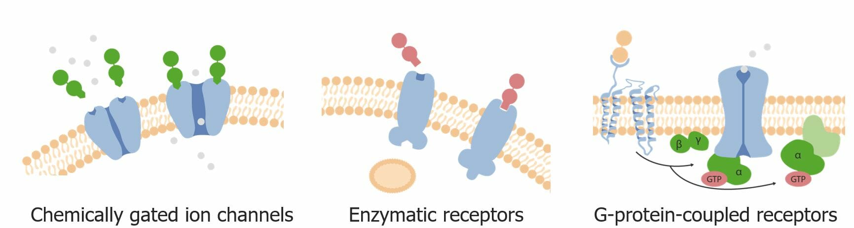
Three primary types of cell surface (i.e., transmembrane) receptors:
chemically gated ion channels, enzymatic receptors, and G-protein-coupled receptors (GPCRs)
Image by Lecturio.
Intracellular receptors
Located within the cell; may be in either:
Cytoplasm
NucleusNucleusWithin a eukaryotic cell, a membrane-limited body which contains chromosomes and one or more nucleoli (cell nucleolus). The nuclear membrane consists of a double unit-type membrane which is perforated by a number of pores; the outermost membrane is continuous with the endoplasmic reticulum. A cell may contain more than one nucleus.The Cell: Organelles
Consists of 3 domains:
Ligand-binding domain
DNA-binding domain
Domain that can interact with other transcription factorsTranscription FactorsEndogenous substances, usually proteins, which are effective in the initiation, stimulation, or termination of the genetic transcription process.Stages of Transcription (e.g., coactivators, repressors)
Ligands are usually small hydrophobic molecules that can cross the cell membraneCell MembraneA cell membrane (also known as the plasma membrane or plasmalemma) is a biological membrane that separates the cell contents from the outside environment. A cell membrane is composed of a phospholipid bilayer and proteins that function to protect cellular DNA and mediate the exchange of ions and molecules. The Cell: Cell Membrane.
Examples of intracellular receptors:
Steroid hormone nuclear receptors
Vitamin DVitamin DA vitamin that includes both cholecalciferols and ergocalciferols, which have the common effect of preventing or curing rickets in animals. It can also be viewed as a hormone since it can be formed in skin by action of ultraviolet rays upon the precursors, 7-dehydrocholesterol and ergosterol, and acts on vitamin D receptors to regulate calcium in opposition to parathyroid hormone.Fat-soluble Vitamins and their Deficiencies receptor
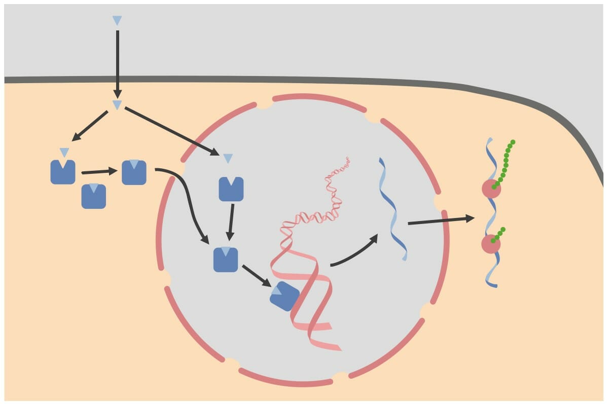
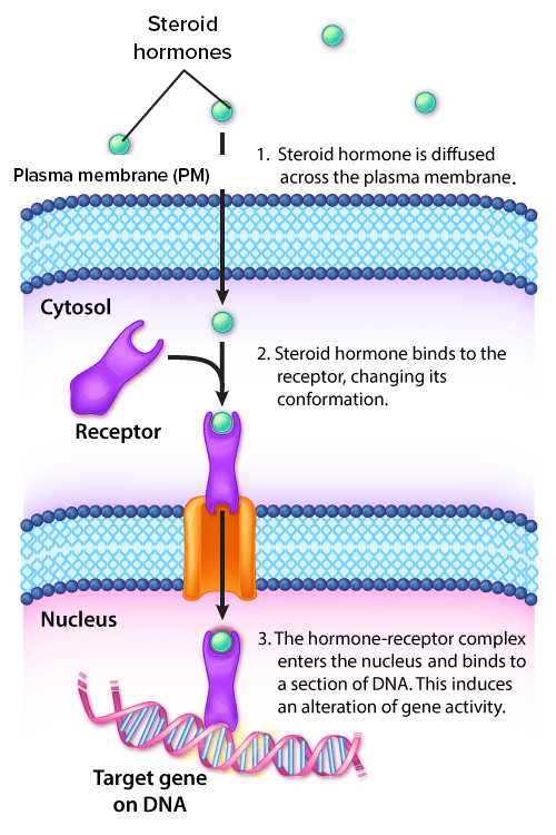
Intracellular receptors: Intracellular receptors may be located within the cytoplasm or the nucleus. Receptors located within the cytoplasm move into the nucleus once they bind with their ligand (i.e., hormone). Within the nucleus, the hormone-receptor complex binds to hormone response elements (HREs), which are specific sequences of DNA. The complex then binds other transcription factors to alter gene expression.
Image by Lecturio.
Table: Receptor types and ligands
Receptor types
Example of ligands
Intracellular receptors
Nuclear receptors
Steroid hormonesSteroid hormonesSteroid hormones produced by the gonads. They stimulate reproductive organs, germ cell maturation, and the secondary sex characteristics in the males and the females. The major sex steroid hormones include estradiol; progesterone; and testosterone.Hormones: Overview and Types (e.g., glucocorticoidsGlucocorticoidsGlucocorticoids are a class within the corticosteroid family. Glucocorticoids are chemically and functionally similar to endogenous cortisol. There are a wide array of indications, which primarily benefit from the antiinflammatory and immunosuppressive effects of this class of drugs.Glucocorticoids)
InsulinInsulinInsulin is a peptide hormone that is produced by the beta cells of the pancreas. Insulin plays a role in metabolic functions such as glucose uptake, glycolysis, glycogenesis, lipogenesis, and protein synthesis. Exogenous insulin may be needed for individuals with diabetes mellitus, in whom there is a deficiency in endogenous insulin or increased insulin resistance. Insulin
AcetylcholineAcetylcholineA neurotransmitter found at neuromuscular junctions, autonomic ganglia, parasympathetic effector junctions, a subset of sympathetic effector junctions, and at many sites in the central nervous system.Receptors and Neurotransmitters of the CNS (AChAChA neurotransmitter found at neuromuscular junctions, autonomic ganglia, parasympathetic effector junctions, a subset of sympathetic effector junctions, and at many sites in the central nervous system.Receptors and Neurotransmitters of the CNS)
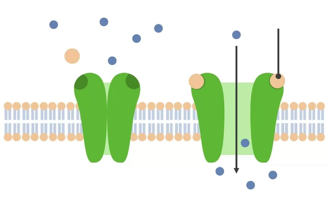
Ligand-gated (chemically gated) ion channel receptors are a subtype of cell surface receptors.
Structure
Consist of multiple transmembrane subunits around a central ion-conducting channel
Have an extracellular (or intracellular) ligand-binding site
Ligand-gated (chemically gated) ion channels: This image demonstrates a ligand (tan ball) binding with a ligand-gated ion channel receptor (green structure), which causes a conformational change in the channel, allowing ions to pass through.
Image by Lecturio.
Physiology
Ligand (e.g., AChAChA neurotransmitter found at neuromuscular junctions, autonomic ganglia, parasympathetic effector junctions, a subset of sympathetic effector junctions, and at many sites in the central nervous system.Receptors and Neurotransmitters of the CNS) binds to the ion channel receptor.
Induces a conformational change → channel opens
Ions flowFlowBlood flows through the heart, arteries, capillaries, and veins in a closed, continuous circuit. Flow is the movement of volume per unit of time. Flow is affected by the pressure gradient and the resistance fluid encounters between 2 points. Vascular resistance is the opposition to flow, which is caused primarily by blood friction against vessel walls.Vascular Resistance, Flow, and Mean Arterial Pressure down their electrochemical gradientElectrochemical gradientThe Cell: Cell Membrane through the channelsChannelsThe Cell: Cell Membrane (e.g., Na+, K+, CaCACondylomata acuminata are a clinical manifestation of genital HPV infection. Condylomata acuminata are described as raised, pearly, flesh-colored, papular, cauliflower-like lesions seen in the anogenital region that may cause itching, pain, or bleeding.Condylomata Acuminata (Genital Warts)2+, and/or Cl−).
This can result in:
Transmission of nerve signals
Muscle contraction
Release of hormonesHormonesHormones are messenger molecules that are synthesized in one part of the body and move through the bloodstream to exert specific regulatory effects on another part of the body. Hormones play critical roles in coordinating cellular activities throughout the body in response to the constant changes in both the internal and external environments. Hormones: Overview and Types
Myasthenia gravisMyasthenia GravisMyasthenia gravis (MG) is an autoimmune neuromuscular disorder characterized by weakness and fatigability of skeletal muscles caused by dysfunction/destruction of acetylcholine receptors at the neuromuscular junction. MG presents with fatigue, ptosis, diplopia, dysphagia, respiratory difficulties, and progressive weakness in the limbs, leading to difficulty in movement. Myasthenia Gravis (MG): an autoimmune neuromuscular disorder characterized by weakness and fatigability of skeletal musclesSkeletal musclesA subtype of striated muscle, attached by tendons to the skeleton. Skeletal muscles are innervated and their movement can be consciously controlled. They are also called voluntary muscles.Muscle Tissue: Histology caused by dysfunction/destruction of acetylcholineAcetylcholineA neurotransmitter found at neuromuscular junctions, autonomic ganglia, parasympathetic effector junctions, a subset of sympathetic effector junctions, and at many sites in the central nervous system.Receptors and Neurotransmitters of the CNS receptors (AChRs) at the neuromuscular junctionNeuromuscular junctionThe synapse between a neuron and a muscle.Skeletal Muscle Contraction (NMJ) (a type of ligand-gated ion channel receptor). When AChAChA neurotransmitter found at neuromuscular junctions, autonomic ganglia, parasympathetic effector junctions, a subset of sympathetic effector junctions, and at many sites in the central nervous system.Receptors and Neurotransmitters of the CNS binds, the channelsChannelsThe Cell: Cell Membrane open, allowing an influx of Na+ into the cell, resulting in depolarizationDepolarizationMembrane Potential that ultimately leads to muscle contraction. Without normal AChRs, muscle contraction is abnormal, and MG presents with fatigueFatigueThe state of weariness following a period of exertion, mental or physical, characterized by a decreased capacity for work and reduced efficiency to respond to stimuli.Fibromyalgia, ptosisPtosisCranial Nerve Palsies, dysphagiaDysphagiaDysphagia is the subjective sensation of difficulty swallowing. Symptoms can range from a complete inability to swallow, to the sensation of solids or liquids becoming “stuck.” Dysphagia is classified as either oropharyngeal or esophageal, with esophageal dysphagia having 2 sub-types: functional and mechanical. Dysphagia, respiratory difficulties, and progressive weakness in the limbs, leading to difficulty in movement.
Enzyme-linked Receptors
Overview
Receptors that have some type of enzymatic activity when “activated” by their ligand
Frequently kinasesKinasesMacrolides and Ketolides: enzymesEnzymesEnzymes are complex protein biocatalysts that accelerate chemical reactions without being consumed by them. Due to the body’s constant metabolic needs, the absence of enzymes would make life unsustainable, as reactions would occur too slowly without these molecules. Basics of Enzymes that catalyze the transfer of a phosphate groupPhosphate groupNucleic Acids from 1 molecule to another
RTKs:
Most common type
Over 90 RTK genesGenesA category of nucleic acid sequences that function as units of heredity and which code for the basic instructions for the development, reproduction, and maintenance of organisms.DNA Types and Structure identified
RTKs are involved in a number of processes frequently associated with general cellular function:
Cell cycleCell cycleThe phases of the cell cycle include interphase (G1, S, and G2) and mitosis (prophase, metaphase, anaphase, and telophase). The cell’s progression through these phases is punctuated by checkpoints regulated by cyclins, cyclin-dependent kinases, tumor suppressors, and their antagonists.Cell Cycle
Cell growth and proliferation
Cell migration
Cell metabolism
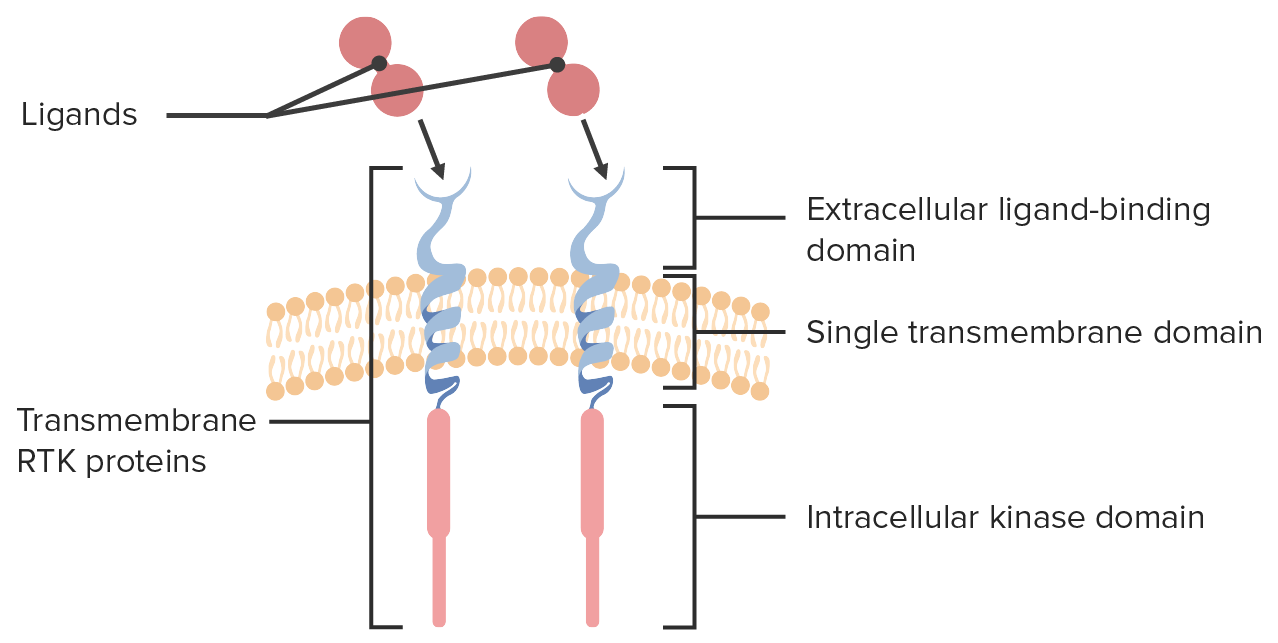
Structure of RTK
Extracellular domain containing the ligand-binding site
Intracellular kinase domain
Single transmembrane helix linking extracellular and intracellular components
The 3-part structure of the receptor tyrosine kinases (RTKs) is shown along with ligands binding to their extracellular binding sites.
Image by Lecturio.
Signaling pathway of RTKs
Ligand binds to the individual RTK receptors, which leads to:
Dimerization: 2 neighboring RTKs join together.
Autophosphorylation:
The dimers phosphorylate each other.
1 RTK monomer transfers a phosphate groupPhosphate groupNucleic Acids from adenosineAdenosineA nucleoside that is composed of adenine and d-ribose. Adenosine or adenosine derivatives play many important biological roles in addition to being components of DNA and RNA. Adenosine itself is a neurotransmitter.Class 5 Antiarrhythmic Drugs triphosphate (ATP) to the other RTK monomer (now known as phosphotyrosines).
Phosphotyrosines act as docking sites for other proteinsProteinsLinear polypeptides that are synthesized on ribosomes and may be further modified, crosslinked, cleaved, or assembled into complex proteins with several subunits. The specific sequence of amino acids determines the shape the polypeptide will take, during protein folding, and the function of the protein.Energy Homeostasis involved in the signal transductionTransductionThe transfer of bacterial DNA by phages from an infected bacterium to another bacterium. This also refers to the transfer of genes into eukaryotic cells by viruses. This naturally occurring process is routinely employed as a gene transfer technique.Bacteriology cascade.
The cellular response of the RTK activation depends on the downstream proteinsProteinsLinear polypeptides that are synthesized on ribosomes and may be further modified, crosslinked, cleaved, or assembled into complex proteins with several subunits. The specific sequence of amino acids determines the shape the polypeptide will take, during protein folding, and the function of the protein.Energy Homeostasis that are present.
The advantage of a kinase cascade is that a small external signal can be amplified many times inside the cell.
Dysfunction of this cascade can lead to cancer due to failure to control general cellular function.
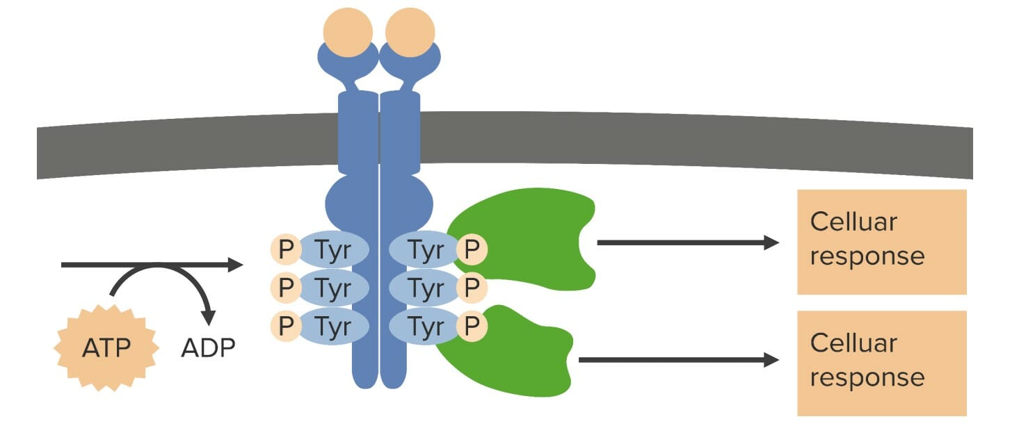
Receptor tyrosine kinase (RTK) function:
When a ligand binds to the extracellular domain of the RTK, 2 RTKs come together in a process known as dimerization. Once dimerized, each RTK transfers a phosphate group from adenosine triphosphate (ATP) to its partner in a process known as autophosphorylation. After autophosphorylation, the phosphotyrosines act as docking and activation sites for other proteins, which are usually other
enzymes. The type of cellular response depends on which additional proteins are present. Tyr: tyrosine P: phosphate
Image by Lecturio.
Examples of RTKs
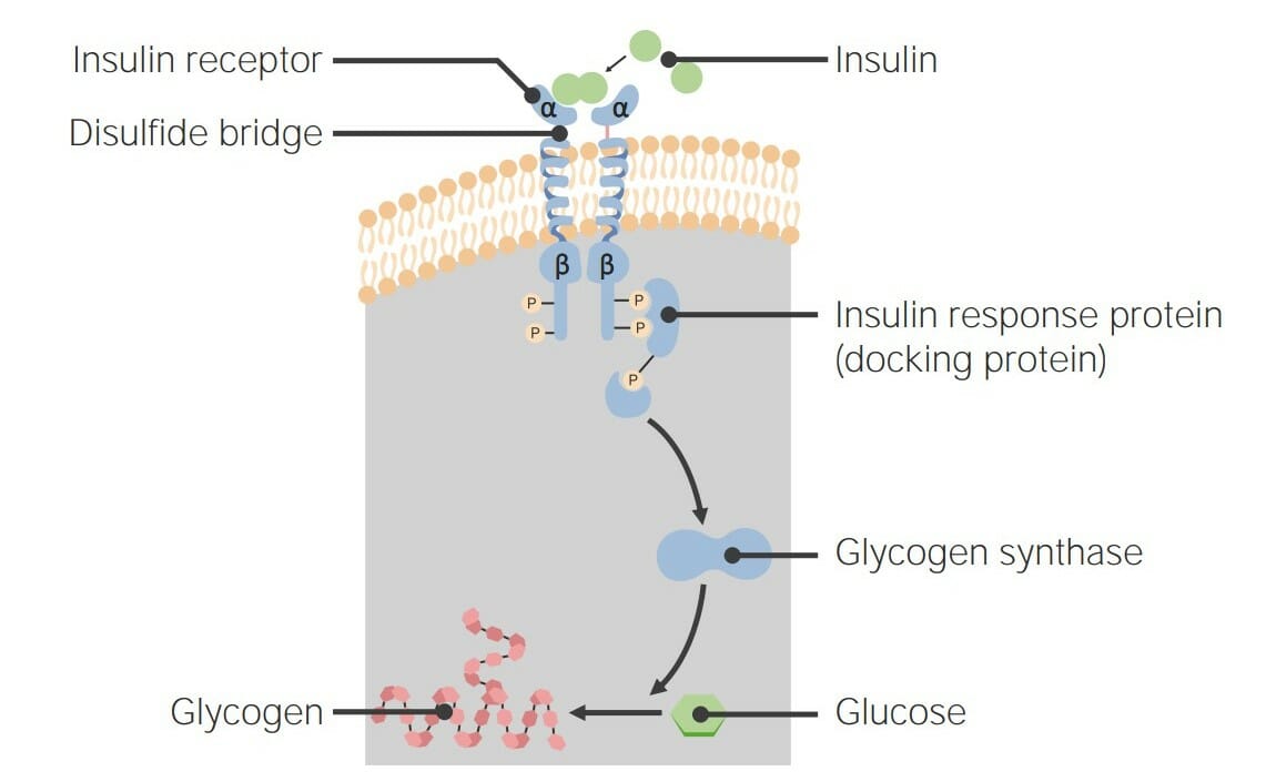
InsulinInsulinInsulin is a peptide hormone that is produced by the beta cells of the pancreas. Insulin plays a role in metabolic functions such as glucose uptake, glycolysis, glycogenesis, lipogenesis, and protein synthesis. Exogenous insulin may be needed for individuals with diabetes mellitus, in whom there is a deficiency in endogenous insulin or increased insulin resistance. Insulin receptors:
Mature insulinInsulinInsulin is a peptide hormone that is produced by the beta cells of the pancreas. Insulin plays a role in metabolic functions such as glucose uptake, glycolysis, glycogenesis, lipogenesis, and protein synthesis. Exogenous insulin may be needed for individuals with diabetes mellitus, in whom there is a deficiency in endogenous insulin or increased insulin resistance. Insulin receptors begin as an inactive dimer, containing 2 alpha and 2 beta subunits that are linked together by disulfide bridges:
Alpha subunits: extracellular domains containing the ligand-binding site
Beta subunits: transmembrane and intracellular tyrosineTyrosineA non-essential amino acid. In animals it is synthesized from phenylalanine. It is also the precursor of epinephrine; thyroid hormones; and melanin.Synthesis of Nonessential Amino Acids kinase domains
InsulinInsulinInsulin is a peptide hormone that is produced by the beta cells of the pancreas. Insulin plays a role in metabolic functions such as glucose uptake, glycolysis, glycogenesis, lipogenesis, and protein synthesis. Exogenous insulin may be needed for individuals with diabetes mellitus, in whom there is a deficiency in endogenous insulin or increased insulin resistance. Insulin binds to RTK alpha domains → triggers rapid autophosphorylation
Signaling pathway:
InsulinInsulinInsulin is a peptide hormone that is produced by the beta cells of the pancreas. Insulin plays a role in metabolic functions such as glucose uptake, glycolysis, glycogenesis, lipogenesis, and protein synthesis. Exogenous insulin may be needed for individuals with diabetes mellitus, in whom there is a deficiency in endogenous insulin or increased insulin resistance. Insulin response substrateSubstrateA substance upon which the enzyme acts.Basics of Enzymes 1 (IRS-1) binds to the phosphotyrosines →
Activates phosphatidylinositol (PI3) kinase →
Results in phosphatidylinositol (3,4,5)-trisphosphate (PIP3) formation →
PIP3 activates 3-phosphoinositide-dependent protein kinaseProtein kinaseA family of enzymes that catalyze the conversion of ATP and a protein to adp and a phosphoprotein.Interferons 1 (PDK1), which activates Akt kinase →
Causes the translocation of glucose transporterGlucose transporterTubular System 4 (GLUT4) (a protein channel that allows glucoseGlucoseA primary source of energy for living organisms. It is naturally occurring and is found in fruits and other parts of plants in its free state. It is used therapeutically in fluid and nutrient replacement.Lactose Intolerance into the cell) to the plasma membranePlasma membraneA cell membrane (also known as the plasma membrane or plasmalemma) is a biological membrane that separates the cell contents from the outside environment. A cell membrane is composed of a phospholipid bilayer and proteins that function to protect cellular DNA and mediate the exchange of ions and molecules.The Cell: Cell Membrane →
↑ GlucoseGlucoseA primary source of energy for living organisms. It is naturally occurring and is found in fruits and other parts of plants in its free state. It is used therapeutically in fluid and nutrient replacement.Lactose Intolerance can enter the cell
Another insulin-signaling pathway results in increased transcriptionTranscriptionTranscription of genetic information is the first step in gene expression. Transcription is the process by which DNA is used as a template to make mRNA. This process is divided into 3 stages: initiation, elongation, and termination. Stages of Transcription and translationTranslationTranslation is the process of synthesizing a protein from a messenger RNA (mRNA) transcript. This process is divided into three primary stages: initiation, elongation, and termination. Translation is catalyzed by structures known as ribosomes, which are large complexes of proteins and ribosomal RNA (rRNA). Stages and Regulation of Translation of glycogen synthaseGlycogen synthaseAn enzyme that catalyzes the transfer of d-glucose from udp glucose into 1, 4-alpha-d-glucosyl chains.Glycogen Metabolism (GS) → ↑ conversion of glucoseGlucoseA primary source of energy for living organisms. It is naturally occurring and is found in fruits and other parts of plants in its free state. It is used therapeutically in fluid and nutrient replacement.Lactose Intolerance to glycogen within the cell
The insulin receptor, a receptor tyrosine kinase (RTK): When insulin binds, the insulin response protein triggers a phosphorylation cascade, ultimately resulting in the activation of glycogen synthase (GS), which converts the extra glucose into glycogen.
Image by Lecturio.
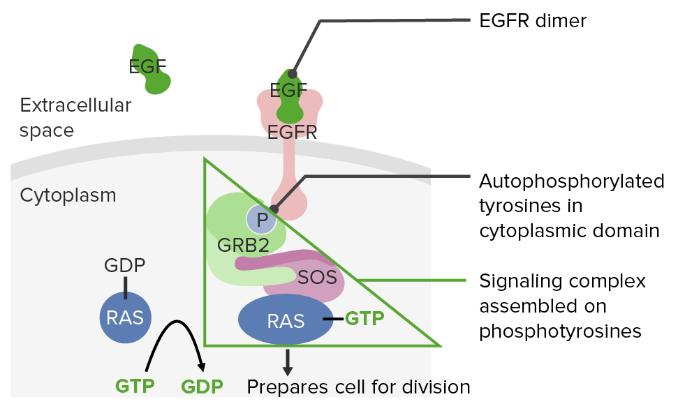
Epidermal growth factor receptors (EGFRs) and the RASRASRenal artery stenosis (RAS) is the narrowing of one or both renal arteries, usually caused by atherosclerotic disease or by fibromuscular dysplasia. If the stenosis is severe enough, the stenosis causes decreased renal blood flow, which activates the renin-angiotensin-aldosterone system (RAAS) and leads to renovascular hypertension (RVH).Renal Artery Stenosis signaling pathway:
Epidermal growth factor (EGF) binds to EGFRs → causes dimerization and autophosphorylation
A signaling complex of proteinsProteinsLinear polypeptides that are synthesized on ribosomes and may be further modified, crosslinked, cleaved, or assembled into complex proteins with several subunits. The specific sequence of amino acids determines the shape the polypeptide will take, during protein folding, and the function of the protein.Energy Homeostasis assembles on the phosphotyrosines.
This signaling complex transfers a phosphate groupPhosphate groupNucleic Acids from guanosine trisphosphate (GTP) to RASRASRenal artery stenosis (RAS) is the narrowing of one or both renal arteries, usually caused by atherosclerotic disease or by fibromuscular dysplasia. If the stenosis is severe enough, the stenosis causes decreased renal blood flow, which activates the renin-angiotensin-aldosterone system (RAAS) and leads to renovascular hypertension (RVH).Renal Artery Stenosis guanosine diphosphate (RAS-GDP), resulting in RAS-GTP, which is the active form of RASRASRenal artery stenosis (RAS) is the narrowing of one or both renal arteries, usually caused by atherosclerotic disease or by fibromuscular dysplasia. If the stenosis is severe enough, the stenosis causes decreased renal blood flow, which activates the renin-angiotensin-aldosterone system (RAAS) and leads to renovascular hypertension (RVH).Renal Artery Stenosis.
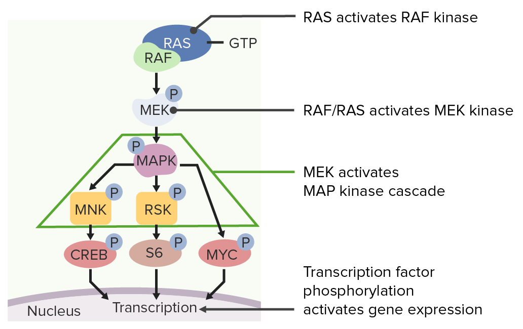
RASRASRenal artery stenosis (RAS) is the narrowing of one or both renal arteries, usually caused by atherosclerotic disease or by fibromuscular dysplasia. If the stenosis is severe enough, the stenosis causes decreased renal blood flow, which activates the renin-angiotensin-aldosterone system (RAAS) and leads to renovascular hypertension (RVH).Renal Artery Stenosis signaling pathway:
MAPK activates MNK and RSK, which phosphorylate specific transcription factorsTranscription FactorsEndogenous substances, usually proteins, which are effective in the initiation, stimulation, or termination of the genetic transcription process.Stages of Transcription required for cell divisionCell DivisionA type of cell nucleus division by means of which the two daughter nuclei normally receive identical complements of the number of chromosomes of the somatic cells of the species.Cell Cycle.
RASRASRenal artery stenosis (RAS) is the narrowing of one or both renal arteries, usually caused by atherosclerotic disease or by fibromuscular dysplasia. If the stenosis is severe enough, the stenosis causes decreased renal blood flow, which activates the renin-angiotensin-aldosterone system (RAAS) and leads to renovascular hypertension (RVH).Renal Artery Stenosis itself is a slow GTPase:
RAS-GTP will slowly cleave off a phosphate groupPhosphate groupNucleic Acids, inactivating itself back to RAS-GDP.
Point mutations can remove its GTPase activity, resulting in RASRASRenal artery stenosis (RAS) is the narrowing of one or both renal arteries, usually caused by atherosclerotic disease or by fibromuscular dysplasia. If the stenosis is severe enough, the stenosis causes decreased renal blood flow, which activates the renin-angiotensin-aldosterone system (RAAS) and leads to renovascular hypertension (RVH).Renal Artery Stenosis that is perpetually activated → promotes continued cell divisionCell DivisionA type of cell nucleus division by means of which the two daughter nuclei normally receive identical complements of the number of chromosomes of the somatic cells of the species.Cell Cycle
RASRASRenal artery stenosis (RAS) is the narrowing of one or both renal arteries, usually caused by atherosclerotic disease or by fibromuscular dysplasia. If the stenosis is severe enough, the stenosis causes decreased renal blood flow, which activates the renin-angiotensin-aldosterone system (RAAS) and leads to renovascular hypertension (RVH).Renal Artery Stenosis mutations are indicated in multiple human cancers.
Epidermal growth factor (EGF) binds to the EGF receptor (EGFR), which results in the phosphorylation of RAS.
Image by Lecturio.
Phosphorylated RAS then activates a signaling cascade which results in cell division.
Image by Lecturio.
Examples of clinical relevance
Abnormalities in RTKs are known to cause a number of different congenital malformation syndromes and cancers, especially with gain of function mutations causing excessive cell divisionCell DivisionA type of cell nucleus division by means of which the two daughter nuclei normally receive identical complements of the number of chromosomes of the somatic cells of the species.Cell Cycle. Some examples include:
Chronic myeloid leukemiaChronic myeloid leukemiaChronic myeloid leukemia is a malignant proliferation of the granulocytic cell line characterized by a fairly normal differentiation. The underlying genetic abnormality is the Philadelphia chromosome, an abbreviated chromosome 22, resulting from reciprocal (9;22)(q34;q11) translocation. Chronic Myeloid Leukemia (CMLCMLChronic myeloid leukemia is a malignant proliferation of the granulocytic cell line characterized by a fairly normal differentiation. The underlying genetic abnormality is the Philadelphia chromosome, an abbreviated chromosome 22, resulting from reciprocal (9;22)(q34;q11) translocation. Chronic Myeloid Leukemia): a malignant proliferation of the granulocytic cell line due to a reciprocal translocation (9;22)(q34;q11). The chromosomeChromosomeIn a prokaryotic cell or in the nucleus of a eukaryotic cell, a structure consisting of or containing DNA which carries the genetic information essential to the cell.Basic Terms of Genetics contains a BCR-ABL1 fusion geneGeneA category of nucleic acid sequences that function as units of heredity and which code for the basic instructions for the development, reproduction, and maintenance of organisms.Basic Terms of Genetics (from ABL1 on chromosome 9Chromosome 9Friedreich Ataxia andBCRBCRLymphocytes: Histology on chromosomeChromosomeIn a prokaryotic cell or in the nucleus of a eukaryotic cell, a structure consisting of or containing DNA which carries the genetic information essential to the cell.Basic Terms of Genetics 22), which produces constitutively active tyrosineTyrosineA non-essential amino acid. In animals it is synthesized from phenylalanine. It is also the precursor of epinephrine; thyroid hormones; and melanin.Synthesis of Nonessential Amino AcidskinasesKinasesMacrolides and Ketolides and, consequently, uncontrolled granulocytic production. PatientsPatientsIndividuals participating in the health care system for the purpose of receiving therapeutic, diagnostic, or preventive procedures.Clinician–Patient Relationship can be asymptomatic or have constitutional symptomsConstitutional SymptomsAntineutrophil Cytoplasmic Antibody (ANCA)-Associated Vasculitis, sternal painPainAn unpleasant sensation induced by noxious stimuli which are detected by nerve endings of nociceptive neurons.Pain: Types and Pathways, and splenomegalySplenomegalySplenomegaly is pathologic enlargement of the spleen that is attributable to numerous causes, including infections, hemoglobinopathies, infiltrative processes, and outflow obstruction of the portal vein. Splenomegaly.
Achondroplasia: an autosomal dominantAutosomal dominantAutosomal inheritance, both dominant and recessive, refers to the transmission of genes from the 22 autosomal chromosomes. Autosomal dominant diseases are expressed when only 1 copy of the dominant allele is inherited. Autosomal Recessive and Autosomal Dominant Inheritancebone dysplasiaBone dysplasiaX-linked Hypophosphatemic Rickets due to gain of function mutations in the fibroblast growth factorFibroblast growth factorA family of small polypeptide growth factors that share several common features including a strong affinity for heparin, and a central barrel-shaped core region of 140 amino acids that is highly homologous between family members. Although originally studied as proteins that stimulate the growth of fibroblasts this distinction is no longer a requirement for membership in the fibroblast growth factor family.X-linked Hypophosphatemic Rickets receptor 3 (FGFR3) geneGeneA category of nucleic acid sequences that function as units of heredity and which code for the basic instructions for the development, reproduction, and maintenance of organisms.Basic Terms of Genetics, which codes for an RTK. As a result, the FGFR3 receptor is permanently activated, inhibiting chondrocyte proliferation, which results in impaired boneBoneBone is a compact type of hardened connective tissue composed of bone cells, membranes, an extracellular mineralized matrix, and central bone marrow. The 2 primary types of bone are compact and spongy. Bones: Structure and Types formation and skeletal anomalies.
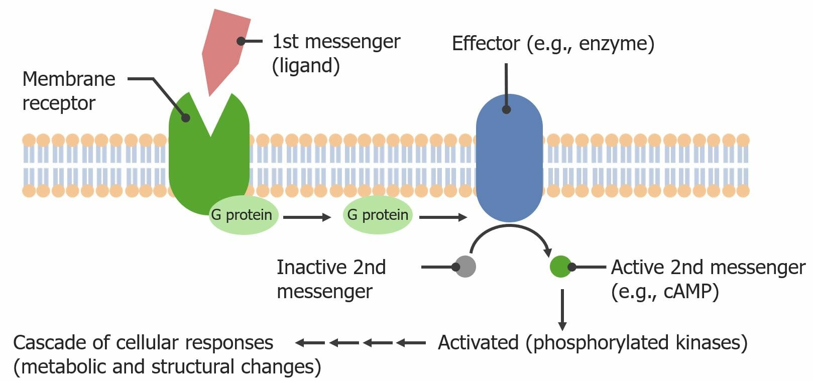
G-protein-coupled receptors (GPCRs) are transmembrane proteinsProteinsLinear polypeptides that are synthesized on ribosomes and may be further modified, crosslinked, cleaved, or assembled into complex proteins with several subunits. The specific sequence of amino acids determines the shape the polypeptide will take, during protein folding, and the function of the protein.Energy Homeostasis that bindBINDHyperbilirubinemia of the Newborn hormone ligands on their extracellular side, which induces a conformational change inside the cell, activating an associated G protein, which then triggers a signaling cascade via 2nd messengers.
Largest family of receptors, with over 800 GPCR genesGenesA category of nucleic acid sequences that function as units of heredity and which code for the basic instructions for the development, reproduction, and maintenance of organisms.DNA Types and Structure identified
Often involved in metabolic and structural changes within a cell
Examples:
Beta-adrenergic receptors
Many olfactory receptors
Structure
Extracellular domain: contains the ligand-binding sites
Transmembrane domain:
Anchors the receptor in the PM
Often composed of 7 alpha helices (known as seven-transmembrane (7TM) receptors)
Intracellular domain: bound to a G protein
G proteinsProteinsLinear polypeptides that are synthesized on ribosomes and may be further modified, crosslinked, cleaved, or assembled into complex proteins with several subunits. The specific sequence of amino acids determines the shape the polypeptide will take, during protein folding, and the function of the protein.Energy Homeostasis:
ProteinsProteinsLinear polypeptides that are synthesized on ribosomes and may be further modified, crosslinked, cleaved, or assembled into complex proteins with several subunits. The specific sequence of amino acids determines the shape the polypeptide will take, during protein folding, and the function of the protein.Energy Homeostasis that bindBINDHyperbilirubinemia of the Newborn to guanineGuanineNucleic AcidsnucleotidesNucleotidesThe monomeric units from which DNA or RNA polymers are constructed. They consist of a purine or pyrimidine base, a pentose sugar, and a phosphate group.Nucleic Acids (GTP and GDP)
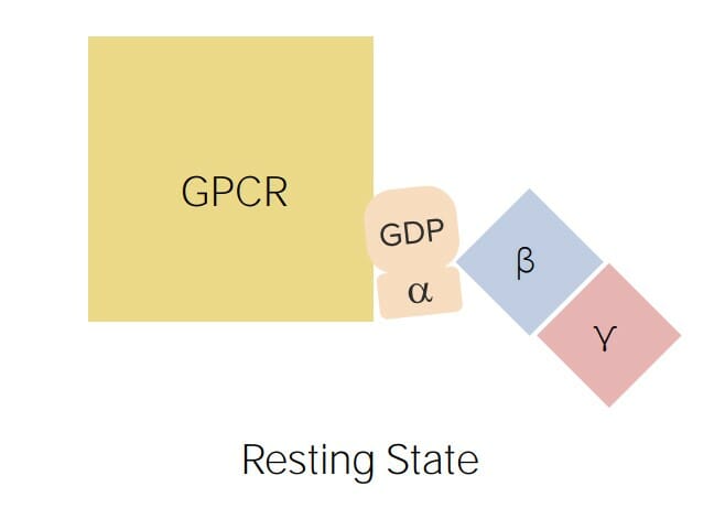
Consist of 3 subunits: alpha, beta, and gamma
Alpha subunit:
Binds the guanineGuanineNucleic AcidsnucleotidesNucleotidesThe monomeric units from which DNA or RNA polymers are constructed. They consist of a purine or pyrimidine base, a pentose sugar, and a phosphate group.Nucleic Acids: GTP (active state) and GDP (inactive state)
Slowly hydrolyzes GTP to GDP → once activated by GTP, the alpha subunit will eventually inactivate itself through hydrolytic conversion to GDP
Beta and gamma: help the alpha‒GDP associate with the GPCR
Diagram depicting a G-protein-coupled receptor (GPCR) bound to a G protein: The G proteins consist of 3 subunits: alpha (which binds to guanosine diphosphate (GDP) in its inactive form and guanosine triphosphate (GTP) in its active form), beta, and gamma (which helps the alpha subunit associate with the GPCR).
Image by Lecturio.
Activation pathway
Ligand binds to its binding site on the external portion of the receptor → induces a conformational change in the GPCR
This causes phosphorylationPhosphorylationThe introduction of a phosphoryl group into a compound through the formation of an ester bond between the compound and a phosphorus moiety.Post-translational Protein Processing of the GDP bound to the alpha subunit → becomes alpha‒GTP (activated G protein)
Alpha‒GTP separates from the beta and gamma subunit → goes and phosphorylates the next protein in the signaling cascade known as the effector protein
Effector proteinsProteinsLinear polypeptides that are synthesized on ribosomes and may be further modified, crosslinked, cleaved, or assembled into complex proteins with several subunits. The specific sequence of amino acids determines the shape the polypeptide will take, during protein folding, and the function of the protein.Energy Homeostasis:
EnzymesEnzymesEnzymes are complex protein biocatalysts that accelerate chemical reactions without being consumed by them. Due to the body’s constant metabolic needs, the absence of enzymes would make life unsustainable, as reactions would occur too slowly without these molecules. Basics of Enzymes, often membrane bound
PhosphorylationPhosphorylationThe introduction of a phosphoryl group into a compound through the formation of an ester bond between the compound and a phosphorus moiety.Post-translational Protein Processing may be activating or inactivating
Frequently generate 2nd messengers
Activation pathway for G-protein-coupled receptors (GPCRs): A ligand binds to the GPCR, inducing a conformational change internally. This conformational change causes the alpha subunit of the G protein to exchange a guanosine diphosphate (GDP) for a guanosine triphosphate (GTP), which activates the G protein. The GTP-bound alpha subunit separates from the beta and gamma subunits and goes to activate an effector enzyme (via phosphorylation from the GTP). The effector enzyme then activates a 2nd messenger (here cyclic adenosine monophosphate (cAMP)), which relays the signal inside the cell.
Image by Lecturio.
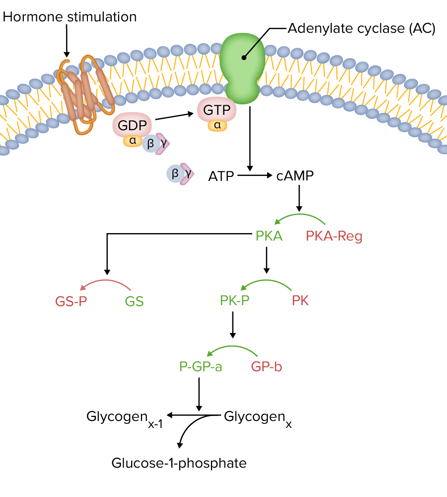
Adenylate cyclase (AC) and the cAMPcAMPAn adenine nucleotide containing one phosphate group which is esterified to both the 3′- and 5′-positions of the sugar moiety. It is a second messenger and a key intracellular regulator, functioning as a mediator of activity for a number of hormones, including epinephrine, glucagon, and acth.Phosphodiesterase Inhibitors 2nd messenger system
Adenylate cyclase (also called adenylyl cyclase) is a common GPCR effector protein. An activated G protein phosphorylates AC, activating it to convert ATP to cAMPcAMPAn adenine nucleotide containing one phosphate group which is esterified to both the 3′- and 5′-positions of the sugar moiety. It is a second messenger and a key intracellular regulator, functioning as a mediator of activity for a number of hormones, including epinephrine, glucagon, and acth.Phosphodiesterase Inhibitors, a common 2nd messenger.
GlucagonGlucagonA 29-amino acid pancreatic peptide derived from proglucagon which is also the precursor of intestinal glucagon-like peptides. Glucagon is secreted by pancreatic alpha cells and plays an important role in regulation of blood glucose concentration, ketone metabolism, and several other biochemical and physiological processes.Gastrointestinal Secretions binds its receptor, which is a GPCR → conformational change
The alpha subunit of the G protein bound to the internal surface of the GPCR exchanges GDP for GTP and separates from the beta and gamma subunits.
Alpha‒GTP (active form) phosphorylates AC, activating it.
AC generates cAMPcAMPAn adenine nucleotide containing one phosphate group which is esterified to both the 3′- and 5′-positions of the sugar moiety. It is a second messenger and a key intracellular regulator, functioning as a mediator of activity for a number of hormones, including epinephrine, glucagon, and acth.Phosphodiesterase Inhibitors from ATP.
cAMPcAMPAn adenine nucleotide containing one phosphate group which is esterified to both the 3′- and 5′-positions of the sugar moiety. It is a second messenger and a key intracellular regulator, functioning as a mediator of activity for a number of hormones, including epinephrine, glucagon, and acth.Phosphodiesterase Inhibitors phosphorylates the regulatory subunits of protein kinaseProtein kinaseA family of enzymes that catalyze the conversion of ATP and a protein to adp and a phosphoprotein.Interferons A (PKA) → this causes the regulatory subunits to disassociate from the catalytic subunits, thus “activating” the PKA
Phosphorylase kinase (PK), activating it → PK activates glycogen phosphorylaseGlycogen phosphorylaseAn enzyme that catalyzes the degradation of glycogen in animals by releasing glucose-1-phosphate from the terminal alpha-1, 4-glycosidic bond. This enzyme exists in two forms: an active phosphorylated form ( phosphorylase A) and an inactive un-phosphorylated form (phosphorylase B). Both A and B forms of phosphorylase exist as homodimers. In mammals, the major isozymes of glycogen phosphorylase are found in muscle, liver and brain tissue.Glycogen Metabolism→ stimulates the breakdown of glycogen
Summary: GlucagonGlucagonA 29-amino acid pancreatic peptide derived from proglucagon which is also the precursor of intestinal glucagon-like peptides. Glucagon is secreted by pancreatic alpha cells and plays an important role in regulation of blood glucose concentration, ketone metabolism, and several other biochemical and physiological processes.Gastrointestinal Secretions stimulates the breakdown of glycogen to glucoseGlucoseA primary source of energy for living organisms. It is naturally occurring and is found in fruits and other parts of plants in its free state. It is used therapeutically in fluid and nutrient replacement.Lactose Intolerance and simultaneously inhibits glycogen synthesisSynthesisPolymerase Chain Reaction (PCR).
G-protein-coupled receptor (GPCR) that is coupled to adenylate cyclase (AC):
Note active enzymes are shown in green while inactive enzymes are shown in red. The AC converts adenosine triphosphate (ATP) to cyclic adenosine monophosphate (cAMP), which then activates
protein kinase A (PKA). PKA then phosphorylates both glycogen synthase (GS), inactivating the GS, and phosphorylase kinase (PK), activating the PK. Activated PK then activates glycogen phosphorylase, which stimulates the breakdown of glycogen to glucose. GTP: guanosine-5′-triphosphate GDP: guanosine diphosphate ATP: adenosine triphosphate cAMP: cyclic adenosine monophosphate
Image by Lecturio.
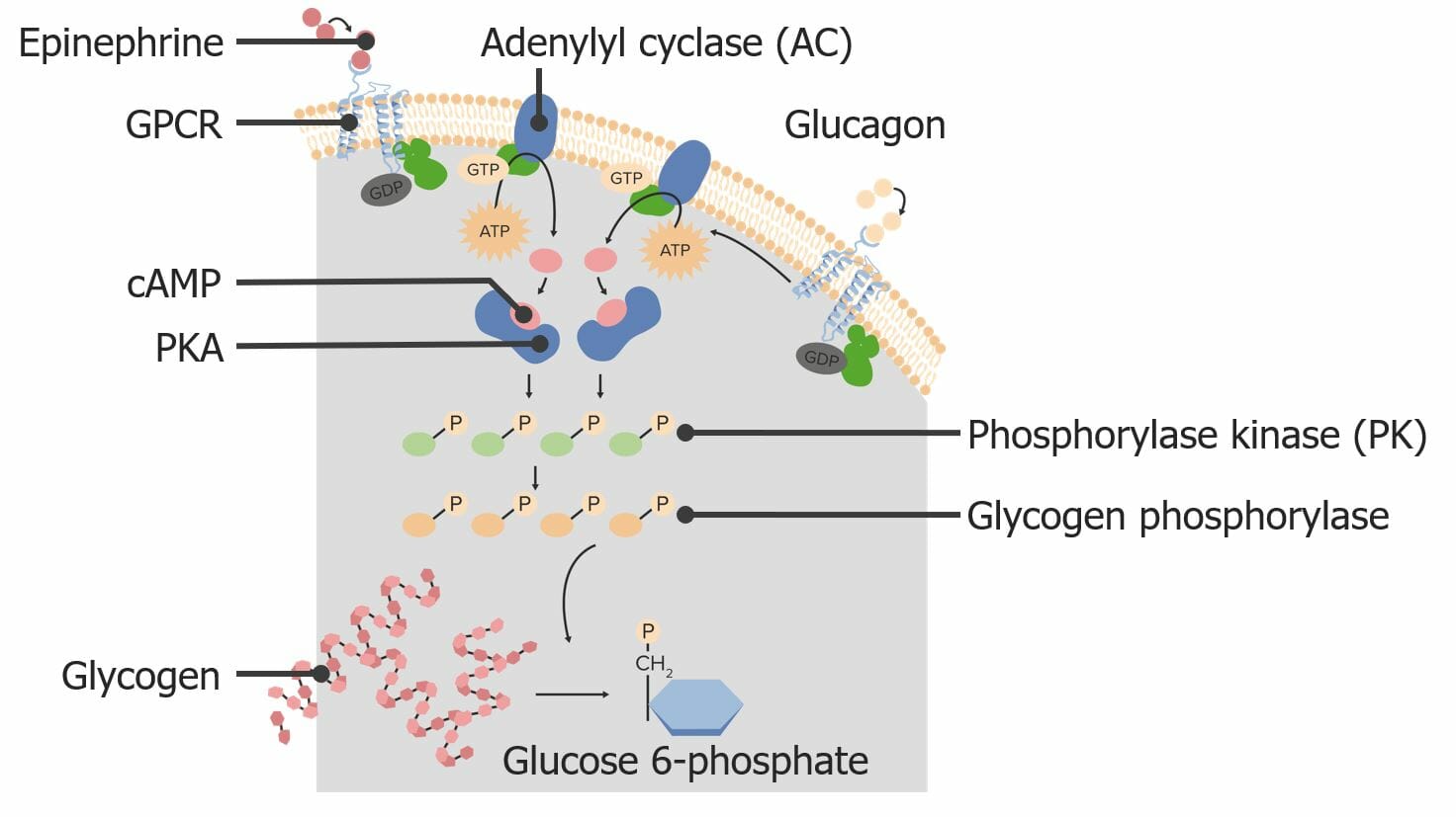
Example 2: EpinephrineEpinephrineThe active sympathomimetic hormone from the adrenal medulla. It stimulates both the alpha- and beta- adrenergic systems, causes systemic vasoconstriction and gastrointestinal relaxation, stimulates the heart, and dilates bronchi and cerebral vessels.Sympathomimetic Drugs pathways (1 molecule having different effects depending on the proteinsProteinsLinear polypeptides that are synthesized on ribosomes and may be further modified, crosslinked, cleaved, or assembled into complex proteins with several subunits. The specific sequence of amino acids determines the shape the polypeptide will take, during protein folding, and the function of the protein.Energy Homeostasis present in the target cell)
In skeletal muscle: epinephrineEpinephrineThe active sympathomimetic hormone from the adrenal medulla. It stimulates both the alpha- and beta- adrenergic systems, causes systemic vasoconstriction and gastrointestinal relaxation, stimulates the heart, and dilates bronchi and cerebral vessels.Sympathomimetic Drugs binds to a GPCR, which activates AC in an identical fashion to glucagonGlucagonA 29-amino acid pancreatic peptide derived from proglucagon which is also the precursor of intestinal glucagon-like peptides. Glucagon is secreted by pancreatic alpha cells and plays an important role in regulation of blood glucose concentration, ketone metabolism, and several other biochemical and physiological processes.Gastrointestinal Secretions → ↑ glycogen breakdown into glucoseGlucoseA primary source of energy for living organisms. It is naturally occurring and is found in fruits and other parts of plants in its free state. It is used therapeutically in fluid and nutrient replacement.Lactose Intolerance
In cardiac muscleCardiac muscleThe muscle tissue of the heart. It is composed of striated, involuntary muscle cells connected to form the contractile pump to generate blood flow.Muscle Tissue: Histology: the cAMPcAMPAn adenine nucleotide containing one phosphate group which is esterified to both the 3′- and 5′-positions of the sugar moiety. It is a second messenger and a key intracellular regulator, functioning as a mediator of activity for a number of hormones, including epinephrine, glucagon, and acth.Phosphodiesterase Inhibitors produced by AC results in ↑ contraction strength
In intestines: epinephrineEpinephrineThe active sympathomimetic hormone from the adrenal medulla. It stimulates both the alpha- and beta- adrenergic systems, causes systemic vasoconstriction and gastrointestinal relaxation, stimulates the heart, and dilates bronchi and cerebral vessels.Sympathomimetic Drugs binds to a GPCR which inhibits AC, causing relaxation of intestinal muscles and slowing of digestionDigestionDigestion refers to the process of the mechanical and chemical breakdown of food into smaller particles, which can then be absorbed and utilized by the body.Digestion and Absorption
Two different G proteins can activate the same internal signal transduction pathway: Here, epinephrine and glucagon can both activate glycogen breakdown into glucose. GPCR: G-protein-coupled receptors cAMP: cyclic adenosine monophosphate PKA: protein kinase A
Image by Lecturio.
Phospholipase CPhospholipase CA subclass of phospholipases that hydrolyze the phosphoester bond found in the third position of glycerophospholipids. Although the singular term phospholipase C specifically refers to an enzyme that catalyzes the hydrolysis of phosphatidylcholine, it is commonly used in the literature to refer to broad variety of enzymes that specifically catalyze the hydrolysis of phosphatidylinositols.Pseudomonas (PLC) (effector protein) and 2nd messengers IP3 and DAGDAGSecond Messengers
Ligand binds GPCR → conformational change
The alpha subunit of the G protein exchanges GDP for GTP and separates from the beta and gamma subunits.
Alpha‒GTP (active) phosphorylates PLC, activating it.
PLC cleaves phosphatidylinositol 4,5-bisphosphate (PIP2) into IP3 and DAGDAGSecond Messengers.
IP3 travels to the ER and binds to a ligand-gated ion channel, causing that channel to open and release CaCACondylomata acuminata are a clinical manifestation of genital HPV infection. Condylomata acuminata are described as raised, pearly, flesh-colored, papular, cauliflower-like lesions seen in the anogenital region that may cause itching, pain, or bleeding.Condylomata Acuminata (Genital Warts) into the cytoplasm.
CaCACondylomata acuminata are a clinical manifestation of genital HPV infection. Condylomata acuminata are described as raised, pearly, flesh-colored, papular, cauliflower-like lesions seen in the anogenital region that may cause itching, pain, or bleeding.Condylomata Acuminata (Genital Warts)2+ then:
Exerts a cellular response
Causes DAGDAGSecond Messengers to activate protein kinaseProtein kinaseA family of enzymes that catalyze the conversion of ATP and a protein to adp and a phosphoprotein.Interferons C (PKC), which goes on to phosphorylate additional proteinsProteinsLinear polypeptides that are synthesized on ribosomes and may be further modified, crosslinked, cleaved, or assembled into complex proteins with several subunits. The specific sequence of amino acids determines the shape the polypeptide will take, during protein folding, and the function of the protein.Energy Homeostasis
E.g., in parietal cellsParietal cellsRounded or pyramidal cells of the gastric glands. They secrete hydrochloric acid and produce gastric intrinsic factor, a glycoprotein that binds vitamin B12.Stomach: Anatomy in the stomachStomachThe stomach is a muscular sac in the upper left portion of the abdomen that plays a critical role in digestion. The stomach develops from the foregut and connects the esophagus with the duodenum. Structurally, the stomach is C-shaped and forms a greater and lesser curvature and is divided grossly into regions: the cardia, fundus, body, and pylorus. Stomach: Anatomy, AChAChA neurotransmitter found at neuromuscular junctions, autonomic ganglia, parasympathetic effector junctions, a subset of sympathetic effector junctions, and at many sites in the central nervous system.Receptors and Neurotransmitters of the CNS binds a muscarinic GPCR → activates PLC → generates IP3 and DAGDAGSecond Messengers → CaCACondylomata acuminata are a clinical manifestation of genital HPV infection. Condylomata acuminata are described as raised, pearly, flesh-colored, papular, cauliflower-like lesions seen in the anogenital region that may cause itching, pain, or bleeding.Condylomata Acuminata (Genital Warts)2+ from the ER and DAGDAGSecond Messengers both help activate the H+/K+ ATPase, which secretes H+ ions into the stomachStomachThe stomach is a muscular sac in the upper left portion of the abdomen that plays a critical role in digestion. The stomach develops from the foregut and connects the esophagus with the duodenum. Structurally, the stomach is C-shaped and forms a greater and lesser curvature and is divided grossly into regions: the cardia, fundus, body, and pylorus. Stomach: Anatomy lumen
A G-protein-coupled receptor (GPCR) activates phospholipase C (PLC), which converts phosphatidylinositol 4,5-bisphosphate (PIP2) into inositol triphosphate (IP3) and diacylglycerol (DAG). The IP3 then binds to a ligand-gated ion channel on the ER, causing the channel to open and Ca to efflux into the cytoplasm, causing a cellular response (e.g., triggers hormone release from endocrine cells). GTP: guanosine-5′-triphosphate GDP: guanosine diphosphate
Image by Lecturio.
Example of clinical relevance
Over 30 different human diseases can be linked to mutations in GPCRs. These mutations may be activating or inactivating. One example is nephrogenic diabetesDiabetesDiabetes mellitus (DM) is a metabolic disease characterized by hyperglycemia and dysfunction of the regulation of glucose metabolism by insulin. Type 1 DM is diagnosed mostly in children and young adults as the result of autoimmune destruction of β cells in the pancreas and the resulting lack of insulin. Type 2 DM has a significant association with obesity and is characterized by insulin resistance.Diabetes Mellitus insipidus (NDI).
Nephrogenic diabetesDiabetesDiabetes mellitus (DM) is a metabolic disease characterized by hyperglycemia and dysfunction of the regulation of glucose metabolism by insulin. Type 1 DM is diagnosed mostly in children and young adults as the result of autoimmune destruction of β cells in the pancreas and the resulting lack of insulin. Type 2 DM has a significant association with obesity and is characterized by insulin resistance.Diabetes Mellitus insipidus: a disorder caused by abnormalities in the antidiuretic hormoneAntidiuretic hormoneAntidiuretic hormones released by the neurohypophysis of all vertebrates (structure varies with species) to regulate water balance and osmolarity. In general, vasopressin is a nonapeptide consisting of a six-amino-acid ring with a cysteine 1 to cysteine 6 disulfide bridge or an octapeptide containing a cystine. All mammals have arginine vasopressin except the pig with a lysine at position 8. Vasopressin, a vasoconstrictor, acts on the kidney collecting ducts to increase water reabsorption, increase blood volume and blood pressure.Hypernatremia (ADH) receptor, leading to ADH resistanceResistancePhysiologically, the opposition to flow of air caused by the forces of friction. As a part of pulmonary function testing, it is the ratio of driving pressure to the rate of air flow.Ventilation: Mechanics of Breathing. The ADH receptor is a GPCR that normally triggers the insertion of aquaporin channelsChannelsThe Cell: Cell Membrane into the membranes of renal collecting ductCollecting ductStraight tubes commencing in the radiate part of the kidney cortex where they receive the curved ends of the distal convoluted tubules. In the medulla the collecting tubules of each pyramid converge to join a central tube (duct of bellini) which opens on the summit of the papilla.Renal Cell Carcinoma cells, allowing for water reabsorption. With an abnormal receptor, the GPCR ineffectively transmits the ADH signal within the cell, resulting in resistanceResistancePhysiologically, the opposition to flow of air caused by the forces of friction. As a part of pulmonary function testing, it is the ratio of driving pressure to the rate of air flow.Ventilation: Mechanics of Breathing to ADH and a decreased ability for individuals to concentrate urine.
Act as ligand-activated transcription factorsTranscription FactorsEndogenous substances, usually proteins, which are effective in the initiation, stimulation, or termination of the genetic transcription process.Stages of Transcription, which ultimately affect geneGeneA category of nucleic acid sequences that function as units of heredity and which code for the basic instructions for the development, reproduction, and maintenance of organisms.Basic Terms of Genetics expression
Although known as nuclear receptors, they are often found in the cytoplasm, which move to the nucleusNucleusWithin a eukaryotic cell, a membrane-limited body which contains chromosomes and one or more nucleoli (cell nucleolus). The nuclear membrane consists of a double unit-type membrane which is perforated by a number of pores; the outermost membrane is continuous with the endoplasmic reticulum. A cell may contain more than one nucleus.The Cell: Organelles.
Structure
C-terminal ligand-binding region
Core DNA-binding domain (DBD), which binds to hormone response elements (HREs) in the DNADNAA deoxyribonucleotide polymer that is the primary genetic material of all cells. Eukaryotic and prokaryotic organisms normally contain DNA in a double-stranded state, yet several important biological processes transiently involve single-stranded regions. DNA, which consists of a polysugar-phosphate backbone possessing projections of purines (adenine and guanine) and pyrimidines (thymine and cytosine), forms a double helix that is held together by hydrogen bonds between these purines and pyrimidines (adenine to thymine and guanine to cytosine).DNA Types and Structure
Regions that interact with other transcription factorsTranscription FactorsEndogenous substances, usually proteins, which are effective in the initiation, stimulation, or termination of the genetic transcription process.Stages of Transcription
Activation pathway
Ligand binds the intracellular receptor (usually in the cytoplasm).
The ligand–receptor complex travels to the nucleusNucleusWithin a eukaryotic cell, a membrane-limited body which contains chromosomes and one or more nucleoli (cell nucleolus). The nuclear membrane consists of a double unit-type membrane which is perforated by a number of pores; the outermost membrane is continuous with the endoplasmic reticulum. A cell may contain more than one nucleus.The Cell: Organelles (if not already there) to bindBINDHyperbilirubinemia of the Newborn the HREs in DNADNAA deoxyribonucleotide polymer that is the primary genetic material of all cells. Eukaryotic and prokaryotic organisms normally contain DNA in a double-stranded state, yet several important biological processes transiently involve single-stranded regions. DNA, which consists of a polysugar-phosphate backbone possessing projections of purines (adenine and guanine) and pyrimidines (thymine and cytosine), forms a double helix that is held together by hydrogen bonds between these purines and pyrimidines (adenine to thymine and guanine to cytosine).DNA Types and Structure.
Interacts with other transcription factorsTranscription FactorsEndogenous substances, usually proteins, which are effective in the initiation, stimulation, or termination of the genetic transcription process.Stages of Transcription to affect geneGeneA category of nucleic acid sequences that function as units of heredity and which code for the basic instructions for the development, reproduction, and maintenance of organisms.Basic Terms of Genetics expression (may act as enhancers or repressors)
Nuclear receptor activation pathway
Image by Kevin Ahern, edited by Lecturio.
Example of clinical relevance
Complete androgen insensitivity syndromeComplete Androgen Insensitivity SyndromeAndrogen Insensitivity Syndrome (AISAISScoliosis): an X-linked recessiveX-Linked RecessiveDuchenne Muscular Dystrophy condition in which a genetic mutationMutationGenetic mutations are errors in DNA that can cause protein misfolding and dysfunction. There are various types of mutations, including chromosomal, point, frameshift, and expansion mutations. Types of Mutations affects the function of androgen receptors, leading to testosteroneTestosteroneA potent androgenic steroid and major product secreted by the leydig cells of the testis. Its production is stimulated by luteinizing hormone from the pituitary gland. In turn, testosterone exerts feedback control of the pituitary LH and FSH secretion. Depending on the tissues, testosterone can be further converted to dihydrotestosterone or estradiol.Androgens and AntiandrogensresistanceResistancePhysiologically, the opposition to flow of air caused by the forces of friction. As a part of pulmonary function testing, it is the ratio of driving pressure to the rate of air flow.Ventilation: Mechanics of Breathing. The androgen receptors are nuclear receptors, located in the cytoplasm, which move into the nucleusNucleusWithin a eukaryotic cell, a membrane-limited body which contains chromosomes and one or more nucleoli (cell nucleolus). The nuclear membrane consists of a double unit-type membrane which is perforated by a number of pores; the outermost membrane is continuous with the endoplasmic reticulum. A cell may contain more than one nucleus.The Cell: Organelles when bound to androgensAndrogensAndrogens are naturally occurring steroid hormones responsible for development and maintenance of the male sex characteristics, including penile, scrotal, and clitoral growth, development of sexual hair, deepening of the voice, and musculoskeletal growth. Androgens and Antiandrogens and increase the transcriptionTranscriptionTranscription of genetic information is the first step in gene expression. Transcription is the process by which DNA is used as a template to make mRNA. This process is divided into 3 stages: initiation, elongation, and termination. Stages of Transcription of proteinsProteinsLinear polypeptides that are synthesized on ribosomes and may be further modified, crosslinked, cleaved, or assembled into complex proteins with several subunits. The specific sequence of amino acids determines the shape the polypeptide will take, during protein folding, and the function of the protein.Energy Homeostasis that cause androgenic effects. With abnormal receptors, individuals will have a 46,XY karyotypeKaryotypeThe full set of chromosomes presented as a systematized array of metaphase chromosomes from a photomicrograph of a single cell nucleus arranged in pairs in descending order of size and according to the position of the centromere.Congenital Malformations of the Female Reproductive System and undescended testesTestesGonadal Hormones, with external female genitalia and breast development (due to peripheral conversion of the excess testosteroneTestosteroneA potent androgenic steroid and major product secreted by the leydig cells of the testis. Its production is stimulated by luteinizing hormone from the pituitary gland. In turn, testosterone exerts feedback control of the pituitary LH and FSH secretion. Depending on the tissues, testosterone can be further converted to dihydrotestosterone or estradiol.Androgens and Antiandrogens to estrogenEstrogenCompounds that interact with estrogen receptors in target tissues to bring about the effects similar to those of estradiol. Estrogens stimulate the female reproductive organs, and the development of secondary female sex characteristics. Estrogenic chemicals include natural, synthetic, steroidal, or non-steroidal compounds.Ovaries: Anatomy).
Alberts, B., Johnson, A., Lewis, J., Morgan, D., Raff, M., Roberts, K., & Walter, P. (2022). Molecular biology of the cell (7th ed.). Garland Science.
Nelson, D. L., & Cox, M. M. (2021). Lehninger principles of biochemistry (8th ed.). W. H. Freeman.
Rogers, J., et al. (2024). Mechanical control of antigen detection and discrimination by T and B cell receptors. Biophysical Journal, 123(15), 2234–2255. https://doi.org/10.1016/j.bpj.2024.05.020
Su, J., et al. (2024). Cell–cell communication: New insights and clinical implications. Signal Transduction and Targeted Therapy, 9(1), 1–52. https://doi.org/10.1038/s41392-024-01888-z