Pulmonary edemaEdemaEdema is a condition in which excess serous fluid accumulates in the body cavity or interstitial space of connective tissues. Edema is a symptom observed in several medical conditions. It can be categorized into 2 types, namely, peripheral (in the extremities) and internal (in an organ or body cavity). Edema is a condition caused by excess fluid within the lung parenchyma and alveoliAlveoliSmall polyhedral outpouchings along the walls of the alveolar sacs, alveolar ducts and terminal bronchioles through the walls of which gas exchange between alveolar air and pulmonary capillary blood takes place.Acute Respiratory Distress Syndrome (ARDS) as a consequence of a disease process. Based on etiology, pulmonary edemaEdemaEdema is a condition in which excess serous fluid accumulates in the body cavity or interstitial space of connective tissues. Edema is a symptom observed in several medical conditions. It can be categorized into 2 types, namely, peripheral (in the extremities) and internal (in an organ or body cavity). Edema is classified as cardiogenic or noncardiogenic. PatientsPatientsIndividuals participating in the health care system for the purpose of receiving therapeutic, diagnostic, or preventive procedures.Clinician–Patient Relationship may present with progressive dyspneaDyspneaDyspnea is the subjective sensation of breathing discomfort. Dyspnea is a normal manifestation of heavy physical or psychological exertion, but also may be caused by underlying conditions (both pulmonary and extrapulmonary). Dyspnea, orthopnea, cough, or respiratory failureRespiratory failureRespiratory failure is a syndrome that develops when the respiratory system is unable to maintain oxygenation and/or ventilation. Respiratory failure may be acute or chronic and is classified as hypoxemic, hypercapnic, or a combination of the two. Respiratory Failure. Pulmonary edemaEdemaEdema is a condition in which excess serous fluid accumulates in the body cavity or interstitial space of connective tissues. Edema is a symptom observed in several medical conditions. It can be categorized into 2 types, namely, peripheral (in the extremities) and internal (in an organ or body cavity). Edema is easily recognized on a chest X-rayX-rayPenetrating electromagnetic radiation emitted when the inner orbital electrons of an atom are excited and release radiant energy. X-ray wavelengths range from 1 pm to 10 nm. Hard x-rays are the higher energy, shorter wavelength x-rays. Soft x-rays or grenz rays are less energetic and longer in wavelength. The short wavelength end of the x-ray spectrum overlaps the gamma rays wavelength range. The distinction between gamma rays and x-rays is based on their radiation source.Pulmonary Function Tests, and an evaluation should be done to identify the underlying cause. Management involves supporting the patient’s respiratory status with oxygen, diureticsDiureticsAgents that promote the excretion of urine through their effects on kidney function.Heart Failure and Chronic Coronary Syndrome Medication, and treatment of the underlying cause.
Pulmonary edemaEdemaEdema is a condition in which excess serous fluid accumulates in the body cavity or interstitial space of connective tissues. Edema is a symptom observed in several medical conditions. It can be categorized into 2 types, namely, peripheral (in the extremities) and internal (in an organ or body cavity). Edema is the accumulation of excess fluid within the lung parenchyma and alveoliAlveoliSmall polyhedral outpouchings along the walls of the alveolar sacs, alveolar ducts and terminal bronchioles through the walls of which gas exchange between alveolar air and pulmonary capillary blood takes place.Acute Respiratory Distress Syndrome (ARDS).
Epidemiology
Frequent cause of hospital admission for patientsPatientsIndividuals participating in the health care system for the purpose of receiving therapeutic, diagnostic, or preventive procedures.Clinician–Patient Relationship with heart failureHeart FailureA heterogeneous condition in which the heart is unable to pump out sufficient blood to meet the metabolic need of the body. Heart failure can be caused by structural defects, functional abnormalities (ventricular dysfunction), or a sudden overload beyond its capacity. Chronic heart failure is more common than acute heart failure which results from sudden insult to cardiac function, such as myocardial infarction.Total Anomalous Pulmonary Venous Return (TAPVR)
Men > women
More often seen in the elderly or critically ill
Etiology
Pulmonary edemaEdemaEdema is a condition in which excess serous fluid accumulates in the body cavity or interstitial space of connective tissues. Edema is a symptom observed in several medical conditions. It can be categorized into 2 types, namely, peripheral (in the extremities) and internal (in an organ or body cavity). Edema is classified based on the underlying etiology.
Cardiogenic pulmonary edemaEdemaEdema is a condition in which excess serous fluid accumulates in the body cavity or interstitial space of connective tissues. Edema is a symptom observed in several medical conditions. It can be categorized into 2 types, namely, peripheral (in the extremities) and internal (in an organ or body cavity). Edema:
Decompensated heart failureHeart FailureA heterogeneous condition in which the heart is unable to pump out sufficient blood to meet the metabolic need of the body. Heart failure can be caused by structural defects, functional abnormalities (ventricular dysfunction), or a sudden overload beyond its capacity. Chronic heart failure is more common than acute heart failure which results from sudden insult to cardiac function, such as myocardial infarction.Total Anomalous Pulmonary Venous Return (TAPVR):
Hypertensive emergencyHypertensive emergencyA condition of markedly elevated blood pressure with diastolic pressure usually greater than 120 mm hg. Malignant hypertension is characterized by widespread vascular damage, papilledema, retinopathy, hypertensive encephalopathy, and renal dysfunction.Uncontrolled Hypertension
Arrhythmia
Acute mechanical cause (e.g., valvular disorder)
Noncardiogenic pulmonary edemaEdemaEdema is a condition in which excess serous fluid accumulates in the body cavity or interstitial space of connective tissues. Edema is a symptom observed in several medical conditions. It can be categorized into 2 types, namely, peripheral (in the extremities) and internal (in an organ or body cavity). Edema:
ARDS
Transfusion-related acute lung injuryTransfusion-related acute lung injuryA rare but serious transfusion-related reaction in which fluid builds up in the lungs unrelated to excessively high infusion rate and/or volume (transfusion-associated circulatory overload). Signs of transfusion-related acute lung injury include pulmonary secretions; hypotension; fever; dyspnea; tachypnea; tachycardia; and cyanosis.Transfusion Reactions (TRALI)
Fluid overload related to renal failureRenal failureConditions in which the kidneys perform below the normal level in the ability to remove wastes, concentrate urine, and maintain electrolyte balance; blood pressure; and calcium metabolism. Renal insufficiency can be classified by the degree of kidney damage (as measured by the level of proteinuria) and reduction in glomerular filtration rate.Crush Syndrome
High-altitude exposure
Severe infection
Inhalation injury
Aspiration
Near drowningDrowningDrowning occurs due to respiratory impairment from submersion or immersion in a liquid medium. Aspiration of water leads to hypoxemia, which affects all organ systems, resulting in respiratory insufficiency and acute respiratory distress syndrome (ARDS), cardiac arrhythmias, and neuronal damage. Drowning
AnaphylaxisAnaphylaxisAn acute hypersensitivity reaction due to exposure to a previously encountered antigen. The reaction may include rapidly progressing urticaria, respiratory distress, vascular collapse, systemic shock, and death.Type I Hypersensitivity Reaction
Lung reexpansion (e.g., after large-volume thoracentesisThoracentesisAspiration of fluid or air from the thoracic cavity. It is coupled sometimes with the administration of drugs into the pleural cavity.Thoracic Surgery)
Medications and drugs
Neurogenic
Epileptic seizuresSeizuresA seizure is abnormal electrical activity of the neurons in the cerebral cortex that can manifest in numerous ways depending on the region of the brain affected. Seizures consist of a sudden imbalance that occurs between the excitatory and inhibitory signals in cortical neurons, creating a net excitation. The 2 major classes of seizures are focal and generalized. Seizures
Intracranial hemorrhageIntracranial hemorrhageSubarachnoid hemorrhage (SAH) is a type of cerebrovascular accident (stroke) resulting from intracranial hemorrhage into the subarachnoid space between the arachnoid and the pia mater layers of the meninges surrounding the brain. Most sahs originate from a saccular aneurysm in the circle of willis but may also occur as a result of trauma, uncontrolled hypertension, vasculitis, anticoagulant use, or stimulant use.Subarachnoid Hemorrhage
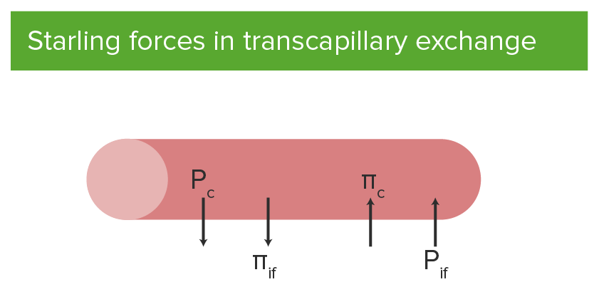
Starling forcesStarling ForcesCapillaries: Histology explain how fluid moves from the blood into extravascular spaces, which leads to pulmonary edemaEdemaEdema is a condition in which excess serous fluid accumulates in the body cavity or interstitial space of connective tissues. Edema is a symptom observed in several medical conditions. It can be categorized into 2 types, namely, peripheral (in the extremities) and internal (in an organ or body cavity). Edema.
PlasmaPlasmaThe residual portion of blood that is left after removal of blood cells by centrifugation without prior blood coagulation.Transfusion ProductscolloidColloidColloid solutions include large proteins or cells that do not readily cross capillary membranes. They remain in the ecf and do not distribute into the icf (similar to crystalloids).Intravenous Fluidsosmotic pressureOsmotic pressureThe pressure required to prevent the passage of solvent through a semipermeable membrane that separates a pure solvent from a solution of the solvent and solute or that separates different concentrations of a solution. It is proportional to the osmolality of the solution.Intravenous Fluids pulls fluid into the capillary.
Interstitial fluidInterstitial fluidBody Fluid CompartmentscolloidColloidColloid solutions include large proteins or cells that do not readily cross capillary membranes. They remain in the ecf and do not distribute into the icf (similar to crystalloids).Intravenous Fluidsosmotic pressureOsmotic pressureThe pressure required to prevent the passage of solvent through a semipermeable membrane that separates a pure solvent from a solution of the solvent and solute or that separates different concentrations of a solution. It is proportional to the osmolality of the solution.Intravenous Fluids pulls fluid out of the capillary.
Starling forces in transcapillary exchange:
Outward forces include hydrostatic pressure of blood in the capillary (Pc) and interstitial fluid colloid osmotic pressure (πif).
Inward forces include hydrostatic pressure of the interstitial fluid (Pif) and plasma colloid osmotic pressure (πc) of the capillary.
Image by Lecturio.
Cardiogenic pulmonary edemaEdemaEdema is a condition in which excess serous fluid accumulates in the body cavity or interstitial space of connective tissues. Edema is a symptom observed in several medical conditions. It can be categorized into 2 types, namely, peripheral (in the extremities) and internal (in an organ or body cavity). Edema
Poor cardiac outputCardiac outputThe volume of blood passing through the heart per unit of time. It is usually expressed as liters (volume) per minute so as not to be confused with stroke volume (volume per beat).Cardiac Mechanics → ↑ atrial pressure
↑ Pulmonary venous pressure
↑ Pulmonary capillary pressure
Fluid moves into the interstitial spaces.
↑ Pressure in the interstitial spaces
Fluid moves into the alveoliAlveoliSmall polyhedral outpouchings along the walls of the alveolar sacs, alveolar ducts and terminal bronchioles through the walls of which gas exchange between alveolar air and pulmonary capillary blood takes place.Acute Respiratory Distress Syndrome (ARDS).
↓ Oxygen diffusionDiffusionThe tendency of a gas or solute to pass from a point of higher pressure or concentration to a point of lower pressure or concentration and to distribute itself throughout the available space. Diffusion, especially facilitated diffusion, is a major mechanism of biological transport.Peritoneal Dialysis and Hemodialysis capacity → hypoxiaHypoxiaSub-optimal oxygen levels in the ambient air of living organisms.Ischemic Cell Damage and symptoms
Noncardiogenic pulmonary edemaEdemaEdema is a condition in which excess serous fluid accumulates in the body cavity or interstitial space of connective tissues. Edema is a symptom observed in several medical conditions. It can be categorized into 2 types, namely, peripheral (in the extremities) and internal (in an organ or body cavity). Edema
Depending on the etiology, the cause of noncardiogenic pulmonary edemaEdemaEdema is a condition in which excess serous fluid accumulates in the body cavity or interstitial space of connective tissues. Edema is a symptom observed in several medical conditions. It can be categorized into 2 types, namely, peripheral (in the extremities) and internal (in an organ or body cavity). Edema may be due to any of the following:
↑ Permeability of pulmonary capillariesCapillariesCapillaries are the primary structures in the circulatory system that allow the exchange of gas, nutrients, and other materials between the blood and the extracellular fluid (ECF). Capillaries are the smallest of the blood vessels. Because a capillary diameter is so small, only 1 RBC may pass through at a time.Capillaries: Histology
Increasing dyspneaDyspneaDyspnea is the subjective sensation of breathing discomfort. Dyspnea is a normal manifestation of heavy physical or psychological exertion, but also may be caused by underlying conditions (both pulmonary and extrapulmonary). Dyspnea (particularly with exertion)
Respiratory distress
Orthopnea
Paroxysmal nocturnal dyspneaDyspneaDyspnea is the subjective sensation of breathing discomfort. Dyspnea is a normal manifestation of heavy physical or psychological exertion, but also may be caused by underlying conditions (both pulmonary and extrapulmonary). Dyspnea
Cough:
Nonproductive
Frothy sputum
Blood tinged
Physical exam
Common findings for pulmonary edemaEdemaEdema is a condition in which excess serous fluid accumulates in the body cavity or interstitial space of connective tissues. Edema is a symptom observed in several medical conditions. It can be categorized into 2 types, namely, peripheral (in the extremities) and internal (in an organ or body cavity). Edema:
TachycardiaTachycardiaAbnormally rapid heartbeat, usually with a heart rate above 100 beats per minute for adults. Tachycardia accompanied by disturbance in the cardiac depolarization (cardiac arrhythmia) is called tachyarrhythmia.Sepsis in Children
Lower extremity edemaEdemaEdema is a condition in which excess serous fluid accumulates in the body cavity or interstitial space of connective tissues. Edema is a symptom observed in several medical conditions. It can be categorized into 2 types, namely, peripheral (in the extremities) and internal (in an organ or body cavity). Edema
Weight gain
Diagnosis
Chest X-rayX-rayPenetrating electromagnetic radiation emitted when the inner orbital electrons of an atom are excited and release radiant energy. X-ray wavelengths range from 1 pm to 10 nm. Hard x-rays are the higher energy, shorter wavelength x-rays. Soft x-rays or grenz rays are less energetic and longer in wavelength. The short wavelength end of the x-ray spectrum overlaps the gamma rays wavelength range. The distinction between gamma rays and x-rays is based on their radiation source.Pulmonary Function Tests
The presence of pulmonary edemaEdemaEdema is a condition in which excess serous fluid accumulates in the body cavity or interstitial space of connective tissues. Edema is a symptom observed in several medical conditions. It can be categorized into 2 types, namely, peripheral (in the extremities) and internal (in an organ or body cavity). Edema can be quickly seen on a chest X-rayX-rayPenetrating electromagnetic radiation emitted when the inner orbital electrons of an atom are excited and release radiant energy. X-ray wavelengths range from 1 pm to 10 nm. Hard x-rays are the higher energy, shorter wavelength x-rays. Soft x-rays or grenz rays are less energetic and longer in wavelength. The short wavelength end of the x-ray spectrum overlaps the gamma rays wavelength range. The distinction between gamma rays and x-rays is based on their radiation source.Pulmonary Function Tests. Subtle differences may be present depending on the etiology.
Cardiogenic pulmonary edemaEdemaEdema is a condition in which excess serous fluid accumulates in the body cavity or interstitial space of connective tissues. Edema is a symptom observed in several medical conditions. It can be categorized into 2 types, namely, peripheral (in the extremities) and internal (in an organ or body cavity). Edema:
Distribution of opacities:
Perihilar (batwing appearance)
Diffuse (central and peripheral)
Lung volume may be ↓
Associated findings:
Prominent vessels; particularly in the upper chest (cephalization)
Kerley B lines:
Thin, linear pulmonary opacities near the chest wallChest wallThe chest wall consists of skin, fat, muscles, bones, and cartilage. The bony structure of the chest wall is composed of the ribs, sternum, and thoracic vertebrae. The chest wall serves as armor for the vital intrathoracic organs and provides the stability necessary for the movement of the shoulders and arms. Chest Wall: Anatomy
CardiomegalyCardiomegalyEnlargement of the heart, usually indicated by a cardiothoracic ratio above 0. 50. Heart enlargement may involve the right, the left, or both heart ventricles or heart atria. Cardiomegaly is a nonspecific symptom seen in patients with chronic systolic heart failure (heart failure) or several forms of cardiomyopathies.Ebstein’s Anomaly
Pleural effusions
Noncardiogenic pulmonary edemaEdemaEdema is a condition in which excess serous fluid accumulates in the body cavity or interstitial space of connective tissues. Edema is a symptom observed in several medical conditions. It can be categorized into 2 types, namely, peripheral (in the extremities) and internal (in an organ or body cavity). Edema:
No cardiomegalyCardiomegalyEnlargement of the heart, usually indicated by a cardiothoracic ratio above 0. 50. Heart enlargement may involve the right, the left, or both heart ventricles or heart atria. Cardiomegaly is a nonspecific symptom seen in patients with chronic systolic heart failure (heart failure) or several forms of cardiomyopathies.Ebstein’s Anomaly
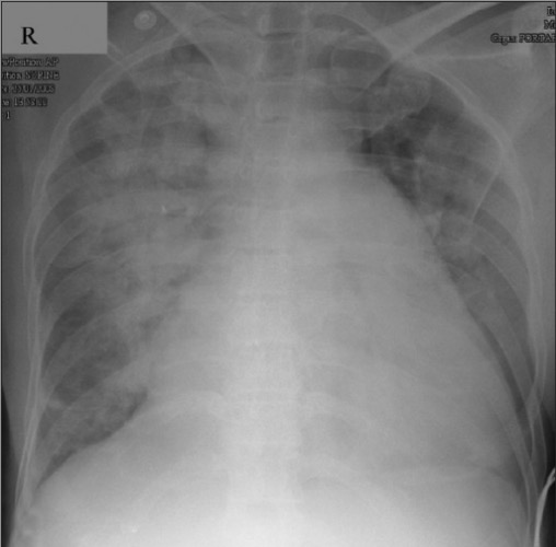
Chest X-ray showing cardiogenic pulmonary edema: Notice the large cardiac silhouette and diffuse lung opacities.
Image: “X-ray chest in postero-anterior view” by Department of Pediatric Cardiology, Amrita Institute of Medical Sciences, Kochi, Kerala, India. License: CC BY 2.0
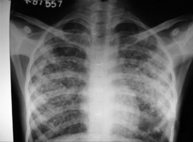
Chest X-ray showing noncardiogenic pulmonary edema: Notice the normal heart size and the distribution of the lung opacities.
Image: “X-ray chest PA view showing pulmonary edema” by Department of Anaesthesiology and Critical Care, R.N.T. Medical College and AG of Hospital, Udaipur – 313 001, Rajasthan, India. License: CC BY 2.0
Supporting workup
Tests can be used to determine the underlying etiology and to rule out a cardiac cause.
Laboratory evaluation:
↑ Troponin → ACS
↑ Creatinine → renal failureRenal failureConditions in which the kidneys perform below the normal level in the ability to remove wastes, concentrate urine, and maintain electrolyte balance; blood pressure; and calcium metabolism. Renal insufficiency can be classified by the degree of kidney damage (as measured by the level of proteinuria) and reduction in glomerular filtration rate.Crush Syndrome
↑ WBC → possible sepsisSepsisSystemic inflammatory response syndrome with a proven or suspected infectious etiology. When sepsis is associated with organ dysfunction distant from the site of infection, it is called severe sepsis. When sepsis is accompanied by hypotension despite adequate fluid infusion, it is called septic shock.Sepsis and Septic Shock or pneumoniaPneumoniaPneumonia or pulmonary inflammation is an acute or chronic inflammation of lung tissue. Causes include infection with bacteria, viruses, or fungi. In more rare cases, pneumonia can also be caused through toxic triggers through inhalation of toxic substances, immunological processes, or in the course of radiotherapy.Pneumonia
BNPBNPA peptide that is secreted by the brain and the heart atria, stored mainly in cardiac ventricular myocardium. It can cause natriuresis; diuresis; vasodilation; and inhibits secretion of renin and aldosterone. It improves heart function. It contains 32 amino acids.Renal Sodium and Water Regulation is usually:
<200 pg/mL in patientsPatientsIndividuals participating in the health care system for the purpose of receiving therapeutic, diagnostic, or preventive procedures.Clinician–Patient Relationship with noncardiogenic pulmonary edemaEdemaEdema is a condition in which excess serous fluid accumulates in the body cavity or interstitial space of connective tissues. Edema is a symptom observed in several medical conditions. It can be categorized into 2 types, namely, peripheral (in the extremities) and internal (in an organ or body cavity). Edema
↑ in heart failureHeart FailureA heterogeneous condition in which the heart is unable to pump out sufficient blood to meet the metabolic need of the body. Heart failure can be caused by structural defects, functional abnormalities (ventricular dysfunction), or a sudden overload beyond its capacity. Chronic heart failure is more common than acute heart failure which results from sudden insult to cardiac function, such as myocardial infarction.Total Anomalous Pulmonary Venous Return (TAPVR)
Imaging:
ECGECGAn electrocardiogram (ECG) is a graphic representation of the electrical activity of the heart plotted against time. Adhesive electrodes are affixed to the skin surface allowing measurement of cardiac impulses from many angles. The ECG provides 3-dimensional information about the conduction system of the heart, the myocardium, and other cardiac structures. Electrocardiogram (ECG):
Evaluate the left ventricular ejection fractionEjection fractionCardiac Cycle (LVEF) → ↓ in left heart failureHeart FailureA heterogeneous condition in which the heart is unable to pump out sufficient blood to meet the metabolic need of the body. Heart failure can be caused by structural defects, functional abnormalities (ventricular dysfunction), or a sudden overload beyond its capacity. Chronic heart failure is more common than acute heart failure which results from sudden insult to cardiac function, such as myocardial infarction.Total Anomalous Pulmonary Venous Return (TAPVR)
Assess for other cardiac abnormalities:
Cardiac tamponadeTamponadePericardial effusion, usually of rapid onset, exceeding ventricular filling pressures and causing collapse of the heart with a markedly reduced cardiac output.Pericarditis
Management of acute pulmonary edemaEdemaEdema is a condition in which excess serous fluid accumulates in the body cavity or interstitial space of connective tissues. Edema is a symptom observed in several medical conditions. It can be categorized into 2 types, namely, peripheral (in the extremities) and internal (in an organ or body cavity). Edema is focused on counteracting the compromised physiologic mechanisms and treating the underlying cause.
Always assess the patient’s airwayAirwayABCDE Assessment, breathing, and circulationCirculationThe movement of the blood as it is pumped through the cardiovascular system.ABCDE Assessment on arrival.
↑ Lung recruitmentRecruitmentSkeletal Muscle Contraction to improve gas exchangeGas exchangeHuman cells are primarily reliant on aerobic metabolism. The respiratory system is involved in pulmonary ventilation and external respiration, while the circulatory system is responsible for transport and internal respiration. Pulmonary ventilation (breathing) represents movement of air into and out of the lungs. External respiration, or gas exchange, is represented by the O2 and CO2 exchange between the lungs and the blood.Gas Exchange
Positive pressure to open the alveoliAlveoliSmall polyhedral outpouchings along the walls of the alveolar sacs, alveolar ducts and terminal bronchioles through the walls of which gas exchange between alveolar air and pulmonary capillary blood takes place.Acute Respiratory Distress Syndrome (ARDS) and prevent collapse due to fluid
Options: continuous positive airwayAirwayABCDE Assessment pressure (CPAPCPAPA technique of respiratory therapy, in either spontaneously breathing or mechanically ventilated patients, in which airway pressure is maintained above atmospheric pressure throughout the respiratory cycle by pressurization of the ventilatory circuit.Noninvasive Ventilation) or bilevel positive airwayAirwayABCDE Assessment pressure (BiPAPBiPAPNoninvasive Ventilation)
Mechanically ventilate those who fail the above options, are obtunded, or are in severe respiratory failureRespiratory failureRespiratory failure is a syndrome that develops when the respiratory system is unable to maintain oxygenation and/or ventilation. Respiratory failure may be acute or chronic and is classified as hypoxemic, hypercapnic, or a combination of the two. Respiratory Failure.
NitroglycerinNitroglycerinA volatile vasodilator which relieves angina pectoris by stimulating guanylate cyclase and lowering cytosolic calcium. It is also sometimes used for tocolysis and explosives.Nitrates:
Use in cardiogenic pulmonary edemaEdemaEdema is a condition in which excess serous fluid accumulates in the body cavity or interstitial space of connective tissues. Edema is a symptom observed in several medical conditions. It can be categorized into 2 types, namely, peripheral (in the extremities) and internal (in an organ or body cavity). Edema for preloadPreloadCardiac Mechanics reduction.
Dilates blood vessels which ↓ pressure on the lungsLungsLungs are the main organs of the respiratory system. Lungs are paired viscera located in the thoracic cavity and are composed of spongy tissue. The primary function of the lungs is to oxygenate blood and eliminate CO2. Lungs: Anatomy
Monitor for hypotensionHypotensionHypotension is defined as low blood pressure, specifically < 90/60 mm Hg, and is most commonly a physiologic response. Hypotension may be mild, serious, or life threatening, depending on the cause. Hypotension.
↑ Urination (removes fluid from the cardiovascular system)
May not be of benefit in some noncardiogenic etiologies
Identify and treat the underlying condition.
PrognosisPrognosisA prediction of the probable outcome of a disease based on a individual’s condition and the usual course of the disease as seen in similar situations.Non-Hodgkin Lymphomas
Depends on the underlying etiology
PatientsPatientsIndividuals participating in the health care system for the purpose of receiving therapeutic, diagnostic, or preventive procedures.Clinician–Patient Relationship with cardiogenic pulmonary edemaEdemaEdema is a condition in which excess serous fluid accumulates in the body cavity or interstitial space of connective tissues. Edema is a symptom observed in several medical conditions. It can be categorized into 2 types, namely, peripheral (in the extremities) and internal (in an organ or body cavity). Edema can quickly improve within a matter of minutes to an hour with rapid action.
PatientsPatientsIndividuals participating in the health care system for the purpose of receiving therapeutic, diagnostic, or preventive procedures.Clinician–Patient Relationship with noncardiogenic pulmonary edemaEdemaEdema is a condition in which excess serous fluid accumulates in the body cavity or interstitial space of connective tissues. Edema is a symptom observed in several medical conditions. It can be categorized into 2 types, namely, peripheral (in the extremities) and internal (in an organ or body cavity). Edema often take longer to improve.
Respiratory and cardiac arrestCardiac arrestCardiac arrest is the sudden, complete cessation of cardiac output with hemodynamic collapse. Patients present as pulseless, unresponsive, and apneic. Rhythms associated with cardiac arrest are ventricular fibrillation/tachycardia, asystole, or pulseless electrical activity. Cardiac Arrest
PneumoniaPneumoniaPneumonia or pulmonary inflammation is an acute or chronic inflammation of lung tissue. Causes include infection with bacteria, viruses, or fungi. In more rare cases, pneumonia can also be caused through toxic triggers through inhalation of toxic substances, immunological processes, or in the course of radiotherapy.Pneumonia: infection of the lung parenchyma most often caused by a bacteriaBacteriaBacteria are prokaryotic single-celled microorganisms that are metabolically active and divide by binary fission. Some of these organisms play a significant role in the pathogenesis of diseases. Bacteriology or virusVirusViruses are infectious, obligate intracellular parasites composed of a nucleic acid core surrounded by a protein capsid. Viruses can be either naked (non-enveloped) or enveloped. The classification of viruses is complex and based on many factors, including type and structure of the nucleoid and capsid, the presence of an envelope, the replication cycle, and the host range. Virology. PatientsPatientsIndividuals participating in the health care system for the purpose of receiving therapeutic, diagnostic, or preventive procedures.Clinician–Patient Relationship present with feverFeverFever is defined as a measured body temperature of at least 38°C (100.4°F). Fever is caused by circulating endogenous and/or exogenous pyrogens that increase levels of prostaglandin E2 in the hypothalamus. Fever is commonly associated with chills, rigors, sweating, and flushing of the skin. Fever, dyspneaDyspneaDyspnea is the subjective sensation of breathing discomfort. Dyspnea is a normal manifestation of heavy physical or psychological exertion, but also may be caused by underlying conditions (both pulmonary and extrapulmonary). Dyspnea, and a productive cough. Chest X-rayX-rayPenetrating electromagnetic radiation emitted when the inner orbital electrons of an atom are excited and release radiant energy. X-ray wavelengths range from 1 pm to 10 nm. Hard x-rays are the higher energy, shorter wavelength x-rays. Soft x-rays or grenz rays are less energetic and longer in wavelength. The short wavelength end of the x-ray spectrum overlaps the gamma rays wavelength range. The distinction between gamma rays and x-rays is based on their radiation source.Pulmonary Function Tests findings usually show lobar consolidationConsolidationPulmonary Function Tests; however, multifocalMultifocalRetinoblastoma infiltrates can be seen in some cases. Management usually involves empiric antibiotics, which can be tailored if the causative organism is identified. Antivirals are used in cases where a viral cause is suspected.
PneumoconiosisPneumoconiosisPneumoconiosis is an occupational disease that results from the inhalation and deposition of mineral dusts and other inorganic particles in the lung. It can be categorized according to the type of causative particle involved or by the type of response provoked.Pneumoconiosis: an occupational disease resulting from inhalation of inorganic particles into the lungsLungsLungs are the main organs of the respiratory system. Lungs are paired viscera located in the thoracic cavity and are composed of spongy tissue. The primary function of the lungs is to oxygenate blood and eliminate CO2. Lungs: Anatomy. PneumoconiosisPneumoconiosisPneumoconiosis is an occupational disease that results from the inhalation and deposition of mineral dusts and other inorganic particles in the lung. It can be categorized according to the type of causative particle involved or by the type of response provoked.Pneumoconiosis can cause chronic inflammationChronic InflammationInflammation and fibrosisFibrosisAny pathological condition where fibrous connective tissue invades any organ, usually as a consequence of inflammation or other injury.Bronchiolitis Obliterans. PatientsPatientsIndividuals participating in the health care system for the purpose of receiving therapeutic, diagnostic, or preventive procedures.Clinician–Patient Relationship will have progressive dyspneaDyspneaDyspnea is the subjective sensation of breathing discomfort. Dyspnea is a normal manifestation of heavy physical or psychological exertion, but also may be caused by underlying conditions (both pulmonary and extrapulmonary). Dyspnea and dry coughDry CoughStrongyloidiasis. Chest X-rayX-rayPenetrating electromagnetic radiation emitted when the inner orbital electrons of an atom are excited and release radiant energy. X-ray wavelengths range from 1 pm to 10 nm. Hard x-rays are the higher energy, shorter wavelength x-rays. Soft x-rays or grenz rays are less energetic and longer in wavelength. The short wavelength end of the x-ray spectrum overlaps the gamma rays wavelength range. The distinction between gamma rays and x-rays is based on their radiation source.Pulmonary Function Tests findings can vary depending on the causative particle but may include ground-glass opacities, calcifications, lung nodules, and pleural irregularities. Management is mainly symptomatic.
Interstitial lung disease: a group of conditions causing progressive fibrosisFibrosisAny pathological condition where fibrous connective tissue invades any organ, usually as a consequence of inflammation or other injury.Bronchiolitis Obliterans of the lungsLungsLungs are the main organs of the respiratory system. Lungs are paired viscera located in the thoracic cavity and are composed of spongy tissue. The primary function of the lungs is to oxygenate blood and eliminate CO2. Lungs: Anatomy. PatientsPatientsIndividuals participating in the health care system for the purpose of receiving therapeutic, diagnostic, or preventive procedures.Clinician–Patient Relationship may be asymptomatic or have a gradual onset of dyspneaDyspneaDyspnea is the subjective sensation of breathing discomfort. Dyspnea is a normal manifestation of heavy physical or psychological exertion, but also may be caused by underlying conditions (both pulmonary and extrapulmonary). Dyspnea and cough. Chest X-rayX-rayPenetrating electromagnetic radiation emitted when the inner orbital electrons of an atom are excited and release radiant energy. X-ray wavelengths range from 1 pm to 10 nm. Hard x-rays are the higher energy, shorter wavelength x-rays. Soft x-rays or grenz rays are less energetic and longer in wavelength. The short wavelength end of the x-ray spectrum overlaps the gamma rays wavelength range. The distinction between gamma rays and x-rays is based on their radiation source.Pulmonary Function Tests findings can vary from the infiltrate pattern to the location within the lungsLungsLungs are the main organs of the respiratory system. Lungs are paired viscera located in the thoracic cavity and are composed of spongy tissue. The primary function of the lungs is to oxygenate blood and eliminate CO2. Lungs: Anatomy, depending on the etiology. CT and biopsyBiopsyRemoval and pathologic examination of specimens from the living body.Ewing Sarcoma may be needed for the diagnosis. Management depends on the etiology but can include steroidsSteroidsA group of polycyclic compounds closely related biochemically to terpenes. They include cholesterol, numerous hormones, precursors of certain vitamins, bile acids, alcohols (sterols), and certain natural drugs and poisons. Steroids have a common nucleus, a fused, reduced 17-carbon atom ring system, cyclopentanoperhydrophenanthrene. Most steroids also have two methyl groups and an aliphatic side-chain attached to the nucleus.Benign Liver Tumors and immunosuppressive therapies.
Diffuse alveolar hemorrhage: pulmonary hemorrhage due to damage to the pulmonary vessels. Etiologies can include systemic vasculitidesVasculitidesVasculitides are a group of conditions characterized by vasculitis, ischemia, and damage to the organs supplied by the affected vessels. The affected arteries are of different sizes and locations and vary by the type of vasculitis. Vasculitides, pulmonary infectionsInfectionsInvasion of the host organism by microorganisms or their toxins or by parasites that can cause pathological conditions or diseases.Chronic Granulomatous Disease, toxic exposures, and coagulation disorders. PatientsPatientsIndividuals participating in the health care system for the purpose of receiving therapeutic, diagnostic, or preventive procedures.Clinician–Patient Relationship may present with dyspneaDyspneaDyspnea is the subjective sensation of breathing discomfort. Dyspnea is a normal manifestation of heavy physical or psychological exertion, but also may be caused by underlying conditions (both pulmonary and extrapulmonary). Dyspnea, hemoptysisHemoptysisHemoptysis is defined as the expectoration of blood originating in the lower respiratory tract. Hemoptysis is a consequence of another disease process and can be classified as either life threatening or non-life threatening. Hemoptysis can result in significant morbidity and mortality due to both drowning (reduced gas exchange as the lungs fill with blood) and hemorrhagic shock. Hemoptysis, or respiratory failureRespiratory failureRespiratory failure is a syndrome that develops when the respiratory system is unable to maintain oxygenation and/or ventilation. Respiratory failure may be acute or chronic and is classified as hypoxemic, hypercapnic, or a combination of the two. Respiratory Failure. A chest X-rayX-rayPenetrating electromagnetic radiation emitted when the inner orbital electrons of an atom are excited and release radiant energy. X-ray wavelengths range from 1 pm to 10 nm. Hard x-rays are the higher energy, shorter wavelength x-rays. Soft x-rays or grenz rays are less energetic and longer in wavelength. The short wavelength end of the x-ray spectrum overlaps the gamma rays wavelength range. The distinction between gamma rays and x-rays is based on their radiation source.Pulmonary Function Tests will show diffuse alveolar infiltrates. BronchoscopyBronchoscopyEndoscopic examination, therapy or surgery of the bronchi.Laryngomalacia and Tracheomalacia confirms the diagnosis. Management includes respiratory support while the underlying cause is determined and treated.
Naum, R., Filatov, A., Alusma-Hibbert, K., & Espinosa, P. S. (2020). Pulmonary Edema and Stunned Myocardium in Subarachnoid Hemorrhage. Cureus, 12(4), e7746. https://doi.org/10.7759/cureus.7746
Siddamreddy, S., Thotakura, R., Dandu, V., Kanuru, S., & Meegada, S. (2020). Corona Virus Disease 2019 (COVID-19) Presenting as Acute ST Elevation Myocardial Infarction. Cureus, 12(4), e7782. https://doi.org/10.7759/cureus.7782