Bundle branch and fascicular blocks occur when the normal electrical activity in the His-Purkinje system is interrupted. These blocks can be due to many etiologies that may affect the structure of the heart or the conduction system directly. The blocks are classified into right bundle branch block, left bundle branch block, left anterior fascicular block, and left posterior fascicular block depending on the location of the disruption. Most individuals are asymptomatic. ECG will provide the diagnosis. Some common ECG findings include a prolonged QRS interval, R-wave changes, axis deviation, and (in some cases) S-wave changes. No specific treatment is indicated.
Last updated: Jan 7, 2026
Bundle branch and fascicular blocks Fascicular Blocks Bundle branch and fascicular blocks occur when the normal electrical activity in the His-Purkinje system is interrupted. These blocks can be due to many etiologies that may affect the structure of the heart or the conduction system directly. Bundle Branch and Fascicular Blocks are classified on the basis of where the disruption occurs within the His-Purkinje system.

Bundle branch and fascicular blocks arise because of obstruction of electrical current through the His-Purkinje system and are named on the basis of the location of that disruption.
Image by Lecturio.These fascicular blocks Fascicular Blocks Bundle branch and fascicular blocks occur when the normal electrical activity in the His-Purkinje system is interrupted. These blocks can be due to many etiologies that may affect the structure of the heart or the conduction system directly. Bundle Branch and Fascicular Blocks can occur because of many of the same causes of RBBB RBBB Bundle Branch and Fascicular Blocks or LBBB LBBB Bundle Branch and Fascicular Blocks, most notably:
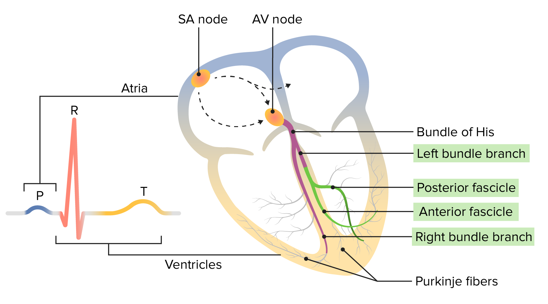
Normal electrophysiology of the heart:
The conduction impulse starts at the sinoatrial (SA) node and travels through the atrium to the atrioventricular (AV) node. From there, it moves through the bundle of His down through both bundle branches (and fascicles) to the Purkinje fibers. The movement of this electrical impulse can be recorded on an ECG.
P wave (blue): depolarization of atrial myocardium
QRS complex (orange): depolarization of ventricular myocardium
T wave (yellow): repolarization of ventricular myocardium
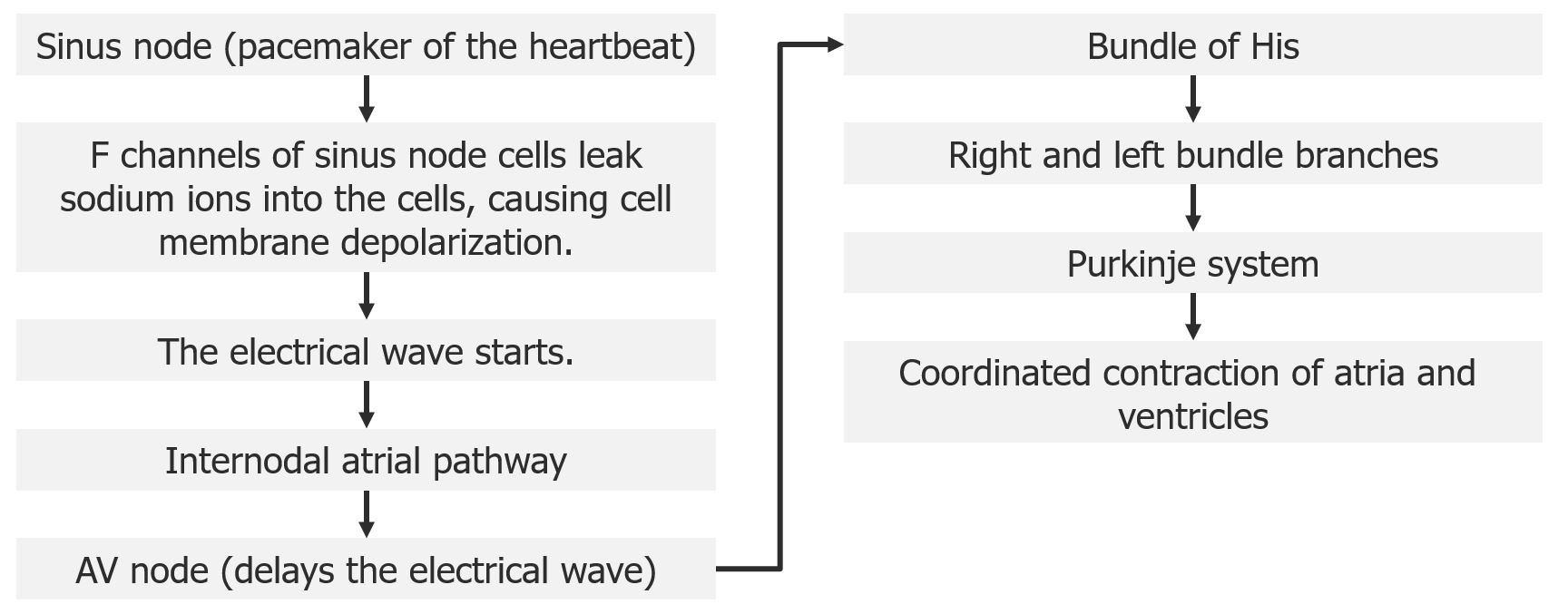
Diagram outlining the electrical pathway through the heart
Image by Lecturio.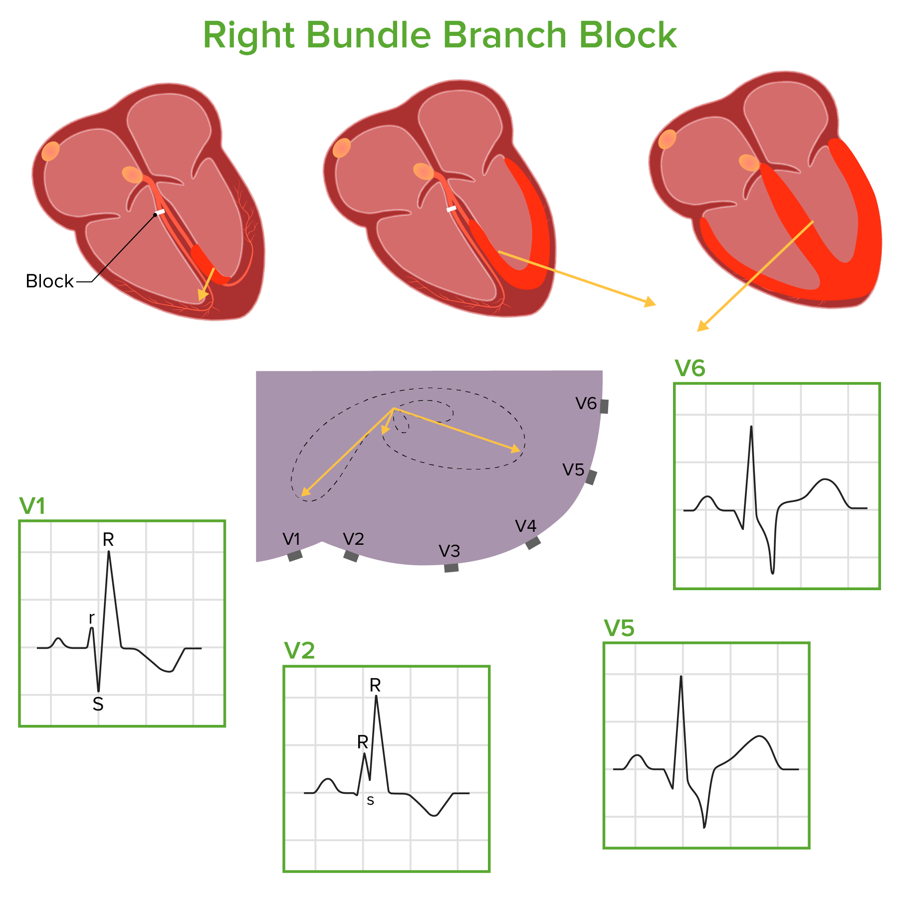
Diagram demonstrating how electrical conduction moves through the ventricles in a right bundle branch block:
The electrical impulse runs through the left bundle branch, through the septum and left ventricle, and then through the right ventricle. These phases result in the electrical conduction vectors shown above (drawn in relation to a cross section of the thorax with the precordial leads attached), which correlate with the corresponding ECG waveforms.
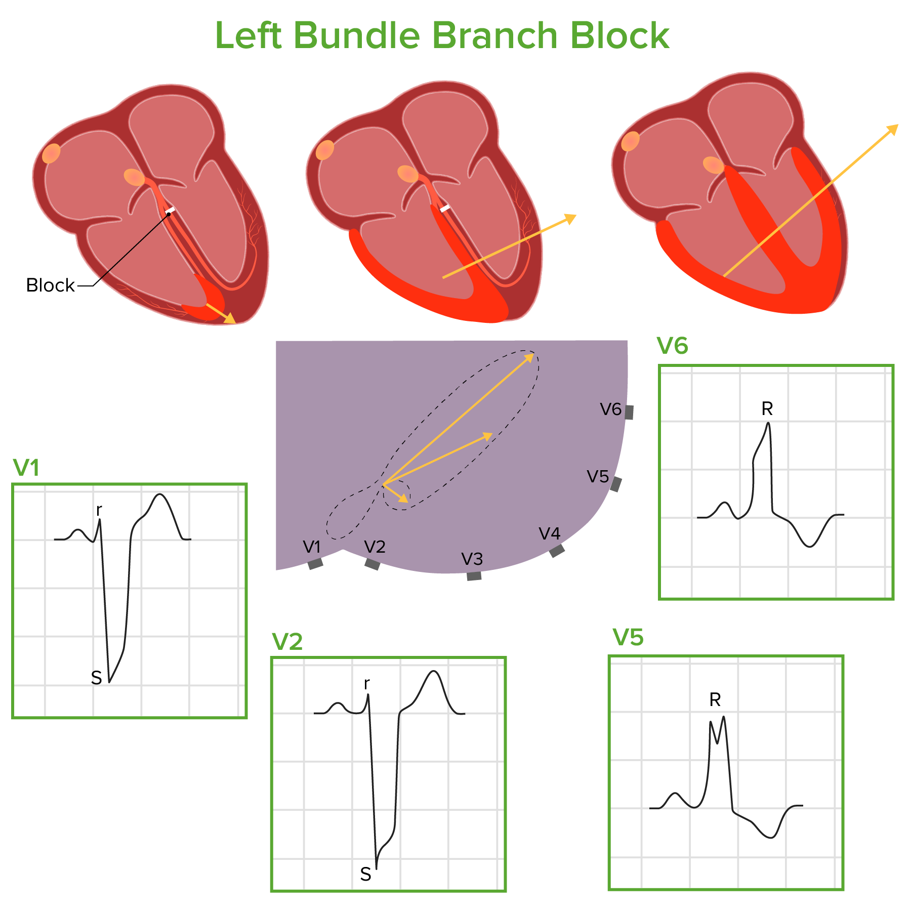
Diagram demonstrating how electrical conduction moves through the ventricles in a left bundle branch block:
The electrical impulse runs through the right bundle branch, through the septum and right ventricle, and then through the left ventricle. These phases result in the electrical conduction vectors shown above (drawn in relation to a cross section of the thorax with the precordial leads attached), which correlate with the corresponding ECG waveforms.
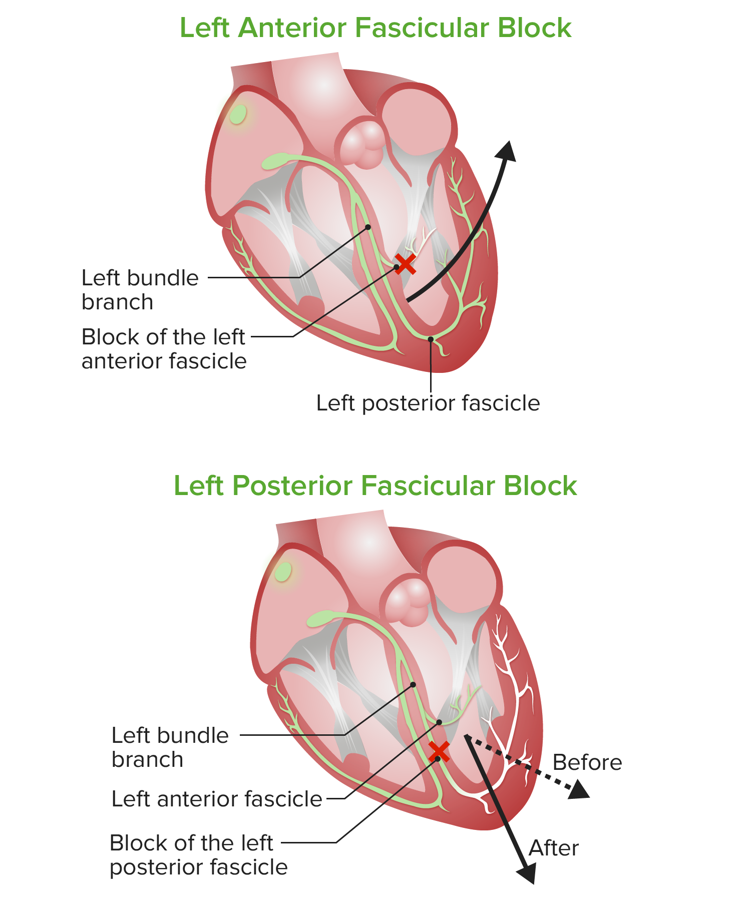
Diagram of left anterior and posterior fascicular blocks:
In left anterior fascicular block or hemiblock (LAFB), the resultant electrical vector results in significant left axis deviation. In left posterior fascicular block or hemiblock (LPFB), the electrical vector is deviated a bit rightward but is not significantly displaced from the normal QRS axis range.
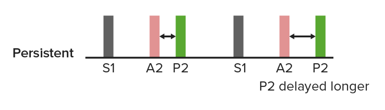
A diagram of a persistent split S2, in which closure of the pulmonic valve is delayed further by inspiration (right). This can occur in a right bundle branch block.
Image by Lecturio.Audio:
This audio clip is an example of a split S2 S2 Heart Sounds in the setting of an RBBB RBBB Bundle Branch and Fascicular Blocks. The 2 sounds occurring during S2 S2 Heart Sounds result from delayed closure Delayed Closure Gastroschisis of the pulmonic valve in relation to the aortic valve Aortic valve The valve between the left ventricle and the ascending aorta which prevents backflow into the left ventricle. Heart: Anatomy.
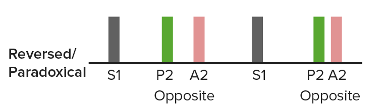
Diagram showing paradoxical splitting in which closure of the aortic valve, which is delayed: The name “paradoxical” is because the split narrows the inspiration (right). The split can be heard in some individuals with a left bundle branch block.
Image by Lecturio.
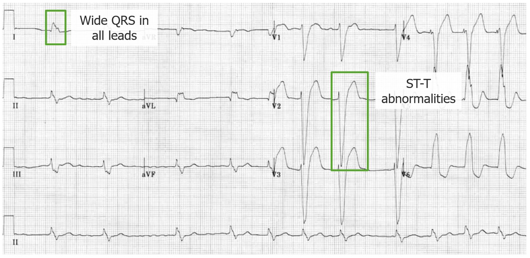
QRS morphology seen in right bundle branch block:
The RsR’ (and variations of this) give the appearance of “rabbit ears.” The S wave in leads I and V6 will appear broad, deep, and slurred.
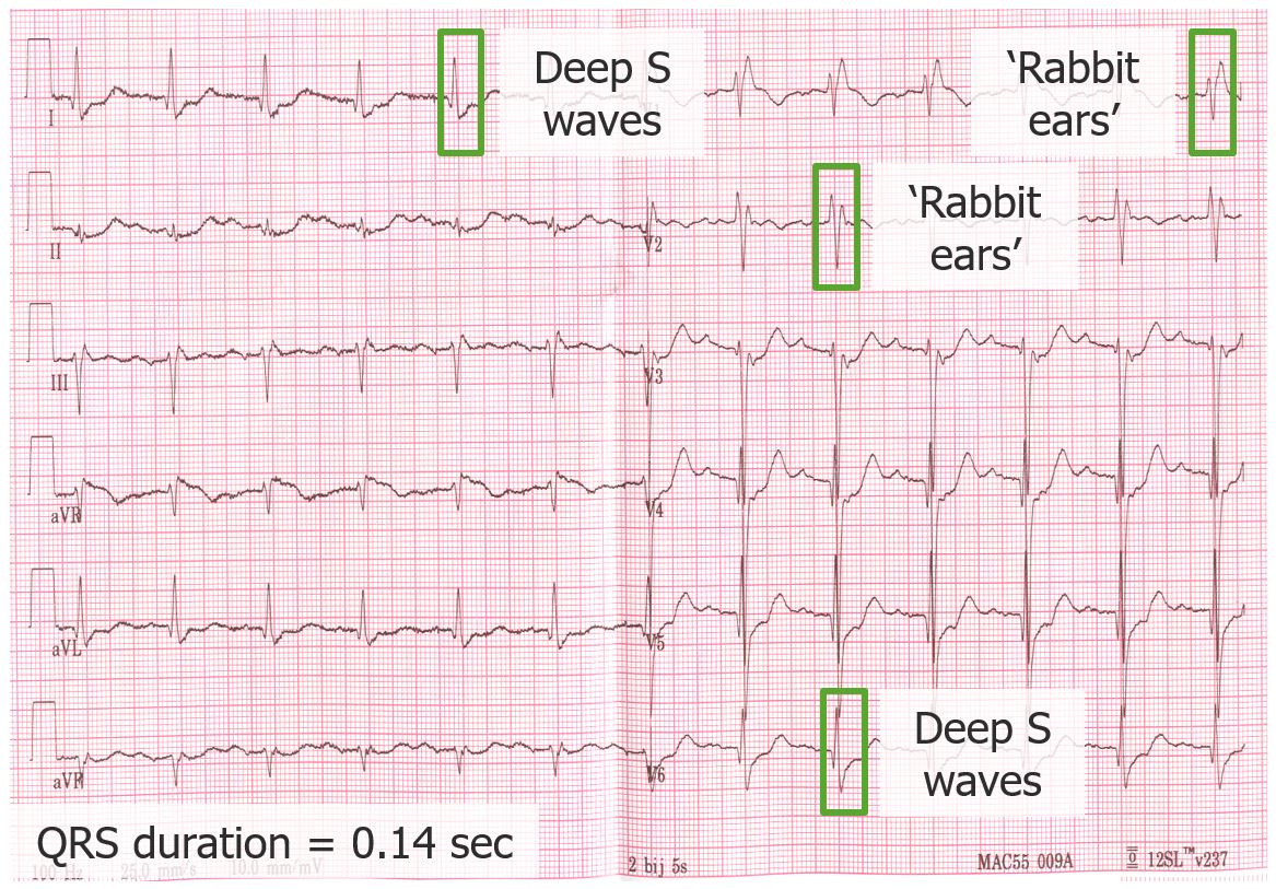
ECG demonstrating a right bundle branch block:
The QRS duration is prolonged, at 140 msec. Note the rSR’ and RSr’ in leads V1 and 2, along with the deep and broad S waves in I and V6.
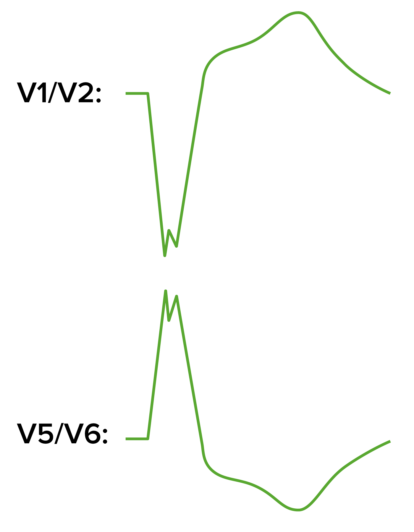
ECG patterns in left bundle branch block:
A large S wave will be seen in V1, while a large, notched R wave occurs in V5 and V6. Note that the ST-segment and T-wave directions are discordant to the QRS.
ECG demonstrating a left bundle branch block:
Note the widened QRS; the large, notched R waves in V5 and V6; and the large, broad S waves in V1 and 2. The ST segments and T waves are also generally discordant to the QRS complex.
A bundle branch block Bundle branch block A form of heart block in which the electrical stimulation of heart ventricles is interrupted at either one of the branches of bundle of His thus preventing the simultaneous depolarization of the two ventricles. Bundle Branch and Fascicular Blocks may be considered incomplete if the usual RBBB RBBB Bundle Branch and Fascicular Blocks or LBBB LBBB Bundle Branch and Fascicular Blocks pattern is seen but the QRS duration is 110–119 msec.

ECG demonstrating left anterior fascicular block:
Here, the axis is deviated to –60 degrees and a small Q wave is noted in aVL. The QRS is slightly prolonged, but still < 120 msec.
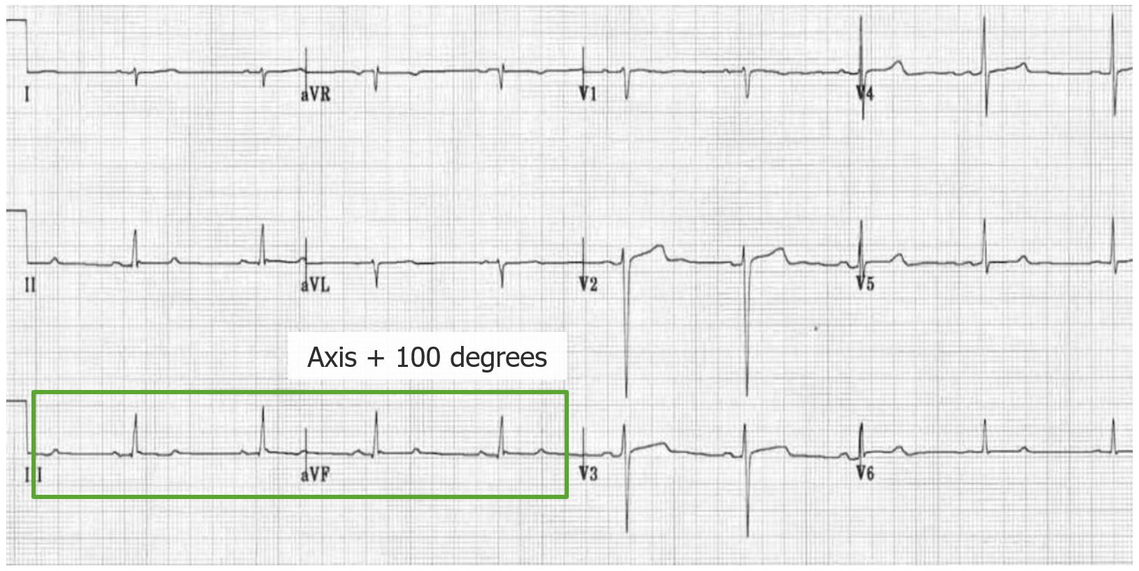
ECG demonstrating a left posterior fascicular block:
There is right axis deviation (+ 100 degrees), small Q waves in II, III, and aVF, rS complexes in I and aVL. The QRS complex duration is also < 120 msec.
Management may vary depending on practice location and is generally guided by cardiology consultation. The following information is based on the most recent US and European guidelines.
Diagnosis Codes:
These codes are used to specify the type of electrical conduction block within the heart’s ventricles, such as a Right Bundle Branch Block Right bundle branch block Bundle Branch and Fascicular Blocks ( RBBB RBBB Bundle Branch and Fascicular Blocks) or a Left Bundle Branch Block Left bundle branch block Bundle Branch and Fascicular Blocks ( LBBB LBBB Bundle Branch and Fascicular Blocks), which are diagnosed via an electrocardiogram Electrocardiogram An electrocardiogram (ECG) is a graphic representation of the electrical activity of the heart plotted against time. Adhesive electrodes are affixed to the skin surface allowing measurement of cardiac impulses from many angles. The ECG provides 3-dimensional information about the conduction system of the heart, the myocardium, and other cardiac structures. Electrocardiogram (ECG).
| Domain | Code | Description |
|---|---|---|
| ICD-10-CM | I44.7 | Left bundle-branch block, unspecified |
| ICD-10-CM | I45.10 | Unspecified right bundle-branch block |
| ICD-10-CM | I44.4 | Left anterior fascicular block Left anterior fascicular block Bundle Branch and Fascicular Blocks |
| ICD-10-CM | I45.0 | Right fascicular block |
| SNOMED CT | 164908000 | Right bundle branch block Right bundle branch block Bundle Branch and Fascicular Blocks (disorder) |
| SNOMED CT | 164909008 | Left bundle branch block Left bundle branch block Bundle Branch and Fascicular Blocks (disorder) |
Evaluation & Workup:
This CPT code is for a 12-lead electrocardiogram Electrocardiogram An electrocardiogram (ECG) is a graphic representation of the electrical activity of the heart plotted against time. Adhesive electrodes are affixed to the skin surface allowing measurement of cardiac impulses from many angles. The ECG provides 3-dimensional information about the conduction system of the heart, the myocardium, and other cardiac structures. Electrocardiogram (ECG) ( ECG ECG An electrocardiogram (ECG) is a graphic representation of the electrical activity of the heart plotted against time. Adhesive electrodes are affixed to the skin surface allowing measurement of cardiac impulses from many angles. The ECG provides 3-dimensional information about the conduction system of the heart, the myocardium, and other cardiac structures. Electrocardiogram (ECG)), which is the essential and definitive diagnostic tool used to identify the characteristic patterns of a bundle branch or fascicular block.
| Domain | Code | Description |
|---|---|---|
| CPT | 93000 | Electrocardiogram Electrocardiogram An electrocardiogram (ECG) is a graphic representation of the electrical activity of the heart plotted against time. Adhesive electrodes are affixed to the skin surface allowing measurement of cardiac impulses from many angles. The ECG provides 3-dimensional information about the conduction system of the heart, the myocardium, and other cardiac structures. Electrocardiogram (ECG), routine ECG ECG An electrocardiogram (ECG) is a graphic representation of the electrical activity of the heart plotted against time. Adhesive electrodes are affixed to the skin surface allowing measurement of cardiac impulses from many angles. The ECG provides 3-dimensional information about the conduction system of the heart, the myocardium, and other cardiac structures. Electrocardiogram (ECG) with at least 12 leads; with interpretation and report |
Procedures/Interventions:
This CPT code is used for the implantation Implantation Endometrial implantation of embryo, mammalian at the blastocyst stage. Fertilization and First Week of a permanent pacemaker Pacemaker A device designed to stimulate, by electric impulses, contraction of the heart muscles. It may be temporary (external) or permanent (internal or internal-external). Bradyarrhythmias, which may be required if a bundle branch block Bundle branch block A form of heart block in which the electrical stimulation of heart ventricles is interrupted at either one of the branches of bundle of His thus preventing the simultaneous depolarization of the two ventricles. Bundle Branch and Fascicular Blocks progresses to a high-grade or complete atrioventricular (AV) block, causing symptomatic bradycardia Bradycardia Bradyarrhythmia is a rhythm in which the heart rate is less than 60/min. Bradyarrhythmia can be physiologic, without symptoms or hemodynamic change. Pathologic bradyarrhythmia results in reduced cardiac output and hemodynamic instability causing syncope, dizziness, or dyspnea. Bradyarrhythmias.
| Domain | Code | Description |
|---|---|---|
| CPT | 33208 | Insertion of new or replacement of permanent pacemaker Pacemaker A device designed to stimulate, by electric impulses, contraction of the heart muscles. It may be temporary (external) or permanent (internal or internal-external). Bradyarrhythmias with transvenous electrode(s); atrial and ventricular |
Complications & Supportive Procedures:
This ICD-10 code is used to document the most significant complication of a bifascicular or trifascicular block, which is the progression to a complete AV block Complete AV block Atrioventricular block (AV block), a serious condition where electrical signals from the atria do not reach the ventricles.
| Domain | Code | Description |
|---|---|---|
| ICD-10-CM | I44.2 | Atrioventricular block Atrioventricular block Atrioventricular (AV) block is a bradyarrhythmia caused by delay, or interruption, in the electrical conduction between the atria and the ventricles. Atrioventricular block occurs due to either anatomic or functional impairment, and is classified into 3 types. Atrioventricular block (AV block), complete |