Horner syndrome is a condition resulting from an interruption of the sympathetic innervation of the eyes. The syndrome is usually idiopathic but can be directly caused by head and neck trauma, cerebrovascular disease, or a tumor of the CNS. Horner syndrome is classified as 1st-order (central), 2nd-order (preganglionic), or 3rd-order (postganglionic) based on the location of the lesion along the sympathetic pathway. Partial ptosis, miosis, and facial anhidrosis are the classical signs of Horner syndrome, making up a characteristic triad. Other associated neurologic signs may also be present depending on the location of the lesion and can aid in determining the cause. The syndrome is diagnosed by using cocaine, apraclonidine, or hydroxyamphetamine eye drops. Management of Horner syndrome requires treatment of the underlying condition.
Horner syndromeHorner syndromeHorner syndrome is a condition resulting from an interruption of the sympathetic innervation of the eyes. The syndrome is usually idiopathic but can be directly caused by head and neck trauma, cerebrovascular disease, or a tumor of the CNS. Horner Syndrome, also known as oculosympathetic paresisParesisA general term referring to a mild to moderate degree of muscular weakness, occasionally used as a synonym for paralysis (severe or complete loss of motor function). In the older literature, paresis often referred specifically to paretic neurosyphilis. ‘general paresis’ and ‘general paralysis’ may still carry that connotation. Bilateral lower extremity paresis is referred to as paraparesis.Spinal Disk Herniation, is a condition resulting from the interruption of the sympathetic innervation to the eyes. The syndrome is characterized by the classic triad of:
MiosisMiosisPupil: Physiology and Abnormalities (constricted pupilPupilThe pupil is the space within the eye that permits light to project onto the retina. Anatomically located in front of the lens, the pupil’s size is controlled by the surrounding iris. The pupil provides insight into the function of the central and autonomic nervous systems. Pupil: Physiology and Abnormalities)
Can affect any age, sexSexThe totality of characteristics of reproductive structure, functions, phenotype, and genotype, differentiating the male from the female organism.Gender Dysphoria, or ethnicity
Frequency: approximately 1 per 6000 individuals
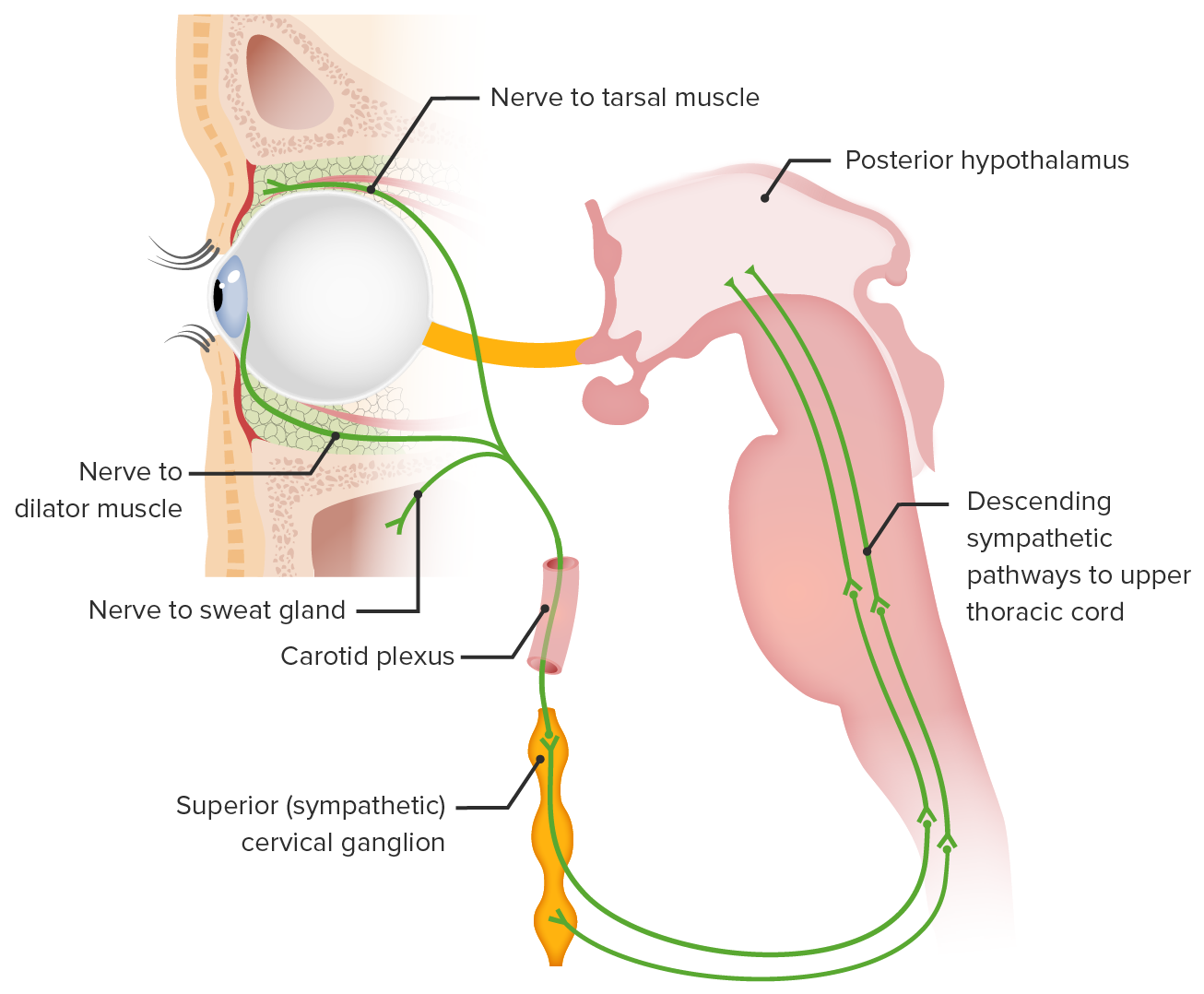
Neuroanatomy[1,4,6,7]
Horner syndromeHorner syndromeHorner syndrome is a condition resulting from an interruption of the sympathetic innervation of the eyes. The syndrome is usually idiopathic but can be directly caused by head and neck trauma, cerebrovascular disease, or a tumor of the CNS. Horner Syndrome can result from a lesion anywhere on the 3-neuron sympathetic pathway supplying the eye. The nerve supply starts from the posterolateral hypothalamusHypothalamusThe hypothalamus is a collection of various nuclei within the diencephalon in the center of the brain. The hypothalamus plays a vital role in endocrine regulation as the primary regulator of the pituitary gland, and it is the major point of integration between the central nervous and endocrine systems.Hypothalamus and ends as the long ciliary nerves that supply the iris dilator and Müller muscles (superior tarsal muscle).
1st-order neuron1st-order neuronOriginates in the hypothalamus and descends to the first synapse in the cervical spinal cord, located at levels C8–T2 (also called the ciliospinal center of Budge).Horner Syndrome: originates in the hypothalamusHypothalamusThe hypothalamus is a collection of various nuclei within the diencephalon in the center of the brain. The hypothalamus plays a vital role in endocrine regulation as the primary regulator of the pituitary gland, and it is the major point of integration between the central nervous and endocrine systems.Hypothalamus and descends to the first synapseSynapseThe junction between 2 neurons is called a synapse. The synapse allows a neuron to pass an electrical or chemical signal to another neuron or target effector cell. Synapses and Neurotransmission in the cervical spinal cordSpinal cordThe spinal cord is the major conduction pathway connecting the brain to the body; it is part of the CNS. In cross section, the spinal cord is divided into an H-shaped area of gray matter (consisting of synapsing neuronal cell bodies) and a surrounding area of white matter (consisting of ascending and descending tracts of myelinated axons). Spinal Cord: Anatomy, located at levels C8–T2 (also called the ciliospinal center of Budge).
2nd-order neuron2nd-order neuronPreganglionic pupillomotor fibers exit the spinal cord at T1, travel through the brachial plexus, over the lung apex, ascending to the superior cervical ganglion located near the angle of the mandible and the bifurcation of the common carotid artery.Horner Syndrome: Preganglionic pupillomotor fibers exit the spinal cordSpinal cordThe spinal cord is the major conduction pathway connecting the brain to the body; it is part of the CNS. In cross section, the spinal cord is divided into an H-shaped area of gray matter (consisting of synapsing neuronal cell bodies) and a surrounding area of white matter (consisting of ascending and descending tracts of myelinated axons). Spinal Cord: Anatomy at T1, travel through the brachial plexusBrachial PlexusThe large network of nerve fibers which distributes the innervation of the upper extremity. The brachial plexus extends from the neck into the axilla. In humans, the nerves of the plexus usually originate from the lower cervical and the first thoracic spinal cord segments (c5-c8 and T1), but variations are not uncommon.Peripheral Nerve Injuries in the Cervicothoracic Region, over the lung apex, ascending to the superior cervical ganglion located near the angle of the mandibleMandibleThe largest and strongest bone of the face constituting the lower jaw. It supports the lower teeth.Jaw and Temporomandibular Joint: Anatomy and the bifurcation of the common carotid arteryCommon carotid arteryThe two principal arteries supplying the structures of the head and neck. They ascend in the neck, one on each side, and at the level of the upper border of the thyroid cartilage, each divides into two branches, the external and internal carotid arteries.Carotid Arterial System: Anatomy.
3rd-order neuron3rd-order neuronPupillomotor fibers ascend along the internal carotid artery and enter the cavernous sinus where it is in close relation with the abducens nerve (cranial nerve (CN) VI). These fibers enter the orbit with the ophthalmic branch (V1) of the trigeminal nerve (CN V) via the long ciliary nerves, which innervate the iris dilator and Müller muscles.Horner Syndrome: Pupillomotor fibers ascend along the internal carotid arteryInternal carotid arteryBranch of the common carotid artery which supplies the anterior part of the brain, the eye and its appendages, the forehead and nose.Carotid Arterial System: Anatomy and enter the cavernous sinus where it is in close relation with the abducens nerveAbducens nerveThe 6th cranial nerve which originates in the abducens nucleus of the pons and sends motor fibers to the lateral rectus muscles of the eye. Damage to the nerve or its nucleus disrupts horizontal eye movement control.The 12 Cranial Nerves: Overview and Functions (cranial nerve (CN) VI). These fibers enter the orbit with the ophthalmic branch (V1) of the trigeminal nerveTrigeminal nerveThe 5th and largest cranial nerve. The trigeminal nerve is a mixed motor and sensory nerve. The larger sensory part forms the ophthalmic, mandibular, and maxillary nerves which carry afferents sensitive to external or internal stimuli from the skin, muscles, and joints of the face and mouth and from the teeth. Most of these fibers originate from cells of the trigeminal ganglion and project to the trigeminal nucleus of the brain stem. The smaller motor part arises from the brain stem trigeminal motor nucleus and innervates the muscles of mastication.The 12 Cranial Nerves: Overview and Functions (CN V) via the long ciliary nerves, which innervate the iris dilator and Müller muscles.
The neural circuitry involved in Horner syndrome:
1) Posterior hypothalamic sympathetic fibers synapse at the ciliospinal center of Budge (C8–T2).
2) Preganglionic fibers traverse the brachial plexus to synapse at the superior cervical ganglion.
3) Postganglionic fibers ascend in the carotid sheath to innervate the target structures.
1st-order, or central, Horner syndromeHorner syndromeHorner syndrome is a condition resulting from an interruption of the sympathetic innervation of the eyes. The syndrome is usually idiopathic but can be directly caused by head and neck trauma, cerebrovascular disease, or a tumor of the CNS. Horner Syndrome: caused by lesions of the sympathetic tracts in the brainstem or cervicothoracic spinal cordSpinal cordThe spinal cord is the major conduction pathway connecting the brain to the body; it is part of the CNS. In cross section, the spinal cord is divided into an H-shaped area of gray matter (consisting of synapsing neuronal cell bodies) and a surrounding area of white matter (consisting of ascending and descending tracts of myelinated axons). Spinal Cord: Anatomy
2nd-order, or preganglionic, Horner syndromeHorner syndromeHorner syndrome is a condition resulting from an interruption of the sympathetic innervation of the eyes. The syndrome is usually idiopathic but can be directly caused by head and neck trauma, cerebrovascular disease, or a tumor of the CNS. Horner Syndrome: caused by lesions that involve the spinal cordSpinal cordThe spinal cord is the major conduction pathway connecting the brain to the body; it is part of the CNS. In cross section, the spinal cord is divided into an H-shaped area of gray matter (consisting of synapsing neuronal cell bodies) and a surrounding area of white matter (consisting of ascending and descending tracts of myelinated axons). Spinal Cord: Anatomy, thoracic outletThoracic OutletThoracic Outlet Syndrome, or lung apex. These lesions are usually acquired through trauma, surgery, or malignancyMalignancyHemothorax (e.g., Pancoast tumorPancoast TumorThoracic Outlet Syndrome).
3rd-order, or postganglionic, Horner syndromeHorner syndromeHorner syndrome is a condition resulting from an interruption of the sympathetic innervation of the eyes. The syndrome is usually idiopathic but can be directly caused by head and neck trauma, cerebrovascular disease, or a tumor of the CNS. Horner Syndrome: caused by lesions of the internal carotid arteryInternal carotid arteryBranch of the common carotid artery which supplies the anterior part of the brain, the eye and its appendages, the forehead and nose.Carotid Arterial System: Anatomy, such as arterial dissectionArterial dissectionArterial dissection is a violation of the structural integrity of the arterial wall that results in blood accumulating between the layers.Dissection of the Carotid and Vertebral Arteries, thrombosisThrombosisFormation and development of a thrombus or blood clot in the blood vessel.Epidemic Typhus, cavernous sinus aneurysmAneurysmAn aneurysm is a bulging, weakened area of a blood vessel that causes an abnormal widening of its diameter > 1.5 times the size of the native vessel. Aneurysms occur more often in arteries than in veins and are at risk of dissection and rupture, which can be life-threatening. Thoracic Aortic Aneurysms, or injuries acquired during carotid artery stenting
Etiology
Most cases of Horner syndromeHorner syndromeHorner syndrome is a condition resulting from an interruption of the sympathetic innervation of the eyes. The syndrome is usually idiopathic but can be directly caused by head and neck trauma, cerebrovascular disease, or a tumor of the CNS. Horner Syndrome are idiopathicIdiopathicDermatomyositis. Of the identified causes, the etiology depends on the location of the lesion. The causes vary between adult and pediatric populations.
1st-order syndrome (central)[1–4,6,7]
HypothalamusHypothalamusThe hypothalamus is a collection of various nuclei within the diencephalon in the center of the brain. The hypothalamus plays a vital role in endocrine regulation as the primary regulator of the pituitary gland, and it is the major point of integration between the central nervous and endocrine systems.Hypothalamus:
DemyelinationDemyelinationMultiple Sclerosis (e.g., multiple sclerosisSclerosisA pathological process consisting of hardening or fibrosis of an anatomical structure, often a vessel or a nerve.Wilms Tumor)
Spinal cordSpinal cordThe spinal cord is the major conduction pathway connecting the brain to the body; it is part of the CNS. In cross section, the spinal cord is divided into an H-shaped area of gray matter (consisting of synapsing neuronal cell bodies) and a surrounding area of white matter (consisting of ascending and descending tracts of myelinated axons). Spinal Cord: Anatomy:
NeckNeckThe part of a human or animal body connecting the head to the rest of the body.Peritonsillar Abscess trauma
MyelitisMyelitisInflammation of the spinal cord. Relatively common etiologies include infections; autoimmune diseases; spinal cord; and ischemia. Clinical features generally include weakness, sensory loss, localized pain, incontinence, and other signs of autonomic dysfunction.Relapsing Fever
SyringomyeliaSyringomyeliaLongitudinal cavities in the spinal cord, most often in the cervical region, which may extend for multiple spinal levels. The cavities are lined by dense, gliogenous tissue and may be associated with spinal cord neoplasms; spinal cord traumatic injuries; and vascular malformations. Syringomyelia is marked clinically by pain and paresthesia, muscular atrophy of the hands, and analgesia with thermoanesthesia of the hands and arms, but with the tactile sense preserved (sensory dissociation). Lower extremity spasticity and incontinence may also develop.Central Cord Syndrome
DemyelinationDemyelinationMultiple Sclerosis (e.g., multiple sclerosisSclerosisA pathological process consisting of hardening or fibrosis of an anatomical structure, often a vessel or a nerve.Wilms Tumor)
Arteriovenous malformationArteriovenous malformationAbnormal formation of blood vessels that shunt arterial blood directly into veins without passing through the capillaries. They usually are crooked, dilated, and with thick vessel walls. A common type is the congenital arteriovenous fistula. The lack of blood flow and oxygen in the capillaries can lead to tissue damage in the affected areas.Erysipelas (AVM)
Infarction
Arnold-Chiari malformation
EncephalitisEncephalitisEncephalitis is inflammation of the brain parenchyma caused by an infection, usually viral. Encephalitis may present with mild symptoms such as headache, fever, fatigue, and muscle and joint pain or with severe symptoms such as seizures, altered consciousness, and paralysis.Encephalitis
ThyroidThyroidThe thyroid gland is one of the largest endocrine glands in the human body. The thyroid gland is a highly vascular, brownish-red gland located in the visceral compartment of the anterior region of the neck.Thyroid Gland: Anatomy malignancies
Birth trauma with injury to lower brachial plexusBrachial PlexusThe large network of nerve fibers which distributes the innervation of the upper extremity. The brachial plexus extends from the neck into the axilla. In humans, the nerves of the plexus usually originate from the lower cervical and the first thoracic spinal cord segments (c5-c8 and T1), but variations are not uncommon.Peripheral Nerve Injuries in the Cervicothoracic Region
Mandibular tooth abscessAbscessAccumulation of purulent material in tissues, organs, or circumscribed spaces, usually associated with signs of infection.Chronic Granulomatous Disease
Lesions of the middle earMiddle earThe space and structures directly internal to the tympanic membrane and external to the inner ear (labyrinth). Its major components include the auditory ossicles and the eustachian tube that connects the cavity of middle ear (tympanic cavity) to the upper part of the throat.Acute Otitis Media (e.g., acute otitis mediaAcute Otitis MediaAcute otitis media is an infection in the middle ear characterized by mucosal inflammation and retention of fluid. The most common pathogens are Streptococcus pneumoniae, Haemophilus influenzae, and Moraxella catarrhalis. The condition can present with fever, otalgia, and diminished hearing. Acute Otitis Media)
IatrogenicIatrogenicAny adverse condition in a patient occurring as the result of treatment by a physician, surgeon, or other health professional, especially infections acquired by a patient during the course of treatment.Anterior Cord Syndrome (e.g., central venous catheterization, chest tube placementTube placementSurgical procedure involving the creation of an opening (stoma) into the chest cavity for drainage; used in the treatment of pleural effusion; pneumothorax; hemothorax; and empyema.Thoracic Surgery, neckNeckThe part of a human or animal body connecting the head to the rest of the body.Peritonsillar Abscess and thoracic surgeries)
3rd-order syndrome (postganglionic)[1–4,6,7]
Internal carotid artery dissectionInternal Carotid Artery DissectionThe splitting of the vessel wall in one or both (left and right) internal carotid arteries. Interstitial hemorrhage into the media of the vessel wall can lead to occlusion of the internal carotid artery and aneurysm formation.Cranial Nerve Palsies
Carotid cavernous fistulaFistulaAbnormal communication most commonly seen between two internal organs, or between an internal organ and the surface of the body.Anal Fistula
Trauma
Herpes zosterHerpes ZosterVaricella-zoster virus (VZV) is a linear, double-stranded DNA virus in the Herpesviridae family. Shingles (also known as herpes zoster) is more common in adults and occurs due to the reactivation of VZV. Varicella-Zoster Virus/Chickenpox
Nasopharyngeal carcinomaNasopharyngeal carcinomaA carcinoma that originates in the epithelium of the nasopharynx and includes four subtypes: keratinizing squamous cell, non-keratinizing, basaloid squamous cell, and papillary adenocarcinoma. It is most prevalent in southeast Asian populations and is associated with Epstein-Barr virus infections. Somatic mutations associated with this cancer have been identified in npcr, bap1, ubap1, ErbB2, ErbB3, mll2, pik3ca, kras, nras, and arid1a genes.Epstein-Barr Virus, lymphomaLymphomaA general term for various neoplastic diseases of the lymphoid tissue.Imaging of the Mediastinum
Cluster or migraineMigraineMigraine headache is a primary headache disorder and is among the most prevalent disorders in the world. Migraine is characterized by episodic, moderate to severe headaches that may be associated with increased sensitivity to light and sound, as well as nausea and/or vomiting. Migraine HeadacheheadacheHeadacheThe symptom of pain in the cranial region. It may be an isolated benign occurrence or manifestation of a wide variety of headache disorders.Brain Abscess
Etiology of Horner syndromeHorner syndromeHorner syndrome is a condition resulting from an interruption of the sympathetic innervation of the eyes. The syndrome is usually idiopathic but can be directly caused by head and neck trauma, cerebrovascular disease, or a tumor of the CNS. Horner Syndrome in children[1,4]
Congenital (diagnosed 4 weeks after birth):
Trauma to the neckNeckThe part of a human or animal body connecting the head to the rest of the body.Peritonsillar Abscess and brainstem during birth
Congenital infectionsInfectionsInvasion of the host organism by microorganisms or their toxins or by parasites that can cause pathological conditions or diseases.Chronic Granulomatous Disease
NeuroblastomaNeuroblastomaNeuroblastoma is a malignancy that arises from the neural crest cell derivatives along the sympathetic chain (neuroblasts) and is most commonly located in the adrenal medulla. The tumor often presents in childhood with a flank mass that crosses the midline.Neuroblastoma
NeuroblastomaNeuroblastomaNeuroblastoma is a malignancy that arises from the neural crest cell derivatives along the sympathetic chain (neuroblasts) and is most commonly located in the adrenal medulla. The tumor often presents in childhood with a flank mass that crosses the midline.Neuroblastoma
Carotid artery thrombosisThrombosisFormation and development of a thrombus or blood clot in the blood vessel.Epidemic Typhus
NeckNeckThe part of a human or animal body connecting the head to the rest of the body.Peritonsillar Abscess trauma
Postsurgical (e.g., after jugular cannulation, thoracic surgeryThoracic SurgeryBasic surgical intervention in the thoracic cavity has the primary goal of alleviating any malady that mechanically affects the function of the heart and lungs, which can be secondary to underlying pathologies or, most commonly, trauma. Interventions include tube thoracostomy, thoracentesis, and emergency thoracotomy.Thoracic Surgery, or neckNeckThe part of a human or animal body connecting the head to the rest of the body.Peritonsillar Abscess surgery)
Horner syndromeHorner syndromeHorner syndrome is a condition resulting from an interruption of the sympathetic innervation of the eyes. The syndrome is usually idiopathic but can be directly caused by head and neck trauma, cerebrovascular disease, or a tumor of the CNS. Horner Syndrome is the result of the disruption of the sympathetic supply to the eye. The symptoms depend on the location of the lesion, and the severity depends on the severity of denervation.[2–4]
Upon exam, anisocoriaAnisocoriaUnequal pupil size, which may represent a benign physiologic variant or a manifestation of disease. Pathologic anisocoria reflects an abnormality in the musculature of the iris (iris diseases) or in the parasympathetic or sympathetic pathways that innervate the pupil. Physiologic anisocoria refers to an asymmetry of pupil diameter, usually less than 2mm, that is not associated with disease.Pupil: Physiology and Abnormalities and a lag of pupilPupilThe pupil is the space within the eye that permits light to project onto the retina. Anatomically located in front of the lens, the pupil’s size is controlled by the surrounding iris. The pupil provides insight into the function of the central and autonomic nervous systems. Pupil: Physiology and Abnormalities dilation can be seen.
Denervation of the nerves that supply the facial sweat glandsSweat glandsSweat-producing structures that are embedded in the dermis. Each gland consists of a single tube, a coiled body, and a superficial duct.Soft Tissue Abscess causes anhidrosis:
Depends on the degree of disruption of the sympathetic supply
Seen in 1st- or 2nd-order lesions, but not a prominent feature of 3rd-order lesions
Clinical Presentation
Horner syndromeHorner syndromeHorner syndrome is a condition resulting from an interruption of the sympathetic innervation of the eyes. The syndrome is usually idiopathic but can be directly caused by head and neck trauma, cerebrovascular disease, or a tumor of the CNS. Horner Syndrome, or oculosympathetic paresisParesisA general term referring to a mild to moderate degree of muscular weakness, occasionally used as a synonym for paralysis (severe or complete loss of motor function). In the older literature, paresis often referred specifically to paretic neurosyphilis. ‘general paresis’ and ‘general paralysis’ may still carry that connotation. Bilateral lower extremity paresis is referred to as paraparesis.Spinal Disk Herniation, is characterized by a triad of signs (see below), but it can present with other accompanying symptoms depending on where the oculosympathetic pathway is interrupted.[1–4,6,7]
Classic triad of Horner syndromeHorner syndromeHorner syndrome is a condition resulting from an interruption of the sympathetic innervation of the eyes. The syndrome is usually idiopathic but can be directly caused by head and neck trauma, cerebrovascular disease, or a tumor of the CNS. Horner Syndrome:
AnisocoriaAnisocoriaUnequal pupil size, which may represent a benign physiologic variant or a manifestation of disease. Pathologic anisocoria reflects an abnormality in the musculature of the iris (iris diseases) or in the parasympathetic or sympathetic pathways that innervate the pupil. Physiologic anisocoria refers to an asymmetry of pupil diameter, usually less than 2mm, that is not associated with disease.Pupil: Physiology and Abnormalities (difference in the pupillary size) that is more prominent in the dark
Dilation lag (the constricted pupilPupilThe pupil is the space within the eye that permits light to project onto the retina. Anatomically located in front of the lens, the pupil’s size is controlled by the surrounding iris. The pupil provides insight into the function of the central and autonomic nervous systems. Pupil: Physiology and Abnormalities takes 15–20 seconds longer to dilate when a light source is moved away from the eye.)
EyelidsEyelidsEach of the upper and lower folds of skin which cover the eye when closed.Blepharitis:
Inverse ptosisPtosisCranial Nerve Palsies of the lower eyelid (lower lid rests at a higher level than normal)
Combined: decreased palpebral aperture compared to the other eye
Extraocular movements may be affected in lesions of the brainstem or the cavernous sinus.
Associated neurologic signs:
Signs of brainstem lesions:
AtaxiaAtaxiaImpairment of the ability to perform smoothly coordinated voluntary movements. This condition may affect the limbs, trunk, eyes, pharynx, larynx, and other structures. Ataxia may result from impaired sensory or motor function. Sensory ataxia may result from posterior column injury or peripheral nerve diseases. Motor ataxia may be associated with cerebellar diseases; cerebral cortex diseases; thalamic diseases; basal ganglia diseases; injury to the red nucleus; and other conditions.Ataxia-telangiectasia
DiplopiaDiplopiaA visual symptom in which a single object is perceived by the visual cortex as two objects rather than one. Disorders associated with this condition include refractive errors; strabismus; oculomotor nerve diseases; trochlear nerve diseases; abducens nerve diseases; and diseases of the brain stem and occipital lobe.Myasthenia Gravis
Lateralized weakness
VertigoVertigoVertigo is defined as the perceived sensation of rotational motion while remaining still. A very common complaint in primary care and the ER, vertigo is more frequently experienced by women and its prevalence increases with age. Vertigo is classified into peripheral or central based on its etiology. Vertigo
Signs of spinal cordSpinal cordThe spinal cord is the major conduction pathway connecting the brain to the body; it is part of the CNS. In cross section, the spinal cord is divided into an H-shaped area of gray matter (consisting of synapsing neuronal cell bodies) and a surrounding area of white matter (consisting of ascending and descending tracts of myelinated axons). Spinal Cord: Anatomy (myelopathic) lesions:
Ipsilateral weakness
Long tract signs
Bowel and bladderBladderA musculomembranous sac along the urinary tract. Urine flows from the kidneys into the bladder via the ureters, and is held there until urination.Pyelonephritis and Perinephric Abscess impairment
Brachial plexopathyPlexopathyNeuropathy is a nerve pathology presenting with sensory, motor, or autonomic impairment secondary to dysfunction of the affected nerve. The peripheral nerves are derived from several plexuses, with the brachial and lumbosacral plexuses supplying the major innervation to the extremities. Mononeuropathy (affecting a single nerve) and plexopathy (affecting the plexus) can occur from trauma, compression, and systemic diseases. Mononeuropathy and Plexopathy: painPainAn unpleasant sensation induced by noxious stimuli which are detected by nerve endings of nociceptive neurons.Pain: Types and Pathways and weakness in the armArmThe arm, or “upper arm” in common usage, is the region of the upper limb that extends from the shoulder to the elbow joint and connects inferiorly to the forearm through the cubital fossa. It is divided into 2 fascial compartments (anterior and posterior).Arm: Anatomy or handHandThe hand constitutes the distal part of the upper limb and provides the fine, precise movements needed in activities of daily living. It consists of 5 metacarpal bones and 14 phalanges, as well as numerous muscles innervated by the median and ulnar nerves. Hand: Anatomy
In infants and children, the Harlequin sign (facial flushing) is more apparent than anhidrosis.
Iris heterochromia (different-colored irides) may be seen in children with congenital Horner syndromeCongenital Horner SyndromeHorner Syndrome. In these cases, the affected eye has a lighter color.
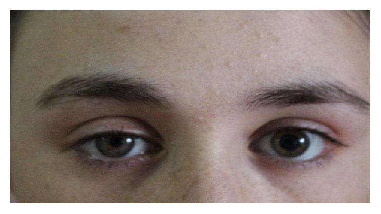
Right-sided miosis and ptosis suggestive of Horner syndrome
Image: “Myosis and eyelid ptosis were noted on the right side” by Case Reports in Endocrinology. License: CC BY 4.0
Diagnosis and Management
Horner syndromeHorner syndromeHorner syndrome is a condition resulting from an interruption of the sympathetic innervation of the eyes. The syndrome is usually idiopathic but can be directly caused by head and neck trauma, cerebrovascular disease, or a tumor of the CNS. Horner Syndrome is a constellation of signs and symptoms. Focus should be placed on diagnosis and management of the underlying cause.
Clinical diagnosis[4–6]
Horner syndromeHorner syndromeHorner syndrome is a condition resulting from an interruption of the sympathetic innervation of the eyes. The syndrome is usually idiopathic but can be directly caused by head and neck trauma, cerebrovascular disease, or a tumor of the CNS. Horner Syndrome is clinically diagnosed by the presence of ptosisPtosisCranial Nerve Palsies and dilation lag in the affected eye.
Anhidrosis is also usually observed on the same side as the affected eye but is not necessary for diagnosis.
Painful acute anisocoriaAnisocoriaUnequal pupil size, which may represent a benign physiologic variant or a manifestation of disease. Pathologic anisocoria reflects an abnormality in the musculature of the iris (iris diseases) or in the parasympathetic or sympathetic pathways that innervate the pupil. Physiologic anisocoria refers to an asymmetry of pupil diameter, usually less than 2mm, that is not associated with disease.Pupil: Physiology and Abnormalities is highly suggestive of Horner syndromeHorner syndromeHorner syndrome is a condition resulting from an interruption of the sympathetic innervation of the eyes. The syndrome is usually idiopathic but can be directly caused by head and neck trauma, cerebrovascular disease, or a tumor of the CNS. Horner Syndrome.
Pharmacologic tests[4–6]
In patientsPatientsIndividuals participating in the health care system for the purpose of receiving therapeutic, diagnostic, or preventive procedures.Clinician–Patient Relationship who present with acute painful anisocoriaAnisocoriaUnequal pupil size, which may represent a benign physiologic variant or a manifestation of disease. Pathologic anisocoria reflects an abnormality in the musculature of the iris (iris diseases) or in the parasympathetic or sympathetic pathways that innervate the pupil. Physiologic anisocoria refers to an asymmetry of pupil diameter, usually less than 2mm, that is not associated with disease.Pupil: Physiology and Abnormalities, or in the setting of trauma or malignancyMalignancyHemothorax, do not delay imaging studies to rule out a cavernous sinus lesion or arterial dissectionArterial dissectionArterial dissection is a violation of the structural integrity of the arterial wall that results in blood accumulating between the layers.Dissection of the Carotid and Vertebral Arteries. Pharmacologic tests are performed in non-acute cases in the outpatient setting, where urgent imaging is not necessary.
For diagnosis confirmation:
Topical cocaineTopical CocainePupil: Physiology and Abnormalities can be used to confirm the diagnosis of Horner syndromeHorner syndromeHorner syndrome is a condition resulting from an interruption of the sympathetic innervation of the eyes. The syndrome is usually idiopathic but can be directly caused by head and neck trauma, cerebrovascular disease, or a tumor of the CNS. Horner Syndrome:
CocaineCocaineAn alkaloid ester extracted from the leaves of plants including coca. It is a local anesthetic and vasoconstrictor and is clinically used for that purpose, particularly in the eye, ear, nose, and throat. It also has powerful central nervous system effects similar to the amphetamines and is a drug of abuse. Cocaine, like amphetamines, acts by multiple mechanisms on brain catecholaminergic neurons; the mechanism of its reinforcing effects is thought to involve inhibition of dopamine uptake.Local Anesthetics blocks reuptake of norepinephrineNorepinephrinePrecursor of epinephrine that is secreted by the adrenal medulla and is a widespread central and autonomic neurotransmitter. Norepinephrine is the principal transmitter of most postganglionic sympathetic fibers, and of the diffuse projection system in the brain that arises from the locus ceruleus.Receptors and Neurotransmitters of the CNS from the synaptic cleftSynaptic cleftSynapses and Neurotransmission and will cause dilation of the pupilPupilThe pupil is the space within the eye that permits light to project onto the retina. Anatomically located in front of the lens, the pupil’s size is controlled by the surrounding iris. The pupil provides insight into the function of the central and autonomic nervous systems. Pupil: Physiology and Abnormalities in case of an intact sympathetic pathway. CocaineCocaineAn alkaloid ester extracted from the leaves of plants including coca. It is a local anesthetic and vasoconstrictor and is clinically used for that purpose, particularly in the eye, ear, nose, and throat. It also has powerful central nervous system effects similar to the amphetamines and is a drug of abuse. Cocaine, like amphetamines, acts by multiple mechanisms on brain catecholaminergic neurons; the mechanism of its reinforcing effects is thought to involve inhibition of dopamine uptake.Local Anesthetics has no effect on eyes with impaired sympathetic innervation.
1 hour after the application of 2 drops of 10% cocaineCocaineAn alkaloid ester extracted from the leaves of plants including coca. It is a local anesthetic and vasoconstrictor and is clinically used for that purpose, particularly in the eye, ear, nose, and throat. It also has powerful central nervous system effects similar to the amphetamines and is a drug of abuse. Cocaine, like amphetamines, acts by multiple mechanisms on brain catecholaminergic neurons; the mechanism of its reinforcing effects is thought to involve inhibition of dopamine uptake.Local Anesthetics, a normal pupilPupilThe pupil is the space within the eye that permits light to project onto the retina. Anatomically located in front of the lens, the pupil’s size is controlled by the surrounding iris. The pupil provides insight into the function of the central and autonomic nervous systems. Pupil: Physiology and Abnormalities dilates more than the Horner pupilPupilThe pupil is the space within the eye that permits light to project onto the retina. Anatomically located in front of the lens, the pupil’s size is controlled by the surrounding iris. The pupil provides insight into the function of the central and autonomic nervous systems. Pupil: Physiology and Abnormalities, increasing the degree of anisocoriaAnisocoriaUnequal pupil size, which may represent a benign physiologic variant or a manifestation of disease. Pathologic anisocoria reflects an abnormality in the musculature of the iris (iris diseases) or in the parasympathetic or sympathetic pathways that innervate the pupil. Physiologic anisocoria refers to an asymmetry of pupil diameter, usually less than 2mm, that is not associated with disease.Pupil: Physiology and Abnormalities.
AnisocoriaAnisocoriaUnequal pupil size, which may represent a benign physiologic variant or a manifestation of disease. Pathologic anisocoria reflects an abnormality in the musculature of the iris (iris diseases) or in the parasympathetic or sympathetic pathways that innervate the pupil. Physiologic anisocoria refers to an asymmetry of pupil diameter, usually less than 2mm, that is not associated with disease.Pupil: Physiology and Abnormalities ≥ 1 mm after cocaineCocaineAn alkaloid ester extracted from the leaves of plants including coca. It is a local anesthetic and vasoconstrictor and is clinically used for that purpose, particularly in the eye, ear, nose, and throat. It also has powerful central nervous system effects similar to the amphetamines and is a drug of abuse. Cocaine, like amphetamines, acts by multiple mechanisms on brain catecholaminergic neurons; the mechanism of its reinforcing effects is thought to involve inhibition of dopamine uptake.Local Anesthetics administration is considered a positive result.
Note on diagnostic role:
Proper, secure storage is necessary, as this is a controlled drug.
Urinary excretion of metabolites can last up to 48 hours (this is important in occupations requiring drug screeningScreeningPreoperative Care).
Topical apraclonidineApraclonidineGlaucoma is used as an alternative agent to confirm Horner syndromeHorner syndromeHorner syndrome is a condition resulting from an interruption of the sympathetic innervation of the eyes. The syndrome is usually idiopathic but can be directly caused by head and neck trauma, cerebrovascular disease, or a tumor of the CNS. Horner Syndrome:
An α-adrenergic agonist that causes pupillary dilation in the Horner pupilPupilThe pupil is the space within the eye that permits light to project onto the retina. Anatomically located in front of the lens, the pupil’s size is controlled by the surrounding iris. The pupil provides insight into the function of the central and autonomic nervous systems. Pupil: Physiology and Abnormalities and mild pupillary constriction in the normal eye by down-regulating norepinephrineNorepinephrinePrecursor of epinephrine that is secreted by the adrenal medulla and is a widespread central and autonomic neurotransmitter. Norepinephrine is the principal transmitter of most postganglionic sympathetic fibers, and of the diffuse projection system in the brain that arises from the locus ceruleus.Receptors and Neurotransmitters of the CNS
A reversal of anisocoriaAnisocoriaUnequal pupil size, which may represent a benign physiologic variant or a manifestation of disease. Pathologic anisocoria reflects an abnormality in the musculature of the iris (iris diseases) or in the parasympathetic or sympathetic pathways that innervate the pupil. Physiologic anisocoria refers to an asymmetry of pupil diameter, usually less than 2mm, that is not associated with disease.Pupil: Physiology and Abnormalities after the application of 2 drops of 0.5% apraclonidineApraclonidineGlaucoma is suggestive of Horner syndromeHorner syndromeHorner syndrome is a condition resulting from an interruption of the sympathetic innervation of the eyes. The syndrome is usually idiopathic but can be directly caused by head and neck trauma, cerebrovascular disease, or a tumor of the CNS. Horner Syndrome.
Safety concerns have been raised owing to CNS effects (e.g., drowsiness), bradycardiaBradycardiaBradyarrhythmia is a rhythm in which the heart rate is less than 60/min. Bradyarrhythmia can be physiologic, without symptoms or hemodynamic change. Pathologic bradyarrhythmia results in reduced cardiac output and hemodynamic instability causing syncope, dizziness, or dyspnea.Bradyarrhythmias, and hypotensionHypotensionHypotension is defined as low blood pressure, specifically < 90/60 mm Hg, and is most commonly a physiologic response. Hypotension may be mild, serious, or life threatening, depending on the cause. Hypotension in some cases.
For localization of lesion:
Topical hydroxyamphetamine is used to differentiate 1st-order and 2nd-order Horner syndrome2nd-order Horner syndromeCaused by lesions that involve the spinal cord, thoracic outlet, or lung apex. These lesions are usually acquired through trauma, surgery, or malignancy (e.g., Pancoast tumor).Horner Syndrome from 3rd-order Horner syndrome3rd-order Horner syndromecaused by lesions of the internal carotid artery, such as arterial dissection, thrombosis, cavernous sinus aneurysm,or injuries acquired during carotid artery stentingHorner Syndrome:
Hydroxyamphetamine causes a release of norepinephrineNorepinephrinePrecursor of epinephrine that is secreted by the adrenal medulla and is a widespread central and autonomic neurotransmitter. Norepinephrine is the principal transmitter of most postganglionic sympathetic fibers, and of the diffuse projection system in the brain that arises from the locus ceruleus.Receptors and Neurotransmitters of the CNS from intact postganglionic adrenergic nerve endings, causing pupillary dilation.
1 hour after the application of 1% hydroxyamphetamine eye drops, dilation of both pupils indicates a lesion of the 1st- or 2nd-order neuron2nd-order neuronPreganglionic pupillomotor fibers exit the spinal cord at T1, travel through the brachial plexus, over the lung apex, ascending to the superior cervical ganglion located near the angle of the mandible and the bifurcation of the common carotid artery.Horner Syndrome.
In 1st- or 2nd-order lesions, the postganglionic nerve endings are not impaired.
Intact postganglionic nerve endings → (+) response to hydroxyamphetamine
If the miotic pupilPupilThe pupil is the space within the eye that permits light to project onto the retina. Anatomically located in front of the lens, the pupil’s size is controlled by the surrounding iris. The pupil provides insight into the function of the central and autonomic nervous systems. Pupil: Physiology and Abnormalities fails to dilate, it indicates a 3rd-order Horner syndrome3rd-order Horner syndromecaused by lesions of the internal carotid artery, such as arterial dissection, thrombosis, cavernous sinus aneurysm,or injuries acquired during carotid artery stentingHorner Syndrome.
There is loss of nerve endings and stores of norepinephrineNorepinephrinePrecursor of epinephrine that is secreted by the adrenal medulla and is a widespread central and autonomic neurotransmitter. Norepinephrine is the principal transmitter of most postganglionic sympathetic fibers, and of the diffuse projection system in the brain that arises from the locus ceruleus.Receptors and Neurotransmitters of the CNS caused by the postganglionic neuron lesion.
Damaged postganglionic nerve endings → no response to hydroxyamphetamine
Pholedrine can be used as an alternative to hydroxyamphetamine. The test is performed using 1% pholedrine.
Imaging[4–7]
General approach:
Different factors (i.e., presentation, availability, associated radiationRadiationEmission or propagation of acoustic waves (sound), electromagnetic energy waves (such as light; radio waves; gamma rays; or x-rays), or a stream of subatomic particles (such as electrons; neutrons; protons; or alpha particles).Osteosarcoma) affect decision-making. There is no firm imaging guideline used, but the traditional approach is outlined.
Imaging is used in conjunction with medical tests to confirm the etiology and locate the site of the lesion.
MRI of the brainBrainThe part of central nervous system that is contained within the skull (cranium). Arising from the neural tube, the embryonic brain is comprised of three major parts including prosencephalon (the forebrain); mesencephalon (the midbrain); and rhombencephalon (the hindbrain). The developed brain consists of cerebrum; cerebellum; and other structures in the brain stem.Nervous System: Anatomy, Structure, and Classification and spinal cordSpinal cordThe spinal cord is the major conduction pathway connecting the brain to the body; it is part of the CNS. In cross section, the spinal cord is divided into an H-shaped area of gray matter (consisting of synapsing neuronal cell bodies) and a surrounding area of white matter (consisting of ascending and descending tracts of myelinated axons). Spinal Cord: Anatomy is indicated in cases with signs indicative of central nervous systemCentral nervous systemThe main information-processing organs of the nervous system, consisting of the brain, spinal cord, and meninges.Nervous System: Anatomy, Structure, and Classification lesions.
Head CT is also advised in cases of Horner syndromeHorner syndromeHorner syndrome is a condition resulting from an interruption of the sympathetic innervation of the eyes. The syndrome is usually idiopathic but can be directly caused by head and neck trauma, cerebrovascular disease, or a tumor of the CNS. Horner Syndrome with a history of or clinical signs suggesting stroke.
Chest X-rayX-rayPenetrating electromagnetic radiation emitted when the inner orbital electrons of an atom are excited and release radiant energy. X-ray wavelengths range from 1 pm to 10 nm. Hard x-rays are the higher energy, shorter wavelength x-rays. Soft x-rays or grenz rays are less energetic and longer in wavelength. The short wavelength end of the x-ray spectrum overlaps the gamma rays wavelength range. The distinction between gamma rays and x-rays is based on their radiation source.Pulmonary Function Tests followed by a CT scan must be performed in patientsPatientsIndividuals participating in the health care system for the purpose of receiving therapeutic, diagnostic, or preventive procedures.Clinician–Patient Relationship when pulmonary malignancyMalignancyHemothorax is suspected.
Imaging based on the location of lesion:
For those presenting with acute-onset painful anisocoriaAnisocoriaUnequal pupil size, which may represent a benign physiologic variant or a manifestation of disease. Pathologic anisocoria reflects an abnormality in the musculature of the iris (iris diseases) or in the parasympathetic or sympathetic pathways that innervate the pupil. Physiologic anisocoria refers to an asymmetry of pupil diameter, usually less than 2mm, that is not associated with disease.Pupil: Physiology and Abnormalities or trauma or those with malignancyMalignancyHemothorax, rule out carotid artery dissectionCarotid artery dissectionThe splitting of the vessel wall in one or both (left and right) internal carotid arteries. Interstitial hemorrhage into the media of the vessel wall can lead to occlusion of the internal carotid artery and aneurysm formation.Dissection of the Carotid and Vertebral Arteries:
CT angiographyAngiographyRadiography of blood vessels after injection of a contrast medium.Cardiac Surgery of the intracranial vessels, carotid arteriesCarotid ArteriesEither of the two principal arteries on both sides of the neck that supply blood to the head and neck; each divides into two branches, the internal carotid artery and the external carotid artery.Carotid Arterial System: Anatomy, and aortic archAortic archMediastinum and Great Vessels: Anatomy (include imaging of the lung apices and orbits)
Can be performed emergently
Widely available
Fat-suppressed MRI and arterial-phase contrast-enhanced MRMRCalculated as the ratio of the total number of people who die due to all causes over a specific time period to the total number of people in the selected population.Measures of Health StatusangiographyAngiographyRadiography of blood vessels after injection of a contrast medium.Cardiac Surgery:
Option if contraindication to contrast
Takes more time and has limited availability
Conventional angiographyAngiographyRadiography of blood vessels after injection of a contrast medium.Cardiac Surgery: gold standard
For those with suspected 1st-order neuron1st-order neuronOriginates in the hypothalamus and descends to the first synapse in the cervical spinal cord, located at levels C8–T2 (also called the ciliospinal center of Budge).Horner Syndrome lesions (which affect the brainBrainThe part of central nervous system that is contained within the skull (cranium). Arising from the neural tube, the embryonic brain is comprised of three major parts including prosencephalon (the forebrain); mesencephalon (the midbrain); and rhombencephalon (the hindbrain). The developed brain consists of cerebrum; cerebellum; and other structures in the brain stem.Nervous System: Anatomy, Structure, and Classification and spinal cordSpinal cordThe spinal cord is the major conduction pathway connecting the brain to the body; it is part of the CNS. In cross section, the spinal cord is divided into an H-shaped area of gray matter (consisting of synapsing neuronal cell bodies) and a surrounding area of white matter (consisting of ascending and descending tracts of myelinated axons). Spinal Cord: Anatomy):
BrainBrainThe part of central nervous system that is contained within the skull (cranium). Arising from the neural tube, the embryonic brain is comprised of three major parts including prosencephalon (the forebrain); mesencephalon (the midbrain); and rhombencephalon (the hindbrain). The developed brain consists of cerebrum; cerebellum; and other structures in the brain stem.Nervous System: Anatomy, Structure, and Classification and/or cervicothoracic MRI: imaging of choice
MRI protocol is determined by the presenting neurologic signs (e.g., hemiparesisHemiparesisThe term hemiparesis refers to mild to moderate weakness involving one side of the body.Epidural Hemorrhage, sensory deficitSensory DeficitAnterior Cord Syndrome, ataxiaAtaxiaImpairment of the ability to perform smoothly coordinated voluntary movements. This condition may affect the limbs, trunk, eyes, pharynx, larynx, and other structures. Ataxia may result from impaired sensory or motor function. Sensory ataxia may result from posterior column injury or peripheral nerve diseases. Motor ataxia may be associated with cerebellar diseases; cerebral cortex diseases; thalamic diseases; basal ganglia diseases; injury to the red nucleus; and other conditions.Ataxia-telangiectasia, ophthalmic deficits).
For those with suspected 2nd-order or 3rd-order neuron3rd-order neuronPupillomotor fibers ascend along the internal carotid artery and enter the cavernous sinus where it is in close relation with the abducens nerve (cranial nerve (CN) VI). These fibers enter the orbit with the ophthalmic branch (V1) of the trigeminal nerve (CN V) via the long ciliary nerves, which innervate the iris dilator and Müller muscles.Horner Syndrome lesions:
CT angiographyAngiographyRadiography of blood vessels after injection of a contrast medium.Cardiac Surgery of the intracranial vessels, carotid arteriesCarotid ArteriesEither of the two principal arteries on both sides of the neck that supply blood to the head and neck; each divides into two branches, the internal carotid artery and the external carotid artery.Carotid Arterial System: Anatomy, and aortic archAortic archMediastinum and Great Vessels: Anatomy (include imaging of the lung apices and orbits)
Chest X-rayX-rayPenetrating electromagnetic radiation emitted when the inner orbital electrons of an atom are excited and release radiant energy. X-ray wavelengths range from 1 pm to 10 nm. Hard x-rays are the higher energy, shorter wavelength x-rays. Soft x-rays or grenz rays are less energetic and longer in wavelength. The short wavelength end of the x-ray spectrum overlaps the gamma rays wavelength range. The distinction between gamma rays and x-rays is based on their radiation source.Pulmonary Function Tests is part of the initial evaluation, but it is important to follow up if results are negative.
Bronchogenic carcinomas are missed on chest X-rayX-rayPenetrating electromagnetic radiation emitted when the inner orbital electrons of an atom are excited and release radiant energy. X-ray wavelengths range from 1 pm to 10 nm. Hard x-rays are the higher energy, shorter wavelength x-rays. Soft x-rays or grenz rays are less energetic and longer in wavelength. The short wavelength end of the x-ray spectrum overlaps the gamma rays wavelength range. The distinction between gamma rays and x-rays is based on their radiation source.Pulmonary Function Tests in 19% of cases.
Evaluation for aortic dissectionAortic dissectionAortic dissection occurs due to shearing stress from pulsatile pressure causing a tear in the tunica intima of the aortic wall. This tear allows blood to flow into the media, creating a “false lumen.” Aortic dissection is most commonly caused by uncontrolled hypertension.Aortic Dissection is not adequate with a chest X-rayX-rayPenetrating electromagnetic radiation emitted when the inner orbital electrons of an atom are excited and release radiant energy. X-ray wavelengths range from 1 pm to 10 nm. Hard x-rays are the higher energy, shorter wavelength x-rays. Soft x-rays or grenz rays are less energetic and longer in wavelength. The short wavelength end of the x-ray spectrum overlaps the gamma rays wavelength range. The distinction between gamma rays and x-rays is based on their radiation source.Pulmonary Function Tests.
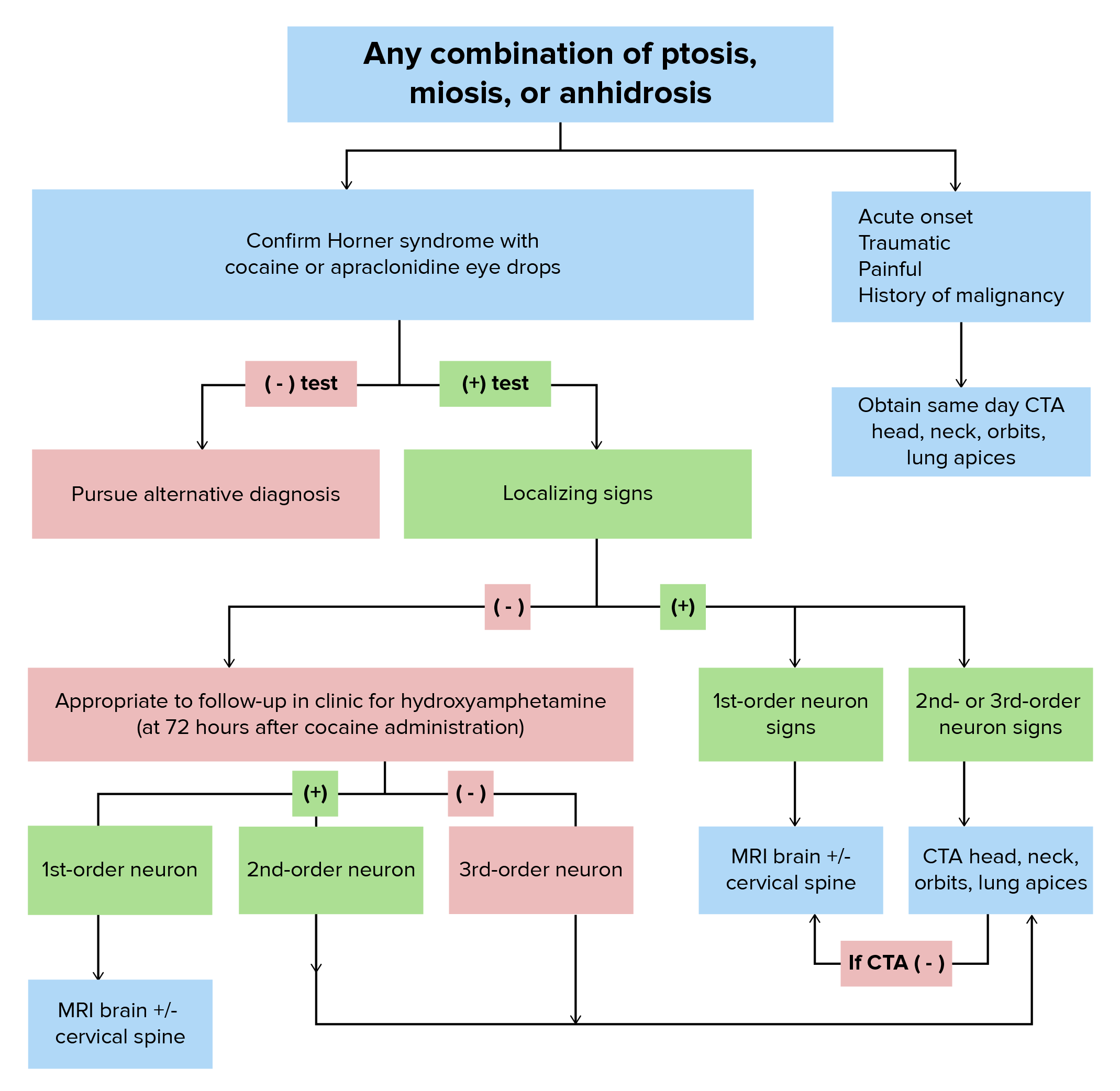
Horner syndrome diagnosis: algorithm for the evaluation of Horner syndrome and localization of the lesion[4–8]
Image by Lecturio.
Management[4–6]
Depends on the underlying cause
Prompt recognition of the condition and diagnosis of the underlying etiology is necessary to reduce worsening of the condition.
The acute painful onset of Horner syndromeHorner syndromeHorner syndrome is a condition resulting from an interruption of the sympathetic innervation of the eyes. The syndrome is usually idiopathic but can be directly caused by head and neck trauma, cerebrovascular disease, or a tumor of the CNS. Horner Syndrome should be considered a medical emergency, as it might indicate a carotid artery dissectionCarotid artery dissectionThe splitting of the vessel wall in one or both (left and right) internal carotid arteries. Interstitial hemorrhage into the media of the vessel wall can lead to occlusion of the internal carotid artery and aneurysm formation.Dissection of the Carotid and Vertebral Arteries.
Vascular surgical care is necessary in cases of carotid artery dissectionCarotid artery dissectionThe splitting of the vessel wall in one or both (left and right) internal carotid arteries. Interstitial hemorrhage into the media of the vessel wall can lead to occlusion of the internal carotid artery and aneurysm formation.Dissection of the Carotid and Vertebral Arteries.
Neurosurgical care should be provided in cases of Horner syndromeHorner syndromeHorner syndrome is a condition resulting from an interruption of the sympathetic innervation of the eyes. The syndrome is usually idiopathic but can be directly caused by head and neck trauma, cerebrovascular disease, or a tumor of the CNS. Horner Syndrome related to aneurysms.
Differential Diagnosis
Adie pupilPupilThe pupil is the space within the eye that permits light to project onto the retina. Anatomically located in front of the lens, the pupil’s size is controlled by the surrounding iris. The pupil provides insight into the function of the central and autonomic nervous systems. Pupil: Physiology and Abnormalities: disorder of parasympathetic denervation of the pupilPupilThe pupil is the space within the eye that permits light to project onto the retina. Anatomically located in front of the lens, the pupil’s size is controlled by the surrounding iris. The pupil provides insight into the function of the central and autonomic nervous systems. Pupil: Physiology and Abnormalities that results in poor light constriction but better accommodationAccommodationRefractive Errors. Adie pupilPupilThe pupil is the space within the eye that permits light to project onto the retina. Anatomically located in front of the lens, the pupil’s size is controlled by the surrounding iris. The pupil provides insight into the function of the central and autonomic nervous systems. Pupil: Physiology and Abnormalities is mostly idiopathicIdiopathicDermatomyositis. The affected pupilPupilThe pupil is the space within the eye that permits light to project onto the retina. Anatomically located in front of the lens, the pupil’s size is controlled by the surrounding iris. The pupil provides insight into the function of the central and autonomic nervous systems. Pupil: Physiology and Abnormalities initially appears abnormally dilated at rest and has a poor pupillary constriction in bright light. PatientsPatientsIndividuals participating in the health care system for the purpose of receiving therapeutic, diagnostic, or preventive procedures.Clinician–Patient Relationship complain of difficulty adapting to dark conditions and photophobiaPhotophobiaAbnormal sensitivity to light. This may occur as a manifestation of eye diseases; migraine; subarachnoid hemorrhage; meningitis; and other disorders. Photophobia may also occur in association with depression and other mental disorders.Migraine Headache. No treatment is required, but topical physostigminePhysostigmineA cholinesterase inhibitor that is rapidly absorbed through membranes. It can be applied topically to the conjunctiva. It also can cross the blood-brain barrier and is used when central nervous system effects are desired, as in the treatment of severe anticholinergic toxicity.Cholinomimetic Drugs may provide relief.
Argyll-Robertson pupilPupilThe pupil is the space within the eye that permits light to project onto the retina. Anatomically located in front of the lens, the pupil’s size is controlled by the surrounding iris. The pupil provides insight into the function of the central and autonomic nervous systems. Pupil: Physiology and Abnormalities: characterized by small and irregular pupils that constrict briskly to near targets but react with little to no constriction to light. Argyll-Robertson pupilPupilThe pupil is the space within the eye that permits light to project onto the retina. Anatomically located in front of the lens, the pupil’s size is controlled by the surrounding iris. The pupil provides insight into the function of the central and autonomic nervous systems. Pupil: Physiology and Abnormalities is frequently associated with iris atrophyAtrophyDecrease in the size of a cell, tissue, organ, or multiple organs, associated with a variety of pathological conditions such as abnormal cellular changes, ischemia, malnutrition, or hormonal changes.Cellular Adaptation and is a highly specific sign of neurosyphilisNeurosyphilisInfections of the central nervous system caused by treponema pallidum which present with a variety of clinical syndromes. The initial phase of infection usually causes a mild or asymptomatic meningeal reaction. The meningovascular form may present acutely as brain infarction. The infection may also remain subclinical for several years. Late syndromes include general paresis; tabes dorsalis; meningeal syphilis; syphilitic optic atrophy; and spinal syphilis. General paresis is characterized by progressive dementia; dysarthria; tremor; myoclonus; seizures; and argyll-robertson pupils.Syphilis and is treated by managing the underlying cause.
Chronic anterior uveitisUveitisUveitis is the inflammation of the uvea, the pigmented middle layer of the eye, which comprises the iris, ciliary body, and choroid. The condition is categorized based on the site of disease; anterior uveitis is the most common. Diseases of the Uvea: condition that affects the anterior uveaUveaThe pigmented vascular coat of the eyeball, consisting of the choroid; ciliary body; and iris, which are continuous with each other.Eye: Anatomy, iris, and ciliary bodyCiliary bodyA ring of tissue extending from the scleral spur to the ora serrata of the retina. It consists of the uveal portion and the epithelial portion. The ciliary muscle is in the uveal portion and the ciliary processes are in the epithelial portion.Eye: Anatomy. The symptoms of uveitisUveitisUveitis is the inflammation of the uvea, the pigmented middle layer of the eye, which comprises the iris, ciliary body, and choroid. The condition is categorized based on the site of disease; anterior uveitis is the most common. Diseases of the Uvea are painPainAn unpleasant sensation induced by noxious stimuli which are detected by nerve endings of nociceptive neurons.Pain: Types and Pathways, rednessRednessInflammation, and photophobiaPhotophobiaAbnormal sensitivity to light. This may occur as a manifestation of eye diseases; migraine; subarachnoid hemorrhage; meningitis; and other disorders. Photophobia may also occur in association with depression and other mental disorders.Migraine Headache. The symptoms respond well to antiinflammatory drugs. Chronic anterior uveitisUveitisUveitis is the inflammation of the uvea, the pigmented middle layer of the eye, which comprises the iris, ciliary body, and choroid. The condition is categorized based on the site of disease; anterior uveitis is the most common. Diseases of the Uvea occurs in association with other chronic inflammatory conditions.
Pupillary sphincter tear: occurs because of trauma and can cause unilateral or bilateral mydriasisMydriasisDilation of pupils to greater than 6 mm combined with failure of the pupils to constrict when stimulated with light. This condition may occur due to injury of the pupillary fibers in the oculomotor nerve, in acute angle-closure glaucoma, and in adie syndrome.Glaucoma. PatientsPatientsIndividuals participating in the health care system for the purpose of receiving therapeutic, diagnostic, or preventive procedures.Clinician–Patient Relationship report glare, haloes in brightly lit conditions, and trouble reading. PatientsPatientsIndividuals participating in the health care system for the purpose of receiving therapeutic, diagnostic, or preventive procedures.Clinician–Patient Relationship with atonic, mydriatic pupils report worse glare at night and inability to focus on near objects. The tear requires surgical repair.
Ocular myasthenia: ocular representation of myasthenia gravisMyasthenia GravisMyasthenia gravis (MG) is an autoimmune neuromuscular disorder characterized by weakness and fatigability of skeletal muscles caused by dysfunction/destruction of acetylcholine receptors at the neuromuscular junction. MG presents with fatigue, ptosis, diplopia, dysphagia, respiratory difficulties, and progressive weakness in the limbs, leading to difficulty in movement. Myasthenia Gravis. Ocular myasthenia results in ocular muscle fatigueFatigueThe state of weariness following a period of exertion, mental or physical, characterized by a decreased capacity for work and reduced efficiency to respond to stimuli.Fibromyalgia and weakness. In myasthenia, antibodiesAntibodiesImmunoglobulins (Igs), also known as antibodies, are glycoprotein molecules produced by plasma cells that act in immune responses by recognizing and binding particular antigens. The various Ig classes are IgG (the most abundant), IgM, IgE, IgD, and IgA, which differ in their biologic features, structure, target specificity, and distribution.Immunoglobulins: Types and Functions that block the acetylcholineAcetylcholineA neurotransmitter found at neuromuscular junctions, autonomic ganglia, parasympathetic effector junctions, a subset of sympathetic effector junctions, and at many sites in the central nervous system.Receptors and Neurotransmitters of the CNSreceptorReceptorReceptors are proteins located either on the surface of or within a cell that can bind to signaling molecules known as ligands (e.g., hormones) and cause some type of response within the cell.Receptors are produced, resulting in muscle fatigueFatigueThe state of weariness following a period of exertion, mental or physical, characterized by a decreased capacity for work and reduced efficiency to respond to stimuli.Fibromyalgia and paralysis in some cases. Diagnosis relies on an edrophoniumEdrophoniumA rapid-onset, short-acting cholinesterase inhibitor used in cardiac arrhythmias and in the diagnosis of myasthenia gravis. It has also been used as an antidote to curare principles.Myasthenia GravissodiumSodiumA member of the alkali group of metals. It has the atomic symbol na, atomic number 11, and atomic weight 23.Hyponatremia test. Treatment involves anticholinesterase agents.
Billing and Coding
Diagnosis Codes:
This code is used to diagnose Horner syndromeHorner syndromeHorner syndrome is a condition resulting from an interruption of the sympathetic innervation of the eyes. The syndrome is usually idiopathic but can be directly caused by head and neck trauma, cerebrovascular disease, or a tumor of the CNS. Horner Syndrome, a classic neurological syndrome characterized by a triad of symptoms: miosisMiosisPupil: Physiology and Abnormalities (constricted pupilPupilThe pupil is the space within the eye that permits light to project onto the retina. Anatomically located in front of the lens, the pupil’s size is controlled by the surrounding iris. The pupil provides insight into the function of the central and autonomic nervous systems. Pupil: Physiology and Abnormalities), ptosisPtosisCranial Nerve Palsies (drooping eyelid), and anhidrosis (decreased sweating) on one side of the face.
Coding System
Code
Description
ICD-10-CM
G90.2
Horner’s syndrome
SNOMED CT
128176002
Horner’s syndrome (disorder)
Evaluation & Workup:
Horner syndromeHorner syndromeHorner syndrome is a condition resulting from an interruption of the sympathetic innervation of the eyes. The syndrome is usually idiopathic but can be directly caused by head and neck trauma, cerebrovascular disease, or a tumor of the CNS. Horner Syndrome is a sign of an underlying problem. This code is for an MRI of the neckNeckThe part of a human or animal body connecting the head to the rest of the body.Peritonsillar Abscess and chest, a critical imaging study to look for the underlying cause, such as a Pancoast tumorPancoast TumorThoracic Outlet Syndrome in the lung apex or a carotid artery dissectionCarotid artery dissectionThe splitting of the vessel wall in one or both (left and right) internal carotid arteries. Interstitial hemorrhage into the media of the vessel wall can lead to occlusion of the internal carotid artery and aneurysm formation.Dissection of the Carotid and Vertebral Arteries.
Coding System
Code
Description
CPT
70540
Magnetic resonance imaging, orbit, face, and/or neckNeckThe part of a human or animal body connecting the head to the rest of the body.Peritonsillar Abscess; without contrast material(s)
Kanagalingam, S., Miller, N. R. (2015). Horner syndrome: clinical perspectives. Eye and Brain, 2015, 35–46. https://doi.org/10.2147/EB.S63633
Davagnanam, I., Fraser, C. L., Miszkiel, K., Daniel, C. S., Plant, G. T. (2013). Adult Horner’s syndrome: a combined clinical, pharmacological, and imaging algorithm. Eye, 27(3), 291–298. https://doi.org/10.1038/eye.2012.281
American College of Radiology. (2017). ACR appropriateness criteria: orbits, vision, and visual loss. Retrieved October 25, 2022, from https://acsearch.acr.org/docs/69486/Narrative
Create your free account or log in to continue reading!