Diphtheria is an infectious disease caused by Corynebacterium diphtheriae that most often results in respiratory disease with membranous inflammation of the pharynx, sore throat, fever, swollen glands, and weakness. The hallmark sign is a sheet of thick, gray material covering the back of the throat. Diphtheria can also manifest as cutaneous disease leading to nonspecific skin lesions. In advanced stages, diphtheria can damage the heart, kidneys, and nervous system. It is diagnosed via a culture of pharyngeal swabs and treated with antibiotic therapy and the diphtheria antitoxin.
DiphtheriaDiphtheriaDiphtheria is an infectious disease caused by Corynebacterium diphtheriae that most often results in respiratory disease with membranous inflammation of the pharynx, sore throat, fever, swollen glands, and weakness. The hallmark sign is a sheet of thick, gray material covering the back of the throat. Diphtheria is caused by the bacterium Corynebacterium diphtheriaeCorynebacterium diphtheriaeDiphtheria is an infectious disease caused by corynebacterium diphtheriae that most often results in respiratory disease with membranous inflammation of the pharynx, sore throat, fever, swollen glands, and weakness. The hallmark sign is a sheet of thick, gray material covering the back of the throat.Diphtheria.
Gram-positiveGram-PositivePenicillinsbacillusBacillusBacillus are aerobic, spore-forming, gram-positive bacilli. Two pathogenic species are Bacillus anthracis (B. anthracis) and B. cereus. Bacillus (non-motile, non-spore-forming)
4 biotypes: gravis, intermedius, mitis, and belfanti
Only strains harboring a lysogenic bacteriophageLysogenic bacteriophageVibrio encoding the diphtheriaDiphtheriaDiphtheria is an infectious disease caused by Corynebacterium diphtheriae that most often results in respiratory disease with membranous inflammation of the pharynx, sore throat, fever, swollen glands, and weakness. The hallmark sign is a sheet of thick, gray material covering the back of the throat. Diphtheria toxin geneGeneA category of nucleic acid sequences that function as units of heredity and which code for the basic instructions for the development, reproduction, and maintenance of organisms.Basic Terms of Genetics (tox) cause clinical diphtheriaDiphtheriaDiphtheria is an infectious disease caused by Corynebacterium diphtheriae that most often results in respiratory disease with membranous inflammation of the pharynx, sore throat, fever, swollen glands, and weakness. The hallmark sign is a sheet of thick, gray material covering the back of the throat. Diphtheria.
Humans are the only known reservoirReservoirAnimate or inanimate sources which normally harbor disease-causing organisms and thus serve as potential sources of disease outbreaks. Reservoirs are distinguished from vectors (disease vectors) and carriers, which are agents of disease transmission rather than continuing sources of potential disease outbreaks. Humans may serve both as disease reservoirs and carriers.Escherichia coli.
Can also be transmitted through skinSkinThe skin, also referred to as the integumentary system, is the largest organ of the body. The skin is primarily composed of the epidermis (outer layer) and dermis (deep layer). The epidermis is primarily composed of keratinocytes that undergo rapid turnover, while the dermis contains dense layers of connective tissue.Skin: Structure and Functions lesions
Immunization has minimized toxigenic strain transmission in resource-rich countries.
Epidemiology[1,7,10]
Remains a widespread disease/endemic in resource-limited countries due to insufficient vaccinationVaccinationVaccination is the administration of a substance to induce the immune system to develop protection against a disease. Unlike passive immunization, which involves the administration of pre-performed antibodies, active immunization constitutes the administration of a vaccine to stimulate the body to produce its own antibodies.Vaccination coverage
5% of populations in endemic areas have positive pharyngeal cultures.
Peak incidenceIncidenceThe number of new cases of a given disease during a given period in a specified population. It also is used for the rate at which new events occur in a defined population. It is differentiated from prevalence, which refers to all cases in the population at a given time.Measures of Disease Frequency in colder months
Pathophysiology
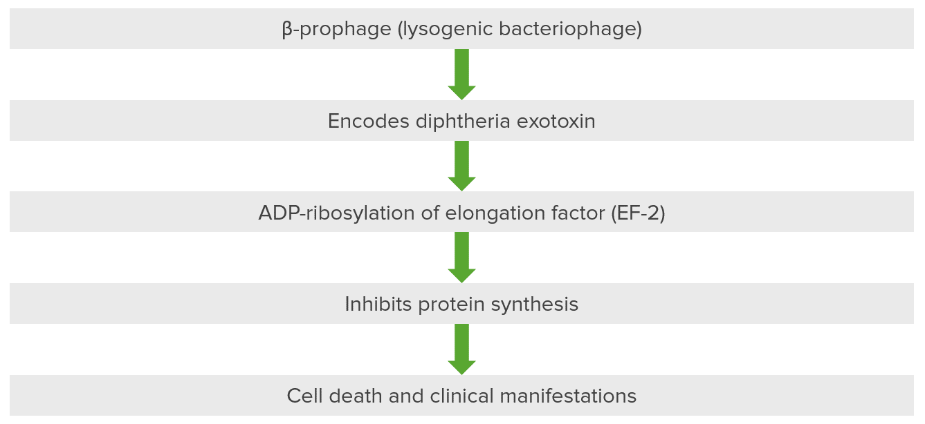
Pathogenesis of diseases associated with Corynebacterium: diphtheria exotoxin inactivates elongation factor (EF-2) via ADP-ribosylation.[1,2,3] This bacteriophage, a beta prophage, introduces itself into the cell and DNA of the host organism, resulting in the encoding of diphtheria exotoxin. The exotoxin has 3 domains: 1 is present in fragment A and is catalytic, while 2 are present in fragment B for receptor binding and membrane insertion and translocation. The exotoxin is the major cause of the disease in diphtheria because it ribosylates ADP present in the elongation factor. The elongation factor, specifically elongation factor 2 (EF-2), is critical for the elongation of protein chains. Diphtheria exotoxin inhibits EF-2 so that protein is not synthesized, resulting in cellular death and secondary clinical manifestations.
Respiratory diphtheriaDiphtheriaDiphtheria is an infectious disease caused by Corynebacterium diphtheriae that most often results in respiratory disease with membranous inflammation of the pharynx, sore throat, fever, swollen glands, and weakness. The hallmark sign is a sheet of thick, gray material covering the back of the throat. Diphtheria[2–4,7,9]
Symptoms begin 2–5 days after infection.
Gradual onset of:
Sore throatSore throatPharyngitis is an inflammation of the back of the throat (pharynx). Pharyngitis is usually caused by an upper respiratory tract infection, which is viral in most cases. It typically results in a sore throat and fever. Other symptoms may include a runny nose, cough, headache, and hoarseness.Pharyngitis
Cervical lymphadenopathyLymphadenopathyLymphadenopathy is lymph node enlargement (> 1 cm) and is benign and self-limited in most patients. Etiologies include malignancy, infection, and autoimmune disorders, as well as iatrogenic causes such as the use of certain medications. Generalized lymphadenopathy often indicates underlying systemic disease. Lymphadenopathy
Mild erythematous pharynxPharynxThe pharynx is a component of the digestive system that lies posterior to the nasal cavity, oral cavity, and larynx. The pharynx can be divided into the oropharynx, nasopharynx, and laryngopharynx. Pharyngeal muscles play an integral role in vital processes such as breathing, swallowing, and speaking. Pharynx: Anatomy
Characterized by a grayish-white membrane covering the posterior pharyngeal wall and/or tonsilsTonsilsTonsillitis that bleeds heavily during attempts at removal owing to its highly vascular nature
Present in at least 1/3 of cases
Formation is induced by the toxin (made up of necrotic fibrinFibrinA protein derived from fibrinogen in the presence of thrombin, which forms part of the blood clot.Rapidly Progressive Glomerulonephritis, WBCs, RBCsRBCsErythrocytes, or red blood cells (RBCs), are the most abundant cells in the blood. While erythrocytes in the fetus are initially produced in the yolk sac then the liver, the bone marrow eventually becomes the main site of production.Erythrocytes: Histology, and epithelial cells)
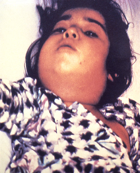
Severe lymphadenopathyLymphadenopathyLymphadenopathy is lymph node enlargement (> 1 cm) and is benign and self-limited in most patients. Etiologies include malignancy, infection, and autoimmune disorders, as well as iatrogenic causes such as the use of certain medications. Generalized lymphadenopathy often indicates underlying systemic disease. Lymphadenopathy with “bull neckNeckThe part of a human or animal body connecting the head to the rest of the body.Peritonsillar Abscess” appearance (pathognomonic)
Foul breath or mouth odor
A clinical sign of a patient with respiratory diphtheria: severe lymphadenopathy with “bull neck” appearance
Image: “Diphtheria bull neck” by CDC/Public Health Image Library (PHIL). License: Public Domain
Pediatric patient with diphtheria presenting with characteristic grayish-white membrane covering the posterior pharyngeal wall
Image: “A doctor with the UK’s Emergency Medical Team checks a child for symptoms of Diphtheria in a makeshift clinic in the Kutupalong camp for Rohingya refugees in Bangladesh” by Russell Watkins/Department for International Development. License: CC BY 2.0
Laryngeal diphtheriaDiphtheriaDiphtheria is an infectious disease caused by Corynebacterium diphtheriae that most often results in respiratory disease with membranous inflammation of the pharynx, sore throat, fever, swollen glands, and weakness. The hallmark sign is a sheet of thick, gray material covering the back of the throat. Diphtheria[2–4,11]
Characteristic swollen neck and throat or “bull neck”
Often accompanied by the following symptoms (historically referred to as “diphtheritic croupCroupCroup, also known as laryngotracheobronchitis, is a disease most commonly caused by a viral infection that leads to severe inflammation of the upper airway. It usually presents in children < 5 years of age. Patients develop a hoarse, "seal-like" barking cough and inspiratory stridor. Croup”):
Nasal diphtheriaDiphtheriaDiphtheria is an infectious disease caused by Corynebacterium diphtheriae that most often results in respiratory disease with membranous inflammation of the pharynx, sore throat, fever, swollen glands, and weakness. The hallmark sign is a sheet of thick, gray material covering the back of the throat. Diphtheria[2,11]
Tracheobronchial diphtheriaDiphtheriaDiphtheria is an infectious disease caused by Corynebacterium diphtheriae that most often results in respiratory disease with membranous inflammation of the pharynx, sore throat, fever, swollen glands, and weakness. The hallmark sign is a sheet of thick, gray material covering the back of the throat. Diphtheria[4]
Develops secondary to membrane spread
Can result in respiratory compromise, especially in children with smaller airways
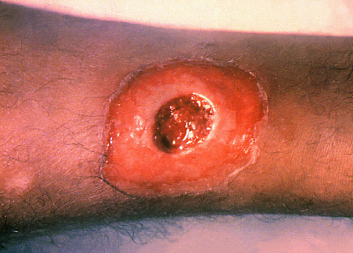
Cutaneous (wound) diphtheriaDiphtheriaDiphtheria is an infectious disease caused by Corynebacterium diphtheriae that most often results in respiratory disease with membranous inflammation of the pharynx, sore throat, fever, swollen glands, and weakness. The hallmark sign is a sheet of thick, gray material covering the back of the throat. Diphtheria[2,4,11]
Local trauma is a frequent precedent (e.g., IV drug use)
Most often seen in homeless patientsPatientsIndividuals participating in the health care system for the purpose of receiving therapeutic, diagnostic, or preventive procedures.Clinician–Patient Relationship or those with a history of drug abuse
Clinical sign of a patient with cutaneous (wound) diphtheria: chronic non-healing ulcer
Image: “A diphtheria skin lesion on the leg” by CDC/Public Health Image Library (PHIL). License: Public Domain
Systemic toxemia[4,7,9,11]
Damage to distant organs: cardiac toxicityToxicityDosage Calculation, neurologic toxicityToxicityDosage Calculation, renal failureRenal failureConditions in which the kidneys perform below the normal level in the ability to remove wastes, concentrate urine, and maintain electrolyte balance; blood pressure; and calcium metabolism. Renal insufficiency can be classified by the degree of kidney damage (as measured by the level of proteinuria) and reduction in glomerular filtration rate.Crush Syndrome
MyocarditisMyocarditisMyocarditis is an inflammatory disease of the myocardium, which may occur alone or in association with a systemic process. There are numerous etiologies of myocarditis, but all lead to inflammation and myocyte injury, most often leading to signs and symptoms of heart failure. Myocarditis:
ST–T-wave changes on ECGECGAn electrocardiogram (ECG) is a graphic representation of the electrical activity of the heart plotted against time. Adhesive electrodes are affixed to the skin surface allowing measurement of cardiac impulses from many angles. The ECG provides 3-dimensional information about the conduction system of the heart, the myocardium, and other cardiac structures. Electrocardiogram (ECG)
QTc prolongation
Heart block
In severe cases:
Complex heart blocks
Heart failureHeart FailureA heterogeneous condition in which the heart is unable to pump out sufficient blood to meet the metabolic need of the body. Heart failure can be caused by structural defects, functional abnormalities (ventricular dysfunction), or a sudden overload beyond its capacity. Chronic heart failure is more common than acute heart failure which results from sudden insult to cardiac function, such as myocardial infarction.Total Anomalous Pulmonary Venous Return (TAPVR)
Poor prognosisPrognosisA prediction of the probable outcome of a disease based on a individual’s condition and the usual course of the disease as seen in similar situations.Non-Hodgkin Lymphomas
DemyelinationDemyelinationMultiple Sclerosis and nerve deficits begin in the posterior oral pharynxPharynxThe pharynx is a component of the digestive system that lies posterior to the nasal cavity, oral cavity, and larynx. The pharynx can be divided into the oropharynx, nasopharynx, and laryngopharynx. Pharyngeal muscles play an integral role in vital processes such as breathing, swallowing, and speaking. Pharynx: Anatomy.
Peripheral neuritis (can take weeks to months to develop)
Renal failureRenal failureConditions in which the kidneys perform below the normal level in the ability to remove wastes, concentrate urine, and maintain electrolyte balance; blood pressure; and calcium metabolism. Renal insufficiency can be classified by the degree of kidney damage (as measured by the level of proteinuria) and reduction in glomerular filtration rate.Crush Syndrome from toxin effect or hypotensionHypotensionHypotension is defined as low blood pressure, specifically < 90/60 mm Hg, and is most commonly a physiologic response. Hypotension may be mild, serious, or life threatening, depending on the cause. Hypotension
Diagnosis
General[2,9]
Consider the diagnosis with relevant clinical manifestations listed above along with epidemiologic risk factors.
Treatment should not be delayed while awaiting laboratory results if the clinical picture strongly suggests diphtheriaDiphtheriaDiphtheria is an infectious disease caused by Corynebacterium diphtheriae that most often results in respiratory disease with membranous inflammation of the pharynx, sore throat, fever, swollen glands, and weakness. The hallmark sign is a sheet of thick, gray material covering the back of the throat. Diphtheria.
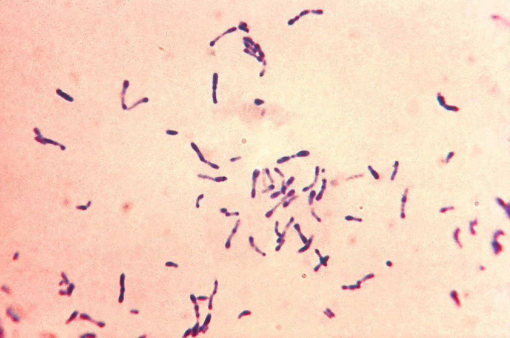
Gram-positive Corynebacterium diphtheriae bacteria with blue metachromatic granules. The bacteria were grown via Loeffler’s medium to enhance the characteristic metachromatic granules. Notice the club-shaped nature and formation into Y and V.
Image: “Gram-positive Corynebacterium diphtheriae bacteria” by Centers for Disease Control and Prevention’s Public Health Image Library (PHIL). License: Public Domain
A throat swab inoculated with Corynebacterium diphtheriae is cultured on a Petri dish plate containing Tinsdale medium supplemented with the agar component Tellurite. The gray-black colonies, each surrounded by a dark brown halo, are characteristic of C. diphtheriae on Tellurite agar.
Image: “Corynebacterium diphtheriae” by Public Health Image Library (PHIL). License: Public Domain
Toxin detection
Elek testElek testDiphtheria performed to determine if strain is toxigenic:[2,3,4,5,7]
C. diphtheriae grows on an agar culture.
An antitoxin-impregnated filter paper is laid over the culture, and toxin production is shown by the development of an immunoprecipitin band.
Sensitive and specific detection of the diphtheriaDiphtheriaDiphtheria is an infectious disease caused by Corynebacterium diphtheriae that most often results in respiratory disease with membranous inflammation of the pharynx, sore throat, fever, swollen glands, and weakness. The hallmark sign is a sheet of thick, gray material covering the back of the throat. Diphtheria toxin
Immunochromatographic strip (ICS) assay:[5,6,7]
Highly sensitive test to detect diphtheriaDiphtheriaDiphtheria is an infectious disease caused by Corynebacterium diphtheriae that most often results in respiratory disease with membranous inflammation of the pharynx, sore throat, fever, swollen glands, and weakness. The hallmark sign is a sheet of thick, gray material covering the back of the throat. Diphtheria toxin
No specialized equipment needed
PCRPCRPolymerase chain reaction (PCR) is a technique that amplifies DNA fragments exponentially for analysis. The process is highly specific, allowing for the targeting of specific genomic sequences, even with minuscule sample amounts. The PCR cycles multiple times through 3 phases: denaturation of the template DNA, annealing of a specific primer to the individual DNA strands, and synthesis/elongation of new DNA molecules.Polymerase Chain Reaction (PCR) testing can identify the geneGeneA category of nucleic acid sequences that function as units of heredity and which code for the basic instructions for the development, reproduction, and maintenance of organisms.Basic Terms of Genetics via subunit A.[3,4,5]
Detects the toxin geneGeneA category of nucleic acid sequences that function as units of heredity and which code for the basic instructions for the development, reproduction, and maintenance of organisms.Basic Terms of Genetics, but does not indicate toxin production
Culture must be run to confirm the presence of active disease due to the toxigenic organism.
Mnemonic
Key points to remember about diphtheriaDiphtheriaDiphtheria is an infectious disease caused by Corynebacterium diphtheriae that most often results in respiratory disease with membranous inflammation of the pharynx, sore throat, fever, swollen glands, and weakness. The hallmark sign is a sheet of thick, gray material covering the back of the throat. Diphtheria: ABCDEFG
ADP-ribosylation
Beta-prophage
Corynebacterium diphtheriae
Elongation factor 2
Granules
Management and Prevention
Management can vary based on location. The following information is based on US and UK public health recommendations:
General overview[2,7,9,11]
When there is clinical suspicion of diphtheriaDiphtheriaDiphtheria is an infectious disease caused by Corynebacterium diphtheriae that most often results in respiratory disease with membranous inflammation of the pharynx, sore throat, fever, swollen glands, and weakness. The hallmark sign is a sheet of thick, gray material covering the back of the throat. Diphtheria, the following are applied:
Respiratory droplet isolation for respiratory illness
Respiratory diphtheriaDiphtheriaDiphtheria is an infectious disease caused by Corynebacterium diphtheriae that most often results in respiratory disease with membranous inflammation of the pharynx, sore throat, fever, swollen glands, and weakness. The hallmark sign is a sheet of thick, gray material covering the back of the throat. Diphtheria: antibiotic therapy + diphtheria antitoxinDiphtheria antitoxinAn antitoxin produced against the toxin of corynebacterium diphtheriae that is used for the treatment of diphtheria.Diphtheria
Antitoxin is generally not given owing to absence of pseudomembranesPseudomembranesRaised yellow or off-white plaques up to 2 cm in diameter that form as a result of mucosal ulcerationPseudomembranous Colitis and cardiac manifestations, but in the United Kingdom, use in large lesions may be justified.[4,8,9]
Antibiotic therapy[2,4,7,9,11]
Suppresses the toxin-producing bacteriaBacteriaBacteria are prokaryotic single-celled microorganisms that are metabolically active and divide by binary fission. Some of these organisms play a significant role in the pathogenesis of diseases. Bacteriology, reducing toxin production and transmission of the infection
Options (total treatment course, 14 days):
ErythromycinErythromycinA bacteriostatic antibiotic macrolide produced by streptomyces erythreus. Erythromycin a is considered its major active component. In sensitive organisms, it inhibits protein synthesis by binding to 50s ribosomal subunits. This binding process inhibits peptidyl transferase activity and interferes with translocation of amino acids during translation and assembly of proteins.Macrolides and Ketolides 500 mg 4 times daily (IV or oral), OR
ProcaineProcaineA local anesthetic of the ester type that has a slow onset and a short duration of action. It is mainly used for infiltration anesthesia, peripheral nerve block, and spinal block.Local AnestheticspenicillinPenicillinRheumatic Fever G
Adults: 1.2 million units intramuscularly (IM) once daily.
Children: 50,000 units/kg IM once daily (maximum 1.2 million units/day)
Switch to oral penicillinPenicillinRheumatic Fever V 500 mg (10 to 15 mg/kg/dose in children) 4 times daily when individual is able to tolerate oral intake.
Diphtheria antitoxinDiphtheria antitoxinAn antitoxin produced against the toxin of corynebacterium diphtheriae that is used for the treatment of diphtheria.Diphtheria (DAT)[2,7,8]
Inactivates the toxin
In the United States, DAT is obtained by coordinating with the state health department and contacting the CDC’s Emergency Operations Center at 770-488-7100.[2]
Precautions:
Prior to administration, a skinSkinThe skin, also referred to as the integumentary system, is the largest organ of the body. The skin is primarily composed of the epidermis (outer layer) and dermis (deep layer). The epidermis is primarily composed of keratinocytes that undergo rapid turnover, while the dermis contains dense layers of connective tissue.Skin: Structure and Functions test is performed to rule out hypersensitivity to the animal serum.
EpinephrineEpinephrineThe active sympathomimetic hormone from the adrenal medulla. It stimulates both the alpha- and beta- adrenergic systems, causes systemic vasoconstriction and gastrointestinal relaxation, stimulates the heart, and dilates bronchi and cerebral vessels.Sympathomimetic Drugs should be on handHandThe hand constitutes the distal part of the upper limb and provides the fine, precise movements needed in activities of daily living. It consists of 5 metacarpal bones and 14 phalanges, as well as numerous muscles innervated by the median and ulnar nerves. Hand: Anatomy because of the risk of anaphylaxisAnaphylaxisAn acute hypersensitivity reaction due to exposure to a previously encountered antigen. The reaction may include rapidly progressing urticaria, respiratory distress, vascular collapse, systemic shock, and death.Type I Hypersensitivity Reaction.
DosageDosageDosage Calculation (given over 60 minutes IV) according to the American Academy of Pediatrics:[4]
Pharyngeal/laryngeal disease of < 48 hours duration: 20,000‒40,000 units
Nasopharyngeal disease: 40,000‒60,000 units
Illness of > 48 hours’ duration or in case of diffuse neck swelling (“bull neck”): 80,000‒120,000 units
Laryngeal/pharyngeal/nasopharyngeal disease of ≤ 48 hours’ duration: 70,000 IU
Laryngeal/pharyngeal/nasopharyngeal disease of > 48 hours’ duration or severe illness (“bull neckNeckThe part of a human or animal body connecting the head to the rest of the body.Peritonsillar Abscess”): 100,000 IU
SkinSkinThe skin, also referred to as the integumentary system, is the largest organ of the body. The skin is primarily composed of the epidermis (outer layer) and dermis (deep layer). The epidermis is primarily composed of keratinocytes that undergo rapid turnover, while the dermis contains dense layers of connective tissue.Skin: Structure and Functions lesions (large size > 2 cm2): 40,000 IU
Monitoring[2,4,7,9,11]
Monitor for signs of cardiac involvement:
Serial electrocardiograms
Cardiac enzymesEnzymesEnzymes are complex protein biocatalysts that accelerate chemical reactions without being consumed by them. Due to the body’s constant metabolic needs, the absence of enzymes would make life unsustainable, as reactions would occur too slowly without these molecules. Basics of Enzymes (helps evaluate degree of myocardial damage)
Check neurologic status
Obtain follow-up cultures:
Cultures done at 24–48 hours and 2 weeks later
Two negative cultures (≥ 24 hours apart) are needed to discontinue isolation.
Individuals who continue to harbor the bacteriaBacteriaBacteria are prokaryotic single-celled microorganisms that are metabolically active and divide by binary fission. Some of these organisms play a significant role in the pathogenesis of diseases. Bacteriology are recommended to get an additional 10 days of oral erythromycinErythromycinA bacteriostatic antibiotic macrolide produced by streptomyces erythreus. Erythromycin a is considered its major active component. In sensitive organisms, it inhibits protein synthesis by binding to 50s ribosomal subunits. This binding process inhibits peptidyl transferase activity and interferes with translocation of amino acids during translation and assembly of proteins.Macrolides and Ketolides[9]
Convalescence period[2,9]
PatientsPatientsIndividuals participating in the health care system for the purpose of receiving therapeutic, diagnostic, or preventive procedures.Clinician–Patient Relationship are given vaccinationVaccinationVaccination is the administration of a substance to induce the immune system to develop protection against a disease. Unlike passive immunization, which involves the administration of pre-performed antibodies, active immunization constitutes the administration of a vaccine to stimulate the body to produce its own antibodies.Vaccination with diphtheriaDiphtheriaDiphtheria is an infectious disease caused by Corynebacterium diphtheriae that most often results in respiratory disease with membranous inflammation of the pharynx, sore throat, fever, swollen glands, and weakness. The hallmark sign is a sheet of thick, gray material covering the back of the throat. DiphtheriatoxoidToxoidPreparations of pathogenic organisms or their derivatives made nontoxic and intended for active immunologic prophylaxis. They include deactivated toxins. Anatoxin toxoids are distinct from anatoxins that are tropanes found in cyanobacteria.Vaccination (as illness does not confer immunity).
Contacts[2,4,7,9,11]
Close contacts (e.g., family, caretakers, medical staff) need to be monitored for development of symptoms for ≥ 1 week.
1 dose of penicillinPenicillinRheumatic Fever G benzathine (600,000 units IM) for those < 6 years of age, OR
1 dose of penicillinPenicillinRheumatic Fever G benzathine (1.2 million units IM) for those ≥ 6 years of age, OR)
Oral erythromycinErythromycinA bacteriostatic antibiotic macrolide produced by streptomyces erythreus. Erythromycin a is considered its major active component. In sensitive organisms, it inhibits protein synthesis by binding to 50s ribosomal subunits. This binding process inhibits peptidyl transferase activity and interferes with translocation of amino acids during translation and assembly of proteins.Macrolides and Ketolides (500 mg 4 times daily) for 7‒10 days
Note: In the United Kingdom, 7 days of antibiotic prophylaxisProphylaxisCephalosporins may be offered.[11]
In contacts whose cultures turn positive, proceed with treatment regimen.
Determine diphtheriaDiphtheriaDiphtheria is an infectious disease caused by Corynebacterium diphtheriae that most often results in respiratory disease with membranous inflammation of the pharynx, sore throat, fever, swollen glands, and weakness. The hallmark sign is a sheet of thick, gray material covering the back of the throat. DiphtheriatoxoidToxoidPreparations of pathogenic organisms or their derivatives made nontoxic and intended for active immunologic prophylaxis. They include deactivated toxins. Anatoxin toxoids are distinct from anatoxins that are tropanes found in cyanobacteria.VaccinationvaccinationVaccinationVaccination is the administration of a substance to induce the immune system to develop protection against a disease. Unlike passive immunization, which involves the administration of pre-performed antibodies, active immunization constitutes the administration of a vaccine to stimulate the body to produce its own antibodies.Vaccination status:
Unknown or < 3 doses: administer diphtheriaDiphtheriaDiphtheria is an infectious disease caused by Corynebacterium diphtheriae that most often results in respiratory disease with membranous inflammation of the pharynx, sore throat, fever, swollen glands, and weakness. The hallmark sign is a sheet of thick, gray material covering the back of the throat. DiphtheriatoxoidToxoidPreparations of pathogenic organisms or their derivatives made nontoxic and intended for active immunologic prophylaxis. They include deactivated toxins. Anatoxin toxoids are distinct from anatoxins that are tropanes found in cyanobacteria.Vaccination and complete series
≥ 3 doses, last one > 5 years ago: administer booster diphtheriaDiphtheriaDiphtheria is an infectious disease caused by Corynebacterium diphtheriae that most often results in respiratory disease with membranous inflammation of the pharynx, sore throat, fever, swollen glands, and weakness. The hallmark sign is a sheet of thick, gray material covering the back of the throat. DiphtheriatoxoidToxoidPreparations of pathogenic organisms or their derivatives made nontoxic and intended for active immunologic prophylaxis. They include deactivated toxins. Anatoxin toxoids are distinct from anatoxins that are tropanes found in cyanobacteria.Vaccination
≥ 3 doses, last one ≤ 5 years ago: vaccinationVaccinationVaccination is the administration of a substance to induce the immune system to develop protection against a disease. Unlike passive immunization, which involves the administration of pre-performed antibodies, active immunization constitutes the administration of a vaccine to stimulate the body to produce its own antibodies.Vaccination not required (except for children with dose due)
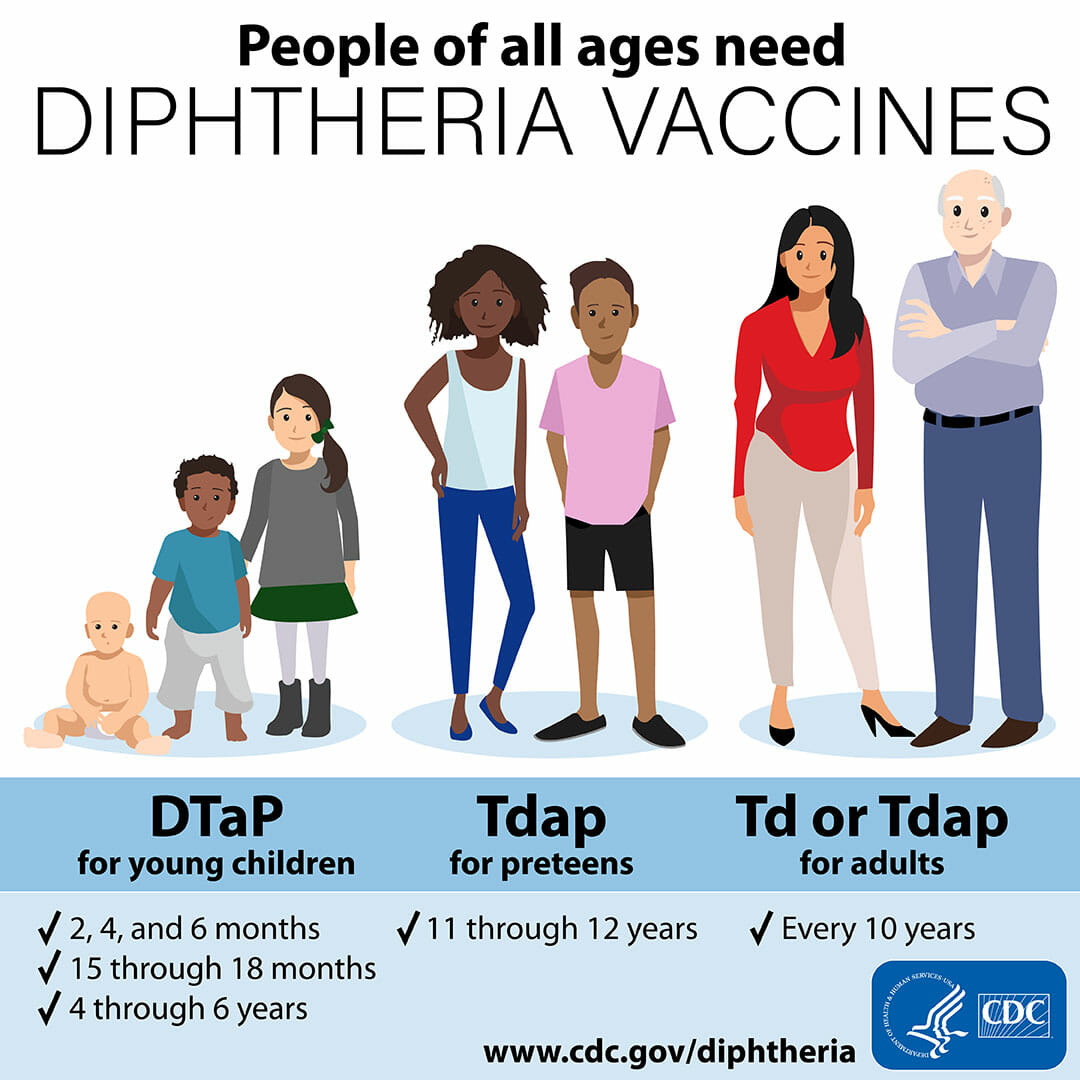
Prevention[2]
ToxoidToxoidPreparations of pathogenic organisms or their derivatives made nontoxic and intended for active immunologic prophylaxis. They include deactivated toxins. Anatoxin toxoids are distinct from anatoxins that are tropanes found in cyanobacteria.Vaccination vaccines (DTaPDTaPCombined vaccines consisting of diphtheria toxoid; tetanus toxoid; and an acellular form of pertussis vaccine. At least five different purified antigens of b. Pertussis have been used in various combinations in these vaccines.Bordetella, DT, Td, Tdap vaccineVaccineSuspensions of killed or attenuated microorganisms (bacteria, viruses, fungi, protozoa), antigenic proteins, synthetic constructs, or other bio-molecular derivatives, administered for the prevention, amelioration, or treatment of infectious and other diseases.Vaccination) given according to age group and schedule:
DTaPDTaPCombined vaccines consisting of diphtheria toxoid; tetanus toxoid; and an acellular form of pertussis vaccine. At least five different purified antigens of b. Pertussis have been used in various combinations in these vaccines.Bordetella: protection against diphtheriaDiphtheriaDiphtheria is an infectious disease caused by Corynebacterium diphtheriae that most often results in respiratory disease with membranous inflammation of the pharynx, sore throat, fever, swollen glands, and weakness. The hallmark sign is a sheet of thick, gray material covering the back of the throat. Diphtheria, tetanusTetanusTetanus is a bacterial infection caused by Clostridium tetani, a gram-positive obligate anaerobic bacterium commonly found in soil that enters the body through a contaminated wound. C. tetani produces a neurotoxin that blocks the release of inhibitory neurotransmitters and causes prolonged tonic muscle contractions. Tetanus, and pertussisPertussisPertussis, or whooping cough, is a potentially life-threatening highly contagious bacterial infection of the respiratory tract caused by Bordetella pertussis. The disease has 3 clinical stages, the second and third of which are characterized by an intense paroxysmal cough, an inspiratory whoop, and post-tussive vomiting. Pertussis (Whooping Cough) (whooping coughWhooping coughPertussis, or whooping cough, is a potentially life-threatening highly contagious bacterial infection of the respiratory tract caused by Bordetella pertussis. The disease has 3 clinical stages, the second and third of which are characterized by an intense paroxysmal cough, an inspiratory whoop, and post-tussive vomiting. Pertussis (Whooping Cough))
DT (discontinued in the U.S.): protection against diphtheriaDiphtheriaDiphtheria is an infectious disease caused by Corynebacterium diphtheriae that most often results in respiratory disease with membranous inflammation of the pharynx, sore throat, fever, swollen glands, and weakness. The hallmark sign is a sheet of thick, gray material covering the back of the throat. Diphtheria and tetanusTetanusTetanus is a bacterial infection caused by Clostridium tetani, a gram-positive obligate anaerobic bacterium commonly found in soil that enters the body through a contaminated wound. C. tetani produces a neurotoxin that blocks the release of inhibitory neurotransmitters and causes prolonged tonic muscle contractions. Tetanus (given to those with adverse reactions to the acellular pertussisPertussisPertussis, or whooping cough, is a potentially life-threatening highly contagious bacterial infection of the respiratory tract caused by Bordetella pertussis. The disease has 3 clinical stages, the second and third of which are characterized by an intense paroxysmal cough, an inspiratory whoop, and post-tussive vomiting. Pertussis (Whooping Cough) incorporated vaccineVaccineSuspensions of killed or attenuated microorganisms (bacteria, viruses, fungi, protozoa), antigenic proteins, synthetic constructs, or other bio-molecular derivatives, administered for the prevention, amelioration, or treatment of infectious and other diseases.Vaccination)
Tdap: protection against tetanusTetanusTetanus is a bacterial infection caused by Clostridium tetani, a gram-positive obligate anaerobic bacterium commonly found in soil that enters the body through a contaminated wound. C. tetani produces a neurotoxin that blocks the release of inhibitory neurotransmitters and causes prolonged tonic muscle contractions. Tetanus, diphtheriaDiphtheriaDiphtheria is an infectious disease caused by Corynebacterium diphtheriae that most often results in respiratory disease with membranous inflammation of the pharynx, sore throat, fever, swollen glands, and weakness. The hallmark sign is a sheet of thick, gray material covering the back of the throat. Diphtheria, and pertussisPertussisPertussis, or whooping cough, is a potentially life-threatening highly contagious bacterial infection of the respiratory tract caused by Bordetella pertussis. The disease has 3 clinical stages, the second and third of which are characterized by an intense paroxysmal cough, an inspiratory whoop, and post-tussive vomiting. Pertussis (Whooping Cough)
Td: protection against tetanusTetanusTetanus is a bacterial infection caused by Clostridium tetani, a gram-positive obligate anaerobic bacterium commonly found in soil that enters the body through a contaminated wound. C. tetani produces a neurotoxin that blocks the release of inhibitory neurotransmitters and causes prolonged tonic muscle contractions. Tetanus and diphtheriaDiphtheriaDiphtheria is an infectious disease caused by Corynebacterium diphtheriae that most often results in respiratory disease with membranous inflammation of the pharynx, sore throat, fever, swollen glands, and weakness. The hallmark sign is a sheet of thick, gray material covering the back of the throat. Diphtheria
Age-group vaccinations:
Children younger than 7 years of age: DTaPDTaPCombined vaccines consisting of diphtheria toxoid; tetanus toxoid; and an acellular form of pertussis vaccine. At least five different purified antigens of b. Pertussis have been used in various combinations in these vaccines.Bordetella or DT
Older children and adults: Tdap and Td
Booster every 10 years
CDC recommendations regarding diphtheria vaccinations for all age groups.
Image: “This graphic highlights CDC’s diphtheria vaccination recommendations for young children, preteens, and adults.” by CDC. License: Public Domain
Complications
The most common complication involves the respiratory system, with severe cases causing airwayAirwayABCDE Assessment obstruction that requires mechanical ventilationVentilationThe total volume of gas inspired or expired per unit of time, usually measured in liters per minute.Ventilation: Mechanics of Breathing.[2–5,10]
The rest of the complications occur in the following order:
Cardiac (atrioventricular (AV) blocks, other arrhythmias)
Death rate is 5%–10%, with the highest rate noted in those < 5 years or > 40 years of age.
Differential Diagnosis
The differential diagnosis of respiratory diphtheriaDiphtheriaDiphtheria is an infectious disease caused by Corynebacterium diphtheriae that most often results in respiratory disease with membranous inflammation of the pharynx, sore throat, fever, swollen glands, and weakness. The hallmark sign is a sheet of thick, gray material covering the back of the throat. Diphtheria includes:
Infectious mononucleosisMononucleosisInfectious mononucleosis (IM), also known as “the kissing disease,” is a highly contagious viral infection caused by the Epstein-Barr virus. Its common name is derived from its main method of transmission: the spread of infected saliva via kissing. Clinical manifestations of IM include fever, tonsillar pharyngitis, and lymphadenopathy. Mononucleosis: a highly contagious viral infection most commonly caused by the Epstein-Barr virusEpstein-Barr VirusEpstein-Barr virus (EBV) is a linear, double-stranded DNA virus belonging to the Herpesviridae family. This highly prevalent virus is mostly transmitted through contact with oropharyngeal secretions from an infected individual. The virus can infect epithelial cells and B lymphocytes, where it can undergo lytic replication or latency. Epstein-Barr Virus. Clinical manifestations include feverFeverFever is defined as a measured body temperature of at least 38°C (100.4°F). Fever is caused by circulating endogenous and/or exogenous pyrogens that increase levels of prostaglandin E2 in the hypothalamus. Fever is commonly associated with chills, rigors, sweating, and flushing of the skin. Fever, tonsillar pharyngitisPharyngitisPharyngitis is an inflammation of the back of the throat (pharynx). Pharyngitis is usually caused by an upper respiratory tract infection, which is viral in most cases. It typically results in a sore throat and fever. Other symptoms may include a runny nose, cough, headache, and hoarseness. Pharyngitis, and lymphadenopathyLymphadenopathyLymphadenopathy is lymph node enlargement (> 1 cm) and is benign and self-limited in most patients. Etiologies include malignancy, infection, and autoimmune disorders, as well as iatrogenic causes such as the use of certain medications. Generalized lymphadenopathy often indicates underlying systemic disease. Lymphadenopathy. Diagnosis is clinical and confirmed through heterophile antibody testing.
Group A streptococcal tonsillopharyngitis: an infection of the pharynxPharynxThe pharynx is a component of the digestive system that lies posterior to the nasal cavity, oral cavity, and larynx. The pharynx can be divided into the oropharynx, nasopharynx, and laryngopharynx. Pharyngeal muscles play an integral role in vital processes such as breathing, swallowing, and speaking. Pharynx: Anatomy caused by group A streptococci. Clinical manifestations of streptococcal pharyngitisStreptococcal PharyngitisRheumatic Fever include the absence of cough, the presence of tonsillar exudates, feverFeverFever is defined as a measured body temperature of at least 38°C (100.4°F). Fever is caused by circulating endogenous and/or exogenous pyrogens that increase levels of prostaglandin E2 in the hypothalamus. Fever is commonly associated with chills, rigors, sweating, and flushing of the skin. Fever, and tender anterior cervical adenopathy. Diagnosis is confirmed via a rapid antigen detectionAntigen detectionRespiratory Syncytial Virus test or culture.
EpiglottitisEpiglottitisEpiglottitis (or “supraglottitis”) is an inflammation of the epiglottis and adjacent supraglottic structures. The majority of cases are caused by bacterial infection. Symptoms are rapid in onset and severe.Epiglottitis: inflammationInflammationInflammation is a complex set of responses to infection and injury involving leukocytes as the principal cellular mediators in the body’s defense against pathogenic organisms. Inflammation is also seen as a response to tissue injury in the process of wound healing. The 5 cardinal signs of inflammation are pain, heat, redness, swelling, and loss of function. Inflammation of the epiglottisEpiglottisA thin leaf-shaped cartilage that is covered with laryngeal mucosa and situated posterior to the root of the tongue and hyoid bone. During swallowing, the epiglottis folds back over the larynx inlet thus prevents foods from entering the airway.Larynx: Anatomy and surrounding structures, most commonly caused by bacteriaBacteriaBacteria are prokaryotic single-celled microorganisms that are metabolically active and divide by binary fission. Some of these organisms play a significant role in the pathogenesis of diseases. Bacteriology. Symptoms are rapid in onset, severe, and include feverFeverFever is defined as a measured body temperature of at least 38°C (100.4°F). Fever is caused by circulating endogenous and/or exogenous pyrogens that increase levels of prostaglandin E2 in the hypothalamus. Fever is commonly associated with chills, rigors, sweating, and flushing of the skin. Fever, sore throatSore throatPharyngitis is an inflammation of the back of the throat (pharynx). Pharyngitis is usually caused by an upper respiratory tract infection, which is viral in most cases. It typically results in a sore throat and fever. Other symptoms may include a runny nose, cough, headache, and hoarseness.Pharyngitis, dysphagiaDysphagiaDysphagia is the subjective sensation of difficulty swallowing. Symptoms can range from a complete inability to swallow, to the sensation of solids or liquids becoming “stuck.” Dysphagia is classified as either oropharyngeal or esophageal, with esophageal dysphagia having 2 sub-types: functional and mechanical. Dysphagia, droolingDroolingPeritonsillar Abscess, and respiratory distress. Diagnosis is mainly clinical but can be confirmed by pharyngoscopy.
Viral pharyngitisPharyngitisPharyngitis is an inflammation of the back of the throat (pharynx). Pharyngitis is usually caused by an upper respiratory tract infection, which is viral in most cases. It typically results in a sore throat and fever. Other symptoms may include a runny nose, cough, headache, and hoarseness. Pharyngitis: inflammationInflammationInflammation is a complex set of responses to infection and injury involving leukocytes as the principal cellular mediators in the body’s defense against pathogenic organisms. Inflammation is also seen as a response to tissue injury in the process of wound healing. The 5 cardinal signs of inflammation are pain, heat, redness, swelling, and loss of function. Inflammation and infection of the pharynxPharynxThe pharynx is a component of the digestive system that lies posterior to the nasal cavity, oral cavity, and larynx. The pharynx can be divided into the oropharynx, nasopharynx, and laryngopharynx. Pharyngeal muscles play an integral role in vital processes such as breathing, swallowing, and speaking. Pharynx: Anatomy and surrounding structures most commonly caused by a virusVirusViruses are infectious, obligate intracellular parasites composed of a nucleic acid core surrounded by a protein capsid. Viruses can be either naked (non-enveloped) or enveloped. The classification of viruses is complex and based on many factors, including type and structure of the nucleoid and capsid, the presence of an envelope, the replication cycle, and the host range. Virology. Common etiologies include:
AdenovirusAdenovirusAdenovirus (member of the family Adenoviridae) is a nonenveloped, double-stranded DNA virus. Adenovirus is transmitted in a variety of ways, and it can have various presentations based on the site of entry. Presentation can include febrile pharyngitis, conjunctivitis, acute respiratory disease, atypical pneumonia, and gastroenteritis. Adenovirus
Coxsackie A virusVirusViruses are infectious, obligate intracellular parasites composed of a nucleic acid core surrounded by a protein capsid. Viruses can be either naked (non-enveloped) or enveloped. The classification of viruses is complex and based on many factors, including type and structure of the nucleoid and capsid, the presence of an envelope, the replication cycle, and the host range. Virology
OrthomyxoviridaeOrthomyxoviridaeA family of RNA viruses causing influenza and other diseases. There are five recognized genera: influenzavirus a; influenzavirus b; influenzavirus c; isavirus; and thogotovirus.Influenza Viruses/Influenza
Epstein-Barr virusEpstein-Barr VirusEpstein-Barr virus (EBV) is a linear, double-stranded DNA virus belonging to the Herpesviridae family. This highly prevalent virus is mostly transmitted through contact with oropharyngeal secretions from an infected individual. The virus can infect epithelial cells and B lymphocytes, where it can undergo lytic replication or latency. Epstein-Barr Virus
Herpes simplexHerpes SimplexA group of acute infections caused by herpes simplex virus type 1 or type 2 that is characterized by the development of one or more small fluid-filled vesicles with a raised erythematous base on the skin or mucous membrane. It occurs as a primary infection or recurs due to a reactivation of a latent infection.Congenital TORCH InfectionsvirusVirusViruses are infectious, obligate intracellular parasites composed of a nucleic acid core surrounded by a protein capsid. Viruses can be either naked (non-enveloped) or enveloped. The classification of viruses is complex and based on many factors, including type and structure of the nucleoid and capsid, the presence of an envelope, the replication cycle, and the host range. Virology
MeaslesMeaslesMeasles (also known as rubeola) is caused by a single-stranded, linear, negative-sense RNA virus of the family Paramyxoviridae. It is highly contagious and spreads by respiratory droplets or direct-contact transmission from an infected person. Typically a disease of childhood, measles classically starts with cough, coryza, and conjunctivitis, followed by a maculopapular rash. Measles VirusvirusVirusViruses are infectious, obligate intracellular parasites composed of a nucleic acid core surrounded by a protein capsid. Viruses can be either naked (non-enveloped) or enveloped. The classification of viruses is complex and based on many factors, including type and structure of the nucleoid and capsid, the presence of an envelope, the replication cycle, and the host range. Virology
RhinovirusRhinovirusRhinovirus is an acid-labile, positive-sense RNA virus of the Picornavirus family. The virus, which causes the common cold, is most often acquired through the airway via the inhalation of aerosols containing rhinovirus and fomites. Rhinovirus
CoronavirusCoronavirusCoronaviruses are a group of related viruses that contain positive-sense, single-stranded RNA. Coronavirus derives its name from “κορώνη korṓnē” in Greek, which translates as “crown,” after the small club-shaped proteins visible as a ring around the viral envelope in electron micrographs. Coronavirus
Respiratory syncytial virusRespiratory Syncytial VirusRespiratory syncytial virus (RSV) is an enveloped, single-stranded, linear, negative-sense RNA virus of the family Paramyxoviridae and the genus Orthopneumovirus. Two subtypes (A and B) are present in outbreaks, but type A causes more severe disease. Respiratory syncytial virus causes infections of the lungs and respiratory tract.Respiratory Syncytial Virus
Parainfluenza virusParainfluenza virusHuman parainfluenza viruses (HPIVs) are single-stranded, linear, negative-sense RNA viruses of the family Paramyxoviridae and the genus Paramyxovirus. Human parainfluenza viruses are the 2nd most common cause of lower respiratory disease in children, after the respiratory syncytial virus.Parainfluenza Virus
Vincent’s angina: an infection of the gums commonly caused by BacillusBacillusBacillus are aerobic, spore-forming, gram-positive bacilli. Two pathogenic species are Bacillus anthracis (B. anthracis) and B. cereus. Bacillus and BorreliaBorreliaBorrelia are gram-negative microaerophilic spirochetes. Owing to their small size, they are not easily seen on Gram stain but can be visualized using dark-field microscopy, Giemsa, or Wright stain. Spirochetes are motile and move in a characteristic spinning fashion due to axial filaments in the periplasmic space. BorreliabacteriaBacteriaBacteria are prokaryotic single-celled microorganisms that are metabolically active and divide by binary fission. Some of these organisms play a significant role in the pathogenesis of diseases. Bacteriology. Clinical manifestations include acute onset of painful, bleeding gums, blunting of interdental papillaePapillaeLips and Tongue: Anatomy, and an ulcerative necrotic slough of the gingiva. Also referred to as acute necrotizing ulcerative gingivitisGingivitisInflammation of gum tissue (gingiva) without loss of connective tissue.Chédiak-Higashi Syndrome.
Oral candidiasisCandidiasisCandida is a genus of dimorphic, opportunistic fungi. Candida albicans is part of the normal human flora and is the most common cause of candidiasis. The clinical presentation varies and can include localized mucocutaneous infections (e.g., oropharyngeal, esophageal, intertriginous, and vulvovaginal candidiasis) and invasive disease (e.g., candidemia, intraabdominal abscess, pericarditis, and meningitis). Candida/Candidiasis: an overgrowth of CandidaCandidaCandida is a genus of dimorphic, opportunistic fungi. Candida albicans is part of the normal human flora and is the most common cause of candidiasis. The clinical presentation varies and can include localized mucocutaneous infections (e.g., oropharyngeal, esophageal, intertriginous, and vulvovaginal candidiasis) and invasive disease (e.g., candidemia, intraabdominal abscess, pericarditis, and meningitis). Candida/Candidiasis in the oropharynxOropharynxThe middle portion of the pharynx that lies posterior to the mouth, inferior to the soft palate, and superior to the base of the tongue and epiglottis. It has a digestive function as food passes from the mouth into the oropharynx before entering esophagus.Pharynx: Anatomy in typically immunocompromisedimmunocompromisedA human or animal whose immunologic mechanism is deficient because of an immunodeficiency disorder or other disease or as the result of the administration of immunosuppressive drugs or radiation.GastroenteritispatientsPatientsIndividuals participating in the health care system for the purpose of receiving therapeutic, diagnostic, or preventive procedures.Clinician–Patient Relationship. Clinical manifestations include white plaques on the buccal mucosaBuccal mucosaOral Cancer, palatePalateThe palate is the structure that forms the roof of the mouth and floor of the nasal cavity. This structure is divided into soft and hard palates. Palate: Anatomy, tongueTongueThe tongue, on the other hand, is a complex muscular structure that permits tasting and facilitates the process of mastication and communication. The blood supply of the tongue originates from the external carotid artery, and the innervation is through cranial nerves.Lips and Tongue: Anatomy, or oropharynxOropharynxThe middle portion of the pharynx that lies posterior to the mouth, inferior to the soft palate, and superior to the base of the tongue and epiglottis. It has a digestive function as food passes from the mouth into the oropharynx before entering esophagus.Pharynx: Anatomy. Diagnosis is confirmed by Gram stainGram stainKlebsiella or potassiumPotassiumAn element in the alkali group of metals with an atomic symbol k, atomic number 19, and atomic weight 39. 10. It is the chief cation in the intracellular fluid of muscle and other cells. Potassium ion is a strong electrolyte that plays a significant role in the regulation of fluid volume and maintenance of the water-electrolyte balance.Hyperkalemia hydroxide preparation showing buddingBuddingMycologyyeastYeastA general term for single-celled rounded fungi that reproduce by budding. Brewers’ and bakers’ yeasts are saccharomyces cerevisiae; therapeutic dried yeast is yeast, dried.Mycology.
Billing and Coding
Diagnosis Codes:
These codes are used to classify diphtheriaDiphtheriaDiphtheria is an infectious disease caused by Corynebacterium diphtheriae that most often results in respiratory disease with membranous inflammation of the pharynx, sore throat, fever, swollen glands, and weakness. The hallmark sign is a sheet of thick, gray material covering the back of the throat. Diphtheria, a serious and potentially fatal bacterial infection caused by Corynebacterium diphtheriaeCorynebacterium diphtheriaeDiphtheria is an infectious disease caused by corynebacterium diphtheriae that most often results in respiratory disease with membranous inflammation of the pharynx, sore throat, fever, swollen glands, and weakness. The hallmark sign is a sheet of thick, gray material covering the back of the throat.Diphtheria, with specific codes for the location of the infection, such as the pharynxPharynxThe pharynx is a component of the digestive system that lies posterior to the nasal cavity, oral cavity, and larynx. The pharynx can be divided into the oropharynx, nasopharynx, and laryngopharynx. Pharyngeal muscles play an integral role in vital processes such as breathing, swallowing, and speaking. Pharynx: Anatomy or skinSkinThe skin, also referred to as the integumentary system, is the largest organ of the body. The skin is primarily composed of the epidermis (outer layer) and dermis (deep layer). The epidermis is primarily composed of keratinocytes that undergo rapid turnover, while the dermis contains dense layers of connective tissue.Skin: Structure and Functions.
Domain
Code
Description
ICD-10-CM
A36.0
Pharyngeal diphtheriaDiphtheriaDiphtheria is an infectious disease caused by Corynebacterium diphtheriae that most often results in respiratory disease with membranous inflammation of the pharynx, sore throat, fever, swollen glands, and weakness. The hallmark sign is a sheet of thick, gray material covering the back of the throat. Diphtheria
ICD-10-CM
A36.2
Laryngeal diphtheriaDiphtheriaDiphtheria is an infectious disease caused by Corynebacterium diphtheriae that most often results in respiratory disease with membranous inflammation of the pharynx, sore throat, fever, swollen glands, and weakness. The hallmark sign is a sheet of thick, gray material covering the back of the throat. Diphtheria
DiphtheriaDiphtheriaDiphtheria is an infectious disease caused by Corynebacterium diphtheriae that most often results in respiratory disease with membranous inflammation of the pharynx, sore throat, fever, swollen glands, and weakness. The hallmark sign is a sheet of thick, gray material covering the back of the throat. Diphtheria (disorder)
Evaluation & Workup:
This CPT code is used for the throatThroatThe pharynx is a component of the digestive system that lies posterior to the nasal cavity, oral cavity, and larynx. The pharynx can be divided into the oropharynx, nasopharynx, and laryngopharynx. Pharyngeal muscles play an integral role in vital processes such as breathing, swallowing, and speaking.Pharynx: Anatomy culture needed to isolate Corynebacterium diphtheriaeCorynebacterium diphtheriaeDiphtheria is an infectious disease caused by corynebacterium diphtheriae that most often results in respiratory disease with membranous inflammation of the pharynx, sore throat, fever, swollen glands, and weakness. The hallmark sign is a sheet of thick, gray material covering the back of the throat.Diphtheria from a patient’s pharynxPharynxThe pharynx is a component of the digestive system that lies posterior to the nasal cavity, oral cavity, and larynx. The pharynx can be divided into the oropharynx, nasopharynx, and laryngopharynx. Pharyngeal muscles play an integral role in vital processes such as breathing, swallowing, and speaking. Pharynx: Anatomy or skinSkinThe skin, also referred to as the integumentary system, is the largest organ of the body. The skin is primarily composed of the epidermis (outer layer) and dermis (deep layer). The epidermis is primarily composed of keratinocytes that undergo rapid turnover, while the dermis contains dense layers of connective tissue.Skin: Structure and Functions lesion, which confirms the clinical diagnosis.
These codes are used to order the essential, life-saving treatments for diphtheriaDiphtheriaDiphtheria is an infectious disease caused by Corynebacterium diphtheriae that most often results in respiratory disease with membranous inflammation of the pharynx, sore throat, fever, swollen glands, and weakness. The hallmark sign is a sheet of thick, gray material covering the back of the throat. Diphtheria: the diphtheria antitoxinDiphtheria antitoxinAn antitoxin produced against the toxin of corynebacterium diphtheriae that is used for the treatment of diphtheria.Diphtheria to neutralize the circulating bacterial toxin and antibiotics like penicillinPenicillinRheumatic Fever or erythromycinErythromycinA bacteriostatic antibiotic macrolide produced by streptomyces erythreus. Erythromycin a is considered its major active component. In sensitive organisms, it inhibits protein synthesis by binding to 50s ribosomal subunits. This binding process inhibits peptidyl transferase activity and interferes with translocation of amino acids during translation and assembly of proteins.Macrolides and Ketolides to eradicate the bacteriaBacteriaBacteria are prokaryotic single-celled microorganisms that are metabolically active and divide by binary fission. Some of these organisms play a significant role in the pathogenesis of diseases. Bacteriology.
Domain
Code
Description
CPT
90296
Diphtheria antitoxinDiphtheria antitoxinAn antitoxin produced against the toxin of corynebacterium diphtheriae that is used for the treatment of diphtheria.Diphtheria
ErythromycinErythromycinA bacteriostatic antibiotic macrolide produced by streptomyces erythreus. Erythromycin a is considered its major active component. In sensitive organisms, it inhibits protein synthesis by binding to 50s ribosomal subunits. This binding process inhibits peptidyl transferase activity and interferes with translocation of amino acids during translation and assembly of proteins.Macrolides and Ketolides (ingredient)
Complications & Supportive Procedures:
These codes are used to document the severe systemic complications caused by the diphtheriaDiphtheriaDiphtheria is an infectious disease caused by Corynebacterium diphtheriae that most often results in respiratory disease with membranous inflammation of the pharynx, sore throat, fever, swollen glands, and weakness. The hallmark sign is a sheet of thick, gray material covering the back of the throat. Diphtheria toxin, including inflammationInflammationInflammation is a complex set of responses to infection and injury involving leukocytes as the principal cellular mediators in the body’s defense against pathogenic organisms. Inflammation is also seen as a response to tissue injury in the process of wound healing. The 5 cardinal signs of inflammation are pain, heat, redness, swelling, and loss of function. Inflammation of the heart muscle (myocarditisMyocarditisMyocarditis is an inflammatory disease of the myocardium, which may occur alone or in association with a systemic process. There are numerous etiologies of myocarditis, but all lead to inflammation and myocyte injury, most often leading to signs and symptoms of heart failure. Myocarditis) and nerve damage (polyneuropathyPolyneuropathyPolyneuropathy is any disease process affecting the function of or causing damage to multiple nerves of the peripheral nervous system. There are numerous etiologies of polyneuropathy, most of which are systemic and the most common of which is diabetic neuropathy. Polyneuropathy).
Domain
Code
Description
ICD-10-CM
I41
MyocarditisMyocarditisMyocarditis is an inflammatory disease of the myocardium, which may occur alone or in association with a systemic process. There are numerous etiologies of myocarditis, but all lead to inflammation and myocyte injury, most often leading to signs and symptoms of heart failure. Myocarditis in diseases classified elsewhere
ICD-10-CM
G63
PolyneuropathyPolyneuropathyPolyneuropathy is any disease process affecting the function of or causing damage to multiple nerves of the peripheral nervous system. There are numerous etiologies of polyneuropathy, most of which are systemic and the most common of which is diabetic neuropathy. Polyneuropathy in diseases classified elsewhere
Riedel, S., Hobden J.A., Miller, S., et al. (Eds.). (2019). Aerobic non–spore-forming gram-positive bacilli: corynebacterium, listeria, erysipelothrix, nocardia, and related pathogens. In Jawetz, Melnick, & Adelberg’s medical microbiology, 28th ed. McGraw-Hill. https://accessmedicine.mhmedical.com/content.aspx?bookid=2629§ionid=217770586
Engler, K.H., Efstratiou, A., Norn, D., et al. (2002). Immunochromatographic strip test for rapid detection of diphtheria toxin: description and multicenter evaluation in areas of low and high prevalence of diphtheria. Journal of Clinical Microbiology 40(1):80–83. https://doi.org/10.1128/JCM.40.1.80-83.2002