Brain death is a legal and clinical term describing the irreversible cessation of all cerebral and brainstem functions, including the ability of the brain stem to regulate vegetative and respiratory activities. Brain death can be due to a variety of etiologies causing catastrophic injuries to the brain, including brain ischemia due to cardiopulmonary arrest, drugs, sepsis, and trauma. The diagnosis is made at the bedside based on the clinical context and performance of a neurological exam. Additional ancillary studies may be needed to support the diagnosis, and diagnostic criteria may vary among states and countries. A diagnosis of brain death must be established prior to consideration of organ donation.
BrainBrainThe part of central nervous system that is contained within the skull (cranium). Arising from the neural tube, the embryonic brain is comprised of three major parts including prosencephalon (the forebrain); mesencephalon (the midbrain); and rhombencephalon (the hindbrain). The developed brain consists of cerebrum; cerebellum; and other structures in the brain stem.Nervous System: Anatomy, Structure, and Classification death is the complete and permanent loss of all cerebral and brainBrainThe part of central nervous system that is contained within the skull (cranium). Arising from the neural tube, the embryonic brain is comprised of three major parts including prosencephalon (the forebrain); mesencephalon (the midbrain); and rhombencephalon (the hindbrain). The developed brain consists of cerebrum; cerebellum; and other structures in the brain stem.Nervous System: Anatomy, Structure, and Classification stem functions, including the ability of the brainBrainThe part of central nervous system that is contained within the skull (cranium). Arising from the neural tube, the embryonic brain is comprised of three major parts including prosencephalon (the forebrain); mesencephalon (the midbrain); and rhombencephalon (the hindbrain). The developed brain consists of cerebrum; cerebellum; and other structures in the brain stem.Nervous System: Anatomy, Structure, and Classification stem to regulate vegetative and respiratory activities.
Definition of “complete and permanent loss” of brainBrainThe part of central nervous system that is contained within the skull (cranium). Arising from the neural tube, the embryonic brain is comprised of three major parts including prosencephalon (the forebrain); mesencephalon (the midbrain); and rhombencephalon (the hindbrain). The developed brain consists of cerebrum; cerebellum; and other structures in the brain stem.Nervous System: Anatomy, Structure, and Classification function:
Hemorrhagic or ischemic strokeIschemic StrokeAn ischemic stroke (also known as cerebrovascular accident) is an acute neurologic injury that occurs as a result of brain ischemia; this condition may be due to cerebral blood vessel occlusion by thrombosis or embolism, or rarely due to systemic hypoperfusion. Ischemic Stroke
Cardiopulmonary arrestCardiopulmonary arrestCardiac arrest is the sudden, complete cessation of cardiac output with hemodynamic collapse. Patients present as pulseless, unresponsive, and apneic. Rhythms associated with cardiac arrest are ventricular fibrillation/tachycardia, asystole, or pulseless electrical activity.Cardiac Arrest with inadequate CPRCPRThe artificial substitution of heart and lung action as indicated for heart arrest resulting from electric shock, drowning, respiratory arrest, or other causes. The two major components of cardiopulmonary resuscitation are artificial ventilation and closed-chest cardiac massage.Cardiac Arrest
BrainBrainThe part of central nervous system that is contained within the skull (cranium). Arising from the neural tube, the embryonic brain is comprised of three major parts including prosencephalon (the forebrain); mesencephalon (the midbrain); and rhombencephalon (the hindbrain). The developed brain consists of cerebrum; cerebellum; and other structures in the brain stem.Nervous System: Anatomy, Structure, and ClassificationtumorTumorInflammation
↑ Intracranial pressureIntracranial PressureIdiopathic Intracranial Hypertension (ICPICPNormal intracranial pressure (ICP) is defined as < 15 mm Hg, whereas pathologically increased ICP is any pressure ≥ 20 mm Hg. Increased ICP may result from several etiologies, including trauma, intracranial hemorrhage, mass lesions, cerebral edema, increased CSF production, and decreased CSF absorption.Increased Intracranial Pressure (ICP))
CNS infection
SepsisSepsisSystemic inflammatory response syndrome with a proven or suspected infectious etiology. When sepsis is associated with organ dysfunction distant from the site of infection, it is called severe sepsis. When sepsis is accompanied by hypotension despite adequate fluid infusion, it is called septic shock.Sepsis and Septic Shock leading to cerebral hypoperfusion and ischemiaIschemiaA hypoperfusion of the blood through an organ or tissue caused by a pathologic constriction or obstruction of its blood vessels, or an absence of blood circulation.Ischemic Cell Damage
Drug overdose
Pathophysiology[1,3]
BrainBrainThe part of central nervous system that is contained within the skull (cranium). Arising from the neural tube, the embryonic brain is comprised of three major parts including prosencephalon (the forebrain); mesencephalon (the midbrain); and rhombencephalon (the hindbrain). The developed brain consists of cerebrum; cerebellum; and other structures in the brain stem.Nervous System: Anatomy, Structure, and Classification injury → brainBrainThe part of central nervous system that is contained within the skull (cranium). Arising from the neural tube, the embryonic brain is comprised of three major parts including prosencephalon (the forebrain); mesencephalon (the midbrain); and rhombencephalon (the hindbrain). The developed brain consists of cerebrum; cerebellum; and other structures in the brain stem.Nervous System: Anatomy, Structure, and ClassificationedemaEdemaEdema is a condition in which excess serous fluid accumulates in the body cavity or interstitial space of connective tissues. Edema is a symptom observed in several medical conditions. It can be categorized into 2 types, namely, peripheral (in the extremities) and internal (in an organ or body cavity). Edema → ↑ ICPICPNormal intracranial pressure (ICP) is defined as < 15 mm Hg, whereas pathologically increased ICP is any pressure ≥ 20 mm Hg. Increased ICP may result from several etiologies, including trauma, intracranial hemorrhage, mass lesions, cerebral edema, increased CSF production, and decreased CSF absorption.Increased Intracranial Pressure (ICP)
EdemaEdemaEdema is a condition in which excess serous fluid accumulates in the body cavity or interstitial space of connective tissues. Edema is a symptom observed in several medical conditions. It can be categorized into 2 types, namely, peripheral (in the extremities) and internal (in an organ or body cavity). Edema can occur by:
Vasogenic disruption of the blood-brain barrierBlood-brain barrierSpecialized non-fenestrated tightly-joined endothelial cells with tight junctions that form a transport barrier for certain substances between the cerebral capillaries and the brain tissue.Systemic and Special Circulations → protein leakage from plasmaPlasmaThe residual portion of blood that is left after removal of blood cells by centrifugation without prior blood coagulation.Transfusion Products to the brainBrainThe part of central nervous system that is contained within the skull (cranium). Arising from the neural tube, the embryonic brain is comprised of three major parts including prosencephalon (the forebrain); mesencephalon (the midbrain); and rhombencephalon (the hindbrain). The developed brain consists of cerebrum; cerebellum; and other structures in the brain stem.Nervous System: Anatomy, Structure, and Classification
CytotoxicCytotoxicParvovirus B19 modifications in cellular osmolalityOsmolalityPlasma osmolality refers to the combined concentration of all solutes in the blood.Renal Sodium and Water Regulation → neuronsNeuronsThe basic cellular units of nervous tissue. Each neuron consists of a body, an axon, and dendrites. Their purpose is to receive, conduct, and transmit impulses in the nervous system.Nervous System: Histology lose the capacity to manage ionic gradients
Cerebral perfusionCerebral PerfusionSyncope pressure (CPPCPPIncreased Intracranial Pressure (ICP)) equals mean arterial pressureMean Arterial PressureMean arterial pressure (MAP) is the average systemic arterial pressure and is directly related to cardiac output (CO) and systemic vascular resistance (SVR). The SVR and MAP are affected by the vascular anatomy as well as a number of local and neurohumoral factors.Vascular Resistance, Flow, and Mean Arterial Pressure (MAP) minus ICPICPNormal intracranial pressure (ICP) is defined as < 15 mm Hg, whereas pathologically increased ICP is any pressure ≥ 20 mm Hg. Increased ICP may result from several etiologies, including trauma, intracranial hemorrhage, mass lesions, cerebral edema, increased CSF production, and decreased CSF absorption.Increased Intracranial Pressure (ICP):
CPPCPPIncreased Intracranial Pressure (ICP) autoregulates to ensure brainBrainThe part of central nervous system that is contained within the skull (cranium). Arising from the neural tube, the embryonic brain is comprised of three major parts including prosencephalon (the forebrain); mesencephalon (the midbrain); and rhombencephalon (the hindbrain). The developed brain consists of cerebrum; cerebellum; and other structures in the brain stem.Nervous System: Anatomy, Structure, and Classification perfusion in normal circumstances.
AutoregulationAutoregulationSystemic and Special Circulations is lost with extremes of MAP or ICPICPNormal intracranial pressure (ICP) is defined as < 15 mm Hg, whereas pathologically increased ICP is any pressure ≥ 20 mm Hg. Increased ICP may result from several etiologies, including trauma, intracranial hemorrhage, mass lesions, cerebral edema, increased CSF production, and decreased CSF absorption.Increased Intracranial Pressure (ICP).
↑ ICPICPNormal intracranial pressure (ICP) is defined as < 15 mm Hg, whereas pathologically increased ICP is any pressure ≥ 20 mm Hg. Increased ICP may result from several etiologies, including trauma, intracranial hemorrhage, mass lesions, cerebral edema, increased CSF production, and decreased CSF absorption.Increased Intracranial Pressure (ICP) → ↓ CPPCPPIncreased Intracranial Pressure (ICP) → brain ischemiaBrain IschemiaLocalized reduction of blood flow to brain tissue due to arterial obstruction or systemic hypoperfusion. This frequently occurs in conjunction with brain hypoxia. Prolonged ischemia is associated with brain infarction.Ischemic Stroke
Severely ↑ ICPICPNormal intracranial pressure (ICP) is defined as < 15 mm Hg, whereas pathologically increased ICP is any pressure ≥ 20 mm Hg. Increased ICP may result from several etiologies, including trauma, intracranial hemorrhage, mass lesions, cerebral edema, increased CSF production, and decreased CSF absorption.Increased Intracranial Pressure (ICP) → brainBrainThe part of central nervous system that is contained within the skull (cranium). Arising from the neural tube, the embryonic brain is comprised of three major parts including prosencephalon (the forebrain); mesencephalon (the midbrain); and rhombencephalon (the hindbrain). The developed brain consists of cerebrum; cerebellum; and other structures in the brain stem.Nervous System: Anatomy, Structure, and ClassificationherniationHerniationOmphalocele
Mitochondrial dysfunction due to cellular hypoxiaHypoxiaSub-optimal oxygen levels in the ambient air of living organisms.Ischemic Cell Damage → free radicalFree RadicalHighly reactive molecules with an unsatisfied electron valence pair. Free radicals are produced in both normal and pathological processes. They are proven or suspected agents of tissue damage in a wide variety of circumstances including radiation, damage from environment chemicals, and aging. Natural and pharmacological prevention of free radical damage is being actively investigated.Nitroimidazoles generation and reduction in cellular energy production → neuronal cell deathCell deathInjurious stimuli trigger the process of cellular adaptation, whereby cells respond to withstand the harmful changes in their environment. Overwhelmed adaptive mechanisms lead to cell injury. Mild stimuli produce reversible injury. If the stimulus is severe or persistent, injury becomes irreversible. Apoptosis is programmed cell death, a mechanism with both physiologic and pathologic effects.Cell Injury and Death:
Initially in the cerebral hemispheres and basal gangliaBasal GangliaBasal ganglia are a group of subcortical nuclear agglomerations involved in movement, and are located deep to the cerebral hemispheres. Basal ganglia include the striatum (caudate nucleus and putamen), globus pallidus, substantia nigra, and subthalamic nucleus. Basal Ganglia: Anatomy
Followed by the thalamusThalamusThe thalamus is a large, ovoid structure in the dorsal part of the diencephalon that is located between the cerebral cortex and midbrain. It consists of several interconnected nuclei of grey matter separated by the laminae of white matter. The thalamus is the main conductor of information that passes between the cerebral cortex and the periphery, spinal cord, or brain stem.Thalamus: Anatomy and brainstem
Clinical Presentation
History[4,5]
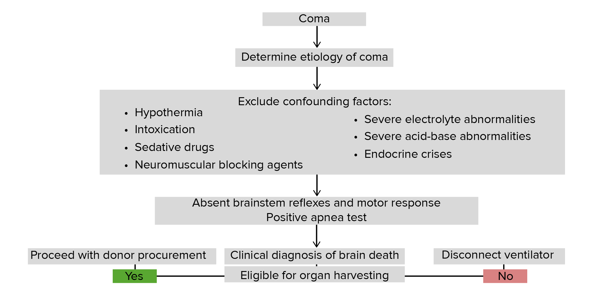
Establish the etiology of the comaComaComa is defined as a deep state of unarousable unresponsiveness, characterized by a score of 3 points on the GCS. A comatose state can be caused by a multitude of conditions, making the precise epidemiology and prognosis of coma difficult to determine. Coma (e.g., overdose, trauma, stroke).
Evaluate for any confounding conditions mimicking irreversible brainBrainThe part of central nervous system that is contained within the skull (cranium). Arising from the neural tube, the embryonic brain is comprised of three major parts including prosencephalon (the forebrain); mesencephalon (the midbrain); and rhombencephalon (the hindbrain). The developed brain consists of cerebrum; cerebellum; and other structures in the brain stem.Nervous System: Anatomy, Structure, and Classification injury:
Severe electrolyte abnormalities or acid-base disorders
Severe endocrine dysfunction
HypothermiaHypothermiaHypothermia can be defined as a drop in the core body temperature below 35°C (95°F) and is classified into mild, moderate, severe, and profound forms based on the degree of temperature decrease. Hypothermia
Circulatory collapse (i.e., severe hypotensionHypotensionHypotension is defined as low blood pressure, specifically < 90/60 mm Hg, and is most commonly a physiologic response. Hypotension may be mild, serious, or life threatening, depending on the cause. Hypotension)
Blood alcohol level > 80 mg/dL
CNS depressants or paralytics
In the presence of hospital-administered sedative drugs and paralytics:
Discontinue medications for 5 eliminationEliminationThe initial damage and destruction of tumor cells by innate and adaptive immunity. Completion of the phase means no cancer growth. Cancer Immunotherapyhalf-lifeHalf-LifeThe time it takes for a substance (drug, radioactive nuclide, or other) to lose half of its pharmacologic, physiologic, or radiologic activity.Pharmacokinetics and Pharmacodynamics periods before testing.
Note: The American College of Medical Toxicology recommends a longer period for a large drug exposure.[7]
May need more time with hepatic or kidney dysfunction
Observation time prior to clinical testing[4,5]
There is insufficient evidence to determine optimal timing for testing.
Anoxic brain injuryAnoxic Brain InjuryPersistent Vegetative State after resuscitated cardiac arrestCardiac arrestCardiac arrest is the sudden, complete cessation of cardiac output with hemodynamic collapse. Patients present as pulseless, unresponsive, and apneic. Rhythms associated with cardiac arrest are ventricular fibrillation/tachycardia, asystole, or pulseless electrical activity. Cardiac Arrest: Wait a minimum of 24 hours.
For other brainBrainThe part of central nervous system that is contained within the skull (cranium). Arising from the neural tube, the embryonic brain is comprised of three major parts including prosencephalon (the forebrain); mesencephalon (the midbrain); and rhombencephalon (the hindbrain). The developed brain consists of cerebrum; cerebellum; and other structures in the brain stem.Nervous System: Anatomy, Structure, and Classification injuries: determined on a case-by-case basis
Clinical neurologic examination[4,6]
The number of, and interval between, neurologic examinations required to establish brainBrainThe part of central nervous system that is contained within the skull (cranium). Arising from the neural tube, the embryonic brain is comprised of three major parts including prosencephalon (the forebrain); mesencephalon (the midbrain); and rhombencephalon (the hindbrain). The developed brain consists of cerebrum; cerebellum; and other structures in the brain stem.Nervous System: Anatomy, Structure, and Classification death may vary depending on the practice location (number often ranges from 1 to 3). Additionally, many places require at least 2 clinicianClinicianA physician, nurse practitioner, physician assistant, or another health professional who is directly involved in patient care and has a professional relationship with patients.Clinician–Patient Relationship evaluations. See your hospital’s designated protocol.
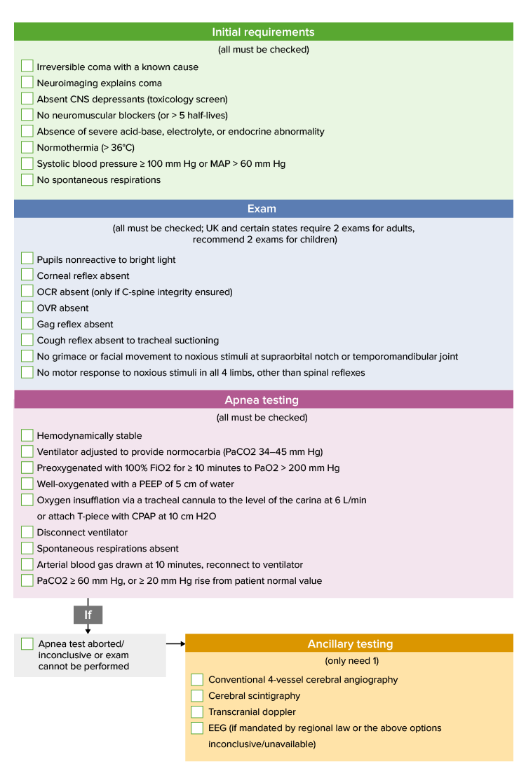
Requirements for assessment:
Minimum core temperature of 36°C is required.
Systolic blood pressure (SBPSBPAscites) > 100 mm Hg or MAP > 60 mm Hg:
Age-appropriate blood pressure targets should be metMETPreoperative Care in children.
Determination of comaComaComa is defined as a deep state of unarousable unresponsiveness, characterized by a score of 3 points on the GCS. A comatose state can be caused by a multitude of conditions, making the precise epidemiology and prognosis of coma difficult to determine. Coma by the bilateral absence of motorMotorNeurons which send impulses peripherally to activate muscles or secretory cells.Nervous System: Histology responses to:
Condyles at the level of the temporomandibular joint (TMJ)
Proximal and distal locations on all limbs
Responses consistent with brainBrainThe part of central nervous system that is contained within the skull (cranium). Arising from the neural tube, the embryonic brain is comprised of three major parts including prosencephalon (the forebrain); mesencephalon (the midbrain); and rhombencephalon (the hindbrain). The developed brain consists of cerebrum; cerebellum; and other structures in the brain stem.Nervous System: Anatomy, Structure, and Classification death:
Pupillary light reflexPupillary Light ReflexConstriction of the pupil in response to light stimulation of the retina. It refers also to any reflex involving the iris, with resultant alteration of the diameter of the pupil.Pupil: Physiology and Abnormalities:
Shine light into each eye and examine for pupillary constriction.
Measure pupilPupilThe pupil is the space within the eye that permits light to project onto the retina. Anatomically located in front of the lens, the pupil’s size is controlled by the surrounding iris. The pupil provides insight into the function of the central and autonomic nervous systems. Pupil: Physiology and Abnormalities diameter.
Responses consistent with brainBrainThe part of central nervous system that is contained within the skull (cranium). Arising from the neural tube, the embryonic brain is comprised of three major parts including prosencephalon (the forebrain); mesencephalon (the midbrain); and rhombencephalon (the hindbrain). The developed brain consists of cerebrum; cerebellum; and other structures in the brain stem.Nervous System: Anatomy, Structure, and Classification death:
Absence of ipsilateral and contralateral pupillary response
Pupils fixed in a midsize or dilated position (4–6 mm) bilaterally
Constricted pupils are not consistent with brainBrainThe part of central nervous system that is contained within the skull (cranium). Arising from the neural tube, the embryonic brain is comprised of three major parts including prosencephalon (the forebrain); mesencephalon (the midbrain); and rhombencephalon (the hindbrain). The developed brain consists of cerebrum; cerebellum; and other structures in the brain stem.Nervous System: Anatomy, Structure, and Classification death.
Notes:
Pupillary response may be affected by previous eye surgery or trauma.
Ensure that no ophthalmic medications may interfere with the test.
Oculocephalic reflex (OCR) and oculovestibular reflex (OVR):
OCR: examined by quickly rotating the head horizontally to both sides:
Make sure there are no concerns about cervical spineSpineThe human spine, or vertebral column, is the most important anatomical and functional axis of the human body. It consists of 7 cervical vertebrae, 12 thoracic vertebrae, and 5 lumbar vertebrae and is limited cranially by the skull and caudally by the sacrum.Vertebral Column: Anatomy integrity.
The term “doll’s eyes” refers to vintage dolls with eyes painted on and unable to move with the head.
Abnormal reflex: Eyes are fixed with head movement as if gazing at a fixed point.
OVR:
Instill 50 mL of cold water into the ear with the head at a 30-degree elevation (for ≥ 60 seconds).
Test both sides.
Ensure auditory canals are patent and tympanic membranes are intact.
Responses consistent with brainBrainThe part of central nervous system that is contained within the skull (cranium). Arising from the neural tube, the embryonic brain is comprised of three major parts including prosencephalon (the forebrain); mesencephalon (the midbrain); and rhombencephalon (the hindbrain). The developed brain consists of cerebrum; cerebellum; and other structures in the brain stem.Nervous System: Anatomy, Structure, and Classification death: absent eye movement with both tests
Note: Eye movement may be impacted by severe orbital or scleral edemaEdemaEdema is a condition in which excess serous fluid accumulates in the body cavity or interstitial space of connective tissues. Edema is a symptom observed in several medical conditions. It can be categorized into 2 types, namely, peripheral (in the extremities) and internal (in an organ or body cavity). Edema.
Corneal reflex:
Examined by touching the corneaCorneaThe transparent anterior portion of the fibrous coat of the eye consisting of five layers: stratified squamous corneal epithelium; bowman membrane; corneal stroma; descemet membrane; and mesenchymal corneal endothelium. It serves as the first refracting medium of the eye.Eye: Anatomy with a cotton swab or squirts of water/saline
Touch the external border of the iris.
Light pressure can be applied.
Be careful not to damage the corneaCorneaThe transparent anterior portion of the fibrous coat of the eye consisting of five layers: stratified squamous corneal epithelium; bowman membrane; corneal stroma; descemet membrane; and mesenchymal corneal endothelium. It serves as the first refracting medium of the eye.Eye: Anatomy.
Responses consistent with brainBrainThe part of central nervous system that is contained within the skull (cranium). Arising from the neural tube, the embryonic brain is comprised of three major parts including prosencephalon (the forebrain); mesencephalon (the midbrain); and rhombencephalon (the hindbrain). The developed brain consists of cerebrum; cerebellum; and other structures in the brain stem.Nervous System: Anatomy, Structure, and Classification death: absent eyelid movement
Note: Response may be impacted by orbital or scleral edemaEdemaEdema is a condition in which excess serous fluid accumulates in the body cavity or interstitial space of connective tissues. Edema is a symptom observed in several medical conditions. It can be categorized into 2 types, namely, peripheral (in the extremities) and internal (in an organ or body cavity). Edema, chemosisChemosisConjunctivitis, or previous corneal transplantationCorneal transplantationPartial or total replacement of the cornea from one human or animal to another.Organ Transplantation.
Examined by touching the posterior pharyngeal wall with a suction device or tongueTongueThe tongue, on the other hand, is a complex muscular structure that permits tasting and facilitates the process of mastication and communication. The blood supply of the tongue originates from the external carotid artery, and the innervation is through cranial nerves.Lips and Tongue: Anatomy depressor
Response consistent with brainBrainThe part of central nervous system that is contained within the skull (cranium). Arising from the neural tube, the embryonic brain is comprised of three major parts including prosencephalon (the forebrain); mesencephalon (the midbrain); and rhombencephalon (the hindbrain). The developed brain consists of cerebrum; cerebellum; and other structures in the brain stem.Nervous System: Anatomy, Structure, and Classification death: absent gag
Cough reflex:
Examined by stimulating the tracheobronchial wall through suctioning
Response consistent with brainBrainThe part of central nervous system that is contained within the skull (cranium). Arising from the neural tube, the embryonic brain is comprised of three major parts including prosencephalon (the forebrain); mesencephalon (the midbrain); and rhombencephalon (the hindbrain). The developed brain consists of cerebrum; cerebellum; and other structures in the brain stem.Nervous System: Anatomy, Structure, and Classification death: absent cough
Note: Response may be impacted by injury to the phrenic nervePhrenic nerveThe motor nerve of the diaphragm. The phrenic nerve fibers originate in the cervical spinal column (mostly C4) and travel through the cervical plexus to the diaphragm.Diaphragm: Anatomy.
Common sequelae[6]
Irreversible cessation of brainstem function is associated with the following abnormalities (which may arise during the clinical course):
HyponatremiaHyponatremiaHyponatremia is defined as a decreased serum sodium (sNa+) concentration less than 135 mmol/L. Serum sodium is the greatest contributor to plasma osmolality, which is very tightly controlled via antidiuretic hormone (ADH) release from the hypothalamus and by the thirst mechanism.Hyponatremia or hypernatremiaHypernatremiaHypernatremia is an elevated serum sodium concentration > 145 mmol/L. Serum sodium is the greatest contributor to plasma osmolality, which is very tightly controlled by the hypothalamus via the thirst mechanism and antidiuretic hormone (ADH) release. Hypernatremia occurs either from a lack of access to water or an excessive intake of sodium.Hypernatremia
DiabetesDiabetesDiabetes mellitus (DM) is a metabolic disease characterized by hyperglycemia and dysfunction of the regulation of glucose metabolism by insulin. Type 1 DM is diagnosed mostly in children and young adults as the result of autoimmune destruction of β cells in the pancreas and the resulting lack of insulin. Type 2 DM has a significant association with obesity and is characterized by insulin resistance.Diabetes Mellitus insipidus
HypothermiaHypothermiaHypothermia can be defined as a drop in the core body temperature below 35°C (95°F) and is classified into mild, moderate, severe, and profound forms based on the degree of temperature decrease. Hypothermia
Cardiac dysrhythmia
Hemodynamic instability
Diagnosis
Outside the clinical examination, additional testing can vary depending on practice location. See your hospital’s designated protocol. The following information is based on US and UK guidelines for adult patientsPatientsIndividuals participating in the health care system for the purpose of receiving therapeutic, diagnostic, or preventive procedures.Clinician–Patient Relationship.
Diagnostic requirements for brainBrainThe part of central nervous system that is contained within the skull (cranium). Arising from the neural tube, the embryonic brain is comprised of three major parts including prosencephalon (the forebrain); mesencephalon (the midbrain); and rhombencephalon (the hindbrain). The developed brain consists of cerebrum; cerebellum; and other structures in the brain stem.Nervous System: Anatomy, Structure, and Classification death[4,5]
Known clinical etiology of brainBrainThe part of central nervous system that is contained within the skull (cranium). Arising from the neural tube, the embryonic brain is comprised of three major parts including prosencephalon (the forebrain); mesencephalon (the midbrain); and rhombencephalon (the hindbrain). The developed brain consists of cerebrum; cerebellum; and other structures in the brain stem.Nervous System: Anatomy, Structure, and Classification death
Absence of confounding conditions
Abnormal brainstem reflexes on neurological examinationNeurological examinationA neurological exam is a systematic assessment of cognitive, sensory, and motor responses to identify pathologies of the nervous system. A neurological exam allows for the localization of neurologic lesions to narrow the differential diagnosis and focus on subsequent laboratory and imaging examinations. The exam should include assessments of the subject’s mental status, speech, cranial nerves, motor system, deep tendon reflexes, sensation, balance, and coordination.Neurological Examination
Unable to perform all aspects of the clinical examination or apnea testApnea TestBrain Death
Uncertainty about clinical response to clinical testing
Cannot completely rule out confounding conditions
May also bolster the clinical diagnosis for grieving families
Laboratory evaluation[4,7]
Laboratory studies are often used to evaluate the etiology and identify confounding factors. The following may be considered (list is not exhaustive and should be guided by the clinical scenario):
ElectrolytesElectrolytesElectrolytes are mineral salts that dissolve in water and dissociate into charged particles called ions, which can be either be positively (cations) or negatively (anions) charged. Electrolytes are distributed in the extracellular and intracellular compartments in different concentrations. Electrolytes are essential for various basic life-sustaining functions.Electrolytes → rule out metabolic disorders and assess acid-base status
Toxicology screen and alcohol level → rule out intoxication:
Specific drug testing should be guided by the patient’s history.
Consult a clinical toxicologist for guidance in cases where intoxication cannot be ruled out by standard testing (e.g., synthetic opioidsOpioidsOpiates are drugs that are derived from the sap of the opium poppy. Opiates have been used since antiquity for the relief of acute severe pain. Opioids are synthetic opiates with properties that are substantially similar to those of opiates. Opioid Analgesics, clonazepamClonazepamAn anticonvulsant used for several types of seizures, including myotonic or atonic seizures, photosensitive epilepsy, and absence seizures, although tolerance may develop. It is seldom effective in generalized tonic-clonic or partial seizures. The mechanism of action appears to involve the enhancement of gamma-aminobutyric acid receptor responses.Benzodiazepines).
CSF analysisCSF analysisMeningitis → not routinely done, but may be performed in the diagnostic workup for an etiology (e.g., CNS infection, subarachnoid hemorrhageSubarachnoid HemorrhageSubarachnoid hemorrhage (SAH) is a type of cerebrovascular accident (stroke) resulting from intracranial hemorrhage into the subarachnoid space between the arachnoid and the pia mater layers of the meninges surrounding the brain. Most SAHs originate from a saccular aneurysm in the circle of Willis but may also occur as a result of trauma, uncontrolled hypertension, vasculitis, anticoagulant use, or stimulant use. Subarachnoid Hemorrhage)
This test is part of all protocols for determination of brainBrainThe part of central nervous system that is contained within the skull (cranium). Arising from the neural tube, the embryonic brain is comprised of three major parts including prosencephalon (the forebrain); mesencephalon (the midbrain); and rhombencephalon (the hindbrain). The developed brain consists of cerebrum; cerebellum; and other structures in the brain stem.Nervous System: Anatomy, Structure, and Classification death. The following is based on US guidelines. The recommended protocol in the UK varies slightly and can be found here.
Performed after all other criteria for brainBrainThe part of central nervous system that is contained within the skull (cranium). Arising from the neural tube, the embryonic brain is comprised of three major parts including prosencephalon (the forebrain); mesencephalon (the midbrain); and rhombencephalon (the hindbrain). The developed brain consists of cerebrum; cerebellum; and other structures in the brain stem.Nervous System: Anatomy, Structure, and Classification death have been metMETPreoperative Care
Before the test:
Ensure SBPSBPAscites ≥ 100 mm Hg or MAP ≥ 60 mm Hg
Temperature should be ≥ 36℃.
Discontinue paralytic drugs.
Place arterial line for easy ABG sampling.
Adjust ventilator settings to a PaCO2 between 35 and 45 mm Hg.
Preoxygenate with 100% O2 for ≥ 10 minutes before the test.
Cannot be done in:
The presence of high cervical spineSpineThe human spine, or vertebral column, is the most important anatomical and functional axis of the human body. It consists of 7 cervical vertebrae, 12 thoracic vertebrae, and 5 lumbar vertebrae and is limited cranially by the skull and caudally by the sacrum.Vertebral Column: Anatomy lesions
Individuals with chronic CO2 retention
Steps:
Discontinue ventilator for 10 minutes.
Provide 100% O2 to a maximum PO2 of 200 mm Hg or until PaCO2 is > 40 mm Hg.
CPAPCPAPA technique of respiratory therapy, in either spontaneously breathing or mechanically ventilated patients, in which airway pressure is maintained above atmospheric pressure throughout the respiratory cycle by pressurization of the ventilatory circuit.Noninvasive Ventilation/PEEPPEEPPressure remaining in the distal airways of the patient at the end of expirationInvasive Mechanical Ventilation can prevent derecruitment and ↓ risk of cardiopulmonary instability.
Oxygen insufflation method via tracheal cannula may also be used.
After 10 minutes, ABG should be sent.
If no spontaneous respiratory efforts → test is terminated after 10 minutes
No respiratory response to PaCO2 ≥ 60 mm Hg, or ≥ 20 mm Hg above baseline values
Final arterial pHpHThe quantitative measurement of the acidity or basicity of a solution.Acid-Base Balance < 7.30
Ancillary testing[4–6]
Brain blood flow studiesBrain Blood Flow StudiesBrain Death: Absent brainBrainThe part of central nervous system that is contained within the skull (cranium). Arising from the neural tube, the embryonic brain is comprised of three major parts including prosencephalon (the forebrain); mesencephalon (the midbrain); and rhombencephalon (the hindbrain). The developed brain consists of cerebrum; cerebellum; and other structures in the brain stem.Nervous System: Anatomy, Structure, and Classificationblood flowBlood flowBlood flow refers to the movement of a certain volume of blood through the vasculature over a given unit of time (e.g., mL per minute).Vascular Resistance, Flow, and Mean Arterial Pressure is consistent with brainBrainThe part of central nervous system that is contained within the skull (cranium). Arising from the neural tube, the embryonic brain is comprised of three major parts including prosencephalon (the forebrain); mesencephalon (the midbrain); and rhombencephalon (the hindbrain). The developed brain consists of cerebrum; cerebellum; and other structures in the brain stem.Nervous System: Anatomy, Structure, and Classification death.
Conventional 4-vessel cerebral angiographyAngiographyRadiography of blood vessels after injection of a contrast medium.Cardiac Surgery/digital subtraction angiographyAngiographyRadiography of blood vessels after injection of a contrast medium.Cardiac Surgery:
Preferred testing method
Should demonstrate absent filling where the internal carotid and vertebral arteriesArteriesArteries are tubular collections of cells that transport oxygenated blood and nutrients from the heart to the tissues of the body. The blood passes through the arteries in order of decreasing luminal diameter, starting in the largest artery (the aorta) and ending in the small arterioles. Arteries are classified into 3 types: large elastic arteries, medium muscular arteries, and small arteries and arterioles. Arteries: Histology enter the skullSkullThe skull (cranium) is the skeletal structure of the head supporting the face and forming a protective cavity for the brain. The skull consists of 22 bones divided into the viscerocranium (facial skeleton) and the neurocranium.Skull: Anatomy base
External carotid circulationCirculationThe movement of the blood as it is pumped through the cardiovascular system.ABCDE Assessment should be patent.
Radionuclide studies (alternative)
Transcranial DopplerDopplerUltrasonography applying the doppler effect, with frequency-shifted ultrasound reflections produced by moving targets (usually red blood cells) in the bloodstream along the ultrasound axis in direct proportion to the velocity of movement of the targets, to determine both direction and velocity of blood flow.Ultrasound (Sonography) (alternative):
If used, it is recommended to perform 2 examinations ≥ 30 minutes apart.
CTACTAA non-invasive method that uses a ct scanner for capturing images of blood vessels and tissues. A contrast material is injected, which helps produce detailed images that aid in diagnosing vascular diseases.Pulmonary Function Tests and MRAMRAImaging of the Heart and Great Vessels are not currently recommended to support the diagnosis of cerebral circulatory arrestCirculatory arrestCardiac arrest is the sudden, complete cessation of cardiac output with hemodynamic collapse. Patients present as pulseless, unresponsive, and apneic. Rhythms associated with cardiac arrest are ventricular fibrillation/tachycardia, asystole, or pulseless electrical activity.Cardiac Arrest.
Required to diagnose brainBrainThe part of central nervous system that is contained within the skull (cranium). Arising from the neural tube, the embryonic brain is comprised of three major parts including prosencephalon (the forebrain); mesencephalon (the midbrain); and rhombencephalon (the hindbrain). The developed brain consists of cerebrum; cerebellum; and other structures in the brain stem.Nervous System: Anatomy, Structure, and Classification death in children
Should not be routinely used in adults unless legally mandated
Consistent with brainBrainThe part of central nervous system that is contained within the skull (cranium). Arising from the neural tube, the embryonic brain is comprised of three major parts including prosencephalon (the forebrain); mesencephalon (the midbrain); and rhombencephalon (the hindbrain). The developed brain consists of cerebrum; cerebellum; and other structures in the brain stem.Nervous System: Anatomy, Structure, and Classification death if no potentials are recorded after 30 minutes
May be falsely negative or falsely positive
Addition studies:
MRI or CT may:
Assist with determining an etiology (e.g., hemorrhage, massMassThree-dimensional lesion that occupies a space within the breastImaging of the Breast lesion)
Demonstrate irreversible, devastating brainBrainThe part of central nervous system that is contained within the skull (cranium). Arising from the neural tube, the embryonic brain is comprised of three major parts including prosencephalon (the forebrain); mesencephalon (the midbrain); and rhombencephalon (the hindbrain). The developed brain consists of cerebrum; cerebellum; and other structures in the brain stem.Nervous System: Anatomy, Structure, and Classification injury:
Evidence of severe edemaEdemaEdema is a condition in which excess serous fluid accumulates in the body cavity or interstitial space of connective tissues. Edema is a symptom observed in several medical conditions. It can be categorized into 2 types, namely, peripheral (in the extremities) and internal (in an organ or body cavity). Edema or herniationHerniationOmphalocele
If seen, brainBrainThe part of central nervous system that is contained within the skull (cranium). Arising from the neural tube, the embryonic brain is comprised of three major parts including prosencephalon (the forebrain); mesencephalon (the midbrain); and rhombencephalon (the hindbrain). The developed brain consists of cerebrum; cerebellum; and other structures in the brain stem.Nervous System: Anatomy, Structure, and ClassificationedemaEdemaEdema is a condition in which excess serous fluid accumulates in the body cavity or interstitial space of connective tissues. Edema is a symptom observed in several medical conditions. It can be categorized into 2 types, namely, peripheral (in the extremities) and internal (in an organ or body cavity). Edema is suggestive of brainBrainThe part of central nervous system that is contained within the skull (cranium). Arising from the neural tube, the embryonic brain is comprised of three major parts including prosencephalon (the forebrain); mesencephalon (the midbrain); and rhombencephalon (the hindbrain). The developed brain consists of cerebrum; cerebellum; and other structures in the brain stem.Nervous System: Anatomy, Structure, and Classification death.
If ICPICPNormal intracranial pressure (ICP) is defined as < 15 mm Hg, whereas pathologically increased ICP is any pressure ≥ 20 mm Hg. Increased ICP may result from several etiologies, including trauma, intracranial hemorrhage, mass lesions, cerebral edema, increased CSF production, and decreased CSF absorption.Increased Intracranial Pressure (ICP) monitor is in place → measured ICPICPNormal intracranial pressure (ICP) is defined as < 15 mm Hg, whereas pathologically increased ICP is any pressure ≥ 20 mm Hg. Increased ICP may result from several etiologies, including trauma, intracranial hemorrhage, mass lesions, cerebral edema, increased CSF production, and decreased CSF absorption.Increased Intracranial Pressure (ICP) ≥ MAP indicates no perfusion gradient for blood flowBlood flowBlood flow refers to the movement of a certain volume of blood through the vasculature over a given unit of time (e.g., mL per minute).Vascular Resistance, Flow, and Mean Arterial Pressure
Algorithm describing the course of action in individuals with brain death
DocumentationDocumentationSystematic organization, storage, retrieval, and dissemination of specialized information, especially of a scientific or technical nature. It often involves authenticating or validating information.Advance Directives required for determination of brainBrainThe part of central nervous system that is contained within the skull (cranium). Arising from the neural tube, the embryonic brain is comprised of three major parts including prosencephalon (the forebrain); mesencephalon (the midbrain); and rhombencephalon (the hindbrain). The developed brain consists of cerebrum; cerebellum; and other structures in the brain stem.Nervous System: Anatomy, Structure, and Classification death[4–6]
Etiology of the comaComaComa is defined as a deep state of unarousable unresponsiveness, characterized by a score of 3 points on the GCS. A comatose state can be caused by a multitude of conditions, making the precise epidemiology and prognosis of coma difficult to determine. Coma
Absence of confounding conditions
Full details and results of clinical testing performed
NeuroimagingNeuroimagingNon-invasive methods of visualizing the central nervous system, especially the brain, by various imaging modalities.Febrile Infant results and timing in relation to clinical testing
Reason for and type of ancillary testing performed and results
Time of death
Identity of the practitioner(s) performing the evaluation
Somatic support after declaration of brainBrainThe part of central nervous system that is contained within the skull (cranium). Arising from the neural tube, the embryonic brain is comprised of three major parts including prosencephalon (the forebrain); mesencephalon (the midbrain); and rhombencephalon (the hindbrain). The developed brain consists of cerebrum; cerebellum; and other structures in the brain stem.Nervous System: Anatomy, Structure, and Classification death[4–7]
Diagnosis made → individual is declared clinically and legally deceased
Physiologic support should be discontinued unless:
Decedent is pregnant and support is needed for the fetus
Notify the local organ procurement organization → notification required in many regions
Organ procurement coordinators review the medical chart.
If appropriate, the coordinators speak with the potential donor’s next of kin.
Family requests due to religious or moral beliefs:
Every effort should be made to address concerns.
Involvement of religious authorities or ethicsEthicsMedical ethics are a set of moral values that guide the decision-making of health care professionals in their daily practice. A sense of ethical responsibility has accompanied the profession of medicine since antiquity, and the Hippocratic oath was the 1st document to codify its core ethical principles.Medical Ethics: Basic Principles committees may be helpful.
Treat every person and family with the utmost respect.
A multidisciplinary approach (e.g., social workers, psychologists, counselors) is advisable.
Prolongation of support > 48 hours should generally be avoided.
Differential Diagnosis
Locked-in syndromeLocked-in syndromeLocked-in syndrome (LIS) is a rare neurological disorder in which patients are awake and conscious but are unable to move their limbs or speak. The disorder is a result of brain injury to the ventral aspect of the pons and caudal ventral midbrain; etiologies include brainstem stroke, tumors, intracranial bleeding, and demyelinating disorders. Locked-in Syndrome(also known as “pseudocomaPseudocomaLocked-in syndrome (LIS) is a rare neurological disorder in which patients are awake and conscious but are unable to move their limbs or speak. The disorder is a result of brain injury to the ventral aspect of the pons and caudal ventral midbrain; etiologies include brainstem stroke, tumors, intracranial bleeding, and demyelinating disorders.Locked-in Syndrome” or “de-efferented state”): a state of quadriplegiaQuadriplegiaSevere or complete loss of motor function in all four limbs which may result from brain diseases; spinal cord diseases; peripheral nervous system diseases; neuromuscular diseases; or rarely muscular diseases. The locked-in syndrome is characterized by quadriplegia in combination with cranial muscle paralysis. Consciousness is spared and the only retained voluntary motor activity may be limited eye movements. This condition is usually caused by a lesion in the upper brain stem which injures the descending corticospinal and corticobulbar tracts.Locked-in Syndrome and paralysis of the lower cranial nervesCranial nervesThere are 12 pairs of cranial nerves (CNs), which run from the brain to various parts of the head, neck, and trunk. The CNs can be sensory or motor or both. The CNs are named and numbered in Roman numerals according to their location, from the front to the back of the brain.The 12 Cranial Nerves: Overview and Functions leading to the inability to move, communicate, or show any facial expression (except for certain eye movements). Locked-in syndromeLocked-in syndromeLocked-in syndrome (LIS) is a rare neurological disorder in which patients are awake and conscious but are unable to move their limbs or speak. The disorder is a result of brain injury to the ventral aspect of the pons and caudal ventral midbrain; etiologies include brainstem stroke, tumors, intracranial bleeding, and demyelinating disorders. Locked-in Syndrome typically results from pontine hemorrhage or infarctInfarctArea of necrotic cells in an organ, arising mainly from hypoxia and ischemiaIschemic Cell Damage. Diagnosis is made clinically with individuals limited to blinking or performing vertical eye movements upon request. Management involves supportive care and prevention of immobilizationImmobilizationDelirium complications. Long-term physical therapyPhysical TherapyBecker Muscular Dystrophy is essential.
HypothermiaHypothermiaHypothermia can be defined as a drop in the core body temperature below 35°C (95°F) and is classified into mild, moderate, severe, and profound forms based on the degree of temperature decrease. Hypothermia: a drop in core body temperatureBody TemperatureThe measure of the level of heat of a human or animal.Heatstroke below 35°C. HypothermiaHypothermiaHypothermia can be defined as a drop in the core body temperature below 35°C (95°F) and is classified into mild, moderate, severe, and profound forms based on the degree of temperature decrease. Hypothermia is classified into mild, moderate, severe, and profound forms based upon the degree of temperature decrease. Populations of individuals with extremes of age, homelessness, mental illness, or substance use disorders may be more vulnerable to accidental hypothermiaAccidental HypothermiaHypothermia. Evaluation includes assessment for associated trauma and contributing medical conditions. Management involves rewarming the individual based on the severity of hypothermiaHypothermiaHypothermia can be defined as a drop in the core body temperature below 35°C (95°F) and is classified into mild, moderate, severe, and profound forms based on the degree of temperature decrease. Hypothermia.
Drug intoxication: intoxication of CNS-suppressant medications or muscle relaxants/paralytics. The drugs may include opioidsOpioidsOpiates are drugs that are derived from the sap of the opium poppy. Opiates have been used since antiquity for the relief of acute severe pain. Opioids are synthetic opiates with properties that are substantially similar to those of opiates. Opioid Analgesics, tricyclics, baclofenBaclofenA gamma-aminobutyric acid derivative that is a specific agonist of gaba-b receptors. It is used in the treatment of muscle spasticity, especially that due to spinal cord injuries. Its therapeutic effects result from actions at spinal and supraspinal sites, generally the reduction of excitatory transmission.Spasmolytics, barbituratesBarbituratesA class of chemicals derived from barbituric acid or thiobarbituric acid. Many of these are gaba modulators used as hypnotics and sedatives, as anesthetics, or as anticonvulsants.Intravenous Anesthetics, paralytics, anticholinergicsAnticholinergicsAnticholinergic drugs block the effect of the neurotransmitter acetylcholine at the muscarinic receptors in the central and peripheral nervous systems. Anticholinergic agents inhibit the parasympathetic nervous system, resulting in effects on the smooth muscle in the respiratory tract, vascular system, urinary tract, GI tract, and pupils of the eyes. Anticholinergic Drugs, and organophosphates, which may significantly suppress consciousness and motorMotorNeurons which send impulses peripherally to activate muscles or secretory cells.Nervous System: Histology/sensorySensoryNeurons which conduct nerve impulses to the central nervous system.Nervous System: Histology responses. The exposure may be either known or unrecognized. Diagnosis is based on history, review of medications, interviews with family, and toxicologic evaluations. Management is based on the diagnosis.
High cervical spineSpineThe human spine, or vertebral column, is the most important anatomical and functional axis of the human body. It consists of 7 cervical vertebrae, 12 thoracic vertebrae, and 5 lumbar vertebrae and is limited cranially by the skull and caudally by the sacrum.Vertebral Column: Anatomy injury: such injuries are associated with quadriparesis, loss of sensation below the level of the injury, inability to breathe, and reduced ability to speak. Although neurologic evaluation should easily distinguish the condition from brainBrainThe part of central nervous system that is contained within the skull (cranium). Arising from the neural tube, the embryonic brain is comprised of three major parts including prosencephalon (the forebrain); mesencephalon (the midbrain); and rhombencephalon (the hindbrain). The developed brain consists of cerebrum; cerebellum; and other structures in the brain stem.Nervous System: Anatomy, Structure, and Classification death, the clinical picture may be confounded by a concurrent traumatic brain injuryTraumatic brain injuryA form of acquired brain injury which occurs when a sudden trauma causes damage to the brain.Le Fort Fractures or the effects of medications. Vigilance and repeated comprehensive neurologic evaluations in combination with neuroimagingNeuroimagingNon-invasive methods of visualizing the central nervous system, especially the brain, by various imaging modalities.Febrile Infant are helpful in establishing the correct diagnosis. Management may be medical or surgical.
Billing and Coding
Diagnosis Codes:
This code is used to document the determination of brainBrainThe part of central nervous system that is contained within the skull (cranium). Arising from the neural tube, the embryonic brain is comprised of three major parts including prosencephalon (the forebrain); mesencephalon (the midbrain); and rhombencephalon (the hindbrain). The developed brain consists of cerebrum; cerebellum; and other structures in the brain stem.Nervous System: Anatomy, Structure, and Classification death, which is the irreversible cessation of all functions of the entire brainBrainThe part of central nervous system that is contained within the skull (cranium). Arising from the neural tube, the embryonic brain is comprised of three major parts including prosencephalon (the forebrain); mesencephalon (the midbrain); and rhombencephalon (the hindbrain). The developed brain consists of cerebrum; cerebellum; and other structures in the brain stem.Nervous System: Anatomy, Structure, and Classification, including the brainstem. Legally and medically, this constitutes death.
Coding System
Code
Description
ICD-10-CM
G93.82
BrainBrainThe part of central nervous system that is contained within the skull (cranium). Arising from the neural tube, the embryonic brain is comprised of three major parts including prosencephalon (the forebrain); mesencephalon (the midbrain); and rhombencephalon (the hindbrain). The developed brain consists of cerebrum; cerebellum; and other structures in the brain stem.Nervous System: Anatomy, Structure, and Classification death
SNOMED CT
110480000
BrainBrainThe part of central nervous system that is contained within the skull (cranium). Arising from the neural tube, the embryonic brain is comprised of three major parts including prosencephalon (the forebrain); mesencephalon (the midbrain); and rhombencephalon (the hindbrain). The developed brain consists of cerebrum; cerebellum; and other structures in the brain stem.Nervous System: Anatomy, Structure, and Classification death (finding)
Procedures/Interventions:
This code is for an electroencephalogram (EEGEEGSeizures), one of the confirmatory tests that can be used to support the clinical diagnosis of brainBrainThe part of central nervous system that is contained within the skull (cranium). Arising from the neural tube, the embryonic brain is comprised of three major parts including prosencephalon (the forebrain); mesencephalon (the midbrain); and rhombencephalon (the hindbrain). The developed brain consists of cerebrum; cerebellum; and other structures in the brain stem.Nervous System: Anatomy, Structure, and Classification death by demonstrating the absence of electrical activity in the brainBrainThe part of central nervous system that is contained within the skull (cranium). Arising from the neural tube, the embryonic brain is comprised of three major parts including prosencephalon (the forebrain); mesencephalon (the midbrain); and rhombencephalon (the hindbrain). The developed brain consists of cerebrum; cerebellum; and other structures in the brain stem.Nervous System: Anatomy, Structure, and Classification.
Greer, D. M., Shemie, S. D., Lewis, A., et al. (2020). Determination of brain death/death by neurologic criteria: the World Brain Death Project. JAMA, 324(11), 1078. https://doi.org/10.1001/jama.2020.11586
Wijdicks, E. F. M., Varelas, P. N., Gronseth, G. S., Greer, D. M. (2010, reaffirmed 2017). Evidence-based guideline update: determining brain death in adults: report of the quality standards subcommittee of the American Academy of Neurology. Neurology, 74(23), 1911–1918. https://doi.org/10.1212/WNL.0b013e3181e242a8