Achieve Mastery of Medical Concepts

Study for medical school and boards with Lecturio

Pyruvate Kinase Deficiency

Pyruvate kinase Pyruvate kinase Atp:pyruvate 2-o-phosphotransferase. A phosphotransferase that catalyzes reversibly the phosphorylation of pyruvate to phosphoenolpyruvate in the presence of ATP. It has four isozymes (l, r, m1, and m2). Glycolysis (PK) deficiency is an autosomal recessive Autosomal recessive Autosomal inheritance, both dominant and recessive, refers to the transmission of genes from the 22 autosomal chromosomes. Autosomal recessive diseases are only expressed when 2 copies of the recessive allele are inherited. Autosomal Recessive and Autosomal Dominant Inheritance enzymatic disorder of erythrocytes Erythrocytes Erythrocytes, or red blood cells (RBCs), are the most abundant cells in the blood. While erythrocytes in the fetus are initially produced in the yolk sac then the liver, the bone marrow eventually becomes the main site of production. Erythrocytes: Histology that results in hemolytic anemia Hemolytic Anemia Hemolytic anemia (HA) is the term given to a large group of anemias that are caused by the premature destruction/hemolysis of circulating red blood cells (RBCs). Hemolysis can occur within (intravascular hemolysis) or outside the blood vessels (extravascular hemolysis). Hemolytic Anemia. PK deficiency is mainly a hereditary disorder, with a defect in the PKLR or PKM gene Gene A category of nucleic acid sequences that function as units of heredity and which code for the basic instructions for the development, reproduction, and maintenance of organisms. Basic Terms of Genetics, but it can be acquired secondary to underlying conditions, such as leukemia. The typical clinical features of PK deficiency are mild to moderate anemia Anemia Anemia is a condition in which individuals have low Hb levels, which can arise from various causes. Anemia is accompanied by a reduced number of RBCs and may manifest with fatigue, shortness of breath, pallor, and weakness. Subtypes are classified by the size of RBCs, chronicity, and etiology. Anemia: Overview and Types, jaundice Jaundice Jaundice is the abnormal yellowing of the skin and/or sclera caused by the accumulation of bilirubin. Hyperbilirubinemia is caused by either an increase in bilirubin production or a decrease in the hepatic uptake, conjugation, or excretion of bilirubin. Jaundice, delay in growth, failure to thrive Failure to Thrive Failure to thrive (FTT), or faltering growth, describes suboptimal weight gain and growth in children. The majority of cases are due to inadequate caloric intake; however, genetic, infectious, and oncological etiologies are also common. Failure to Thrive, and frontal bossing Frontal Bossing Hydrocephalus in Children. However, more severe presentations are possible, especially in younger patients Patients Individuals participating in the health care system for the purpose of receiving therapeutic, diagnostic, or preventive procedures. Clinician–Patient Relationship. Diagnosis involves ruling out other, more common, causes of hemolytic anemia Hemolytic Anemia Hemolytic anemia (HA) is the term given to a large group of anemias that are caused by the premature destruction/hemolysis of circulating red blood cells (RBCs). Hemolysis can occur within (intravascular hemolysis) or outside the blood vessels (extravascular hemolysis). Hemolytic Anemia. Biochemical assays and/or genetic testing Genetic Testing Detection of a mutation; genotype; karyotype; or specific alleles associated with genetic traits, heritable diseases, or predisposition to a disease, or that may lead to the disease in descendants. It includes prenatal genetic testing. Myotonic Dystrophies can confirm the diagnosis. Management may include supportive treatment, mitapivat (a PK activator recently approved by the FDA), exchange transfusion, phototherapy Phototherapy Treatment of disease by exposure to light, especially by variously concentrated light rays or specific wavelengths. Hyperbilirubinemia of the Newborn, or splenectomy Splenectomy Surgical procedure involving either partial or entire removal of the spleen. Rupture of the Spleen. Prognosis Prognosis A prediction of the probable outcome of a disease based on a individual's condition and the usual course of the disease as seen in similar situations. Non-Hodgkin Lymphomas is age-dependent, with older age at diagnosis associated with a better prognosis Prognosis A prediction of the probable outcome of a disease based on a individual's condition and the usual course of the disease as seen in similar situations. Non-Hodgkin Lymphomas.

Last updated: Apr 5, 2023

Editorial responsibility: Stanley Oiseth, Lindsay Jones, Evelin Maza

Overview

Definition

Pyruvate kinase Pyruvate kinase Atp:pyruvate 2-o-phosphotransferase. A phosphotransferase that catalyzes reversibly the phosphorylation of pyruvate to phosphoenolpyruvate in the presence of ATP. It has four isozymes (l, r, m1, and m2). Glycolysis (PK) deficiency is an autosomal recessive Autosomal recessive Autosomal inheritance, both dominant and recessive, refers to the transmission of genes from the 22 autosomal chromosomes. Autosomal recessive diseases are only expressed when 2 copies of the recessive allele are inherited. Autosomal Recessive and Autosomal Dominant Inheritance enzymatic disorder of erythrocytes Erythrocytes Erythrocytes, or red blood cells (RBCs), are the most abundant cells in the blood. While erythrocytes in the fetus are initially produced in the yolk sac then the liver, the bone marrow eventually becomes the main site of production. Erythrocytes: Histology that results in chronic hemolysis due to a defect in the PKLR or PKM gene Gene A category of nucleic acid sequences that function as units of heredity and which code for the basic instructions for the development, reproduction, and maintenance of organisms. Basic Terms of Genetics.

Epidemiology

  • Extremely rare, but most common defect in the glycolysis Glycolysis Glycolysis is a central metabolic pathway responsible for the breakdown of glucose and plays a vital role in generating free energy for the cell and metabolites for further oxidative degradation. Glucose primarily becomes available in the blood as a result of glycogen breakdown or from its synthesis from noncarbohydrate precursors (gluconeogenesis) and is imported into cells by specific transport proteins. Glycolysis pathway
  • Prevalence Prevalence The total number of cases of a given disease in a specified population at a designated time. It is differentiated from incidence, which refers to the number of new cases in the population at a given time. Measures of Disease Frequency is unknown; estimates have ranged from 1 in 20,000 to 1 in 115,000.
  • Worldwide distribution
  • More common in:
    • People of Northern European and Chinese ancestry
    • Isolated populations with a history of consanguinity Consanguinity The magnitude of inbreeding in humans. Basic Terms of Genetics due to the founder effect

Etiology and Pathophysiology

Classification and etiology

  • Hereditary ( genetic mutations Genetic Mutations Carcinogenesis in the PKLR or PKM gene Gene A category of nucleic acid sequences that function as units of heredity and which code for the basic instructions for the development, reproduction, and maintenance of organisms. Basic Terms of Genetics):
    • Homozygous individuals present with clinical manifestations.
    • Heterozygous individuals are usually asymptomatic.
    • PKLR gene Gene A category of nucleic acid sequences that function as units of heredity and which code for the basic instructions for the development, reproduction, and maintenance of organisms. Basic Terms of Genetics located on chromosome Chromosome In a prokaryotic cell or in the nucleus of a eukaryotic cell, a structure consisting of or containing DNA which carries the genetic information essential to the cell. Basic Terms of Genetics 1q21
    • PKM gene Gene A category of nucleic acid sequences that function as units of heredity and which code for the basic instructions for the development, reproduction, and maintenance of organisms. Basic Terms of Genetics located on chromosome Chromosome In a prokaryotic cell or in the nucleus of a eukaryotic cell, a structure consisting of or containing DNA which carries the genetic information essential to the cell. Basic Terms of Genetics 15q22
  • Acquired (secondary to underlying conditions):
    • Leukemia 
    • Refractory sideroblastic anemia Sideroblastic anemia Sideroblastic anemias are a heterogeneous group of bone marrow disorders characterized by abnormal iron accumulation in the mitochondria of erythroid precursors. The accumulated iron appears as granules in a ringlike distribution around the nucleus, giving rise to the characteristic morphological feature of a ring sideroblast. Sideroblastic Anemia 
    • Complication of chemotherapy Chemotherapy Osteosarcoma

Normal physiology

Pyruvate kinase Pyruvate kinase Atp:pyruvate 2-o-phosphotransferase. A phosphotransferase that catalyzes reversibly the phosphorylation of pyruvate to phosphoenolpyruvate in the presence of ATP. It has four isozymes (l, r, m1, and m2). Glycolysis is an enzyme required for the conversion of phosphoenolpyruvate Phosphoenolpyruvate A monocarboxylic acid anion derived from selective deprotonation of the carboxy group of phosphoenolpyruvic acid. It is a metabolic intermediate in glycolysis; gluconeogenesis; and other pathways. Glycolysis ( PEP PEP A monocarboxylic acid anion derived from selective deprotonation of the carboxy group of phosphoenolpyruvic acid. It is a metabolic intermediate in glycolysis; gluconeogenesis; and other pathways. Glycolysis) into pyruvate Pyruvate Derivatives of pyruvic acid, including its salts and esters. Glycolysis and ATP in the energy-producing glycolysis Glycolysis Glycolysis is a central metabolic pathway responsible for the breakdown of glucose and plays a vital role in generating free energy for the cell and metabolites for further oxidative degradation. Glucose primarily becomes available in the blood as a result of glycogen breakdown or from its synthesis from noncarbohydrate precursors (gluconeogenesis) and is imported into cells by specific transport proteins. Glycolysis pathway.

  • RBCs RBCs Erythrocytes, or red blood cells (RBCs), are the most abundant cells in the blood. While erythrocytes in the fetus are initially produced in the yolk sac then the liver, the bone marrow eventually becomes the main site of production. Erythrocytes: Histology are dependent on glycolysis Glycolysis Glycolysis is a central metabolic pathway responsible for the breakdown of glucose and plays a vital role in generating free energy for the cell and metabolites for further oxidative degradation. Glucose primarily becomes available in the blood as a result of glycogen breakdown or from its synthesis from noncarbohydrate precursors (gluconeogenesis) and is imported into cells by specific transport proteins. Glycolysis for ATP production (lack mitochondria Mitochondria Semiautonomous, self-reproducing organelles that occur in the cytoplasm of all cells of most, but not all, eukaryotes. Each mitochondrion is surrounded by a double limiting membrane. The inner membrane is highly invaginated, and its projections are called cristae. Mitochondria are the sites of the reactions of oxidative phosphorylation, which result in the formation of ATP. They contain distinctive ribosomes, transfer RNAs; amino Acyl tRNA synthetases; and elongation and termination factors. Mitochondria depend upon genes within the nucleus of the cells in which they reside for many essential messenger RNAs. Mitochondria are believed to have arisen from aerobic bacteria that established a symbiotic relationship with primitive protoeukaryotes. The Cell: Organelles).
  • ATP is needed for the maintenance of cell function and structural integrity.
Gluconeogenesis as the reverse of glycolysis

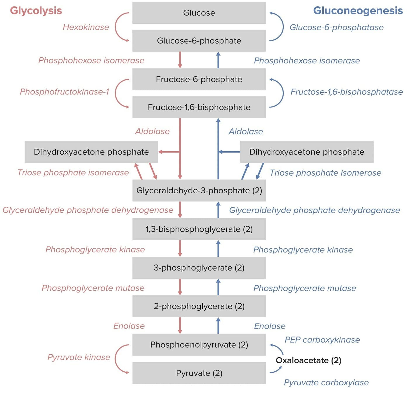
Glycolysis and gluconeogenesis pathways:
Note that the conversion of phosphoenolpyruvate to pyruvate by pyruvate kinase is the final step in the glycolysis pathway.

Image by Lecturio.

Pathophysiology

Effect of PK deficiency: 

  • Buildup of precursors within the glycolysis Glycolysis Glycolysis is a central metabolic pathway responsible for the breakdown of glucose and plays a vital role in generating free energy for the cell and metabolites for further oxidative degradation. Glucose primarily becomes available in the blood as a result of glycogen breakdown or from its synthesis from noncarbohydrate precursors (gluconeogenesis) and is imported into cells by specific transport proteins. Glycolysis pathway, notably 2,3-diphosphoglycerate 2,3-diphosphoglycerate A highly anionic organic phosphate which is present in human red blood cells at about the same molar ratio as hemoglobin. It binds to deoxyhemoglobin but not the oxygenated form, therefore diminishing the oxygen affinity of hemoglobin. This is essential in enabling hemoglobin to unload oxygen in tissue capillaries. It is also an intermediate in the conversion of 3-phosphoglycerate to 2-phosphoglycerate by phosphoglycerate mutase. Glycolysis (2,3-DPG)
  • ↓ Production of:
    • Pyruvate Pyruvate Derivatives of pyruvic acid, including its salts and esters. Glycolysis
    • ATP
    • Lactate 

Consequences of PK deficiency:

  • Hemolysis:
    • Accumulation of intermediate precursors and glycolytic metabolites within RBCs RBCs Erythrocytes, or red blood cells (RBCs), are the most abundant cells in the blood. While erythrocytes in the fetus are initially produced in the yolk sac then the liver, the bone marrow eventually becomes the main site of production. Erythrocytes: Histology:  
    • ↓ RBC membrane plasticity and cell dehydration Dehydration The condition that results from excessive loss of water from a living organism. Volume Depletion and Dehydration premature Premature Childbirth before 37 weeks of pregnancy (259 days from the first day of the mother’s last menstrual period, or 245 days after fertilization). Necrotizing Enterocolitis destruction of RBCs RBCs Erythrocytes, or red blood cells (RBCs), are the most abundant cells in the blood. While erythrocytes in the fetus are initially produced in the yolk sac then the liver, the bone marrow eventually becomes the main site of production. Erythrocytes: Histology in the spleen Spleen The spleen is the largest lymphoid organ in the body, located in the LUQ of the abdomen, superior to the left kidney and posterior to the stomach at the level of the 9th-11th ribs just below the diaphragm. The spleen is highly vascular and acts as an important blood filter, cleansing the blood of pathogens and damaged erythrocytes. Spleen: Anatomy
    • Hemolysis is usually extravascular, but severe hemolysis may be intravascular. 
  • ↑ Oxygen delivery:
    • ↑ 2,3-DPG → rightward shift in the oxygen dissociation Dissociation Defense Mechanisms curve (the RBCs RBCs Erythrocytes, or red blood cells (RBCs), are the most abundant cells in the blood. While erythrocytes in the fetus are initially produced in the yolk sac then the liver, the bone marrow eventually becomes the main site of production. Erythrocytes: Histology have a harder time “holding on” to the O2) → results in ↑ delivery of oxygen to peripheral tissue per volume of hemoglobin
    • Allows for higher physiologic tolerance Tolerance Pharmacokinetics and Pharmacodynamics to anemia Anemia Anemia is a condition in which individuals have low Hb levels, which can arise from various causes. Anemia is accompanied by a reduced number of RBCs and may manifest with fatigue, shortness of breath, pallor, and weakness. Subtypes are classified by the size of RBCs, chronicity, and etiology. Anemia: Overview and Types with PK deficiency
  • Iron overload Iron overload An excessive accumulation of iron in the body due to a greater than normal absorption of iron from the gastrointestinal tract or from parenteral injection. This may arise from idiopathic hemochromatosis, excessive iron intake, chronic alcoholism, certain types of refractory anemia, or transfusional hemosiderosis. Hereditary Hemochromatosis:
    • Ineffective erythropoiesis Erythropoiesis The production of red blood cells (erythrocytes). In humans, erythrocytes are produced by the yolk sac in the first trimester; by the liver in the second trimester; by the bone marrow in the third trimester and after birth. In normal individuals, the erythrocyte count in the peripheral blood remains relatively constant implying a balance between the rate of erythrocyte production and rate of destruction. Erythrocytes: Histology → ↑ production of early erythrocyte precursors
    • Hepcidin Hepcidin Forms of hepcidin, a cationic amphipathic peptide synthesized in the liver as a prepropeptide which is first processed into prohepcidin and then into the biologically active hepcidin forms, including in human the 20-, 22-, and 25-amino acid residue peptide forms. Hepcidin acts as a homeostatic regulators of iron metabolism and also possesses antimicrobial activity. Hereditary Hemochromatosis, which leads to:
      • Iron absorption Iron absorption Digestion and Absorption
      • Iron Iron A metallic element with atomic symbol fe, atomic number 26, and atomic weight 55. 85. It is an essential constituent of hemoglobins; cytochromes; and iron-binding proteins. It plays a role in cellular redox reactions and in the transport of oxygen. Trace Elements retention in macrophages Macrophages The relatively long-lived phagocytic cell of mammalian tissues that are derived from blood monocytes. Main types are peritoneal macrophages; alveolar macrophages; histiocytes; kupffer cells of the liver; and osteoclasts. They may further differentiate within chronic inflammatory lesions to epithelioid cells or may fuse to form foreign body giant cells or langhans giant cells. Innate Immunity: Phagocytes and Antigen Presentation

Clinical Presentation and Complications

Clinical presentation

The age and severity at presentation depend on the extent of hemolysis and anemia Anemia Anemia is a condition in which individuals have low Hb levels, which can arise from various causes. Anemia is accompanied by a reduced number of RBCs and may manifest with fatigue, shortness of breath, pallor, and weakness. Subtypes are classified by the size of RBCs, chronicity, and etiology. Anemia: Overview and Types. Additionally, the age at presentation may vary.

Signs and symptoms in newborns:

  • Neonatal jaundice Neonatal jaundice Yellow discoloration of the skin; mucous membrane; and sclera in the newborn. It is a sign of neonatal hyperbilirubinemia. Most cases are transient self-limiting (physiological neonatal jaundice) occurring in the first week of life, but some can be a sign of pathological disorders, particularly liver diseases. Jaundice/ kernicterus Kernicterus A term used pathologically to describe bilirubin staining of the basal ganglia; brain stem; and cerebellum and clinically to describe a syndrome associated with hyperbilirubinemia. Clinical features include athetosis, muscle spasticity or hypotonia, impaired vertical gaze, and deafness. Nonconjugated bilirubin enters the brain and acts as a neurotoxin, often in association with conditions that impair the blood-brain barrier (e.g., sepsis). This condition occurs primarily in neonates, but may rarely occur in adults. Hyperbilirubinemia of the Newborn
  • Delay in growth, poor weight gain
  • Failure to thrive Failure to Thrive Failure to thrive (FTT), or faltering growth, describes suboptimal weight gain and growth in children. The majority of cases are due to inadequate caloric intake; however, genetic, infectious, and oncological etiologies are also common. Failure to Thrive, lethargy Lethargy A general state of sluggishness, listless, or uninterested, with being tired, and having difficulty concentrating and doing simple tasks. It may be related to depression or drug addiction. Hyponatremia
  • Frontal bossing Frontal Bossing Hydrocephalus in Children
  • Extramedullary hematopoiesis Extramedullary Hematopoiesis Primary Myelofibrosis of the skin Skin The skin, also referred to as the integumentary system, is the largest organ of the body. The skin is primarily composed of the epidermis (outer layer) and dermis (deep layer). The epidermis is primarily composed of keratinocytes that undergo rapid turnover, while the dermis contains dense layers of connective tissue. Skin: Structure and Functions
  • Hydrops Hydrops Cholecystitis fetalis (due to severe anemia Anemia Anemia is a condition in which individuals have low Hb levels, which can arise from various causes. Anemia is accompanied by a reduced number of RBCs and may manifest with fatigue, shortness of breath, pallor, and weakness. Subtypes are classified by the size of RBCs, chronicity, and etiology. Anemia: Overview and Types in utero)

Signs and symptoms in older children and adults:

  • Findings consistent with chronic hemolytic anemia Chronic Hemolytic Anemia Anemia: Overview and Types
  • Pallor 
  • Jaundice Jaundice Jaundice is the abnormal yellowing of the skin and/or sclera caused by the accumulation of bilirubin. Hyperbilirubinemia is caused by either an increase in bilirubin production or a decrease in the hepatic uptake, conjugation, or excretion of bilirubin. Jaundice/ icterus Icterus A clinical manifestation of hyperbilirubinemia, characterized by the yellowish staining of the skin, mucous membranes, and sclera. Clinical jaundice usually is a sign of liver dysfunction. Jaundice 
  • Mild to severe splenomegaly Splenomegaly Splenomegaly is pathologic enlargement of the spleen that is attributable to numerous causes, including infections, hemoglobinopathies, infiltrative processes, and outflow obstruction of the portal vein. Splenomegaly
  • Pigment ( bilirubin Bilirubin A bile pigment that is a degradation product of heme. Heme Metabolism) gallstones Gallstones Cholelithiasis (gallstones) is the presence of stones in the gallbladder. Most gallstones are cholesterol stones, while the rest are composed of bilirubin (pigment stones) and other mixed components. Patients are commonly asymptomatic but may present with biliary colic (intermittent pain in the right upper quadrant). Cholelithiasis
  • Chronic leg Leg The lower leg, or just “leg” in anatomical terms, is the part of the lower limb between the knee and the ankle joint. The bony structure is composed of the tibia and fibula bones, and the muscles of the leg are grouped into the anterior, lateral, and posterior compartments by extensions of fascia. Leg: Anatomy ulcers
Jaundice_in_newborn

A 6-week-old girl with symptoms of jaundice due to hypothyroidism

Image: “Jaundice in newborn” by Dr. Hudson (CDC). License: Public Domain

Complications

  • Heart failure Heart Failure A heterogeneous condition in which the heart is unable to pump out sufficient blood to meet the metabolic need of the body. Heart failure can be caused by structural defects, functional abnormalities (ventricular dysfunction), or a sudden overload beyond its capacity. Chronic heart failure is more common than acute heart failure which results from sudden insult to cardiac function, such as myocardial infarction. Total Anomalous Pulmonary Venous Return (TAPVR)
  • Liver Liver The liver is the largest gland in the human body. The liver is found in the superior right quadrant of the abdomen and weighs approximately 1.5 kilograms. Its main functions are detoxification, metabolism, nutrient storage (e.g., iron and vitamins), synthesis of coagulation factors, formation of bile, filtration, and storage of blood. Liver: Anatomy disease
  • Endocrine dysfunction (e.g., diabetes Diabetes Diabetes mellitus (DM) is a metabolic disease characterized by hyperglycemia and dysfunction of the regulation of glucose metabolism by insulin. Type 1 DM is diagnosed mostly in children and young adults as the result of autoimmune destruction of β cells in the pancreas and the resulting lack of insulin. Type 2 DM has a significant association with obesity and is characterized by insulin resistance. Diabetes Mellitus mellitus, hypogonadism Hypogonadism Hypogonadism is a condition characterized by reduced or no sex hormone production by the testes or ovaries. Hypogonadism can result from primary (hypergonadotropic) or secondary (hypogonadotropic) failure. Symptoms include infertility, increased risk of osteoporosis, erectile dysfunction, decreased libido, and regression (or absence) of secondary sexual characteristics. Hypogonadism)
  • Arthropathies
  • Sepsis Sepsis Systemic inflammatory response syndrome with a proven or suspected infectious etiology. When sepsis is associated with organ dysfunction distant from the site of infection, it is called severe sepsis. When sepsis is accompanied by hypotension despite adequate fluid infusion, it is called septic shock. Sepsis and Septic Shock post-splenectomy (higher incidence Incidence The number of new cases of a given disease during a given period in a specified population. It also is used for the rate at which new events occur in a defined population. It is differentiated from prevalence, which refers to all cases in the population at a given time. Measures of Disease Frequency in children)
  • Thromboembolic disease post-splenectomy (higher incidence Incidence The number of new cases of a given disease during a given period in a specified population. It also is used for the rate at which new events occur in a defined population. It is differentiated from prevalence, which refers to all cases in the population at a given time. Measures of Disease Frequency in adults)
  • Pulmonary hypertension Pulmonary Hypertension Pulmonary hypertension (PH) or pulmonary arterial hypertension (PAH) is characterized by elevated pulmonary arterial pressure, which can lead to chronic progressive right heart failure. Pulmonary hypertension is grouped into 5 categories based on etiology, which include primary PAH, and PH due to cardiac disease, lung or hypoxic disease, chronic thromboembolic disease, and multifactorial or unclear etiologies. Pulmonary Hypertension
  • Increased risk of aplastic anemia Aplastic Anemia Aplastic anemia (AA) is a rare, life-threatening condition characterized by pancytopenia and hypocellularity of the bone marrow (in the absence of any abnormal cells) reflecting damage to hematopoietic stem cells. Aplastic anemia can be acquired or inherited, however, most cases of AA are acquired and caused by autoimmune damage to hematopoietic stem cells. Aplastic Anemia with certain viral infections Infections Invasion of the host organism by microorganisms or their toxins or by parasites that can cause pathological conditions or diseases. Chronic Granulomatous Disease (e.g., parvovirus B19 Parvovirus B19 Primate erythroparvovirus 1 (generally referred to as parvovirus B19, B19 virus, or sometimes erythrovirus B19) ranks among the smallest DNA viruses. Parvovirus B19 is of the family Parvoviridae and genus Erythrovirus. In immunocompetent humans, parvovirus B19 classically results in erythema infectiosum (5th disease) or “slapped cheek syndrome.” Parvovirus B19
  • Ischemic stroke Ischemic Stroke An ischemic stroke (also known as cerebrovascular accident) is an acute neurologic injury that occurs as a result of brain ischemia; this condition may be due to cerebral blood vessel occlusion by thrombosis or embolism, or rarely due to systemic hypoperfusion. Ischemic Stroke
  • Risks associated with repeat blood transfusions Blood transfusions The introduction of whole blood or blood component directly into the bloodstream. Transfusion Products

Diagnosis

Pyruvate kinase Pyruvate kinase Atp:pyruvate 2-o-phosphotransferase. A phosphotransferase that catalyzes reversibly the phosphorylation of pyruvate to phosphoenolpyruvate in the presence of ATP. It has four isozymes (l, r, m1, and m2). Glycolysis deficiency is rare; ruling out other causes of hemolytic anemia Hemolytic Anemia Hemolytic anemia (HA) is the term given to a large group of anemias that are caused by the premature destruction/hemolysis of circulating red blood cells (RBCs). Hemolysis can occur within (intravascular hemolysis) or outside the blood vessels (extravascular hemolysis). Hemolytic Anemia is important.

History and physical examination

  • Clinical features of hemolytic anemia Hemolytic Anemia Hemolytic anemia (HA) is the term given to a large group of anemias that are caused by the premature destruction/hemolysis of circulating red blood cells (RBCs). Hemolysis can occur within (intravascular hemolysis) or outside the blood vessels (extravascular hemolysis). Hemolytic Anemia 
  • Family history Family History Adult Health Maintenance of:
    • Similar symptoms 
    • Unknown cause of anemia Anemia Anemia is a condition in which individuals have low Hb levels, which can arise from various causes. Anemia is accompanied by a reduced number of RBCs and may manifest with fatigue, shortness of breath, pallor, and weakness. Subtypes are classified by the size of RBCs, chronicity, and etiology. Anemia: Overview and Types
    • Pyruvate kinase Pyruvate kinase Atp:pyruvate 2-o-phosphotransferase. A phosphotransferase that catalyzes reversibly the phosphorylation of pyruvate to phosphoenolpyruvate in the presence of ATP. It has four isozymes (l, r, m1, and m2). Glycolysis deficiency

Basic laboratory evaluation

CBC with differential:

  • ↓ Hemoglobin and hematocrit Hematocrit The volume of packed red blood cells in a blood specimen. The volume is measured by centrifugation in a tube with graduated markings, or with automated blood cell counters. It is an indicator of erythrocyte status in disease. For example, anemia shows a low value; polycythemia, a high value. Neonatal Polycythemia
  • Normal or ↑ mean corpuscular volume (MCV)
  • ↑ Reticulocytes (particularly post-splenectomy)
  • Normal or ↑ platelets Platelets Platelets are small cell fragments involved in hemostasis. Thrombopoiesis takes place primarily in the bone marrow through a series of cell differentiation and is influenced by several cytokines. Platelets are formed after fragmentation of the megakaryocyte cytoplasm. Platelets: Histology (reactive thrombocytosis)

Chemistry:

Antiglobulin testing:

  • Negative direct antiglobulin test (DAT; also known as the Coombs test)
  • Negative indirect antiglobulin test

Peripheral blood smear Peripheral Blood Smear Anemia: Overview and Types, focusing on red cell morphology:

  • Characteristic findings of other causes of inherited hemolytic anemia Hemolytic Anemia Hemolytic anemia (HA) is the term given to a large group of anemias that are caused by the premature destruction/hemolysis of circulating red blood cells (RBCs). Hemolysis can occur within (intravascular hemolysis) or outside the blood vessels (extravascular hemolysis). Hemolytic Anemia are absent. 
  • Burr cells
  • Accelerated erythropoiesis Erythropoiesis The production of red blood cells (erythrocytes). In humans, erythrocytes are produced by the yolk sac in the first trimester; by the liver in the second trimester; by the bone marrow in the third trimester and after birth. In normal individuals, the erythrocyte count in the peripheral blood remains relatively constant implying a balance between the rate of erythrocyte production and rate of destruction. Erythrocytes: Histology:
    • Polychromasia
    • Anisocytosis
    • Poikilocytosis
    • Nucleated RBCs RBCs Erythrocytes, or red blood cells (RBCs), are the most abundant cells in the blood. While erythrocytes in the fetus are initially produced in the yolk sac then the liver, the bone marrow eventually becomes the main site of production. Erythrocytes: Histology

Confirmatory testing

Diagnosis is confirmed if a patient with hemolytic anemia Hemolytic Anemia Hemolytic anemia (HA) is the term given to a large group of anemias that are caused by the premature destruction/hemolysis of circulating red blood cells (RBCs). Hemolysis can occur within (intravascular hemolysis) or outside the blood vessels (extravascular hemolysis). Hemolytic Anemia/compensated hemolysis has abnormalities on either biochemical or genetic testing Genetic Testing Detection of a mutation; genotype; karyotype; or specific alleles associated with genetic traits, heritable diseases, or predisposition to a disease, or that may lead to the disease in descendants. It includes prenatal genetic testing. Myotonic Dystrophies.

Biochemical testing:

  • Gold standard
  • Assesses the activity of pyruvate kinase Pyruvate kinase Atp:pyruvate 2-o-phosphotransferase. A phosphotransferase that catalyzes reversibly the phosphorylation of pyruvate to phosphoenolpyruvate in the presence of ATP. It has four isozymes (l, r, m1, and m2). Glycolysis in RBCs RBCs Erythrocytes, or red blood cells (RBCs), are the most abundant cells in the blood. While erythrocytes in the fetus are initially produced in the yolk sac then the liver, the bone marrow eventually becomes the main site of production. Erythrocytes: Histology
  • Pyruvate kinase Pyruvate kinase Atp:pyruvate 2-o-phosphotransferase. A phosphotransferase that catalyzes reversibly the phosphorylation of pyruvate to phosphoenolpyruvate in the presence of ATP. It has four isozymes (l, r, m1, and m2). Glycolysis activity is expected in PK deficiency.
  • A negative result does not eliminate the possibility of PK deficiency:
    • WBCs and platelets Platelets Platelets are small cell fragments involved in hemostasis. Thrombopoiesis takes place primarily in the bone marrow through a series of cell differentiation and is influenced by several cytokines. Platelets are formed after fragmentation of the megakaryocyte cytoplasm. Platelets: Histology also have PK activity. If these are not removed from the hemolysate prior to testing, results can be skewed.
    • If PK deficiency is still suspected, consider specialized assay or genetic testing Genetic Testing Detection of a mutation; genotype; karyotype; or specific alleles associated with genetic traits, heritable diseases, or predisposition to a disease, or that may lead to the disease in descendants. It includes prenatal genetic testing. Myotonic Dystrophies.
  • Note: Biochemical testing should be deferred for 2‒3 months after a transfusion.

Genetic testing Genetic Testing Detection of a mutation; genotype; karyotype; or specific alleles associated with genetic traits, heritable diseases, or predisposition to a disease, or that may lead to the disease in descendants. It includes prenatal genetic testing. Myotonic Dystrophies:

  • Looks for mutations in PKLR
  • Not done as an initial test; the importance of some mutations is unclear.

Indications for genetic testing Genetic Testing Detection of a mutation; genotype; karyotype; or specific alleles associated with genetic traits, heritable diseases, or predisposition to a disease, or that may lead to the disease in descendants. It includes prenatal genetic testing. Myotonic Dystrophies:

  • An individual with unexplained hemolysis or anemia Anemia Anemia is a condition in which individuals have low Hb levels, which can arise from various causes. Anemia is accompanied by a reduced number of RBCs and may manifest with fatigue, shortness of breath, pallor, and weakness. Subtypes are classified by the size of RBCs, chronicity, and etiology. Anemia: Overview and Types who has a sibling with a known PK deficiency
  • Patients Patients Individuals participating in the health care system for the purpose of receiving therapeutic, diagnostic, or preventive procedures. Clinician–Patient Relationship with hemolytic anemia Hemolytic Anemia Hemolytic anemia (HA) is the term given to a large group of anemias that are caused by the premature destruction/hemolysis of circulating red blood cells (RBCs). Hemolysis can occur within (intravascular hemolysis) or outside the blood vessels (extravascular hemolysis). Hemolytic Anemia with a negative DAT when other causes have been ruled out
  • Prenatal testing of pregnant individuals who already have a child with:
    • Hydrops Hydrops Cholecystitis fetalis 
    • Severe transfusion-dependent anemia Anemia Anemia is a condition in which individuals have low Hb levels, which can arise from various causes. Anemia is accompanied by a reduced number of RBCs and may manifest with fatigue, shortness of breath, pallor, and weakness. Subtypes are classified by the size of RBCs, chronicity, and etiology. Anemia: Overview and Types, not resolved by splenectomy Splenectomy Surgical procedure involving either partial or entire removal of the spleen. Rupture of the Spleen

Management and Prognosis

Management

Management is dependent on the age at diagnosis and the severity. 

  • Intrauterine period (for fetal hydrops Hydrops Cholecystitis due to severe anemia Anemia Anemia is a condition in which individuals have low Hb levels, which can arise from various causes. Anemia is accompanied by a reduced number of RBCs and may manifest with fatigue, shortness of breath, pallor, and weakness. Subtypes are classified by the size of RBCs, chronicity, and etiology. Anemia: Overview and Types): intrauterine transfusion
  • Neonatal period (presence of hyperbilirubinemia Hyperbilirubinemia A condition characterized by an abnormal increase of bilirubin in the blood, which may result in jaundice. Bilirubin, a breakdown product of heme, is normally excreted in the bile or further catabolized before excretion in the urine. Jaundice):
  • Infancy through adulthood (presence of severe anemia Anemia Anemia is a condition in which individuals have low Hb levels, which can arise from various causes. Anemia is accompanied by a reduced number of RBCs and may manifest with fatigue, shortness of breath, pallor, and weakness. Subtypes are classified by the size of RBCs, chronicity, and etiology. Anemia: Overview and Types):
    • RBC transfusions 
    • Pharmacotherapy:
      • Mitapivat: a pyruvate kinase Pyruvate kinase Atp:pyruvate 2-o-phosphotransferase. A phosphotransferase that catalyzes reversibly the phosphorylation of pyruvate to phosphoenolpyruvate in the presence of ATP. It has four isozymes (l, r, m1, and m2). Glycolysis activator recently approved by the FDA for the treatment of symptomatic hemolytic anemia Hemolytic Anemia Hemolytic anemia (HA) is the term given to a large group of anemias that are caused by the premature destruction/hemolysis of circulating red blood cells (RBCs). Hemolysis can occur within (intravascular hemolysis) or outside the blood vessels (extravascular hemolysis). Hemolytic Anemia
      • Folic acid
    • Splenectomy Splenectomy Surgical procedure involving either partial or entire removal of the spleen. Rupture of the Spleen: indicated for severe, transfusion-dependent cases unresponsive to mitapivat
    • Iron Iron A metallic element with atomic symbol fe, atomic number 26, and atomic weight 55. 85. It is an essential constituent of hemoglobins; cytochromes; and iron-binding proteins. It plays a role in cellular redox reactions and in the transport of oxygen. Trace Elements chelation (particularly in cases requiring long-term treatment with transfusions) 
    • Investigational therapies:
      • Hematopoietic stem-cell transplantation (for patients Patients Individuals participating in the health care system for the purpose of receiving therapeutic, diagnostic, or preventive procedures. Clinician–Patient Relationship with severe anemia Anemia Anemia is a condition in which individuals have low Hb levels, which can arise from various causes. Anemia is accompanied by a reduced number of RBCs and may manifest with fatigue, shortness of breath, pallor, and weakness. Subtypes are classified by the size of RBCs, chronicity, and etiology. Anemia: Overview and Types requiring multiple transfusions after splenectomy Splenectomy Surgical procedure involving either partial or entire removal of the spleen. Rupture of the Spleen)
      • Gene Gene A category of nucleic acid sequences that function as units of heredity and which code for the basic instructions for the development, reproduction, and maintenance of organisms. Basic Terms of Genetics therapy with CRISPR CRISPR Repetitive nucleic acid sequences that are principal components of the archaeal and bacterial CRISPR-CAS systems, which function as adaptive antiviral defense systems. RNA Types and Structure correction of genetic mutations Genetic Mutations Carcinogenesis

Monitoring

Patients Patients Individuals participating in the health care system for the purpose of receiving therapeutic, diagnostic, or preventive procedures. Clinician–Patient Relationship should be monitored for: 

  • Symptoms and clinical signs of anemia Anemia Anemia is a condition in which individuals have low Hb levels, which can arise from various causes. Anemia is accompanied by a reduced number of RBCs and may manifest with fatigue, shortness of breath, pallor, and weakness. Subtypes are classified by the size of RBCs, chronicity, and etiology. Anemia: Overview and Types 
  • Growth and development 
  • Labs:
    • CBC with differential  
    • Reticulocyte Reticulocyte Immature erythrocytes. In humans, these are erythroid cells that have just undergone extrusion of their cell nucleus. They still contain some organelles that gradually decrease in number as the cells mature. Ribosomes are last to disappear. Certain staining techniques cause components of the ribosomes to precipitate into characteristic ‘reticulum’ (not the same as the endoplasmic reticulum), hence the name reticulocytes. Erythrocytes: Histology count 
    • Ferritin Ferritin Iron-containing proteins that are widely distributed in animals, plants, and microorganisms. Their major function is to store iron in a nontoxic bioavailable form. Each ferritin molecule consists of ferric iron in a hollow protein shell (apoferritins) made of 24 subunits of various sequences depending on the species and tissue types. Hereditary Hemochromatosis levels

Prognosis Prognosis A prediction of the probable outcome of a disease based on a individual’s condition and the usual course of the disease as seen in similar situations. Non-Hodgkin Lymphomas

  • Mild and moderate forms of PK deficiency: excellent prognosis Prognosis A prediction of the probable outcome of a disease based on a individual’s condition and the usual course of the disease as seen in similar situations. Non-Hodgkin Lymphomas
  • Severe forms PK deficiency: prognosis Prognosis A prediction of the probable outcome of a disease based on a individual’s condition and the usual course of the disease as seen in similar situations. Non-Hodgkin Lymphomas age-dependent
    • Primarily symptomatic in early childhood
    • The older the patient is at diagnosis, the better the prognosis Prognosis A prediction of the probable outcome of a disease based on a individual’s condition and the usual course of the disease as seen in similar situations. Non-Hodgkin Lymphomas.

Differential Diagnosis

  • Glucose-6-phosphate dehydrogenase Glucose-6-phosphate dehydrogenase Pentose Phosphate Pathway ( G6PD G6PD Pentose Phosphate Pathway) deficiency: X-linked recessive X-Linked Recessive Duchenne Muscular Dystrophy inherited disorder causing hemolytic anemia Hemolytic Anemia Hemolytic anemia (HA) is the term given to a large group of anemias that are caused by the premature destruction/hemolysis of circulating red blood cells (RBCs). Hemolysis can occur within (intravascular hemolysis) or outside the blood vessels (extravascular hemolysis). Hemolytic Anemia due to deficiencies in the G6PD G6PD Pentose Phosphate Pathway enzyme, which normally helps RBCs RBCs Erythrocytes, or red blood cells (RBCs), are the most abundant cells in the blood. While erythrocytes in the fetus are initially produced in the yolk sac then the liver, the bone marrow eventually becomes the main site of production. Erythrocytes: Histology generate NADPH NADPH Nicotinamide adenine dinucleotide phosphate. A coenzyme composed of ribosylnicotinamide 5′-phosphate (nmn) coupled by pyrophosphate linkage to the 5′-phosphate adenosine 2. Pentose Phosphate Pathway and protects them from oxidative injury. It presents with symptoms of hemolytic anemia Hemolytic Anemia Hemolytic anemia (HA) is the term given to a large group of anemias that are caused by the premature destruction/hemolysis of circulating red blood cells (RBCs). Hemolysis can occur within (intravascular hemolysis) or outside the blood vessels (extravascular hemolysis). Hemolytic Anemia, such as jaundice Jaundice Jaundice is the abnormal yellowing of the skin and/or sclera caused by the accumulation of bilirubin. Hyperbilirubinemia is caused by either an increase in bilirubin production or a decrease in the hepatic uptake, conjugation, or excretion of bilirubin. Jaundice, splenomegaly Splenomegaly Splenomegaly is pathologic enlargement of the spleen that is attributable to numerous causes, including infections, hemoglobinopathies, infiltrative processes, and outflow obstruction of the portal vein. Splenomegaly, and pallor. Diagnosis is by clinical presentation, blood smear Blood smear Myeloperoxidase Deficiency analysis, and measurement of G6PD G6PD Pentose Phosphate Pathway enzyme level. Treatment is symptomatic and includes the avoidance of triggers. 
  • Sickle cell disease Sickle cell disease Sickle cell disease (SCD) is a group of genetic disorders in which an abnormal Hb molecule (HbS) transforms RBCs into sickle-shaped cells, resulting in chronic anemia, vasoocclusive episodes, pain, and organ damage. Sickle Cell Disease: a group of genetic disorders in which an abnormal hemoglobin molecule transforms RBCs RBCs Erythrocytes, or red blood cells (RBCs), are the most abundant cells in the blood. While erythrocytes in the fetus are initially produced in the yolk sac then the liver, the bone marrow eventually becomes the main site of production. Erythrocytes: Histology into a sickle-shaped cell. This results in chronic anemia Anemia Anemia is a condition in which individuals have low Hb levels, which can arise from various causes. Anemia is accompanied by a reduced number of RBCs and may manifest with fatigue, shortness of breath, pallor, and weakness. Subtypes are classified by the size of RBCs, chronicity, and etiology. Anemia: Overview and Types, vaso-occlusive episodes, pain Pain An unpleasant sensation induced by noxious stimuli which are detected by nerve endings of nociceptive neurons. Pain: Types and Pathways, and organ damage. Patients Patients Individuals participating in the health care system for the purpose of receiving therapeutic, diagnostic, or preventive procedures. Clinician–Patient Relationship are susceptible to infection, infarction of various organs, and bone marrow Bone marrow The soft tissue filling the cavities of bones. Bone marrow exists in two types, yellow and red. Yellow marrow is found in the large cavities of large bones and consists mostly of fat cells and a few primitive blood cells. Red marrow is a hematopoietic tissue and is the site of production of erythrocytes and granular leukocytes. Bone marrow is made up of a framework of connective tissue containing branching fibers with the frame being filled with marrow cells. Bone Marrow: Composition and Hematopoiesis aplasia Aplasia Cranial Nerve Palsies. Lung involvement in acute chest syndrome can be rapidly fatal. Sickle cells can usually be seen on the peripheral smear, but hemoglobin electrophoresis Electrophoresis An electrochemical process in which macromolecules or colloidal particles with a net electric charge migrate in a solution under the influence of an electric current. Blotting Techniques is needed for diagnosis. Management includes symptomatic treatment, hydroxyurea Hydroxyurea An antineoplastic agent that inhibits DNA synthesis through the inhibition of ribonucleoside diphosphate reductase. Antimetabolite Chemotherapy, elimination Elimination The initial damage and destruction of tumor cells by innate and adaptive immunity. Completion of the phase means no cancer growth. Cancer Immunotherapy of triggers, and analgesics. 
  • Hyperbilirubinemia Hyperbilirubinemia A condition characterized by an abnormal increase of bilirubin in the blood, which may result in jaundice. Bilirubin, a breakdown product of heme, is normally excreted in the bile or further catabolized before excretion in the urine. Jaundice of the newborn Newborn An infant during the first 28 days after birth. Physical Examination of the Newborn: elevated total serum bilirubin Bilirubin A bile pigment that is a degradation product of heme. Heme Metabolism, which presents clinically with yellowish discoloration of the skin Skin The skin, also referred to as the integumentary system, is the largest organ of the body. The skin is primarily composed of the epidermis (outer layer) and dermis (deep layer). The epidermis is primarily composed of keratinocytes that undergo rapid turnover, while the dermis contains dense layers of connective tissue. Skin: Structure and Functions, sclera Sclera The white, opaque, fibrous, outer tunic of the eyeball, covering it entirely excepting the segment covered anteriorly by the cornea. It is essentially avascular but contains apertures for vessels, lymphatics, and nerves. Eye: Anatomy, and mucous membranes. Although this disorder is typically physiologic jaundice Jaundice Jaundice is the abnormal yellowing of the skin and/or sclera caused by the accumulation of bilirubin. Hyperbilirubinemia is caused by either an increase in bilirubin production or a decrease in the hepatic uptake, conjugation, or excretion of bilirubin. Jaundice, it should be distinguished from pathologic jaundice Jaundice Jaundice is the abnormal yellowing of the skin and/or sclera caused by the accumulation of bilirubin. Hyperbilirubinemia is caused by either an increase in bilirubin production or a decrease in the hepatic uptake, conjugation, or excretion of bilirubin. Jaundice. Diagnosis is based on clinical findings and blood analysis. Management may include phototherapy Phototherapy Treatment of disease by exposure to light, especially by variously concentrated light rays or specific wavelengths. Hyperbilirubinemia of the Newborn and exchange transfusion.
  • Hereditary spherocytosis Hereditary Spherocytosis Hereditary spherocytosis (HS) is the most common type of hereditary hemolytic anemia. The condition is caused by a cytoskeletal protein deficiency in the RBC membrane. This results in loss of membrane stability and deformability of the RBC, giving the cell its spherical shape (spherocyte). Hereditary Spherocytosis: disorder caused by a cytoskeletal protein deficiency Cytoskeletal Protein Deficiency Hereditary Spherocytosis in the RBC membrane. This results in loss of membrane stability and deformability of the RBC, giving the cell a spherical shape (spherocyte). These cells are predisposed to splenic degradation, leading to hemolysis. The condition presents with mild to moderate anemia Anemia Anemia is a condition in which individuals have low Hb levels, which can arise from various causes. Anemia is accompanied by a reduced number of RBCs and may manifest with fatigue, shortness of breath, pallor, and weakness. Subtypes are classified by the size of RBCs, chronicity, and etiology. Anemia: Overview and Types, hyperbilirubinemia Hyperbilirubinemia A condition characterized by an abnormal increase of bilirubin in the blood, which may result in jaundice. Bilirubin, a breakdown product of heme, is normally excreted in the bile or further catabolized before excretion in the urine. Jaundice, reticulocytosis, raised MCHC, and spherocytes on the peripheral blood smear Peripheral Blood Smear Anemia: Overview and Types. Management includes folic acid and splenectomy Splenectomy Surgical procedure involving either partial or entire removal of the spleen. Rupture of the Spleen (definitive treatment).

References

  1. Bianchi, P., et al. (2018). Addressing the diagnostic gaps in pyruvate kinase deficiency: consensus recommendations on the diagnosis of pyruvate kinase deficiency. American Journal of Hematology, 94, 149‒161. https://doi.org/10.1002/ajh.25325 
  2. Grace, R. F., Barcellini, W. (2020). Management of pyruvate kinase deficiency in children and adults. Blood, 136(11), 1241–1249. https://doi.org/10.1182/blood.2019000945 
  3. Al-Samkari, H., Galactéros, F., et al. (2022). Mitapivat versus placebo for pyruvate kinase deficiency. New England Journal of Medicine, 386(15), 1432–1442. https://doi.org/10.1056/NEJMoa2116634 
  4. Enegela, O. A., Anjum, F. (2022). Pyruvate kinase deficiency. StatPearls. Retrieved January 5, 2023, from https://www.ncbi.nlm.nih.gov/books/NBK560581/ 
  5. Prachl, J. T., Brodsky, R. A., Tirnauer, J. S. (2022). Pyruvate kinase deficiency. UpToDate. Retrieved January 5, 2023, from https://www.uptodate.com/contents/pyruvate-kinase-deficiency
  6. Yaish, H. M., Frye, R. E., DeLoughery, T. G. (2022). Pyruvate kinase deficiency. Medscape. Retrieved January 5, 2023, from https://emedicine.medscape.com/article/2196589-overview

Create your free account or log in to continue reading!

Sign up now and get free access to Lecturio with concept pages, medical videos, and questions for your medical education.

User Reviews

Details