Medulloblastomas are malignant embryonal tumors that arise in the posterior fossa in children. Medulloblastomas are the most common malignant brainBrainThe part of central nervous system that is contained within the skull (cranium). Arising from the neural tube, the embryonic brain is comprised of three major parts including prosencephalon (the forebrain); mesencephalon (the midbrain); and rhombencephalon (the hindbrain). The developed brain consists of cerebrum; cerebellum; and other structures in the brain stem.Nervous System: Anatomy, Structure, and Classification tumors in children. PatientsPatientsIndividuals participating in the health care system for the purpose of receiving therapeutic, diagnostic, or preventive procedures.Clinician–Patient Relationship with medulloblastoma present with symptoms of increased intracranial pressureIntracranial PressureIdiopathic Intracranial Hypertension and cerebellar signs, which both evolve and worsen over weeks to a few months. Magnetic resonance imaging is the imaging modality of choice and typically shows a midline or paramedian cerebellar massMassThree-dimensional lesion that occupies a space within the breastImaging of the Breast that enhances with contrast, but histopathological analysis after surgical resection is required for diagnosis. Treatment involves a combination of surgery, radiationRadiationEmission or propagation of acoustic waves (sound), electromagnetic energy waves (such as light; radio waves; gamma rays; or x-rays), or a stream of subatomic particles (such as electrons; neutrons; protons; or alpha particles).Osteosarcoma therapy, and chemotherapyChemotherapyOsteosarcoma. PrognosisPrognosisA prediction of the probable outcome of a disease based on a individual's condition and the usual course of the disease as seen in similar situations.Non-Hodgkin Lymphomas depends on a variety of factors, but with modern multimodality therapy, roughly 75% of children diagnosed with medulloblastoma survive into adulthood.
Medulloblastoma is a highly malignant embryonal tumorTumorInflammation that develops in the posterior fossa in children. It is an embryonal tumorTumorInflammation, arising from neuronal progenitor cells, and generally has a poor prognosisPrognosisA prediction of the probable outcome of a disease based on a individual’s condition and the usual course of the disease as seen in similar situations.Non-Hodgkin Lymphomas.
Classification of nervous systemNervous systemThe nervous system is a small and complex system that consists of an intricate network of neural cells (or neurons) and even more glial cells (for support and insulation). It is divided according to its anatomical components as well as its functional characteristics. The brain and spinal cord are referred to as the central nervous system, and the branches of nerves from these structures are referred to as the peripheral nervous system.Nervous System: Anatomy, Structure, and Classification tumors
Table: Classification of nervous systemNervous systemThe nervous system is a small and complex system that consists of an intricate network of neural cells (or neurons) and even more glial cells (for support and insulation). It is divided according to its anatomical components as well as its functional characteristics. The brain and spinal cord are referred to as the central nervous system, and the branches of nerves from these structures are referred to as the peripheral nervous system.Nervous System: Anatomy, Structure, and Classification tumors
Categories
Specific tumors
Neuroepithelial tumors in the CNS
Astrocytomas, including glioblastoma multiformeGlioblastoma multiformeGlioblastoma multiforme is a high-grade astrocytoma, an aggressive brain tumor arising from astrocytes, with an unknown cause and a poorly understood link to risk factors. There are two main types: primary, a more aggressive form seen more commonly in older patients, and secondary, developing from lower-grade astrocytomas and seen more commonly in younger patients.Glioblastoma Multiforme
OligodendrogliomaOligodendrogliomaOligodendrogliomas are malignant CNS tumors arising from neural glial cell precursors. Oligodendrogliomas often arise in the frontal lobes of the brain and have a generally favorable prognosis when compared to other gliomas. Oligodendrogliomas are the 3rd most common CNS tumor. The most frequent presenting symptom is a seizure.Oligodendroglioma
EpendymomaEpendymomaEpendymomas are glial cell tumors arising from CSF-producing ependymal cells lining the ventricular system. Ependymomas most commonly occur within the posterior fossa in contact with the 4th ventricle, or within the intramedullary spinal cord. Ependymoma and choroid-plexus tumors
Medulloblastomas (embryonal tumors)
Meningeal tumors
Meningiomas
HemangioblastomasHemangioblastomasA benign tumor of the nervous system that may occur sporadically or in association with von Hippel-Lindau disease. It accounts for approximately 2% of intracranial tumors, arising most frequently in the cerebellar hemispheres and vermis. Histologically, the tumors are composed of multiple capillary and sinusoidal channels lined with endothelial cells and clusters of lipid-laden pseudoxanthoma cells. Usually solitary, these tumors can be multiple and may also occur in the brain stem, spinal cord, retina, and supratentorial compartment. Cerebellar hemangioblastomas usually present in the third decade with intracranial hypertension, and ataxia.Von Hippel-Lindau Disease
Sellar region tumors
CraniopharyngiomaCraniopharyngiomaCraniopharyngiomas are rare squamous epithelial tumors with a solid and/or cystic structure that arise from the remnants of Rathke’s pouch along the pituitary stalk, in the suprasellar region. Craniopharyngiomas are histologically benign but tend to invade surrounding structures; thus, they should be treated as low-grade malignancies.Craniopharyngioma
PituitaryPituitaryA small, unpaired gland situated in the sella turcica. It is connected to the hypothalamus by a short stalk which is called the infundibulum.Hormones: Overview and Types adenoma
MetastasisMetastasisThe transfer of a neoplasm from one organ or part of the body to another remote from the primary site.Grading, Staging, and Metastasis to the brainBrainThe part of central nervous system that is contained within the skull (cranium). Arising from the neural tube, the embryonic brain is comprised of three major parts including prosencephalon (the forebrain); mesencephalon (the midbrain); and rhombencephalon (the hindbrain). The developed brain consists of cerebrum; cerebellum; and other structures in the brain stem.Nervous System: Anatomy, Structure, and Classification (5x more common than primary brainBrainThe part of central nervous system that is contained within the skull (cranium). Arising from the neural tube, the embryonic brain is comprised of three major parts including prosencephalon (the forebrain); mesencephalon (the midbrain); and rhombencephalon (the hindbrain). The developed brain consists of cerebrum; cerebellum; and other structures in the brain stem.Nervous System: Anatomy, Structure, and Classification tumors)
Most commonly arising from:
Lung, breast, and renal cell carcinomas
MelanomaMelanomaMelanoma is a malignant tumor arising from melanocytes, the melanin-producing cells of the epidermis. These tumors are most common in fair-skinned individuals with a history of excessive sun exposure and sunburns. Melanoma
Peripheral tumors
Schwannomas, including acoustic neuromaAcoustic neuromaAcoustic neuroma, also referred to as vestibular schwannoma, is a benign tumor arising from Schwann cells of the vestibular component of the cranial nerve VIII. Acoustic neuroma forms within the internal auditory meatus and extends into the cerebellopontine angle. Acoustic Neuroma
NeuroblastomaNeuroblastomaNeuroblastoma is a malignancy that arises from the neural crest cell derivatives along the sympathetic chain (neuroblasts) and is most commonly located in the adrenal medulla. The tumor often presents in childhood with a flank mass that crosses the midline.Neuroblastoma
Epidemiology
IncidenceIncidenceThe number of new cases of a given disease during a given period in a specified population. It also is used for the rate at which new events occur in a defined population. It is differentiated from prevalence, which refers to all cases in the population at a given time.Measures of Disease Frequency:
Approximately 0.5 per 100,000 children in the United States.
Most common malignant brainBrainThe part of central nervous system that is contained within the skull (cranium). Arising from the neural tube, the embryonic brain is comprised of three major parts including prosencephalon (the forebrain); mesencephalon (the midbrain); and rhombencephalon (the hindbrain). The developed brain consists of cerebrum; cerebellum; and other structures in the brain stem.Nervous System: Anatomy, Structure, and ClassificationtumorTumorInflammation in children
Approximately 10%–20% of all primary CNS tumors in children ≤ 18 years of age
Age:
Peak incidenceIncidenceThe number of new cases of a given disease during a given period in a specified population. It also is used for the rate at which new events occur in a defined population. It is differentiated from prevalence, which refers to all cases in the population at a given time.Measures of Disease Frequency is between 5 and 9 years of age.
70%–80% of patientsPatientsIndividuals participating in the health care system for the purpose of receiving therapeutic, diagnostic, or preventive procedures.Clinician–Patient Relationship are diagnosed before age 20.
The disease is extremely rare in patientsPatientsIndividuals participating in the health care system for the purpose of receiving therapeutic, diagnostic, or preventive procedures.Clinician–Patient Relationship over 30.
SexSexThe totality of characteristics of reproductive structure, functions, phenotype, and genotype, differentiating the male from the female organism.Gender Dysphoria: male-to-female ratio is approximately 1.5 to 1.
Race: higher rates in whites than blacks (80% white)
Classification
The WHO has developed both genetic and histologic classifications for medulloblastomas.
Medulloblastomas, genetically defined:
Wingless-related integration site (Wnt) activated
Sonic hedgehog (SHH) activated:
SHH activated and TP53 mutant
SHH activated and TP53 wild type
Non-Wnt/non-SHH:
Group 3: characterized by amplification of the oncogene MYC
Group 4: characterized by amplification of oncogenesOncogenesGenes whose gain-of-function alterations lead to neoplastic cell transformation. They include, for example, genes for activators or stimulators of cell proliferation such as growth factors, growth factor receptors, protein kinases, signal transducers, nuclear phosphoproteins, and transcription factors. A prefix of ‘v-‘ before oncogene symbols indicates oncogenes captured and transmitted by retroviruses; the prefix ‘c-‘ before the gene symbol of an oncogene indicates it is the cellular homolog (proto-oncogenes) of a v-oncogene.CarcinogenesisMYCN and cyclin-dependent kinase 6 (CDK6)
Medulloblastomas, histologically defined:
Classic medulloblastoma
Desmoplastic/nodular medulloblastoma
Medulloblastoma with extensive nodularity
Large-cell/anaplastic medulloblastoma
Medulloblastoma, not otherwise specified
Etiology and pathophysiology
Most cases occur sporadically.
Most cases arise in the posterior fossa.
Metastasize through the CNS and to boneBoneBone is a compact type of hardened connective tissue composed of bone cells, membranes, an extracellular mineralized matrix, and central bone marrow. The 2 primary types of bone are compact and spongy. Bones: Structure and Types
Wnt: a family of signaling oncoproteins involved in patterning decisions during embryonic development
SHH: stimulates proliferation of the granule cell progenitors in the brainBrainThe part of central nervous system that is contained within the skull (cranium). Arising from the neural tube, the embryonic brain is comprised of three major parts including prosencephalon (the forebrain); mesencephalon (the midbrain); and rhombencephalon (the hindbrain). The developed brain consists of cerebrum; cerebellum; and other structures in the brain stem.Nervous System: Anatomy, Structure, and Classification
MYC: an oncogene encoding a multifunctional nuclear protein involved in regulating cell growth, cell divisionCell DivisionA type of cell nucleus division by means of which the two daughter nuclei normally receive identical complements of the number of chromosomes of the somatic cells of the species.Cell Cycle, apoptosisApoptosisA regulated cell death mechanism characterized by distinctive morphologic changes in the nucleus and cytoplasm, including the endonucleolytic cleavage of genomic DNA, at regularly spaced, internucleosomal sites, I.e., DNA fragmentation. It is genetically-programmed and serves as a balance to mitosis in regulating the size of animal tissues and in mediating pathologic processes associated with tumor growth.Ischemic Cell Damage, and metabolism
Approximately 5%–6% of patientsPatientsIndividuals participating in the health care system for the purpose of receiving therapeutic, diagnostic, or preventive procedures.Clinician–Patient Relationship have a cancer predisposition syndrome, including:
Familial adenomatous polyposisFamilial Adenomatous PolyposisFamilial adenomatous polyposis (FAP) is an autosomal dominant inherited genetic disorder that presents with numerous adenomatous polyps in the colon. Familial adenomatous polyposis is the most common of the polyposis syndromes, which is a group of inherited or acquired conditions characterized by the growth of polyps in the GI tract, associated with other extracolonic features. Familial Adenomatous Polyposis (FAPFAPFamilial adenomatous polyposis (FAP) is an autosomal dominant inherited genetic disorder that presents with numerous adenomatous polyps in the colon. Familial adenomatous polyposis is the most common of the polyposis syndromes, which is a group of inherited or acquired conditions characterized by the growth of polyps in the GI tract, associated with other extracolonic features. Familial Adenomatous Polyposis): caused by inactivating mutations in the APCAPCA polyposis syndrome due to an autosomal dominant mutation of the apc genes on chromosome 5. The syndrome is characterized by the development of hundreds of adenomatous polyps in the colon and rectum of affected individuals by early adulthood.Familial Adenomatous PolyposisgeneGeneA category of nucleic acid sequences that function as units of heredity and which code for the basic instructions for the development, reproduction, and maintenance of organisms.Basic Terms of Genetics (part of a complex in the Wnt-signaling pathway)
Nevoid basal cellBasal CellErythema Multiforme carcinoma syndrome (NBCCS): caused by germline mutations in the PTCH1PTCH1A patched receptor for several hedgehog proteins that associates with the smoothened receptor to modulate hedgehog signaling. It is also a tumor suppressor protein; mutations in the patched-1 gene are associated with basal cell nevus syndrome; squamous cell carcinoma of the esophagus; trichoepitheliomas, and carcinoma, transitional cell of the urinary bladder.Basal Cell Carcinoma (BCC)geneGeneA category of nucleic acid sequences that function as units of heredity and which code for the basic instructions for the development, reproduction, and maintenance of organisms.Basic Terms of Genetics (involved in SHH signaling)
Medulloblastomas typically arise in the cerebellumCerebellumThe cerebellum, Latin for “little brain,” is located in the posterior cranial fossa, dorsal to the pons and midbrain, and its principal role is in the coordination of movements. The cerebellum consists of 3 lobes on either side of its 2 hemispheres and is connected in the middle by the vermis. Cerebellum: Anatomy, so patientsPatientsIndividuals participating in the health care system for the purpose of receiving therapeutic, diagnostic, or preventive procedures.Clinician–Patient Relationship typically present with cerebellar symptoms and—since the most common complication of medulloblastoma is hydrocephalusHydrocephalusExcessive accumulation of cerebrospinal fluid within the cranium which may be associated with dilation of cerebral ventricles, intracranial.Subarachnoid Hemorrhage—signs of increased intracranial pressureIntracranial PressureIdiopathic Intracranial Hypertension (ICPICPNormal intracranial pressure (ICP) is defined as < 15 mm Hg, whereas pathologically increased ICP is any pressure ≥ 20 mm Hg. Increased ICP may result from several etiologies, including trauma, intracranial hemorrhage, mass lesions, cerebral edema, increased CSF production, and decreased CSF absorption.Increased Intracranial Pressure (ICP)).
Cerebellar signs and symptoms:
Midline cerebellar tumors:
Gait ataxiaGait ataxiaImpairment of the ability to coordinate the movements required for normal ambulation (walking) which may result from impairments of motor function or sensory feedback. This condition may be associated with brain diseases (including cerebellar diseases and basal ganglia diseases); spinal cord diseases; or peripheral nervous system diseases.Friedreich Ataxia: broad-based gaitGaitManner or style of walking.Neurological Examination and/or difficulty with heel-to-toe walking
Truncal instability
Head titubation (bobbing)
NystagmusNystagmusInvoluntary movements of the eye that are divided into two types, jerk and pendular. Jerk nystagmus has a slow phase in one direction followed by a corrective fast phase in the opposite direction, and is usually caused by central or peripheral vestibular dysfunction. Pendular nystagmus features oscillations that are of equal velocity in both directions and this condition is often associated with visual loss early in life.Albinism
Lateral cerebellar tumors: limb clumsiness or incoordination
Signs and symptoms of ↑ ICPICPNormal intracranial pressure (ICP) is defined as < 15 mm Hg, whereas pathologically increased ICP is any pressure ≥ 20 mm Hg. Increased ICP may result from several etiologies, including trauma, intracranial hemorrhage, mass lesions, cerebral edema, increased CSF production, and decreased CSF absorption.Increased Intracranial Pressure (ICP):
Nocturnal or morning headaches
NauseaNauseaAn unpleasant sensation in the stomach usually accompanied by the urge to vomit. Common causes are early pregnancy, sea and motion sickness, emotional stress, intense pain, food poisoning, and various enteroviruses.Antiemetics and potentially severe vomitingVomitingThe forcible expulsion of the contents of the stomach through the mouth.Hypokalemia
PapilledemaPapilledemaSwelling of the optic disk, usually in association with increased intracranial pressure, characterized by hyperemia, blurring of the disk margins, microhemorrhages, blind spot enlargement, and engorgement of retinal veins. Chronic papilledema may cause optic atrophy and visual loss.Idiopathic Intracranial Hypertension
Brainstem involvement may lead to cranial nerve deficits:
DiplopiaDiplopiaA visual symptom in which a single object is perceived by the visual cortex as two objects rather than one. Disorders associated with this condition include refractive errors; strabismus; oculomotor nerve diseases; trochlear nerve diseases; abducens nerve diseases; and diseases of the brain stem and occipital lobe.Myasthenia Gravis
Hearing lossHearing lossHearing loss, also known as hearing impairment, is any degree of impairment in the ability to apprehend sound as determined by audiometry to be below normal hearing thresholds. Clinical presentation may occur at birth or as a gradual loss of hearing with age, including a short-term or sudden loss at any point. Hearing Loss
Spinal cordSpinal cordThe spinal cord is the major conduction pathway connecting the brain to the body; it is part of the CNS. In cross section, the spinal cord is divided into an H-shaped area of gray matter (consisting of synapsing neuronal cell bodies) and a surrounding area of white matter (consisting of ascending and descending tracts of myelinated axons). Spinal Cord: AnatomycompressionCompressionBlunt Chest Trauma → paraplegia
Nerve root compressionCompressionBlunt Chest Trauma → radiculopathyRadiculopathyDisease involving a spinal nerve root which may result from compression related to intervertebral disk displacement; spinal cord injuries; spinal diseases; and other conditions. Clinical manifestations include radicular pain, weakness, and sensory loss referable to structures innervated by the involved nerve root.Rheumatoid Arthritis
Diagnosis
Imaging
Magnetic resonance imaging is the imaging test of choice. Due to the fact that 20%–25% of patientsPatientsIndividuals participating in the health care system for the purpose of receiving therapeutic, diagnostic, or preventive procedures.Clinician–Patient Relationship have spinal involvement at presentation, MRI of both the brainBrainThe part of central nervous system that is contained within the skull (cranium). Arising from the neural tube, the embryonic brain is comprised of three major parts including prosencephalon (the forebrain); mesencephalon (the midbrain); and rhombencephalon (the hindbrain). The developed brain consists of cerebrum; cerebellum; and other structures in the brain stem.Nervous System: Anatomy, Structure, and Classification and the spinal cordSpinal cordThe spinal cord is the major conduction pathway connecting the brain to the body; it is part of the CNS. In cross section, the spinal cord is divided into an H-shaped area of gray matter (consisting of synapsing neuronal cell bodies) and a surrounding area of white matter (consisting of ascending and descending tracts of myelinated axons). Spinal Cord: Anatomy should be performed. Findings include:
Contrast-enhancing midline or paramedian cerebellar massMassThree-dimensional lesion that occupies a space within the breastImaging of the Breast
MassMassThree-dimensional lesion that occupies a space within the breastImaging of the Breast often compresses the 4th ventricle → may lead to hydrocephalusHydrocephalusExcessive accumulation of cerebrospinal fluid within the cranium which may be associated with dilation of cerebral ventricles, intracranial.Subarachnoid Hemorrhage
Regions of necrosisNecrosisThe death of cells in an organ or tissue due to disease, injury or failure of the blood supply.Ischemic Cell Damage, hemorrhage, or cysticCysticFibrocystic Change changes may be present.
May have nodular or linear enhancement in the spinal cordSpinal cordThe spinal cord is the major conduction pathway connecting the brain to the body; it is part of the CNS. In cross section, the spinal cord is divided into an H-shaped area of gray matter (consisting of synapsing neuronal cell bodies) and a surrounding area of white matter (consisting of ascending and descending tracts of myelinated axons). Spinal Cord: Anatomy:
Known as leptomeningeal dissemination
Can reach cauda equinaCauda EquinaThe lower part of the spinal cord consisting of the lumbar, sacral, and coccygeal nerve roots.Spinal Cord Injuries in a phenomenon known as drop metastasisMetastasisThe transfer of a neoplasm from one organ or part of the body to another remote from the primary site.Grading, Staging, and Metastasis
A CT scan may be performed if an MRI is contraindicated, but CT scans are less sensitive.
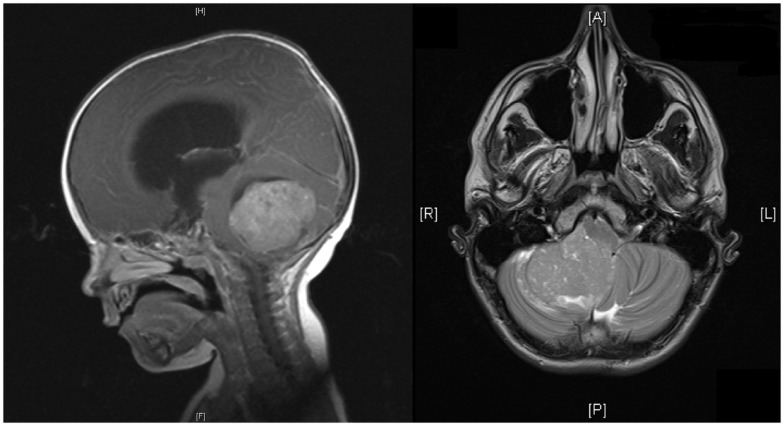
Head MRI showing a cerebellar medulloblastoma in sagittal and horizontal views: Sagittal view shows a midline posterior fossa medulloblastoma with intermediate signal intensity. There is an obstruction to the flow of CSF, marked hydrocephalus, and edema. Horizontal view shows a homogenous enhancing medulloblastoma arising from the right cerebellar hemisphere with displacement of the vermis.
Image: “Pediatric medulloblastoma – update on molecular classification driving targeted therapies” by DeSouza RM, Jones BR, Lowis SP, Kurian KM. License: CC BY 3.0
Cerebrospinal fluidCerebrospinal FluidA watery fluid that is continuously produced in the choroid plexus and circulates around the surface of the brain; spinal cord; and in the cerebral ventricles.Ventricular System: Anatomy analysis
Cerebrospinal fluidCerebrospinal FluidA watery fluid that is continuously produced in the choroid plexus and circulates around the surface of the brain; spinal cord; and in the cerebral ventricles.Ventricular System: Anatomy analysis is important in the evaluation for metastasisMetastasisThe transfer of a neoplasm from one organ or part of the body to another remote from the primary site.Grading, Staging, and Metastasissince approximately ⅓ of medulloblastomas metastasize through the CSF. These medulloblastomas show:
↑ Risk of relapseRelapseRelapsing Fever and poorer prognosisPrognosisA prediction of the probable outcome of a disease based on a individual’s condition and the usual course of the disease as seen in similar situations.Non-Hodgkin Lymphomas
Note: Most patientsPatientsIndividuals participating in the health care system for the purpose of receiving therapeutic, diagnostic, or preventive procedures.Clinician–Patient Relationship present with ↑ ICPICPNormal intracranial pressure (ICP) is defined as < 15 mm Hg, whereas pathologically increased ICP is any pressure ≥ 20 mm Hg. Increased ICP may result from several etiologies, including trauma, intracranial hemorrhage, mass lesions, cerebral edema, increased CSF production, and decreased CSF absorption.Increased Intracranial Pressure (ICP) and/or hydrocephalusHydrocephalusExcessive accumulation of cerebrospinal fluid within the cranium which may be associated with dilation of cerebral ventricles, intracranial.Subarachnoid Hemorrhage, requiring lumbar punctureLumbar PunctureFebrile Infant to be delayed until after surgical resection to prevent brainBrainThe part of central nervous system that is contained within the skull (cranium). Arising from the neural tube, the embryonic brain is comprised of three major parts including prosencephalon (the forebrain); mesencephalon (the midbrain); and rhombencephalon (the hindbrain). The developed brain consists of cerebrum; cerebellum; and other structures in the brain stem.Nervous System: Anatomy, Structure, and ClassificationherniationHerniationOmphalocele.
Histopathology
Diagnosis requires confirmation by histopathologic examination at the time of surgical resection.
Gross characteristics:
Well-circumscribed, gray, soft, friable tumors
Usually have associated necrosisNecrosisThe death of cells in an organ or tissue due to disease, injury or failure of the blood supply.Ischemic Cell Damage
Microscopic characteristics:
Highly cellular tumors with abundant dark staining
Often have abundant mitosisMitosisA type of cell nucleus division by means of which the two daughter nuclei normally receive identical complements of the number of chromosomes of the somatic cells of the species.Cell Cycle
Homer Wright rosettes found in up to 40% of patientsPatientsIndividuals participating in the health care system for the purpose of receiving therapeutic, diagnostic, or preventive procedures.Clinician–Patient Relationship.
Markers expressed in histopathological studies:
SynaptophysinSynaptophysinA marvel domain-containing protein found in the presynaptic vesicles of neurons and neuroendocrine cells. It is commonly used as an immunocytochemical marker for neuroendocrine differentiation.Gastrinoma
Neuron-specific enolaseEnolaseA hydro-lyase that catalyzes the dehydration of 2-phosphoglycerate to form phosphoenolpyruvate. Several different isoforms of this enzyme exist, each with its own tissue specificity.Glycolysis
Nestin (a marker of primitive neuroepithelial cells)
Nuclear β-catenin staining: present in Wnt pathway tumors
P53 mutations
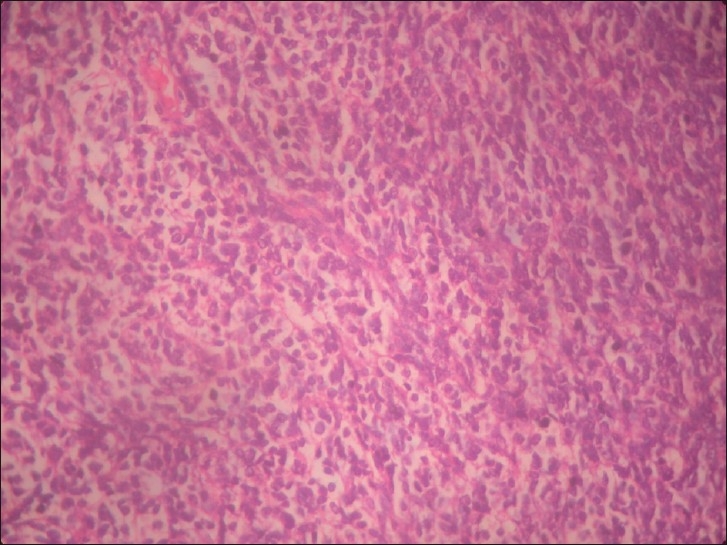
Microphotograph of medulloblastoma (H&E, ×20): The image shows a highly cellular tumor composed of rosettes of small round cells, with high nucleus-cytoplasm ratio.
Image: “Extra axial adult cerebellopontine angle medulloblastoma: An extremely rare site of tumor with metastasis” by Singh M, Cugati G, Symss NP, Pande A, Vasudevan MC, Ramamurthi R. License: CC BY 2.0
Management and Prognosis
Management
The standard of careStandard of careThe minimum acceptable patient care, based on statutes, court decisions, policies, or professional guidelines.Malpractice for management of medulloblastoma is combined modality therapy, including surgery, chemotherapyChemotherapyOsteosarcoma, and radiationRadiationEmission or propagation of acoustic waves (sound), electromagnetic energy waves (such as light; radio waves; gamma rays; or x-rays), or a stream of subatomic particles (such as electrons; neutrons; protons; or alpha particles).Osteosarcoma.
Alleviate ↑ ICPICPNormal intracranial pressure (ICP) is defined as < 15 mm Hg, whereas pathologically increased ICP is any pressure ≥ 20 mm Hg. Increased ICP may result from several etiologies, including trauma, intracranial hemorrhage, mass lesions, cerebral edema, increased CSF production, and decreased CSF absorption.Increased Intracranial Pressure (ICP)
Craniospinal radiationRadiationEmission or propagation of acoustic waves (sound), electromagnetic energy waves (such as light; radio waves; gamma rays; or x-rays), or a stream of subatomic particles (such as electrons; neutrons; protons; or alpha particles).Osteosarcoma therapy (RT):
Higher doses are associated with better tumorTumorInflammation control.
Craniospinal RT in children can cause:
Significant neurocognitive impairment
Hearing lossHearing lossHearing loss, also known as hearing impairment, is any degree of impairment in the ability to apprehend sound as determined by audiometry to be below normal hearing thresholds. Clinical presentation may occur at birth or as a gradual loss of hearing with age, including a short-term or sudden loss at any point. Hearing Loss
Growth abnormalities
HypothyroidismHypothyroidismHypothyroidism is a condition characterized by a deficiency of thyroid hormones. Iodine deficiency is the most common cause worldwide, but Hashimoto’s disease (autoimmune thyroiditis) is the leading cause in non-iodine-deficient regions. Hypothyroidism
Adrenal insufficiencyAdrenal InsufficiencyConditions in which the production of adrenal corticosteroids falls below the requirement of the body. Adrenal insufficiency can be caused by defects in the adrenal glands, the pituitary gland, or the hypothalamus.Adrenal Insufficiency and Addison Disease
HypogonadismHypogonadismHypogonadism is a condition characterized by reduced or no sex hormone production by the testes or ovaries. Hypogonadism can result from primary (hypergonadotropic) or secondary (hypogonadotropic) failure. Symptoms include infertility, increased risk of osteoporosis, erectile dysfunction, decreased libido, and regression (or absence) of secondary sexual characteristics.Hypogonadism and infertilityInfertilityInfertility is the inability to conceive in the context of regular intercourse. The most common causes of infertility in women are related to ovulatory dysfunction or tubal obstruction, whereas, in men, abnormal sperm is a common cause. Infertility
RT may be avoided or given in lower doses in infants and very young children.
For high-risk disease (metastatic, unresectable, or recurrent disease):
Optimal therapy is unknown.
PatientsPatientsIndividuals participating in the health care system for the purpose of receiving therapeutic, diagnostic, or preventive procedures.Clinician–Patient Relationship should be included in a clinical study if possible.
PrognosisPrognosisA prediction of the probable outcome of a disease based on a individual’s condition and the usual course of the disease as seen in similar situations.Non-Hodgkin Lymphomas
Survival in children:
With modern combined modality therapy, approximately 75% of children who are diagnosed with medulloblastoma survive into adulthood.
5-year survival:
Children diagnosed before age 3: 40%–50%
If the disease is disseminated at the time of diagnosis: 15%–30%
Survival in adults:
5-year survival rate: 65%
10-year survival rate: 52%
Wnt tumors are associated with an excellent prognosisPrognosisA prediction of the probable outcome of a disease based on a individual’s condition and the usual course of the disease as seen in similar situations.Non-Hodgkin Lymphomas.
Factors associated with poor prognosisPrognosisA prediction of the probable outcome of a disease based on a individual’s condition and the usual course of the disease as seen in similar situations.Non-Hodgkin Lymphomas:
In 80% of survivors, there are treatment-related morbidities, especially those caused by the craniospinal radiationRadiationEmission or propagation of acoustic waves (sound), electromagnetic energy waves (such as light; radio waves; gamma rays; or x-rays), or a stream of subatomic particles (such as electrons; neutrons; protons; or alpha particles).Osteosarcoma therapy listed above.
Differential Diagnosis
The following posterior fossa tumors are differential diagnoses for medulloblastoma:
Pilocytic astrocytomaPilocytic AstrocytomaAstrocytoma: 2nd most common brainBrainThe part of central nervous system that is contained within the skull (cranium). Arising from the neural tube, the embryonic brain is comprised of three major parts including prosencephalon (the forebrain); mesencephalon (the midbrain); and rhombencephalon (the hindbrain). The developed brain consists of cerebrum; cerebellum; and other structures in the brain stem.Nervous System: Anatomy, Structure, and ClassificationtumorTumorInflammation in children. Magnetic resonance imaging can help distinguish this tumorTumorInflammation from others. For example, pilocytic astrocytomas are usually solitary cysticCysticFibrocystic Change lesions with a mural noduleNoduleChalazion, or they have central necrosisNecrosisThe death of cells in an organ or tissue due to disease, injury or failure of the blood supply.Ischemic Cell Damage with a thick rim of enhancing tissue. In contrast, medulloblastomas tend to have multiple smaller cystsCystsAny fluid-filled closed cavity or sac that is lined by an epithelium. Cysts can be of normal, abnormal, non-neoplastic, or neoplastic tissues.Fibrocystic Change, if cystsCystsAny fluid-filled closed cavity or sac that is lined by an epithelium. Cysts can be of normal, abnormal, non-neoplastic, or neoplastic tissues.Fibrocystic Change are present at all.
EpendymomaEpendymomaEpendymomas are glial cell tumors arising from CSF-producing ependymal cells lining the ventricular system. Ependymomas most commonly occur within the posterior fossa in contact with the 4th ventricle, or within the intramedullary spinal cord. Ependymoma: a tumorTumorInflammation that arises from the ependymal lining of the ventricular systemVentricular SystemThe ventricular system is an extension of the subarachnoid space into the brain consisting of a series of interconnecting spaces and channels. Four chambers are filled with cerebrospinal fluid (CSF): the paired lateral ventricles, the unpaired 3rd ventricle, and the unpaired 4th ventricle. Ventricular System: Anatomy, typically at the base of the 4th ventricle. Tumors are usually intracranial in children and in the spinal cordSpinal cordThe spinal cord is the major conduction pathway connecting the brain to the body; it is part of the CNS. In cross section, the spinal cord is divided into an H-shaped area of gray matter (consisting of synapsing neuronal cell bodies) and a surrounding area of white matter (consisting of ascending and descending tracts of myelinated axons). Spinal Cord: Anatomy in adults. Ependymomas more commonly extend through the foramen of MagendieForamen of MagendieVentricular System: Anatomy (inferiorly) or Luschka (laterally), while this type of extensionExtensionExamination of the Upper Limbs is rare in medulloblastoma. Magnetic resonance imaging is the standard imaging technique, but histologic confirmation is required for diagnosis. Treatment involves surgical resection and adjuvantAdjuvantSubstances that augment, stimulate, activate, potentiate, or modulate the immune response at either the cellular or humoral level. The classical agents (freund’s adjuvant, bcg, corynebacterium parvum, et al.) contain bacterial antigens. Some are endogenous (e.g., histamine, interferon, transfer factor, tuftsin, interleukin-1). Their mode of action is either non-specific, resulting in increased immune responsiveness to a wide variety of antigens, or antigen-specific, i.e., affecting a restricted type of immune response to a narrow group of antigens. The therapeutic efficacy of many biological response modifiers is related to their antigen-specific immunoadjuvanticity.VaccinationradiationRadiationEmission or propagation of acoustic waves (sound), electromagnetic energy waves (such as light; radio waves; gamma rays; or x-rays), or a stream of subatomic particles (such as electrons; neutrons; protons; or alpha particles).Osteosarcoma and/or chemotherapyChemotherapyOsteosarcoma (based on age).
Atypical teratoid/rhabdoid tumors(AT/RT): a rare, aggressive primary CNS tumorTumorInflammation arising from embryonal cells that can appear similar to a medulloblastoma on MRI. Atypical teratoid/rhabdoid tumors can form anywhere in the CNS but typically develop in the cerebellumCerebellumThe cerebellum, Latin for “little brain,” is located in the posterior cranial fossa, dorsal to the pons and midbrain, and its principal role is in the coordination of movements. The cerebellum consists of 3 lobes on either side of its 2 hemispheres and is connected in the middle by the vermis. Cerebellum: Anatomy, most commonly in children < 3 years old. These tumors contain neuroepithelial, rhabdoid, epithelial, and mesenchymal cells. Compared with medulloblastomas, AT/RTs are more likely to involve the lateral hemispheres or cerebellopontine angleCerebellopontine angleJunction between the cerebellum and the pons.Acoustic Neuroma and contain hemorrhage within the tumorTumorInflammation.
References
AlRayahi, J., Alwalid, O., Mubarak, W., Maaz, A. U. R., & Mifsud, W. (2023). Pediatric brain tumors in the molecular era: Updates for the radiologist. Seminars in Roentgenology, 58(1), 47–66. https://doi.org/10.1053/j.ro.2022.09.004
Pfister, S, et al. (2009). Outcome prediction in pediatric medulloblastoma based on DNA copy-number aberrations of chromosomes 6q and 17q and the MYC and MYCN loci. J Clin Oncol. 27(10), 1627‒1636. https://pubmed.ncbi.nlm.nih.gov/19255330/
Create your free account or log in to continue reading!