Overview
Definition
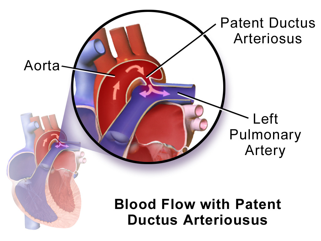
The ductus arteriosus (DA) is a fetal blood vessel connecting the left pulmonary artery to the aorta; bypassing the pulmonary circulation. Failure of the vessel to close and involute within 72 hours of birth results in a condition called patent ductus arteriosus (PDA).
Epidemiology
- Population statistics:
- 5%–10% of all congenital heart defects (CHDs)
- Incidence: 2 in 10,000 live term births
- M:F ratio is 1:2
- Risk factors:
- Prematurity
- Hypoxia:
- History of perinatal asphyxia
- Birth at high altitude
- Maternal factors:
- Rubella infection
- Phenytoin use
- Genetic predisposition:
- Positive family history of PDA
- DiGeorge syndrome
- Cri-du-chat syndrome
- Noonan syndrome
- CHARGE syndrome
The DA:
The DA is a fetal blood vessel connecting the aorta (usually at the arch) to the left pulmonary artery. When this vessel persists after birth, it is termed PDA.
Related videos
Embryology and Pathophysiology
Embryology
- Fetal circulation:
- In utero, lungs are fluid filled and their vasculature is vasoconstricted (oxygen comes from maternal circulation in the placenta).
- DA shunts blood from the right (R) to left (L) in the heart, bypassing high-pressure pulmonary circulation.
- After birth, DA closure is triggered by:
- Increased systemic oxygen (O2) concentration → decreased pulmonary blood pressure
- Reduced prostaglandin E2 (PGE2) levels due to separation from placenta
- Closure starts at the pulmonary end and finishes at the aortic end:
- Functional closure by 15 hours of life
- Anatomical closure by 3rd week of life
- Histological changes turn ductus into ligamentum arteriosum.
- Failure or disruption of transition from fetal to extrauterine life results in the persistence of PDA.
Pathophysiology
- In PDA, the shunt reverses to L-R as pulmonary vascular resistance falls.
- Increased end-diastolic volume in left atrium and left ventricle → volume overload → dilation → heart failure
- Increased pressure in pulmonary artery → pulmonary hypertension → respiratory failure
- Eisenmenger’s syndrome:
- Pulmonary hypertension → right ventricle hypertrophy→ increasing right-sided pressure
- Shunt reverses to R-L in the prenatal period.
Clinical Presentation
The severity of symptoms depends primarily on the degree of L-R shunt, which is dictated by size and length of the defect.
Degree of shunt | Clinical presentation | Physical examination |
---|---|---|
Small shunt |
| Murmur:
|
Moderate shunt |
|
|
Large shunt | Infant:
|
|
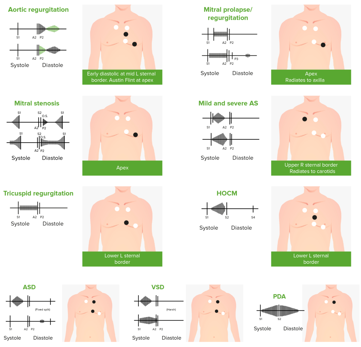
Phonocardiograms of abnormal heart sounds caused by the following cardiac defects:
aortic regurgitation, mitral valve prolapse, mitral stenosis (MS), aortic stenosis (AS), tricuspid regurgitation, hypertrophic obstructive cardiomyopathy (HOCM), atrial septal defect (ASD), ventricular septal defect (VSD), and patent ductus arteriosus (PDA)
Diagnosis
- Clinical findings are confirmed by echocardiogram.
- Echocardiography:
- Evaluates:
- Presence of defect
- Hemodynamics of left atrium and ventricle
- Doppler → degree of shunt and pulmonary arterial pressure
- All preterm infants should be screened.
- Evaluates:
- Chest X-ray:
- Normal in small shunts
- Large shunts may show:
- Increased pulmonary vascularization
- Cardiomegaly
- Electrocardiogram (ECG):
- Biventricular or right ventricular hypertrophy in advanced cases
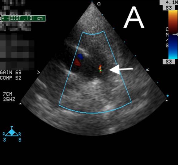
Echocardiogram showing PDA:
An echocardiogram depicting PDA with a small shunt (white arrow) found incidentally in a patient with Kawasaki disease
Management, Complications, and Prognosis
Management
- Observation indicated in smaller defects with no physiological changes:
- Requires close follow-up
- Closure indicated in:
- Moderate and large PDA
- Small PDA with audible murmur
- Presence of pulmonary hypertension
- Heart failure
- Respiratory compromise
- History of infective endocarditis
- Medical therapy (pharmacological closure):
- Decrease prostaglandin synthesis:
- Indomethacin
- Ibuprofen
- Follow-up after 48 hours with echocardiography to ensure closure
- Contraindicated in:
- Cardiac defects where DA required to maintain perfusion for systemic circulation
- Active bleeding
- Suspicious or diagnosed necrotizing enterocolitis
- Impaired renal function
- Decrease prostaglandin synthesis:
- Surgical therapy:
- Indicated when medical therapy fails and/or infant > 6 kg
- Includes percutaneous closure or surgical ligation
Complications
- Pulmonary hypertension
- Bronchopulmonary dysplasia
- Congestive heart failure
- Infective endocarditis
- Post-surgery → paralysis of recurrent laryngeal nerve and stridor
Prognosis
- Spontaneous closure in up to 75% of infants if < 3 months of age
- Isolated PDA has an excellent prognosis (especially small defects).
Differential Diagnosis
Continuous murmur of PDA should be differentiated from:
- Venous hum: benign, common murmur secondary to forces hitting venous walls; heard in children. A continuous murmur best heard on the right side and changes with position (unlike PDA). An echocardiogram is done to confirm no pathology.
- Coronary artery fistula: congenital vascular abnormality whereby the coronary artery drains directly into chambers of heart without a capillary bed. Presentation includes a continuous murmur in the lower pericardium. An echocardiogram with Doppler will show direct drainage into the chambers.
- Aortic regurgitation: a defect of the aortic valve wherein the leaflets do not oppose perfectly, allowing blood to backflow during diastole. Patients may present with a continuous murmur and bounding pulses. Aortic regurgitation is usually seen in older patients. An echocardiogram can diagnose and quantify the severity of valvular defect.
References
- Thomas D. and Anne K. (2020). Clinical manifestations and diagnosis of patent ductus arteriosus in term infants, children, and adults. UpToDate. Retrieved January 09, 2021, from https://www.uptodate.com/contents/clinical-manifestations-and-diagnosis-of-patent-ductus-arteriosus-in-term-infants-children-and-adults
- Bernstein, D. (2020). General principles of treatment of congenital heart disease. In R. M. Kliegman MD, J. W. St Geme MD, N. J. Blum MD, Shah, Samir S., MD, MSCE, Tasker, Robert C., MBBS, MD & Wilson, Karen M., MD, MPH (Eds.), Nelson textbook of pediatrics (pp. 242–2436.e1) doi:http://dx.doi.org/10.1016/B978-0-323-52950-1.00461-2. Retrieved January 31, 2021, from https://www.clinicalkey.es/#!/content/3-s2.0-B9780323529501004612
- Benitz, W. E., & Bhombal, S. (2020). Patent ductus arteriosus. In Martin, Richard J., MBBS, FRACP, Fanaroff, Avroy A., MD, FRCPE, FRCPCH & Walsh, Michele C., MD, MSE (Eds.), Fanaroff and martin’s neonatal-perinatal medicine (pp. 1334–1341) doi:http://dx.doi.org/10.1016/B978-0-323-56711-4.00074-2. Retrieved January 31, 2021, from https://www.clinicalkey.es/#!/content/3-s2.0-B9780323567114000742
- Gillam-Krakauer, M., & Reese, J. (2018). Diagnosis and Management of Patent Ductus Arteriosus. NeoReviews, 19(7), e394–e402. DOI:10.1542/neo.19-7-e394. Retrieved January 31, 2021, from https://pubmed.ncbi.nlm.nih.gov/30505242/