Donovanosis (also known as granuloma inguinale) is an STD caused by Klebsiella granulomatis and is mainly seen in tropical regions. The condition is characterized by chronic, progressive, ulcerating disease mostly affecting the genital region. The patient presents with painless nodular lesions that ulcerate, commonly with a "beefy-red" base. There is no associated inguinal lymphadenopathy. Diagnosis is via history, clinical findings, and tissue smear or biopsy showing Donovan bodies, which are intracellular inclusion bodies inside macrophages. Treatment is with a prolonged course of antibiotics until lesions are healed, with monitoring for recurrence.
DonovanosisDonovanosisDonovanosis (also known as granuloma inguinale) is an STD caused by Klebsiella granulomatis and is mainly seen in tropical regions. The condition is characterized by chronic, progressive, ulcerating disease mostly affecting the genital region. Donovanosis, or granuloma inguinaleGranuloma inguinaleDonovanosis (also known as granuloma inguinale) is an STD caused by Klebsiella granulomatis and is mainly seen in tropical regions. The condition is characterized by chronic, progressive, ulcerating disease mostly affecting the genital region.Donovanosis, is an STD characterized by chronic progressive ulcers affecting the genital region.
Epidemiology[1,2,5,6]
Rare in the United States
Generally, incidenceIncidenceThe number of new cases of a given disease during a given period in a specified population. It also is used for the rate at which new events occur in a defined population. It is differentiated from prevalence, which refers to all cases in the population at a given time.Measures of Disease Frequency is declining worldwide, partly due to the focus on HIVHIVAnti-HIV Drugs prevention and the role genital ulcers play in the disease’s transmission.
Most occurrences are in the following areas:
Papua New Guinea
Southern Africa
French Guiana
India
Caribbean and Brazil
Aboriginal communities in Australia
Etiology[1,2,5,7]
Caused by KlebsiellaKlebsiellaKlebsiella are encapsulated gram-negative, lactose-fermenting bacilli. They form pink colonies on MacConkey agar due to lactose fermentation. The main virulence factor is a polysaccharide capsule. Klebsiella pneumoniae is the most important pathogenic species.Klebsiella granulomatis (formerly Calymmatobacterium granulomatis):
Minimal trauma leads to ulcerative formation without lymphadenopathyLymphadenopathyLymphadenopathy is lymph node enlargement (> 1 cm) and is benign and self-limited in most patients. Etiologies include malignancy, infection, and autoimmune disorders, as well as iatrogenic causes such as the use of certain medications. Generalized lymphadenopathy often indicates underlying systemic disease. Lymphadenopathy.
Eventually, the lesion progressively grows outward, with borders developing a “snake-like” appearance.
IncubationIncubationThe amount time between exposure to an infectious agent and becoming symptomatic.Rabies Virus period: average of 50 days (and up to a year) postinoculation [4]
Transmission:
Generally sexually transmitted[3‒5]
Rare cases of non-sexual contact have been reported.[2]
Most common in women: labia minoraLabia minoraVagina, Vulva, and Pelvic Floor: Anatomy, cervixCervixThe uterus, cervix, and fallopian tubes are part of the internal female reproductive system. The most inferior portion of the uterus is the cervix, which connects the uterine cavity to the vagina. Externally, the cervix is lined by stratified squamous cells; however, the cervical canal is lined by columnar epithelium.Uterus, Cervix, and Fallopian Tubes: Anatomy, and upper genital tract
6% of patientsPatientsIndividuals participating in the health care system for the purpose of receiving therapeutic, diagnostic, or preventive procedures.Clinician–Patient Relationship may have extragenital lesions (lipsLipsThe lips are the soft and movable most external parts of the oral cavity. The blood supply of the lips originates from the external carotid artery, and the innervation is through cranial nerves.Lips and Tongue: Anatomy, palatePalateThe palate is the structure that forms the roof of the mouth and floor of the nasal cavity. This structure is divided into soft and hard palates. Palate: Anatomy, pharynxPharynxThe pharynx is a component of the digestive system that lies posterior to the nasal cavity, oral cavity, and larynx. The pharynx can be divided into the oropharynx, nasopharynx, and laryngopharynx. Pharyngeal muscles play an integral role in vital processes such as breathing, swallowing, and speaking. Pharynx: Anatomy, larynxLarynxThe larynx, also commonly called the voice box, is a cylindrical space located in the neck at the level of the C3-C6 vertebrae. The major structures forming the framework of the larynx are the thyroid cartilage, cricoid cartilage, and epiglottis. The larynx serves to produce sound (phonation), conducts air to the trachea, and prevents large molecules from reaching the lungs.Larynx: Anatomy).
No regional lymphadenopathyLymphadenopathyLymphadenopathy is lymph node enlargement (> 1 cm) and is benign and self-limited in most patients. Etiologies include malignancy, infection, and autoimmune disorders, as well as iatrogenic causes such as the use of certain medications. Generalized lymphadenopathy often indicates underlying systemic disease. Lymphadenopathy[5,6]
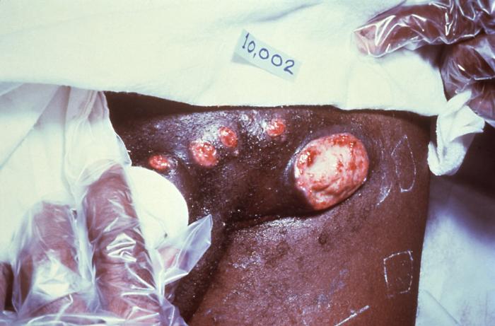
Ulcerative lesions of Donovanosis: a patient’s left inguinal region, revealing the presence of a number of ulcerative erythematous lesions, which were determined to be due to a case of donovanosis
Image: “5365” by CDC/Dr. Tabua, Chief Medical Officer from Port Moresby, Papua, New Guinea. License: Public Domain
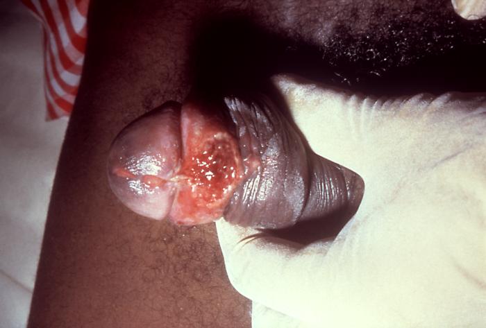
An ulcerogranulomatous lesion due to donovanosis: Note the beefy-red base at the frenulum of the penis.
Image: “5363” by CDC/Dr. Tabua, Chief Medical Officer from Port Moresby, Papua, New Guinea. License: Public Domain
Complications[3–6]
Healing of donovanosisDonovanosisDonovanosis (also known as granuloma inguinale) is an STD caused by Klebsiella granulomatis and is mainly seen in tropical regions. The condition is characterized by chronic, progressive, ulcerating disease mostly affecting the genital region. Donovanosis ulcers can leave scars or hypertrophic lesions.
Neoplastic change: Squamous cell carcinomaSquamous cell carcinomaCutaneous squamous cell carcinoma (cSCC) is caused by malignant proliferation of atypical keratinocytes. This condition is the 2nd most common skin malignancy and usually affects sun-exposed areas of fair-skinned patients. The cancer presents as a firm, erythematous, keratotic plaque or papule. Squamous Cell Carcinoma (SCC) may occur in long-standing untreated lesions.
PolyarthritisPolyarthritisRheumatoid Arthritis or osteomyelitisOsteomyelitisOsteomyelitis is an infection of the bone that results from the spread of microorganisms from the blood (hematogenous), nearby infected tissue, or open wounds (non-hematogenous). Infections are most commonly caused by Staphylococcus aureus.Osteomyelitis
The following information is based on US, UK, and European guidelines. Please see your local guidelines for additional guidance.
Diagnosis
Causative agent is difficult to culture.[4,5]
PCRPCRPolymerase chain reaction (PCR) is a technique that amplifies DNA fragments exponentially for analysis. The process is highly specific, allowing for the targeting of specific genomic sequences, even with minuscule sample amounts. The PCR cycles multiple times through 3 phases: denaturation of the template DNA, annealing of a specific primer to the individual DNA strands, and synthesis/elongation of new DNA molecules.Polymerase Chain Reaction (PCR) is used only in researchResearchCritical and exhaustive investigation or experimentation, having for its aim the discovery of new facts and their correct interpretation, the revision of accepted conclusions, theories, or laws in the light of newly discovered facts, or the practical application of such new or revised conclusions, theories, or laws.Conflict of Interest laboratories and specialised/reference laboratories.[5,6]
SerologySerologyThe study of serum, especially of antigen-antibody reactions in vitro.Yellow Fever Virus is not reliable for diagnosis.[4]
High index of suspicion needed, especially in endemic areas
Diagnosis is made by a microscopic examination of a tissue smear (from the ulcer), tissue crush preparation, or biopsyBiopsyRemoval and pathologic examination of specimens from the living body.Ewing Sarcoma:[4–6]
Giemsa or Wright stain is preferred.
Donovan bodiesDonovan BodiesDonovanosis: bipolar-staining cytoplasmic inclusion bodies within macrophagesMacrophagesThe relatively long-lived phagocytic cell of mammalian tissues that are derived from blood monocytes. Main types are peritoneal macrophages; alveolar macrophages; histiocytes; kupffer cells of the liver; and osteoclasts. They may further differentiate within chronic inflammatory lesions to epithelioid cells or may fuse to form foreign body giant cells or langhans giant cells.Innate Immunity: Phagocytes and Antigen Presentation, which may have a safety-pin appearance
Other histologic changes:
InflammationInflammationInflammation is a complex set of responses to infection and injury involving leukocytes as the principal cellular mediators in the body’s defense against pathogenic organisms. Inflammation is also seen as a response to tissue injury in the process of wound healing. The 5 cardinal signs of inflammation are pain, heat, redness, swelling, and loss of function. Inflammation with plasmaPlasmaThe residual portion of blood that is left after removal of blood cells by centrifugation without prior blood coagulation.Transfusion Products cell and neutrophil infiltration
UlcerationUlcerationCorneal Abrasions, Erosion, and Ulcers, microabscesses, and elongationElongationPolymerase Chain Reaction (PCR) of rete ridgesRete RidgesLentigo Maligna in the epitheliumEpitheliumThe epithelium is a complex of specialized cellular organizations arranged into sheets and lining cavities and covering the surfaces of the body. The cells exhibit polarity, having an apical and a basal pole. Structures important for the epithelial integrity and function involve the basement membrane, the semipermeable sheet on which the cells rest, and interdigitations, as well as cellular junctions. Surface Epithelium: Histology
Samples should also be cultured for Haemophilus ducreyiHaemophilus ducreyiA species of Haemophilus that appears to be the pathogen or causative agent of the sexually transmitted disease, chancroid.Haemophilus → exclude chancroidChancroidChancroid is a highly transmissible STD caused by Haemophilus ducreyi. The disease presents with painful ulcer(s) on the genital tract (termed chancroid or “soft chancre”). Up to 50% of patients will develop painful inguinal lymphadenopathy. Chancroid[5]
HIVHIVAnti-HIV Drugs testing is also recommended for all patientsPatientsIndividuals participating in the health care system for the purpose of receiving therapeutic, diagnostic, or preventive procedures.Clinician–Patient Relationship with a diagnosis of donovanosisDonovanosisDonovanosis (also known as granuloma inguinale) is an STD caused by Klebsiella granulomatis and is mainly seen in tropical regions. The condition is characterized by chronic, progressive, ulcerating disease mostly affecting the genital region. Donovanosis.[3,4]
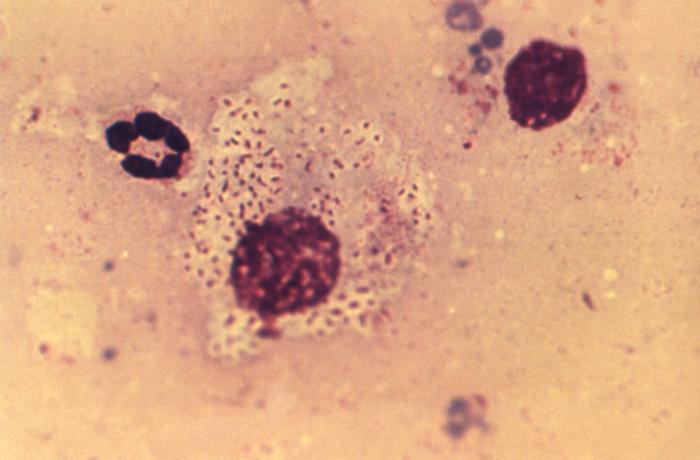
Tissue sample from an ulcer: a white blood cell (WBC) that contained the pathognomonic finding of Donovan bodies, which were encapsulated, Gram-negative rods representing the responsible bacterium Klebsiella granulomatis
Management involves a prolonged course of antibiotics until re-epithelization of the ulcer has occurred (≥ 3 weeks or until the ulcer is completely healed).[3–5]
1st-line: azithromycinAzithromycinA semi-synthetic macrolide antibiotic structurally related to erythromycin. It has been used in the treatment of Mycobacterium avium intracellulare infections, toxoplasmosis, and cryptosporidiosis.Macrolides and Ketolides 1 g orally once a week or 500 mg orally once a day[3–6]
ErythromycinErythromycinA bacteriostatic antibiotic macrolide produced by streptomyces erythreus. Erythromycin a is considered its major active component. In sensitive organisms, it inhibits protein synthesis by binding to 50s ribosomal subunits. This binding process inhibits peptidyl transferase activity and interferes with translocation of amino acids during translation and assembly of proteins.Macrolides and Ketolides (if patient is pregnant) 500 mg orally 4 times daily[3–6]
TrimethoprimTrimethoprimThe sulfonamides are a class of antimicrobial drugs inhibiting folic acid synthesize in pathogens. The prototypical drug in the class is sulfamethoxazole. Although not technically sulfonamides, trimethoprim, dapsone, and pyrimethamine are also important antimicrobial agents inhibiting folic acid synthesis. The agents are often combined with sulfonamides, resulting in a synergistic effect. Sulfonamides and Trimethoprim–sulfamethoxazoleSulfamethoxazoleA bacteriostatic antibacterial agent that interferes with folic acid synthesis in susceptible bacteria. Its broad spectrum of activity has been limited by the development of resistance.Sulfonamides and Trimethoprim 1 double-strength tablet (160 mg/800 mg) orally twice daily[3–6]
Consider adding gentamicinGentamicinAminoglycosides if the ulcer is not beginning to heal by day 7.[4,5]
PregnancyPregnancyThe status during which female mammals carry their developing young (embryos or fetuses) in utero before birth, beginning from fertilization to birth.Pregnancy: Diagnosis, Physiology, and Care: Macrolide options (erythromycinErythromycinA bacteriostatic antibiotic macrolide produced by streptomyces erythreus. Erythromycin a is considered its major active component. In sensitive organisms, it inhibits protein synthesis by binding to 50s ribosomal subunits. This binding process inhibits peptidyl transferase activity and interferes with translocation of amino acids during translation and assembly of proteins.Macrolides and Ketolides or azithromycinAzithromycinA semi-synthetic macrolide antibiotic structurally related to erythromycin. It has been used in the treatment of Mycobacterium avium intracellulare infections, toxoplasmosis, and cryptosporidiosis.Macrolides and Ketolides) are preferred.[3,5]
Follow up with patientsPatientsIndividuals participating in the health care system for the purpose of receiving therapeutic, diagnostic, or preventive procedures.Clinician–Patient Relationship until lesions completely heal. There is a high chance of recurrence at 6–18 months, which requires reinitiating antibiotics.[4,5]
Additional considerations[3,5]
Surgical management is reserved for extensive tissue damage.
Patient education on safe sexSexThe totality of characteristics of reproductive structure, functions, phenotype, and genotype, differentiating the male from the female organism.Gender Dysphoria is important to reduce incidenceIncidenceThe number of new cases of a given disease during a given period in a specified population. It also is used for the rate at which new events occur in a defined population. It is differentiated from prevalence, which refers to all cases in the population at a given time.Measures of Disease Frequency.
PatientsPatientsIndividuals participating in the health care system for the purpose of receiving therapeutic, diagnostic, or preventive procedures.Clinician–Patient Relationship should be screened for other STDs, including:
Hepatitis BHepatitis BHepatitis B virus (HBV) is a partially double-stranded DNA virus, which belongs to the Orthohepadnavirus genus and the Hepadnaviridae family. Most individuals with acute HBV infection are asymptomatic or have mild, self-limiting symptoms. Chronic infection can be asymptomatic or create hepatic inflammation, leading to liver cirrhosis and hepatocellular carcinoma (HCC). Hepatitis B Virus
GonorrheaGonorrheaGonorrhea is a sexually transmitted infection (STI) caused by the gram-negative bacteria Neisseria gonorrhoeae (N. gonorrhoeae). Gonorrhea may be asymptomatic but commonly manifests as cervicitis or urethritis with less common presentations such as proctitis, conjunctivitis, or pharyngitis. Gonorrhea
ChlamydiaChlamydiaChlamydiae are obligate intracellular gram-negative bacteria. They lack a peptidoglycan layer and are best visualized using Giemsa stain. The family of Chlamydiaceae comprises 3 pathogens that can infect humans: Chlamydia trachomatis, Chlamydia psittaci, and Chlamydia pneumoniae.Chlamydia
ChancroidChancroidChancroid is a highly transmissible STD caused by Haemophilus ducreyi. The disease presents with painful ulcer(s) on the genital tract (termed chancroid or “soft chancre”). Up to 50% of patients will develop painful inguinal lymphadenopathy. Chancroid
SyphilisSyphilisSyphilis is a bacterial infection caused by the spirochete Treponema pallidum pallidum (T. p. pallidum), which is usually spread through sexual contact. Syphilis has 4 clinical stages: primary, secondary, latent, and tertiary. Syphilis
Offer (age-appropriate) immunizations, if not already completed:[5]
Hepatitis BHepatitis BHepatitis B virus (HBV) is a partially double-stranded DNA virus, which belongs to the Orthohepadnavirus genus and the Hepadnaviridae family. Most individuals with acute HBV infection are asymptomatic or have mild, self-limiting symptoms. Chronic infection can be asymptomatic or create hepatic inflammation, leading to liver cirrhosis and hepatocellular carcinoma (HCC). Hepatitis B Virus
HPVHPVHuman papillomavirus (HPV) is a nonenveloped, circular, double-stranded DNA virus belonging to the Papillomaviridae family. Humans are the only reservoir, and transmission occurs through close skin-to-skin or sexual contact. Human papillomaviruses infect basal epithelial cells and can affect cell-regulatory proteins to result in cell proliferation. Papillomavirus (HPV)
UK: Examine all sexual partners for the past 6 months.[6]
For children born to affected mothers:[4,6]
Provide a 3-day course of azithromycinAzithromycinA semi-synthetic macrolide antibiotic structurally related to erythromycin. It has been used in the treatment of Mycobacterium avium intracellulare infections, toxoplasmosis, and cryptosporidiosis.Macrolides and Ketolides 20 mg/kg daily (European and UK guidelines)
Differential Diagnosis
ChancroidChancroidChancroid is a highly transmissible STD caused by Haemophilus ducreyi. The disease presents with painful ulcer(s) on the genital tract (termed chancroid or “soft chancre”). Up to 50% of patients will develop painful inguinal lymphadenopathy. Chancroid: an STD caused by Haemophilus ducreyiHaemophilus ducreyiA species of Haemophilus that appears to be the pathogen or causative agent of the sexually transmitted disease, chancroid.Haemophilus. The disease presents with painful ulcer(s) on the genital tract (chancroidChancroidChancroid is a highly transmissible STD caused by Haemophilus ducreyi. The disease presents with painful ulcer(s) on the genital tract (termed chancroid or “soft chancre”). Up to 50% of patients will develop painful inguinal lymphadenopathy. Chancroid or “soft chancreSoft chancreChancroid is a highly transmissible STD caused by Haemophilus ducreyi. The disease presents with painful ulcer(s) on the genital tract (termed chancroid or “soft chancre”). Up to 50% of patients will develop painful inguinal lymphadenopathy.Chancroid”), which can be accompanied by painful inguinal lymphadenopathyInguinal LymphadenopathyLymphadenopathy. PatientsPatientsIndividuals participating in the health care system for the purpose of receiving therapeutic, diagnostic, or preventive procedures.Clinician–Patient Relationship may develop the complication of suppurative lymph nodesLymph NodesThey are oval or bean shaped bodies (1 – 30 mm in diameter) located along the lymphatic system.Lymphatic Drainage System: Anatomy. Diagnosis is clinical and with tests ruling out syphilisSyphilisSyphilis is a bacterial infection caused by the spirochete Treponema pallidum pallidum (T. p. pallidum), which is usually spread through sexual contact. Syphilis has 4 clinical stages: primary, secondary, latent, and tertiary. Syphilis and herpes. AzithromycinAzithromycinA semi-synthetic macrolide antibiotic structurally related to erythromycin. It has been used in the treatment of Mycobacterium avium intracellulare infections, toxoplasmosis, and cryptosporidiosis.Macrolides and Ketolides or ceftriaxoneCeftriaxoneA broad-spectrum cephalosporin antibiotic and cefotaxime derivative with a very long half-life and high penetrability to meninges, eyes and inner ears.Cephalosporins is the treatment of choice. PatientsPatientsIndividuals participating in the health care system for the purpose of receiving therapeutic, diagnostic, or preventive procedures.Clinician–Patient Relationship and their contacts must both be treated.
Herpes simplexHerpes SimplexA group of acute infections caused by herpes simplex virus type 1 or type 2 that is characterized by the development of one or more small fluid-filled vesicles with a raised erythematous base on the skin or mucous membrane. It occurs as a primary infection or recurs due to a reactivation of a latent infection.Congenital TORCH Infections: common STD caused by herpes simplexHerpes SimplexA group of acute infections caused by herpes simplex virus type 1 or type 2 that is characterized by the development of one or more small fluid-filled vesicles with a raised erythematous base on the skin or mucous membrane. It occurs as a primary infection or recurs due to a reactivation of a latent infection.Congenital TORCH InfectionsvirusVirusViruses are infectious, obligate intracellular parasites composed of a nucleic acid core surrounded by a protein capsid. Viruses can be either naked (non-enveloped) or enveloped. The classification of viruses is complex and based on many factors, including type and structure of the nucleoid and capsid, the presence of an envelope, the replication cycle, and the host range. Virologytype 1Type 1Spinal Muscular Atrophy or 2. Prodromal symptoms often precede clusters of painful, fluid-filled vesiclesVesiclesFemale Genitourinary Examination on an erythematous base. These vesiclesVesiclesFemale Genitourinary Examination eventually form ulcers that can coalesce. LymphadenopathyLymphadenopathyLymphadenopathy is lymph node enlargement (> 1 cm) and is benign and self-limited in most patients. Etiologies include malignancy, infection, and autoimmune disorders, as well as iatrogenic causes such as the use of certain medications. Generalized lymphadenopathy often indicates underlying systemic disease. Lymphadenopathy, dysuriaDysuriaPainful urination. It is often associated with infections of the lower urinary tract.Urinary Tract Infections (UTIs), and severe neuralgia can occur. The diagnosis is generally clinical but confirmed with PCRPCRPolymerase chain reaction (PCR) is a technique that amplifies DNA fragments exponentially for analysis. The process is highly specific, allowing for the targeting of specific genomic sequences, even with minuscule sample amounts. The PCR cycles multiple times through 3 phases: denaturation of the template DNA, annealing of a specific primer to the individual DNA strands, and synthesis/elongation of new DNA molecules.Polymerase Chain Reaction (PCR) and serologic testing. Management includes antiviralAntiviralAntivirals for Hepatitis B therapy.
SyphilisSyphilisSyphilis is a bacterial infection caused by the spirochete Treponema pallidum pallidum (T. p. pallidum), which is usually spread through sexual contact. Syphilis has 4 clinical stages: primary, secondary, latent, and tertiary. Syphilis: an STD caused by Treponema pallidumTreponema pallidumThe causative agent of venereal and non-venereal syphilis as well as yaws.Treponema. The disease has 4 clinical stages: Primary syphilisPrimary SyphilisSyphilis begins with a solitary, painless ulcer on the genitals (chancreChancreThe primary sore of syphilis, a painless indurated, eroded papule, occurring at the site of entry of the infection.Syphilis). Progression to secondary syphilisSecondary SyphilisSyphilis manifests as a generalized maculopapularMaculopapularDermatologic ExaminationrashRashRocky Mountain Spotted Fever, which includes the palms and soles. The development of tertiary syphilisTertiary SyphilisSyphilis can cause severe neurologic (neurosyphilisNeurosyphilisInfections of the central nervous system caused by treponema pallidum which present with a variety of clinical syndromes. The initial phase of infection usually causes a mild or asymptomatic meningeal reaction. The meningovascular form may present acutely as brain infarction. The infection may also remain subclinical for several years. Late syndromes include general paresis; tabes dorsalis; meningeal syphilis; syphilitic optic atrophy; and spinal syphilis. General paresis is characterized by progressive dementia; dysarthria; tremor; myoclonus; seizures; and argyll-robertson pupils.Syphilis), cardiovascular, and/or gummatous disease. The dormant period between secondary and tertiary syphilisTertiary SyphilisSyphilis is the latent stageLatent StageBenign Bone Tumors.
Lymphogranuloma venereumLymphogranuloma venereumSubacute inflammation of the inguinal lymph glands caused by certain immunotypes of Chlamydia trachomatis. It is a sexually transmitted disease in the U.S. But is more widespread in developing countries. It is distinguished from granuloma venereum, which is caused by calymmatobacterium granulomatis.Chlamydial Infections (LGVLGVSubacute inflammation of the inguinal lymph glands caused by certain immunotypes of Chlamydia trachomatis. It is a sexually transmitted disease in the U.S. But is more widespread in developing countries. It is distinguished from granuloma venereum, which is caused by calymmatobacterium granulomatis.Chlamydial Infections): an STD caused by 3 strains of Chlamydia trachomatisChlamydia trachomatisType species of Chlamydia causing a variety of ocular and urogenital diseases.Chlamydia. PatientsPatientsIndividuals participating in the health care system for the purpose of receiving therapeutic, diagnostic, or preventive procedures.Clinician–Patient Relationship may have a small, self-limited genital ulcer followed by painful inguinal and/or femoral lymphadenopathyLymphadenopathyLymphadenopathy is lymph node enlargement (> 1 cm) and is benign and self-limited in most patients. Etiologies include malignancy, infection, and autoimmune disorders, as well as iatrogenic causes such as the use of certain medications. Generalized lymphadenopathy often indicates underlying systemic disease. Lymphadenopathy. Diagnosis is clinical; although PCRPCRPolymerase chain reaction (PCR) is a technique that amplifies DNA fragments exponentially for analysis. The process is highly specific, allowing for the targeting of specific genomic sequences, even with minuscule sample amounts. The PCR cycles multiple times through 3 phases: denaturation of the template DNA, annealing of a specific primer to the individual DNA strands, and synthesis/elongation of new DNA molecules.Polymerase Chain Reaction (PCR) testing can help, availability is limited. Management involves tetracyclinesTetracyclinesTetracyclines are a class of broad-spectrum antibiotics indicated for a wide variety of bacterial infections. These medications bind the 30S ribosomal subunit to inhibit protein synthesis of bacteria. Tetracyclines cover gram-positive and gram-negative organisms, as well as atypical bacteria such as chlamydia, mycoplasma, spirochetes, and even protozoa. Tetracyclines or erythromycinErythromycinA bacteriostatic antibiotic macrolide produced by streptomyces erythreus. Erythromycin a is considered its major active component. In sensitive organisms, it inhibits protein synthesis by binding to 50s ribosomal subunits. This binding process inhibits peptidyl transferase activity and interferes with translocation of amino acids during translation and assembly of proteins.Macrolides and Ketolides.
Billing and Coding
Diagnosis Codes:
This code is used to diagnose DonovanosisDonovanosisDonovanosis (also known as granuloma inguinale) is an STD caused by Klebsiella granulomatis and is mainly seen in tropical regions. The condition is characterized by chronic, progressive, ulcerating disease mostly affecting the genital region. Donovanosis (granuloma inguinaleGranuloma inguinaleDonovanosis (also known as granuloma inguinale) is an STD caused by Klebsiella granulomatis and is mainly seen in tropical regions. The condition is characterized by chronic, progressive, ulcerating disease mostly affecting the genital region.Donovanosis), a rare, chronic bacterial infection characterized by painless, progressive ulcerative lesions on the genitals or perineumPerineumThe body region lying between the genital area and the anus on the surface of the trunk, and to the shallow compartment lying deep to this area that is inferior to the pelvic diaphragm. The surface area is between the vulva and the anus in the female, and between the scrotum and the anus in the male.Vagina, Vulva, and Pelvic Floor: Anatomy.
Coding System
Code
Description
ICD-10-CM
A58
Granuloma inguinaleGranuloma inguinaleDonovanosis (also known as granuloma inguinale) is an STD caused by Klebsiella granulomatis and is mainly seen in tropical regions. The condition is characterized by chronic, progressive, ulcerating disease mostly affecting the genital region.Donovanosis
SNOMED CT
54092008
Granuloma inguinaleGranuloma inguinaleDonovanosis (also known as granuloma inguinale) is an STD caused by Klebsiella granulomatis and is mainly seen in tropical regions. The condition is characterized by chronic, progressive, ulcerating disease mostly affecting the genital region.Donovanosis (disorder)
Evaluation & Workup:
The diagnosis is confirmed by a biopsyBiopsyRemoval and pathologic examination of specimens from the living body.Ewing Sarcoma of the lesion, where microscopic examination of a tissue smear reveals characteristic Donovan bodiesDonovan BodiesDonovanosis (bacteriaBacteriaBacteria are prokaryotic single-celled microorganisms that are metabolically active and divide by binary fission. Some of these organisms play a significant role in the pathogenesis of diseases. Bacteriology within macrophagesMacrophagesThe relatively long-lived phagocytic cell of mammalian tissues that are derived from blood monocytes. Main types are peritoneal macrophages; alveolar macrophages; histiocytes; kupffer cells of the liver; and osteoclasts. They may further differentiate within chronic inflammatory lesions to epithelioid cells or may fuse to form foreign body giant cells or langhans giant cells.Innate Immunity: Phagocytes and Antigen Presentation).
Coding System
Code
Description
CPT
11102
Tangential biopsyBiopsyRemoval and pathologic examination of specimens from the living body.Ewing Sarcoma of skinSkinThe skin, also referred to as the integumentary system, is the largest organ of the body. The skin is primarily composed of the epidermis (outer layer) and dermis (deep layer). The epidermis is primarily composed of keratinocytes that undergo rapid turnover, while the dermis contains dense layers of connective tissue.Skin: Structure and Functions; single lesion
Medications:
This code is for azithromycinAzithromycinA semi-synthetic macrolide antibiotic structurally related to erythromycin. It has been used in the treatment of Mycobacterium avium intracellulare infections, toxoplasmosis, and cryptosporidiosis.Macrolides and Ketolides, the preferred antibiotic for treating donovanosisDonovanosisDonovanosis (also known as granuloma inguinale) is an STD caused by Klebsiella granulomatis and is mainly seen in tropical regions. The condition is characterized by chronic, progressive, ulcerating disease mostly affecting the genital region. Donovanosis, typically given as a prolonged course until the lesions have completely healed.
Coding System
Code
Description
RxNorm
18631
AzithromycinAzithromycinA semi-synthetic macrolide antibiotic structurally related to erythromycin. It has been used in the treatment of Mycobacterium avium intracellulare infections, toxoplasmosis, and cryptosporidiosis.Macrolides and Ketolides (ingredient)
References
O’Farrell N. (2018). Donovanosis. Jameson J, Fauci AS, Kasper DL, Hauser SL, Longo DL, Loscalzo J. (Eds.). Harrison’s Principles of Internal Medicine, 20th ed. McGraw-Hill.
International Union against Sexually Transmitted Infections. (2016). Donovanosis: treatment guidelines (Europe). Retrieved Nov 11, 2025, from https://iusti.org/treatment-guideline
American Academy of Pediatrics. (2018). Granuloma inguinale. In Kimberlin, D. W., Brady, M. T., Jackson, M. A., Long, S. S. (Eds.). Red book: 2018 report of the Committee on Infectious Diseases (31st ed., pp. 365–366). Itasca, IL: American Academy of Pediatrics.