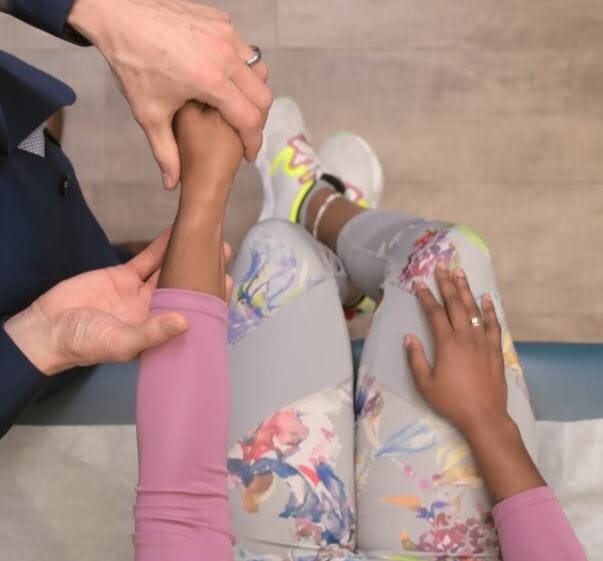
Examination of the upper limbs is the portion of physical examination involving the assessment of the shoulder, elbow, forearmForearmThe forearm is the region of the upper limb between the elbow and the wrist. The term "forearm" is used in anatomy to distinguish this area from the arm, a term that is commonly used to describe the entire upper limb. The forearm consists of 2 long bones (the radius and the ulna), the interosseous membrane, and multiple arteries, nerves, and muscles. Forearm: Anatomy, wrist, and handHandThe hand constitutes the distal part of the upper limb and provides the fine, precise movements needed in activities of daily living. It consists of 5 metacarpal bones and 14 phalanges, as well as numerous muscles innervated by the median and ulnar nerves. Hand: Anatomy to evaluate for signs of pathology. The examination includes inspectionInspectionDermatologic Examination, palpationPalpationApplication of fingers with light pressure to the surface of the body to determine consistency of parts beneath in physical diagnosis; includes palpation for determining the outlines of organs.Dermatologic Examination, tests of range of movement, and provocative maneuvers. A good history should be taken and concurrently used with the exam findings to obtain a presumptive diagnosis.
Briefly explain each step of the examination to the individual and obtain consent.
Position the individual in a sitting position.
Expose the shoulders, armArmThe arm, or “upper arm” in common usage, is the region of the upper limb that extends from the shoulder to the elbow joint and connects inferiorly to the forearm through the cubital fossa. It is divided into 2 fascial compartments (anterior and posterior).Arm: Anatomy, forearmForearmThe forearm is the region of the upper limb between the elbow and the wrist. The term “forearm” is used in anatomy to distinguish this area from the arm, a term that is commonly used to describe the entire upper limb. The forearm consists of 2 long bones (the radius and the ulna), the interosseous membrane, and multiple arteries, nerves, and muscles. Forearm: Anatomy, and handHandThe hand constitutes the distal part of the upper limb and provides the fine, precise movements needed in activities of daily living. It consists of 5 metacarpal bones and 14 phalanges, as well as numerous muscles innervated by the median and ulnar nerves. Hand: Anatomy adequately.
PalpationPalpationApplication of fingers with light pressure to the surface of the body to determine consistency of parts beneath in physical diagnosis; includes palpation for determining the outlines of organs.Dermatologic Examination
Range of motion
Special tests
Anatomy
The upper limb is divided into 3 regions:
ArmArmThe arm, or “upper arm” in common usage, is the region of the upper limb that extends from the shoulder to the elbow joint and connects inferiorly to the forearm through the cubital fossa. It is divided into 2 fascial compartments (anterior and posterior).Arm: Anatomy
ForearmForearmThe forearm is the region of the upper limb between the elbow and the wrist. The term “forearm” is used in anatomy to distinguish this area from the arm, a term that is commonly used to describe the entire upper limb. The forearm consists of 2 long bones (the radius and the ulna), the interosseous membrane, and multiple arteries, nerves, and muscles. Forearm: Anatomy
HandHandThe hand constitutes the distal part of the upper limb and provides the fine, precise movements needed in activities of daily living. It consists of 5 metacarpal bones and 14 phalanges, as well as numerous muscles innervated by the median and ulnar nerves. Hand: Anatomy
The joints involved include:
Shoulder joint
Elbow jointElbow jointThe elbow is the synovial hinge joint between the humerus in the upper arm and the radius and ulna in the forearm. The elbow consists of 3 joints, which form a functional unit enclosed within a single articular capsule. The elbow is the link between the powerful motions of the shoulder and the intricate fine-motor function of the hand. Elbow Joint: Anatomy
Wrist jointWrist jointThe wrist connects the forearm to the hand. It consists of 8 carpal bones, multiple joints, and various supporting ligaments, as well as the distal bones of the forearm and the proximal portion of the 5 metacarpal bones of the hand. Wrist Joint: Anatomy
These joints are surrounded and supported by many muscles, tendons, ligaments, and fibrocartilaginous structures to ensure support and stability and to absorb shockShockShock is a life-threatening condition associated with impaired circulation that results in tissue hypoxia. The different types of shock are based on the underlying cause: distributive (↑ cardiac output (CO), ↓ systemic vascular resistance (SVR)), cardiogenic (↓ CO, ↑ SVR), hypovolemic (↓ CO, ↑ SVR), obstructive (↓ CO), and mixed. Types of Shock during locomotion.
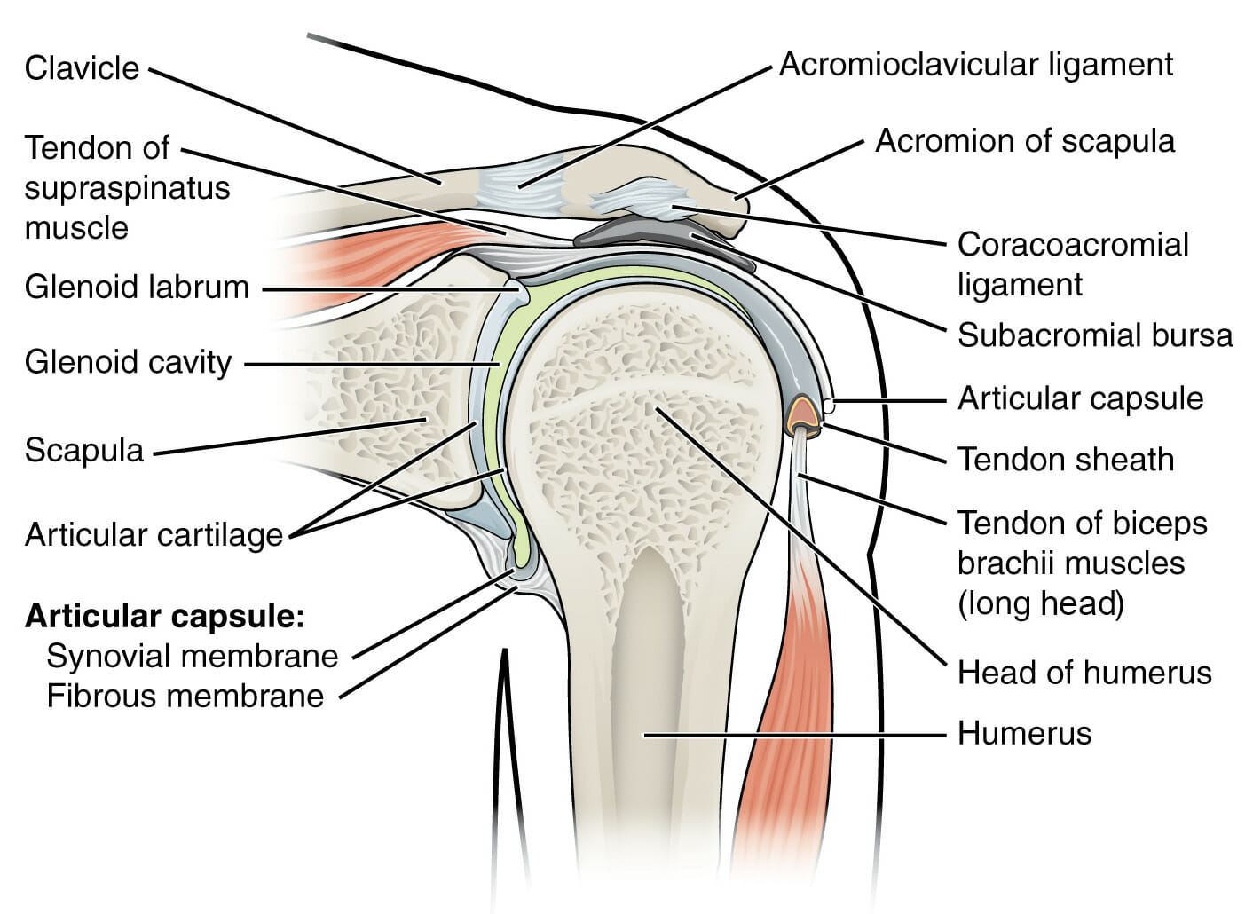
Anatomy of the shoulder
Image: “Shoulder Joint” by OpenStax College. License: CC BY 3.0, edited by Lecturio.
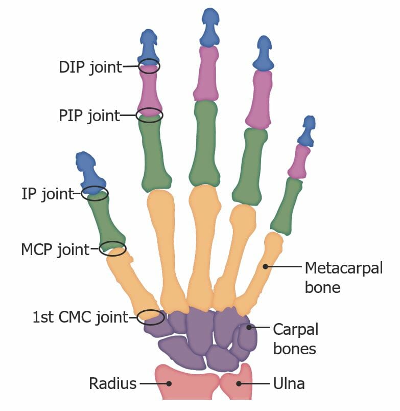
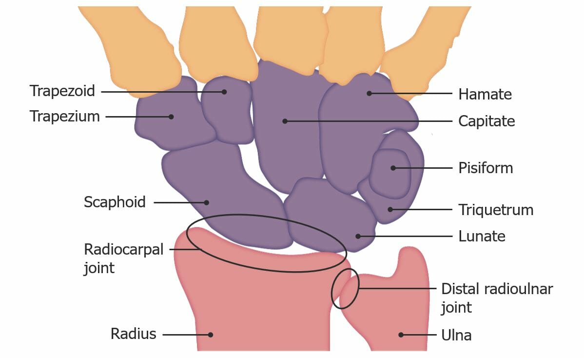
Bones and joints in the hand: CMC: carpometacarpal DIP: distal interphalangeal IP: interphalangeal MCP: metacarpophalangeal PIP: proximal interphalangeal
Muscle atrophyAtrophyDecrease in the size of a cell, tissue, organ, or multiple organs, associated with a variety of pathological conditions such as abnormal cellular changes, ischemia, malnutrition, or hormonal changes.Cellular Adaptation or fasciculationsFasciculationsInvoluntary contraction of the muscle fibers innervated by a motor unit. Fasciculations may be visualized as a muscle twitch or dimpling under the skin, but usually do not generate sufficient force to move a limb. They may represent a benign condition or occur as a manifestation of motor neuron disease or peripheral nervous system diseases.Polyneuropathy
Abnormal positioning
Winging
PalpationPalpationApplication of fingers with light pressure to the surface of the body to determine consistency of parts beneath in physical diagnosis; includes palpation for determining the outlines of organs.Dermatologic Examination
Palpate the various components of the shoulder girdle, including:
Sternoclavicular joint
ClavicleClavicleA bone on the ventral side of the shoulder girdle, which in humans is commonly called the collar bone.Clavicle Fracture
Acromioclavicular joint
Acromion
Coracoid process of the scapula
Head of the humerusHead of The HumerusThe upper rounded extremity of the humerus fitting into the glenoid cavity of the scapula.Arm: Anatomy
Greater tubercle of the humerusHumerusBone in humans and primates extending from the shoulder joint to the elbow joint.Arm: Anatomy
SpineSpineThe human spine, or vertebral column, is the most important anatomical and functional axis of the human body. It consists of 7 cervical vertebrae, 12 thoracic vertebrae, and 5 lumbar vertebrae and is limited cranially by the skull and caudally by the sacrum.Vertebral Column: Anatomy of the scapula
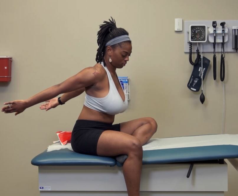
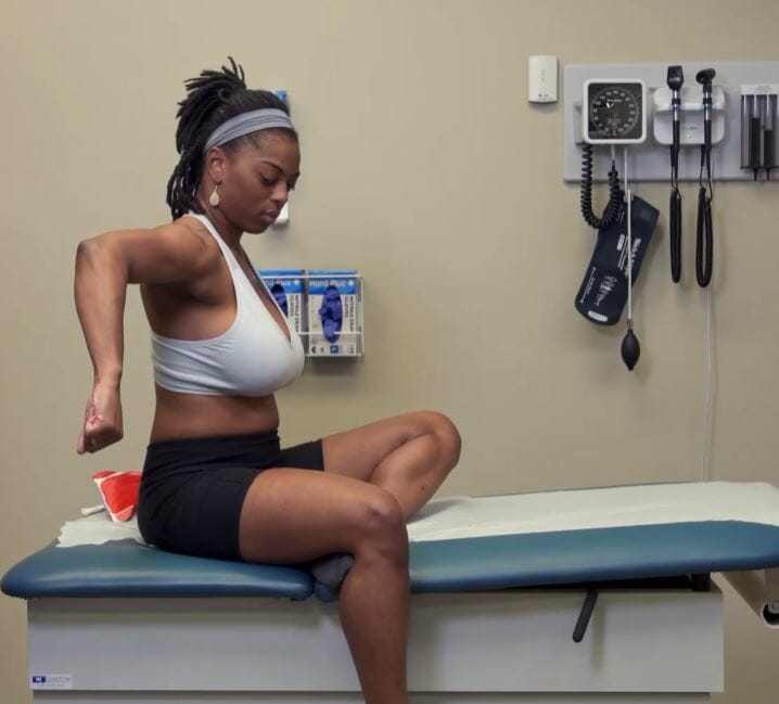
External rotationRotationMotion of an object in which either one or more points on a line are fixed. It is also the motion of a particle about a fixed point.X-rays and abduction of the shoulder joint:
Instruct individual to put the hands behind the head.
Elbows point out to the side.
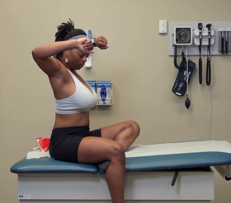
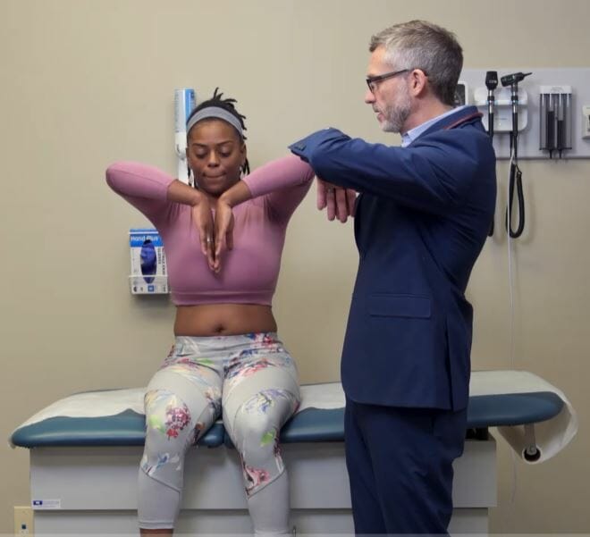
Internal rotationRotationMotion of an object in which either one or more points on a line are fixed. It is also the motion of a particle about a fixed point.X-rays and adduction of the shoulder joint:
Instruct individual to place the hands behind the lower back.
Reach as far up the spineSpineThe human spine, or vertebral column, is the most important anatomical and functional axis of the human body. It consists of 7 cervical vertebrae, 12 thoracic vertebrae, and 5 lumbar vertebrae and is limited cranially by the skull and caudally by the sacrum.Vertebral Column: Anatomy as possible.
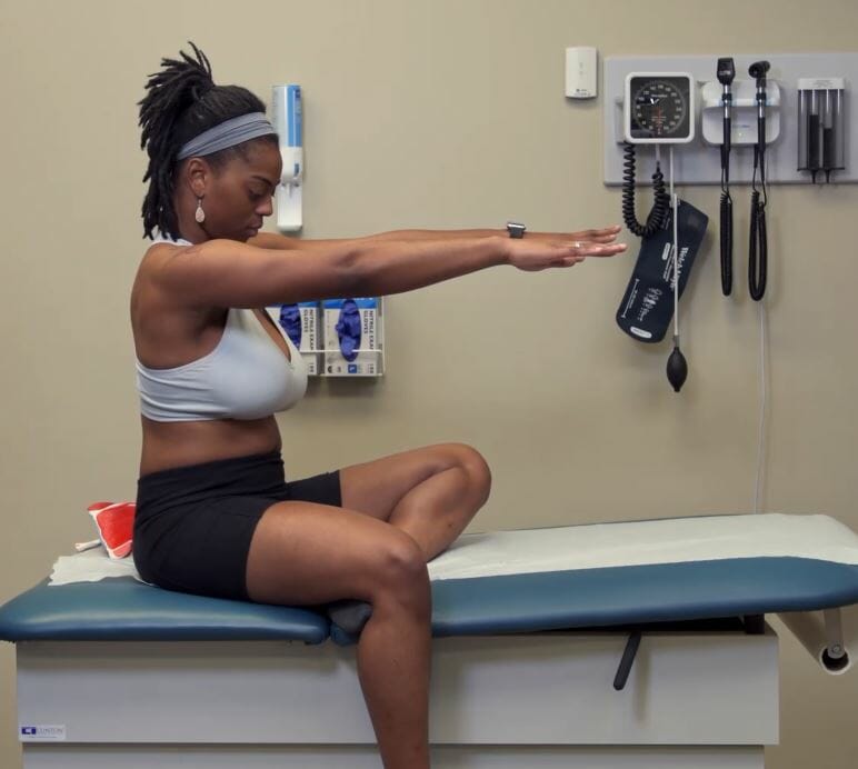
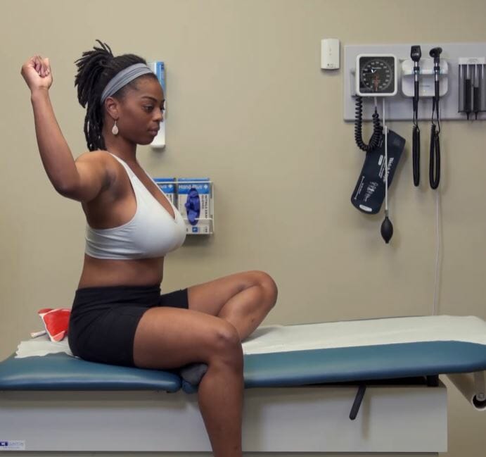
Shoulder flexion:
Individual raises the arms forward.
Normal range of movement: 150–180 degrees
Shoulder extension:
Individual stretches out the arms behind them.
Normal range of movement: 40 degrees
Shoulder abduction:
Individual raises the arms out to the side in an arc-like motion until the hands touch above the head.
Normal range of movement: 180 degrees
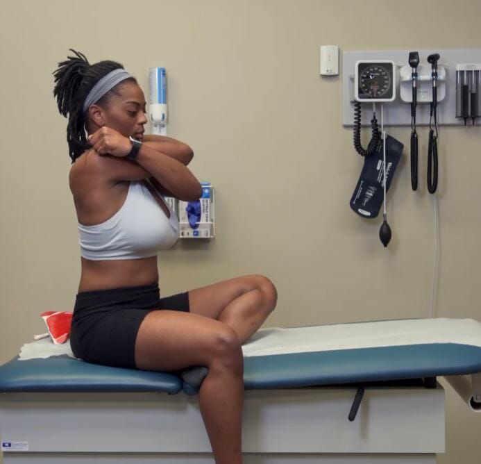
Shoulder adduction:
Individual keeps the arms straight and moves them across the front of the body to the opposite side.
Normal range of movement: 30–40 degrees
Shoulder external rotationRotationMotion of an object in which either one or more points on a line are fixed. It is also the motion of a particle about a fixed point.X-rays:
Individual keeps elbows by the sides flexed at 90 degrees while they move the forearmsForearmsPart of the upper extremity in humans and primates extending from the elbow to the wrist.Bowen Disease and Erythroplasia of Queyrat outward in an arc-like motion.
Normal range of movement: 80–90 degrees
Shoulder internal rotationRotationMotion of an object in which either one or more points on a line are fixed. It is also the motion of a particle about a fixed point.X-rays:
Ask the individual to place each handHandThe hand constitutes the distal part of the upper limb and provides the fine, precise movements needed in activities of daily living. It consists of 5 metacarpal bones and 14 phalanges, as well as numerous muscles innervated by the median and ulnar nerves. Hand: Anatomy behind the back and reach as far up the spineSpineThe human spine, or vertebral column, is the most important anatomical and functional axis of the human body. It consists of 7 cervical vertebrae, 12 thoracic vertebrae, and 5 lumbar vertebrae and is limited cranially by the skull and caudally by the sacrum.Vertebral Column: Anatomy as possible.
Normal range of movement: able to reach level T4T4The major hormone derived from the thyroid gland. Thyroxine is synthesized via the iodination of tyrosines (monoiodotyrosine) and the coupling of iodotyrosines (diiodotyrosine) in the thyroglobulin. Thyroxine is released from thyroglobulin by proteolysis and secreted into the blood. Thyroxine is peripherally deiodinated to form triiodothyronine which exerts a broad spectrum of stimulatory effects on cell metabolism.Thyroid Hormones–T8
Stabilize the scapula by placing one handHandThe hand constitutes the distal part of the upper limb and provides the fine, precise movements needed in activities of daily living. It consists of 5 metacarpal bones and 14 phalanges, as well as numerous muscles innervated by the median and ulnar nerves. Hand: Anatomy on the spineSpineThe human spine, or vertebral column, is the most important anatomical and functional axis of the human body. It consists of 7 cervical vertebrae, 12 thoracic vertebrae, and 5 lumbar vertebrae and is limited cranially by the skull and caudally by the sacrum.Vertebral Column: Anatomy of the scapula and the coracoid process.
Manipulate humeral headHumeral headThe upper rounded extremity of the humerus fitting into the glenoid cavity of the scapula.Arm: Anatomy anteriorly with the other handHandThe hand constitutes the distal part of the upper limb and provides the fine, precise movements needed in activities of daily living. It consists of 5 metacarpal bones and 14 phalanges, as well as numerous muscles innervated by the median and ulnar nerves. Hand: Anatomy.
Positive result: joint laxity compared to the contralateral side
Test significance: possible ligamentous, tendinous, or capsular injury
Inferior drawer test:
Positioning: supine, prone, or seated
Procedure:
Stabilize the scapula by placing one handHandThe hand constitutes the distal part of the upper limb and provides the fine, precise movements needed in activities of daily living. It consists of 5 metacarpal bones and 14 phalanges, as well as numerous muscles innervated by the median and ulnar nerves. Hand: Anatomy on the spineSpineThe human spine, or vertebral column, is the most important anatomical and functional axis of the human body. It consists of 7 cervical vertebrae, 12 thoracic vertebrae, and 5 lumbar vertebrae and is limited cranially by the skull and caudally by the sacrum.Vertebral Column: Anatomy of the scapula and the coracoid process.
Apply distal traction on the elbow.
Positive result:
Visible depression between the edge of the acromion and the humeral headHumeral headThe upper rounded extremity of the humerus fitting into the glenoid cavity of the scapula.Arm: Anatomy (positive sulcus sign)
Joint laxity compared to the contralateral side
Test significance: possible ligamentous, tendinous, or capsular injury
Anterior apprehension test:
Positioning: seated
Procedure:
Stabilize the scapula by placing one handHandThe hand constitutes the distal part of the upper limb and provides the fine, precise movements needed in activities of daily living. It consists of 5 metacarpal bones and 14 phalanges, as well as numerous muscles innervated by the median and ulnar nerves. Hand: Anatomy on the spineSpineThe human spine, or vertebral column, is the most important anatomical and functional axis of the human body. It consists of 7 cervical vertebrae, 12 thoracic vertebrae, and 5 lumbar vertebrae and is limited cranially by the skull and caudally by the sacrum.Vertebral Column: Anatomy of the scapula and the coracoid process.
Abduct shoulder to 90 degrees with the elbow flexed at 90 degrees.
Test significance: possible ligamentous, tendinous, or capsular injury
Posterior apprehension test:
Positioning: seated
Procedure:
Stabilize the scapula by placing one handHandThe hand constitutes the distal part of the upper limb and provides the fine, precise movements needed in activities of daily living. It consists of 5 metacarpal bones and 14 phalanges, as well as numerous muscles innervated by the median and ulnar nerves. Hand: Anatomy on the spineSpineThe human spine, or vertebral column, is the most important anatomical and functional axis of the human body. It consists of 7 cervical vertebrae, 12 thoracic vertebrae, and 5 lumbar vertebrae and is limited cranially by the skull and caudally by the sacrum.Vertebral Column: Anatomy of the scapula and the coracoid process.
Flex shoulder and elbow to 90 degrees and rotate internally.
Test significance: possible ligamentous, tendinous, or capsular injury
Rotator cuff tests
Lateral Jobe test:
Positioning: seated
Procedure:
Abduct the shoulder to 60 degrees.
Internally rotate the shoulder with the thumb pointing down toward the floor.
Push down on the armArmThe arm, or “upper arm” in common usage, is the region of the upper limb that extends from the shoulder to the elbow joint and connects inferiorly to the forearm through the cubital fossa. It is divided into 2 fascial compartments (anterior and posterior).Arm: Anatomy as the individual resists.
Positive result: painPainAn unpleasant sensation induced by noxious stimuli which are detected by nerve endings of nociceptive neurons.Pain: Types and Pathways or weakness against examiner’s resistanceResistancePhysiologically, the opposition to flow of air caused by the forces of friction. As a part of pulmonary function testing, it is the ratio of driving pressure to the rate of air flow.Ventilation: Mechanics of Breathing
Test significance: suggests a tear in the supraspinatus tendon or muscle
Lift-off test:
Positioning: seated
Procedure:
Individual places the dorsum of the handHandThe hand constitutes the distal part of the upper limb and provides the fine, precise movements needed in activities of daily living. It consists of 5 metacarpal bones and 14 phalanges, as well as numerous muscles innervated by the median and ulnar nerves. Hand: Anatomy on the lower back with the elbow flexed to 90 degrees.
The examiner lifts the handHandThe hand constitutes the distal part of the upper limb and provides the fine, precise movements needed in activities of daily living. It consists of 5 metacarpal bones and 14 phalanges, as well as numerous muscles innervated by the median and ulnar nerves. Hand: Anatomy off the back against the individual’s resistanceResistancePhysiologically, the opposition to flow of air caused by the forces of friction. As a part of pulmonary function testing, it is the ratio of driving pressure to the rate of air flow.Ventilation: Mechanics of Breathing (the individual is asked to maintain the handHandThe hand constitutes the distal part of the upper limb and provides the fine, precise movements needed in activities of daily living. It consists of 5 metacarpal bones and 14 phalanges, as well as numerous muscles innervated by the median and ulnar nerves. Hand: Anatomy in this position).
Positive result: inability to maintain the above position against resistanceResistancePhysiologically, the opposition to flow of air caused by the forces of friction. As a part of pulmonary function testing, it is the ratio of driving pressure to the rate of air flow.Ventilation: Mechanics of Breathing
Test significance: suggests a tear in the subscapularis tendon or muscle
Resisted external rotationRotationMotion of an object in which either one or more points on a line are fixed. It is also the motion of a particle about a fixed point.X-rays:
Positioning: seated
Procedure:
Individual adducts and flexes the arms to 90 degrees with the thumb turned up.
The examiner stabilizes the elbow with one handHandThe hand constitutes the distal part of the upper limb and provides the fine, precise movements needed in activities of daily living. It consists of 5 metacarpal bones and 14 phalanges, as well as numerous muscles innervated by the median and ulnar nerves. Hand: Anatomy and applies pressure proximal to the individual’s wrist.
The individual presses the wrists outward in external rotationRotationMotion of an object in which either one or more points on a line are fixed. It is also the motion of a particle about a fixed point.X-rays.
Positive result: painPainAn unpleasant sensation induced by noxious stimuli which are detected by nerve endings of nociceptive neurons.Pain: Types and Pathways or weakness against examiner’s resistanceResistancePhysiologically, the opposition to flow of air caused by the forces of friction. As a part of pulmonary function testing, it is the ratio of driving pressure to the rate of air flow.Ventilation: Mechanics of Breathing
Test significance: suggests a tear in the infraspinatus tendon or muscle
Impingement tests
Painful arc test:
Positioning: seated
Procedure:
Stabilize the scapula by placing one handHandThe hand constitutes the distal part of the upper limb and provides the fine, precise movements needed in activities of daily living. It consists of 5 metacarpal bones and 14 phalanges, as well as numerous muscles innervated by the median and ulnar nerves. Hand: Anatomy on the spineSpineThe human spine, or vertebral column, is the most important anatomical and functional axis of the human body. It consists of 7 cervical vertebrae, 12 thoracic vertebrae, and 5 lumbar vertebrae and is limited cranially by the skull and caudally by the sacrum.Vertebral Column: Anatomy of the scapula and the coracoid process.
Actively and passively carry the shoulder from 0 degrees to 180 degrees of abduction.
Positive result: shoulder painPainAn unpleasant sensation induced by noxious stimuli which are detected by nerve endings of nociceptive neurons.Pain: Types and Pathways at 60–120 degrees
Test significance: suggests subacromial impingement
Neer test:
Positioning: seated
Procedure:
Stabilize the scapula by placing one handHandThe hand constitutes the distal part of the upper limb and provides the fine, precise movements needed in activities of daily living. It consists of 5 metacarpal bones and 14 phalanges, as well as numerous muscles innervated by the median and ulnar nerves. Hand: Anatomy on the spineSpineThe human spine, or vertebral column, is the most important anatomical and functional axis of the human body. It consists of 7 cervical vertebrae, 12 thoracic vertebrae, and 5 lumbar vertebrae and is limited cranially by the skull and caudally by the sacrum.Vertebral Column: Anatomy of the scapula and the coracoid process.
Adduct shoulder to 90 degrees and rotate internally. Extend elbow; forearmForearmThe forearm is the region of the upper limb between the elbow and the wrist. The term “forearm” is used in anatomy to distinguish this area from the arm, a term that is commonly used to describe the entire upper limb. The forearm consists of 2 long bones (the radius and the ulna), the interosseous membrane, and multiple arteries, nerves, and muscles. Forearm: Anatomy is pronated.
Actively and passively raise the armArmThe arm, or “upper arm” in common usage, is the region of the upper limb that extends from the shoulder to the elbow joint and connects inferiorly to the forearm through the cubital fossa. It is divided into 2 fascial compartments (anterior and posterior).Arm: Anatomy (shoulder flexion).
Positive result: painPainAn unpleasant sensation induced by noxious stimuli which are detected by nerve endings of nociceptive neurons.Pain: Types and Pathways at 90–120 degrees
Test significance: suggests subacromial impingement
Hawkins-Kennedy test:
Positioning: seated
Procedure:
Shoulder and elbow are flexed at 90 degrees.
Stabilize the elbow and passively rotate the shoulder joint internally.
Positive result: painPainAn unpleasant sensation induced by noxious stimuli which are detected by nerve endings of nociceptive neurons.Pain: Types and Pathways on internal rotationRotationMotion of an object in which either one or more points on a line are fixed. It is also the motion of a particle about a fixed point.X-rays
Test significance: suggests subacromial impingement
Other provocative tests
Cross-body adduction test:
Positioning: seated
Procedure:
Adduct the armArmThe arm, or “upper arm” in common usage, is the region of the upper limb that extends from the shoulder to the elbow joint and connects inferiorly to the forearm through the cubital fossa. It is divided into 2 fascial compartments (anterior and posterior).Arm: Anatomy(s) across the chest.
Assess the acromioclavicular and sternoclavicular joints.
Positive result: painPainAn unpleasant sensation induced by noxious stimuli which are detected by nerve endings of nociceptive neurons.Pain: Types and Pathways with adduction
Test significance: acromioclavicular or sternoclavicular dysfunction
Yegason’s test:
Positioning: seated
Procedure:
The individual flexes the elbow to 90 degrees.
The individual supinates the wrist and internally rotates the shoulder against the examiner’s resistanceResistancePhysiologically, the opposition to flow of air caused by the forces of friction. As a part of pulmonary function testing, it is the ratio of driving pressure to the rate of air flow.Ventilation: Mechanics of Breathing.
Positive result: painPainAn unpleasant sensation induced by noxious stimuli which are detected by nerve endings of nociceptive neurons.Pain: Types and Pathways in the bicipital groove or movement of the bicepsBicepsArm: Anatomy tendon out of the bicipital groove
The individual flexes the shoulder to 90 degrees with the elbow extended and the forearmForearmThe forearm is the region of the upper limb between the elbow and the wrist. The term “forearm” is used in anatomy to distinguish this area from the arm, a term that is commonly used to describe the entire upper limb. The forearm consists of 2 long bones (the radius and the ulna), the interosseous membrane, and multiple arteries, nerves, and muscles. Forearm: Anatomy supinated.
The examiner applies a downward force as the individual resists.
Positive result: painPainAn unpleasant sensation induced by noxious stimuli which are detected by nerve endings of nociceptive neurons.Pain: Types and Pathways elicited in the bicipital groove
Muscle atrophyAtrophyDecrease in the size of a cell, tissue, organ, or multiple organs, associated with a variety of pathological conditions such as abnormal cellular changes, ischemia, malnutrition, or hormonal changes.Cellular Adaptation or fasciculationsFasciculationsInvoluntary contraction of the muscle fibers innervated by a motor unit. Fasciculations may be visualized as a muscle twitch or dimpling under the skin, but usually do not generate sufficient force to move a limb. They may represent a benign condition or occur as a manifestation of motor neuron disease or peripheral nervous system diseases.Polyneuropathy
Abnormal positioning
PalpationPalpationApplication of fingers with light pressure to the surface of the body to determine consistency of parts beneath in physical diagnosis; includes palpation for determining the outlines of organs.Dermatologic Examination
Palpate the various components of the elbow, including:
Tenderness: Palpate tender areas last and compare with nontender areas.
Range of motion
Flexion:
Individual bends the elbow to touch the shoulder.
Normal range of motion: 0–145 degrees
Extension:
Individual straightens the arms as far out as possible.
Normal range of motion: 0 degrees
Pronation:
Individual turns the forearmForearmThe forearm is the region of the upper limb between the elbow and the wrist. The term “forearm” is used in anatomy to distinguish this area from the arm, a term that is commonly used to describe the entire upper limb. The forearm consists of 2 long bones (the radius and the ulna), the interosseous membrane, and multiple arteries, nerves, and muscles. Forearm: Anatomy so that the palm is facing down.
Individual turns the forearmForearmThe forearm is the region of the upper limb between the elbow and the wrist. The term “forearm” is used in anatomy to distinguish this area from the arm, a term that is commonly used to describe the entire upper limb. The forearm consists of 2 long bones (the radius and the ulna), the interosseous membrane, and multiple arteries, nerves, and muscles. Forearm: Anatomy so that the palm is facing up.
Individual flexes the elbow to 90 degrees with wrist pronated and flexed completely.
Individual is asked to extend the wrist against the examiner’s resistanceResistancePhysiologically, the opposition to flow of air caused by the forces of friction. As a part of pulmonary function testing, it is the ratio of driving pressure to the rate of air flow.Ventilation: Mechanics of Breathing.
Test significance: suggests lateral epicondylitis and/or wrist extensor tendinopathy
Golfer’s elbow (medial epicondylitis):
Positioning: seated
Procedure:
Individual flexes the elbow to 90 degrees with the wrist supinated and extended completely.
Individual is asked to make a fist and flex the wrist against the examiner’s resistanceResistancePhysiologically, the opposition to flow of air caused by the forces of friction. As a part of pulmonary function testing, it is the ratio of driving pressure to the rate of air flow.Ventilation: Mechanics of Breathing.
Muscle atrophyAtrophyDecrease in the size of a cell, tissue, organ, or multiple organs, associated with a variety of pathological conditions such as abnormal cellular changes, ischemia, malnutrition, or hormonal changes.Cellular Adaptation or fasciculationsFasciculationsInvoluntary contraction of the muscle fibers innervated by a motor unit. Fasciculations may be visualized as a muscle twitch or dimpling under the skin, but usually do not generate sufficient force to move a limb. They may represent a benign condition or occur as a manifestation of motor neuron disease or peripheral nervous system diseases.Polyneuropathy
Abnormal positioning
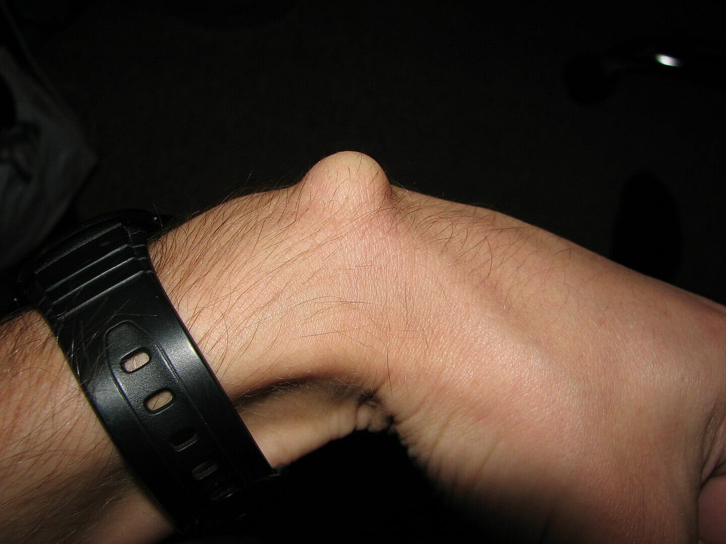
Ganglion cyst:
Synovial cystsCystsAny fluid-filled closed cavity or sac that is lined by an epithelium. Cysts can be of normal, abnormal, non-neoplastic, or neoplastic tissues.Fibrocystic Change that contain mucinous fluid.
Most commonly found on the dorsal aspect of the wrist
Believed to arise from repetitive microtraumaMicrotraumaSmall injuries caused by external force applied to the body including bones, muscles, nerves and tendons.Osgood-Schlatter Disease resulting in mucinous degeneration of connective tissueConnective tissueConnective tissues originate from embryonic mesenchyme and are present throughout the body except inside the brain and spinal cord. The main function of connective tissues is to provide structural support to organs. Connective tissues consist of cells and an extracellular matrix.Connective Tissue: Histology
Majority are asymptomatic.
May present with:
PainPainAn unpleasant sensation induced by noxious stimuli which are detected by nerve endings of nociceptive neurons.Pain: Types and Pathways and tenderness
PalpationPalpationApplication of fingers with light pressure to the surface of the body to determine consistency of parts beneath in physical diagnosis; includes palpation for determining the outlines of organs.Dermatologic Examination
Palpate the various components of the wrist, including:
Tenderness: Palpate tender areas last and compare with nontender areas.
Range of motion
Flexion:
Individual puts the backs of the hands together and flexes the wrist fully.
Normal range of motion: 0–90 degrees
Extension:
Individual puts the backs of the hands together and extends the wrist fully.
Normal range of motion: 0–90 degrees
Adduction:
With palm facing down, individual brings the fingers toward the midline.
Normal range of motion: 0–30 degrees
Abduction:
With palm facing down, individual brings the fingers away from the midline.
Normal range of motion: 0–30 degrees
Pronation:
Individual turns the forearmForearmThe forearm is the region of the upper limb between the elbow and the wrist. The term “forearm” is used in anatomy to distinguish this area from the arm, a term that is commonly used to describe the entire upper limb. The forearm consists of 2 long bones (the radius and the ulna), the interosseous membrane, and multiple arteries, nerves, and muscles. Forearm: Anatomy so the palm is facing down.
Individual turns the forearmForearmThe forearm is the region of the upper limb between the elbow and the wrist. The term “forearm” is used in anatomy to distinguish this area from the arm, a term that is commonly used to describe the entire upper limb. The forearm consists of 2 long bones (the radius and the ulna), the interosseous membrane, and multiple arteries, nerves, and muscles. Forearm: Anatomy so that the palm is facing up.
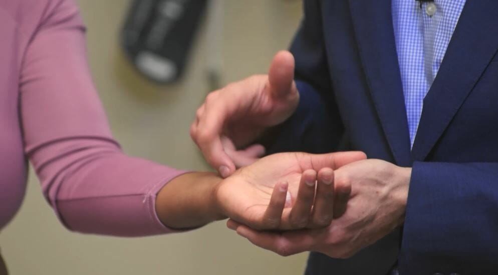
Tinel testTinel testFirm percussion over the course of the median nerve proximal to or on top of the carpal tunnel . A positive test is defined as pain and/or paresthesia in median innervated fingers on percussion over the median nerve.Carpal Tunnel Syndrome:
Positioning: seated
Procedure:
Individual flexes the elbow to 90 degrees with the wrist supinated and extended.
Examiner taps in the center of the transverse carpal ligament.
Positive test: painPainAn unpleasant sensation induced by noxious stimuli which are detected by nerve endings of nociceptive neurons.Pain: Types and Pathways or paresthesia in the distribution of the median nerveMedian NerveA major nerve of the upper extremity. In humans, the fibers of the median nerve originate in the lower cervical and upper thoracic spinal cord (usually C6 to T1), travel via the brachial plexus, and supply sensory and motor innervation to parts of the forearm and hand.Cubital Fossa: Anatomy
Individual flexes the elbow to 90 degrees with the dorsal aspect of the wrists pressed together in the completely flexed position.
Individual is asked to extend the wrist against the examiner’s resistanceResistancePhysiologically, the opposition to flow of air caused by the forces of friction. As a part of pulmonary function testing, it is the ratio of driving pressure to the rate of air flow.Ventilation: Mechanics of Breathing.
Positive test: painPainAn unpleasant sensation induced by noxious stimuli which are detected by nerve endings of nociceptive neurons.Pain: Types and Pathways, paresthesia, or numbness in the distribution of the median nerveMedian NerveA major nerve of the upper extremity. In humans, the fibers of the median nerve originate in the lower cervical and upper thoracic spinal cord (usually C6 to T1), travel via the brachial plexus, and supply sensory and motor innervation to parts of the forearm and hand.Cubital Fossa: Anatomy
Individual adducts the thumb into the palm and closes a fist around it and then ulnarly deviates the wrist.
Individual attempts to abduct the wrist and thumb against the examiner’s resistanceResistancePhysiologically, the opposition to flow of air caused by the forces of friction. As a part of pulmonary function testing, it is the ratio of driving pressure to the rate of air flow.Ventilation: Mechanics of Breathing.
Positive test: painPainAn unpleasant sensation induced by noxious stimuli which are detected by nerve endings of nociceptive neurons.Pain: Types and Pathways over the radial aspect of the wrist
Examiner grasps the thumb and moves the wrist into ulnar deviation
Positive test: painPainAn unpleasant sensation induced by noxious stimuli which are detected by nerve endings of nociceptive neurons.Pain: Types and Pathways over the radial aspect of the wrist/first dorsal compartment
Generally considered more accurate and preferred due to its higher specificity and lower likelihood of producing false positives than the Eichhoff test
Finkelstein test to aid in the diagnosis of de Quervain tenosynovitis
Inspect the following structures of the handHandThe hand constitutes the distal part of the upper limb and provides the fine, precise movements needed in activities of daily living. It consists of 5 metacarpal bones and 14 phalanges, as well as numerous muscles innervated by the median and ulnar nerves. Hand: Anatomy bilaterally:
Bony structures:
MetacarpalsMetacarpalsThe five cylindrical bones of the metacarpus, articulating with the carpal bones proximally and the phalanges of fingers distally.Wrist Joint: Anatomy
Proximal phalangesPhalangesBones that make up the skeleton of the fingers, consisting of two for the thumb, and three for each of the other fingers.Hand: Anatomy
Distal phalangesPhalangesBones that make up the skeleton of the fingers, consisting of two for the thumb, and three for each of the other fingers.Hand: Anatomy
Muscle atrophyAtrophyDecrease in the size of a cell, tissue, organ, or multiple organs, associated with a variety of pathological conditions such as abnormal cellular changes, ischemia, malnutrition, or hormonal changes.Cellular Adaptation or fasciculationsFasciculationsInvoluntary contraction of the muscle fibers innervated by a motor unit. Fasciculations may be visualized as a muscle twitch or dimpling under the skin, but usually do not generate sufficient force to move a limb. They may represent a benign condition or occur as a manifestation of motor neuron disease or peripheral nervous system diseases.Polyneuropathy
Abnormal positioning
Common abnormalities
Deformity:
Rotational deformity due to phalangeal fractureFractureA fracture is a disruption of the cortex of any bone and periosteum and is commonly due to mechanical stress after an injury or accident. Open fractures due to trauma can be a medical emergency. Fractures are frequently associated with automobile accidents, workplace injuries, and trauma.Overview of Bone Fractures
Mallet finger:
Flexion at DIP
Can be passively reversed
Boutonnière deformity:
PIP: fixed flexion
DIP: hyperextension
Swan-neck deformity:
PIP: hyperextension
DIP: flexion
Dupuytren contracture:
Fixed flexion of the MCP and PIP joints
Usually affects the little and ring finger
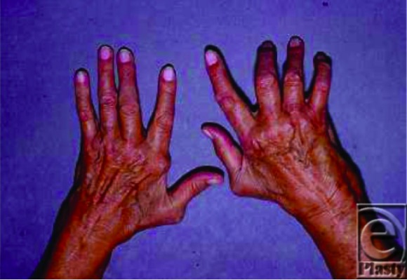
Physical exam findings in rheumatoid arthritis (RA): An individual with RA with several classic deformities: ulnar deviation (left hand) and boutonnière deformity (3rd, 4th, and 5th fingers of the right hand)
Image: “Rheumatoid hand” by Division of Plastic and Reconstructive Surgery, New Jersey Medical School, University of Medicine and Dentistry of New Jersey, Newark. License: CC BY 2.0
Swan-neck deformity of the 5th finger in an individual with RA
Image: “Swan-neck deformity” by Khatam-al-Anbia Eye Research Center, Mashhad University of Medical Sciences, Mashhad, Iran. License: CC BY 2.5
Ganglion cystsCystsAny fluid-filled closed cavity or sac that is lined by an epithelium. Cysts can be of normal, abnormal, non-neoplastic, or neoplastic tissues.Fibrocystic Change
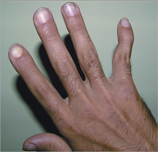
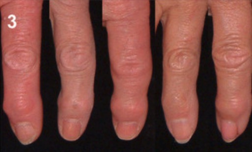
Heberden nodes in osteoarthritis: Bony growth spurs at the distal interphalangeal joints due to osteophytes
Image: “Bony growth spurts” by School of Dentistry, Franciscan University Center, Andradas Street, 1614, 97010-032 Santa Maria, RS, Brazil. License: CC BY 3.0
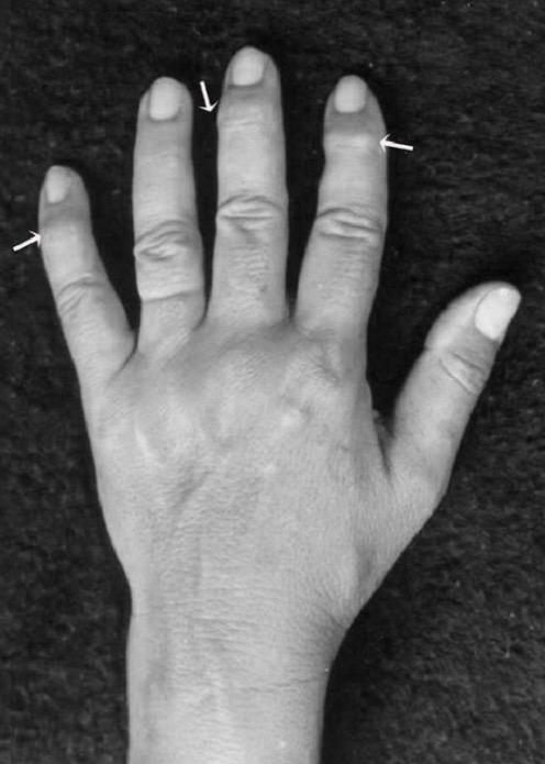
Bouchard nodes in osteoarthritis: Osteophytes of the proximal interphalangeal joints
Image: “Grading of osteoarthritis” by Landspitalinn University Hospital, University of Iceland, IS-108 Fossvogur, Reykjavik, ICELAND. License: CC BY 2.0, edited by Lecturio.
Thickening of the flexor tendons or flexion contracturesContracturesProlonged shortening of the muscle or other soft tissue around a joint, preventing movement of the joint.Wound Healing in the fingers
PalpationPalpationApplication of fingers with light pressure to the surface of the body to determine consistency of parts beneath in physical diagnosis; includes palpation for determining the outlines of organs.Dermatologic Examination
Palpate the various components of the handHandThe hand constitutes the distal part of the upper limb and provides the fine, precise movements needed in activities of daily living. It consists of 5 metacarpal bones and 14 phalanges, as well as numerous muscles innervated by the median and ulnar nerves. Hand: Anatomy, including:
Tenderness (palpate tender areas last and compare with nontender areas)
Palms up:
Palpate the radial and ulnar pulses to confirm adequate blood supply to the handHandThe hand constitutes the distal part of the upper limb and provides the fine, precise movements needed in activities of daily living. It consists of 5 metacarpal bones and 14 phalanges, as well as numerous muscles innervated by the median and ulnar nerves. Hand: Anatomy.
Palpate the muscle bulk of the thenar and hypothenar eminences. Wasting is due to:
Disuse atrophyAtrophyDecrease in the size of a cell, tissue, organ, or multiple organs, associated with a variety of pathological conditions such as abnormal cellular changes, ischemia, malnutrition, or hormonal changes.Cellular Adaptation
Palpate the palm to detect the typical bands of thickened palmar fasciaFasciaLayers of connective tissue of variable thickness. The superficial fascia is found immediately below the skin; the deep fascia invests muscles, nerves, and other organs.Cellulitis associated with Dupuytren contracture.
Assess median and ulnar nerveUlnar NerveA major nerve of the upper extremity. In humans, the fibers of the ulnar nerve originate in the lower cervical and upper thoracic spinal cord (usually C7 to T1), travel via the medial cord of the brachial plexus, and supply sensory and motor innervation to parts of the hand and forearm.Axilla and Brachial Plexus: Anatomy sensation:
Median nerveMedian NerveA major nerve of the upper extremity. In humans, the fibers of the median nerve originate in the lower cervical and upper thoracic spinal cord (usually C6 to T1), travel via the brachial plexus, and supply sensory and motor innervation to parts of the forearm and hand.Cubital Fossa: Anatomy: over the thenar eminence and index finger
Ulnar nerveUlnar NerveA major nerve of the upper extremity. In humans, the fibers of the ulnar nerve originate in the lower cervical and upper thoracic spinal cord (usually C7 to T1), travel via the medial cord of the brachial plexus, and supply sensory and motor innervation to parts of the hand and forearm.Axilla and Brachial Plexus: Anatomy: over the hypothenar eminence and little finger
Palms down:
Assess radial nerveRadial NerveA major nerve of the upper extremity. In humans the fibers of the radial nerve originate in the lower cervical and upper thoracic spinal cord (usually C5 to T1), travel via the posterior cord of the brachial plexus, and supply motor innervation to extensor muscles of the arm and cutaneous sensory fibers to extensor regions of the arm and hand.Axilla and Brachial Plexus: Anatomy sensation over the 1st dorsal web space.
Gently squeeze across the MCP joints to elicit tenderness (suggestive of active inflammatory arthropathyArthropathyOsteoarthritis).
Palpate the joints of the handHandThe hand constitutes the distal part of the upper limb and provides the fine, precise movements needed in activities of daily living. It consists of 5 metacarpal bones and 14 phalanges, as well as numerous muscles innervated by the median and ulnar nerves. Hand: Anatomy:
Rotator cuff tears: Rotator cuff pathology is the most common condition of the shoulder for which individuals seek treatment. Rotator cuff tears are caused by degeneration, impingement, and overload. Presentation is with painPainAn unpleasant sensation induced by noxious stimuli which are detected by nerve endings of nociceptive neurons.Pain: Types and Pathways and weakness at the shoulder joint. On examination, there may be a positive painful arc sign, positive drop arm testDrop arm testFunctional Neurological Symptom Disorder (Conversion Disorder), or weakness in external rotationRotationMotion of an object in which either one or more points on a line are fixed. It is also the motion of a particle about a fixed point.X-rays. Physical examination findings are dependent on the muscles involved. Imaging studies such as musculoskeletal ultrasonography and MRI can be done to confirm the diagnosis. Management is with physical therapyPhysical TherapyBecker Muscular Dystrophy or surgical repair.
Carpal tunnelCarpal TunnelThe carpal tunnel is formed by the transverse carpal ligament (flexor retinaculum) superiorly and the carpal bones inferiorly.Carpal Tunnel Syndrome syndrome: complex of signs and symptoms caused by compressionCompressionBlunt Chest Trauma of the median nerveMedian NerveA major nerve of the upper extremity. In humans, the fibers of the median nerve originate in the lower cervical and upper thoracic spinal cord (usually C6 to T1), travel via the brachial plexus, and supply sensory and motor innervation to parts of the forearm and hand.Cubital Fossa: Anatomy as it crosses the carpal tunnelCarpal TunnelThe carpal tunnel is formed by the transverse carpal ligament (flexor retinaculum) superiorly and the carpal bones inferiorly.Carpal Tunnel Syndrome. Presentation may be with painPainAn unpleasant sensation induced by noxious stimuli which are detected by nerve endings of nociceptive neurons.Pain: Types and Pathways, paresthesia, weakness, and atrophyAtrophyDecrease in the size of a cell, tissue, organ, or multiple organs, associated with a variety of pathological conditions such as abnormal cellular changes, ischemia, malnutrition, or hormonal changes.Cellular Adaptation over the median nerveMedian NerveA major nerve of the upper extremity. In humans, the fibers of the median nerve originate in the lower cervical and upper thoracic spinal cord (usually C6 to T1), travel via the brachial plexus, and supply sensory and motor innervation to parts of the forearm and hand.Cubital Fossa: AnatomydermatomeDermatomeSpinal Disk Herniation and myotomeMyotomeDevelopment of the Limbs distribution. On examination, a positive Tinel testTinel testFirm percussion over the course of the median nerve proximal to or on top of the carpal tunnel . A positive test is defined as pain and/or paresthesia in median innervated fingers on percussion over the median nerve.Carpal Tunnel Syndrome and Phalen test are elicited. Management can be with splinting, physical therapyPhysical TherapyBecker Muscular Dystrophy, or surgical correction.
Rheumatoid arthritisArthritisAcute or chronic inflammation of joints.Osteoarthritis: chronic, systemic, autoimmune, inflammatory disorder of unknown etiology that primarily involves synovial joints. The arthritisArthritisAcute or chronic inflammation of joints.Osteoarthritis is usually symmetricalSymmetricalDermatologic Examination and leads to erosionErosionPartial-thickness loss of the epidermisGeneralized and Localized Rashes of cartilageCartilageCartilage is a type of connective tissue derived from embryonic mesenchyme that is responsible for structural support, resilience, and the smoothness of physical actions. Perichondrium (connective tissue membrane surrounding cartilage) compensates for the absence of vasculature in cartilage by providing nutrition and support. Cartilage: Histology and boneBoneBone is a compact type of hardened connective tissue composed of bone cells, membranes, an extracellular mineralized matrix, and central bone marrow. The 2 primary types of bone are compact and spongy. Bones: Structure and Types, causing joint deformities. During the onset of disease, individuals present with joint painPainAn unpleasant sensation induced by noxious stimuli which are detected by nerve endings of nociceptive neurons.Pain: Types and Pathways, stiffness (especially morning stiffness), and swellingSwellingInflammation of many joints. On examination, joint tenderness and swellingSwellingInflammation may be noted on the small joints of the hands, wrist, and forefoot. Individuals with poorly controlled disease have significant joint deformities (e.g., swan-neck deformity). Management is with early use of disease-modifying antirheumatic drugsDisease-modifying antirheumatic drugsDisease-modifying antirheumatic drugs are antiinflammatory medications used to manage rheumatoid arthritis. The medications slow, but do not cure, the progression of the disease. The medications are classified as either synthetic or biologic agents and each has unique mechanisms of action and side effects.Disease-Modifying Antirheumatic Drugs (DMARDs) (DMARDsDMARDsDisease-modifying antirheumatic drugs are antiinflammatory medications used to manage rheumatoid arthritis. The medications slow, but do not cure, the progression of the disease. The medications are classified as either synthetic or biologic agents and each has unique mechanisms of action and side effects.Disease-Modifying Antirheumatic Drugs (DMARDs)) and antiinflammatory agents.
Elbow tendinopathy: Chronic painChronic painAching sensation that persists for more than a few months. It may or may not be associated with trauma or disease, and may persist after the initial injury has healed. Its localization, character, and timing are more vague than with acute pain.Pain Management at the lateral or medial epicondyleMedial epicondyleArm: Anatomy of the elbow is a relatively common condition, particularly among tennis players and golfers, respectively, and among manual laborers. On examination, there is localized tenderness over the lateral or medial epicondyleMedial epicondyleArm: Anatomy, with painPainAn unpleasant sensation induced by noxious stimuli which are detected by nerve endings of nociceptive neurons.Pain: Types and Pathways elicited during resisted wrist flexion or extension. The diagnosis is primarily clinical. Management may include activity modification, bracing, oral analgesics, or physical therapyPhysical TherapyBecker Muscular Dystrophy.
Ganglion cyst: one of the most common soft tissueSoft TissueSoft Tissue Abscess swellings in the handHandThe hand constitutes the distal part of the upper limb and provides the fine, precise movements needed in activities of daily living. It consists of 5 metacarpal bones and 14 phalanges, as well as numerous muscles innervated by the median and ulnar nerves. Hand: Anatomy and the wrist. A ganglion cyst is a fluid-filled swellingSwellingInflammation overlying a joint or tendon sheath. These cystsCystsAny fluid-filled closed cavity or sac that is lined by an epithelium. Cysts can be of normal, abnormal, non-neoplastic, or neoplastic tissues.Fibrocystic Change are benignBenignFibroadenoma but may occasionally lead to painPainAn unpleasant sensation induced by noxious stimuli which are detected by nerve endings of nociceptive neurons.Pain: Types and Pathways, weakness, and loss of functionLoss of FunctionInflammation. On examination, an obvious swellingSwellingInflammation that is firm, smooth, rounded, rubbery, and at times tender is noted. Management is with reassuranceReassuranceClinician–Patient Relationship for asymptomatic cystsCystsAny fluid-filled closed cavity or sac that is lined by an epithelium. Cysts can be of normal, abnormal, non-neoplastic, or neoplastic tissues.Fibrocystic Change, aspiration, or surgical therapy.
References
Bickley, L.S. (2017). The musculoskeletal system. In: Bates’ Guide to Physical Examination and History Taking, 12th ed. Wolters Kluwer, pp. 645–666.