Achieve Mastery of Medical Concepts

Study for medical school and boards with Lecturio

Acute Bronchiolitis (Clinical)

Acute bronchiolitis is primarily a pediatric respiratory condition caused by inflammation of the bronchioles in response to a viral infection. The condition is a common cause of hospitalization in children in the United States, with the majority of cases caused by respiratory syncytial virus (RSV). Patients usually present with upper respiratory symptoms, such as cough and congestion, and later develop lower respiratory signs, including dyspnea, wheezing, crackles, and hypoxia, for up to 10 days. Diagnosis is clinical and treatment is directed at improving oxygenation and hydration. As the disease course is self-limiting, acute bronchiolitis has good prognosis with appropriate management.

Last updated: Mar 4, 2024

Editorial responsibility: Stanley Oiseth, Lindsay Jones, Evelin Maza

Overview

Definition

Acute bronchiolitis Bronchiolitis Inflammation of the bronchioles. Pediatric Chest Abnormalities is a clinical constellation of respiratory symptoms (increased work of breathing Work of breathing Respiratory muscle contraction during inhalation. The work is accomplished in three phases: lung compliance work, that required to expand the lungs against its elastic forces; tissue resistance work, that required to overcome the viscosity of the lung and chest wall structures; and airway resistance work, that required to overcome airway resistance during the movement of air into the lungs. Work of breathing does not refer to expiration, which is entirely a passive process caused by elastic recoil of the lung and chest cage. Pulmonary Examination, wheezing Wheezing Wheezing is an abnormal breath sound characterized by a whistling noise that can be relatively high-pitched and shrill (more common) or coarse. Wheezing is produced by the movement of air through narrowed or compressed small (intrathoracic) airways. Wheezing, and crackles) caused by acute inflammation Acute Inflammation Inflammation of the small airways (small bronchi Bronchi The larger air passages of the lungs arising from the terminal bifurcation of the trachea. They include the largest two primary bronchi which branch out into secondary bronchi, and tertiary bronchi which extend into bronchioles and pulmonary alveoli. Bronchial Tree: Anatomy and bronchioles Bronchioles The small airways branching off the tertiary bronchi. Terminal bronchioles lead into several orders of respiratory bronchioles which in turn lead into alveolar ducts and then into pulmonary alveoli. Bronchial Tree: Anatomy), typically secondary to viral infections Infections Invasion of the host organism by microorganisms or their toxins or by parasites that can cause pathological conditions or diseases. Chronic Granulomatous Disease.

Epidemiology[2,3,6]

  • Population statistics:
    • Peak incidence Incidence The number of new cases of a given disease during a given period in a specified population. It also is used for the rate at which new events occur in a defined population. It is differentiated from prevalence, which refers to all cases in the population at a given time. Measures of Disease Frequency between 2 and 6 months of age
    • Leading cause of hospitalization Hospitalization The confinement of a patient in a hospital. Delirium of infants in the United States
  • Seasonal preference:
    • Fall and winter Winter Pityriasis Rosea 
    • Respiratory syncytial virus Respiratory Syncytial Virus Respiratory syncytial virus (RSV) is an enveloped, single-stranded, linear, negative-sense RNA virus of the family Paramyxoviridae and the genus Orthopneumovirus. Two subtypes (A and B) are present in outbreaks, but type A causes more severe disease. Respiratory syncytial virus causes infections of the lungs and respiratory tract. Respiratory Syncytial Virus ( RSV RSV Respiratory syncytial virus (RSV) is an enveloped, single-stranded, linear, negative-sense RNA virus of the family Paramyxoviridae and the genus Orthopneumovirus. Two subtypes (A and B) are present in outbreaks, but type A causes more severe disease. Respiratory syncytial virus causes infections of the lungs and respiratory tract. Respiratory Syncytial Virus)–related
  • Risk factors for severe disease:
Seasonal behaviour of bronchiolitis

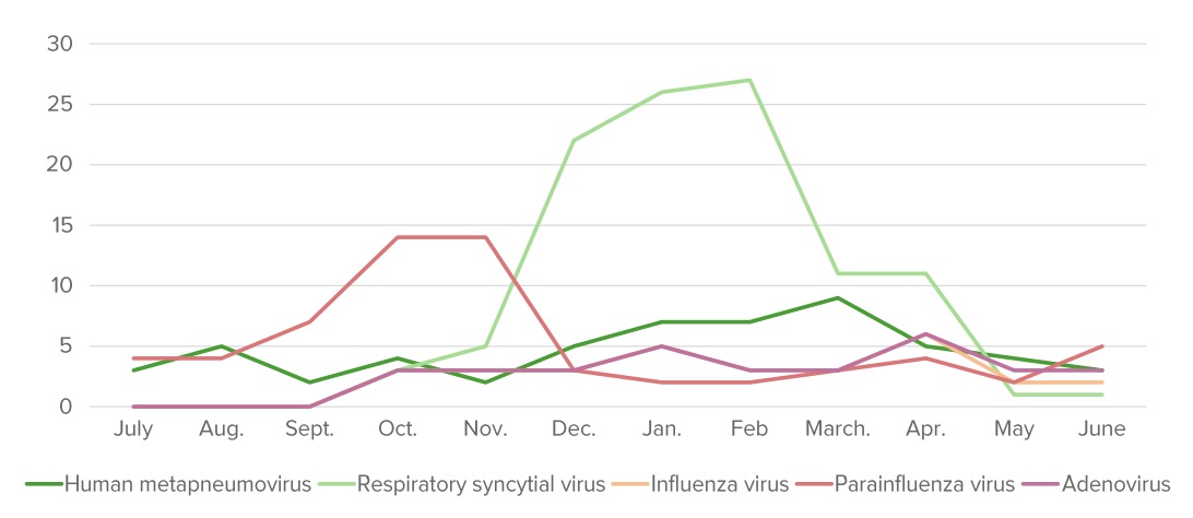
The annual distribution (%) of pathogens that cause bronchiolitis. “Bronchiolitis season” starts around October and finishes around May in the Northern Hemisphere.

Image by Lecturio.

Etiology[5,6]

  • RSV RSV Respiratory syncytial virus (RSV) is an enveloped, single-stranded, linear, negative-sense RNA virus of the family Paramyxoviridae and the genus Orthopneumovirus. Two subtypes (A and B) are present in outbreaks, but type A causes more severe disease. Respiratory syncytial virus causes infections of the lungs and respiratory tract. Respiratory Syncytial Virus: estimated to be ⅓ of cases of bronchiolitis Bronchiolitis Inflammation of the bronchioles. Pediatric Chest Abnormalities
  • Human rhinovirus Rhinovirus Rhinovirus is an acid-labile, positive-sense RNA virus of the Picornavirus family. The virus, which causes the common cold, is most often acquired through the airway via the inhalation of aerosols containing rhinovirus and fomites. Rhinovirus
  • Coronavirus Coronavirus Coronaviruses are a group of related viruses that contain positive-sense, single-stranded RNA. Coronavirus derives its name from “κορώνη korṓnē” in Greek, which translates as “crown,” after the small club-shaped proteins visible as a ring around the viral envelope in electron micrographs. Coronavirus
  • Human bocavirus
  • Less common:
    • Parainfluenza virus Parainfluenza virus Human parainfluenza viruses (HPIVs) are single-stranded, linear, negative-sense RNA viruses of the family Paramyxoviridae and the genus Paramyxovirus. Human parainfluenza viruses are the 2nd most common cause of lower respiratory disease in children, after the respiratory syncytial virus. Parainfluenza Virus
    • Human metapneumovirus Human Metapneumovirus Acute Bronchiolitis
    • Influenza Influenza Influenza viruses are members of the Orthomyxoviridae family and the causative organisms of influenza, a highly contagious febrile respiratory disease. There are 3 primary influenza viruses (A, B, and C) and various subtypes, which are classified based on their virulent surface antigens, hemagglutinin (HA) and neuraminidase (NA). Influenza typically presents with a fever, myalgia, headache, and symptoms of an upper respiratory infection. Influenza Viruses/Influenza virus Virus Viruses are infectious, obligate intracellular parasites composed of a nucleic acid core surrounded by a protein capsid. Viruses can be either naked (non-enveloped) or enveloped. The classification of viruses is complex and based on many factors, including type and structure of the nucleoid and capsid, the presence of an envelope, the replication cycle, and the host range. Virology
    • Adenovirus Adenovirus Adenovirus (member of the family Adenoviridae) is a nonenveloped, double-stranded DNA virus. Adenovirus is transmitted in a variety of ways, and it can have various presentations based on the site of entry. Presentation can include febrile pharyngitis, conjunctivitis, acute respiratory disease, atypical pneumonia, and gastroenteritis. Adenovirus
    • Mycoplasma pneumoniae Mycoplasma pneumoniae Short filamentous organism of the genus mycoplasma, which binds firmly to the cells of the respiratory epithelium. It is one of the etiologic agents of non-viral primary atypical pneumonia in man. Mycoplasma 
    • Pertussis Pertussis Pertussis, or whooping cough, is a potentially life-threatening highly contagious bacterial infection of the respiratory tract caused by Bordetella pertussis. The disease has 3 clinical stages, the second and third of which are characterized by an intense paroxysmal cough, an inspiratory whoop, and post-tussive vomiting. Pertussis (Whooping Cough)
  • In 30% of cases, 2 viruses Viruses Minute infectious agents whose genomes are composed of DNA or RNA, but not both. They are characterized by a lack of independent metabolism and the inability to replicate outside living host cells. Virology are found.
Causes of bronchiolitis

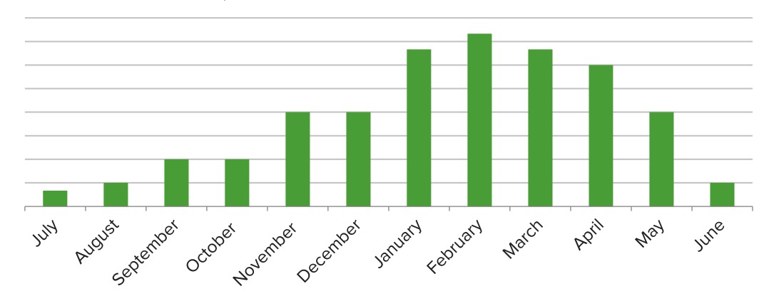
Seasonal variations in the number of cases that can be attributed to the respiratory syncytial virus (RSV): At its peak, RSV accounts for about 1/3 of bronchiolitis cases.

Image by Lecturio.

Pathophysiology and Clinical Presentation

Pathophysiology[3,6,10]

Pathologic changes are noted within 24 hours of contact with a pathogen:

  • Virus Virus Viruses are infectious, obligate intracellular parasites composed of a nucleic acid core surrounded by a protein capsid. Viruses can be either naked (non-enveloped) or enveloped. The classification of viruses is complex and based on many factors, including type and structure of the nucleoid and capsid, the presence of an envelope, the replication cycle, and the host range. Virology enters epithelial cells of terminal bronchioles Bronchioles The small airways branching off the tertiary bronchi. Terminal bronchioles lead into several orders of respiratory bronchioles which in turn lead into alveolar ducts and then into pulmonary alveoli. Bronchial Tree: Anatomy.
  • Inoculation causes inflammation Inflammation Inflammation is a complex set of responses to infection and injury involving leukocytes as the principal cellular mediators in the body’s defense against pathogenic organisms. Inflammation is also seen as a response to tissue injury in the process of wound healing. The 5 cardinal signs of inflammation are pain, heat, redness, swelling, and loss of function. Inflammation edema Edema Edema is a condition in which excess serous fluid accumulates in the body cavity or interstitial space of connective tissues. Edema is a symptom observed in several medical conditions. It can be categorized into 2 types, namely, peripheral (in the extremities) and internal (in an organ or body cavity). Edema, mucus secretion Secretion Coagulation Studies, and epithelium Epithelium The epithelium is a complex of specialized cellular organizations arranged into sheets and lining cavities and covering the surfaces of the body. The cells exhibit polarity, having an apical and a basal pole. Structures important for the epithelial integrity and function involve the basement membrane, the semipermeable sheet on which the cells rest, and interdigitations, as well as cellular junctions. Surface Epithelium: Histology sloughing:
    • Narrowing and obstruction of small airways → atelectasis Atelectasis Atelectasis is the partial or complete collapse of a part of the lung. Atelectasis is almost always a secondary phenomenon from conditions causing bronchial obstruction, external compression, surfactant deficiency, or scarring. Atelectasis and intermittent air trapping
    • ↓ Lung tissue compliance Compliance Distensibility measure of a chamber such as the lungs (lung compliance) or bladder. Compliance is expressed as a change in volume per unit change in pressure. Veins: Histology
    • Diffusion Diffusion The tendency of a gas or solute to pass from a point of higher pressure or concentration to a point of lower pressure or concentration and to distribute itself throughout the available space. Diffusion, especially facilitated diffusion, is a major mechanism of biological transport. Peritoneal Dialysis and Hemodialysis of gases across alveolar/capillary membranes

Clinical presentation[4–6,11]

Symptoms are on a spectrum based on the severity of the disease:

  • Initial 1–3 days (upper respiratory tract symptoms):
  • Peaks on day 3–5, lasts, on average, for 10 days (lower respiratory tract symptoms):
    • Symptoms of bronchiolitis Bronchiolitis Inflammation of the bronchioles. Pediatric Chest Abnormalities coincide with multiple other respiratory conditions; pattern of symptoms can sometimes be more suggestive of this disease:
      • Clinical course may vary depending on preexisting/co-occurring medical conditions, patient age, and severity of illness.[5]
      • Prodrome Prodrome Symptoms that appear 24–48 hours prior to migraine onset. Migraine Headache: 3 days of upper respiratory symptoms 
      • Peak illness: lasts 2 days, lower respiratory symptoms predominant[6]
      • Resolution: Symptoms gradually pass, usually without intervention, but this may take as long as 28 days in nonhospitalized patients Patients Individuals participating in the health care system for the purpose of receiving therapeutic, diagnostic, or preventive procedures. Clinician–Patient Relationship.[5]
      • Variable Variable Variables represent information about something that can change. The design of the measurement scales, or of the methods for obtaining information, will determine the data gathered and the characteristics of that data. As a result, a variable can be qualitative or quantitative, and may be further classified into subgroups. Types of Variables wheezing Wheezing Wheezing is an abnormal breath sound characterized by a whistling noise that can be relatively high-pitched and shrill (more common) or coarse. Wheezing is produced by the movement of air through narrowed or compressed small (intrathoracic) airways. Wheezing is one of the longest-lasting symptoms.
    • Wheezing Wheezing Wheezing is an abnormal breath sound characterized by a whistling noise that can be relatively high-pitched and shrill (more common) or coarse. Wheezing is produced by the movement of air through narrowed or compressed small (intrathoracic) airways. Wheezing and diffuse crackles on lung exam:
      • Differentiated from reactive airway Airway ABCDE Assessment disease (RAD) because wheezing Wheezing Wheezing is an abnormal breath sound characterized by a whistling noise that can be relatively high-pitched and shrill (more common) or coarse. Wheezing is produced by the movement of air through narrowed or compressed small (intrathoracic) airways. Wheezing in RAD is fixed in intensity and position, whereas in bronchiolitis Bronchiolitis Inflammation of the bronchioles. Pediatric Chest Abnormalities it is polyphonic, shifts, and appears and disappears.[4]
    • Fever Fever Fever is defined as a measured body temperature of at least 38°C (100.4°F). Fever is caused by circulating endogenous and/or exogenous pyrogens that increase levels of prostaglandin E2 in the hypothalamus. Fever is commonly associated with chills, rigors, sweating, and flushing of the skin. Fever ≤ 101°F (38.3°C )
    • Shortness of breath Shortness of breath Dyspnea is the subjective sensation of breathing discomfort. Dyspnea is a normal manifestation of heavy physical or psychological exertion, but also may be caused by underlying conditions (both pulmonary and extrapulmonary). Dyspnea
  • Severe cases:

Diagnosis and Management

Diagnosis[1,3,6,7]

  • Diagnosis based on clinical suspicion:
    • Characteristic lower respiratory tract symptoms
    • Patients Patients Individuals participating in the health care system for the purpose of receiving therapeutic, diagnostic, or preventive procedures. Clinician–Patient Relationship < 2 years
    • Presenting during the fall and winter Winter Pityriasis Rosea seasons
  • Routine testing of any kind is discouraged, as it provides no additional therapeutic value.
  • Investigation in severe cases to evaluate comorbid conditions or superimposed infection:[7]
    • Symptoms prompting further investigation:
      • High fever Fever Fever is defined as a measured body temperature of at least 38°C (100.4°F). Fever is caused by circulating endogenous and/or exogenous pyrogens that increase levels of prostaglandin E2 in the hypothalamus. Fever is commonly associated with chills, rigors, sweating, and flushing of the skin. Fever
      • Severe presentation 
      • History of comorbidities Comorbidities The presence of co-existing or additional diseases with reference to an initial diagnosis or with reference to the index condition that is the subject of study. Comorbidity may affect the ability of affected individuals to function and also their survival; it may be used as a prognostic indicator for length of hospital stay, cost factors, and outcome or survival. St. Louis Encephalitis Virus
    • Tests include:
      • Complete blood count (CBC): leukocytosis Leukocytosis A transient increase in the number of leukocytes in a body fluid. West Nile Virus 
      • Chest X-ray X-ray Penetrating electromagnetic radiation emitted when the inner orbital electrons of an atom are excited and release radiant energy. X-ray wavelengths range from 1 pm to 10 nm. Hard x-rays are the higher energy, shorter wavelength x-rays. Soft x-rays or grenz rays are less energetic and longer in wavelength. The short wavelength end of the x-ray spectrum overlaps the gamma rays wavelength range. The distinction between gamma rays and x-rays is based on their radiation source. Pulmonary Function Tests: hyperinflation Hyperinflation Imaging of the Lungs and Pleura with atelectasis Atelectasis Atelectasis is the partial or complete collapse of a part of the lung. Atelectasis is almost always a secondary phenomenon from conditions causing bronchial obstruction, external compression, surfactant deficiency, or scarring. Atelectasis 
      • RSV RSV Respiratory syncytial virus (RSV) is an enveloped, single-stranded, linear, negative-sense RNA virus of the family Paramyxoviridae and the genus Orthopneumovirus. Two subtypes (A and B) are present in outbreaks, but type A causes more severe disease. Respiratory syncytial virus causes infections of the lungs and respiratory tract. Respiratory Syncytial Virus testing on aspirate

Risk stratification[3]

Risk stratification at admission is important to determine the appropriate level of care. 

Management[1,3,6,8,11]

Management depends on severity:

  • Mild-to-moderate cases → supportive:
    • Caregiver education on upright positioning during sleeping and feeds
    • Use of cool-mist humidifier and antipyretic Antipyretic Acetaminophen
    • Bulb suctioning of oral and nasal secretions
    • Maintain hydration and feeding
    • Follow-up if patient worsens
  • Severe (< 28 days old, apnea, respiratory distress, lethargic):
    • Admit for inpatient care
    • Humidified oxygen and nebulized hypertonic saline Hypertonic saline Hypertonic sodium chloride solution. A solution having an osmotic pressure greater than that of physiologic salt solution (0. 9 g NaCl in 100 ml purified water). Hyponatremia
    • Suctioning of secretions
    • IV hydration Iv Hydration Crush Syndrome
    • No evidence to support use of albuterol Albuterol A short-acting beta-2 adrenergic agonist that is primarily used as a bronchodilator agent to treat asthma. Sympathomimetic Drugs, epinephrine Epinephrine The active sympathomimetic hormone from the adrenal medulla. It stimulates both the alpha- and beta- adrenergic systems, causes systemic vasoconstriction and gastrointestinal relaxation, stimulates the heart, and dilates bronchi and cerebral vessels. Sympathomimetic Drugs, or corticosteroids Corticosteroids Chorioretinitis
  • Complications, such as pneumonia Pneumonia Pneumonia or pulmonary inflammation is an acute or chronic inflammation of lung tissue. Causes include infection with bacteria, viruses, or fungi. In more rare cases, pneumonia can also be caused through toxic triggers through inhalation of toxic substances, immunological processes, or in the course of radiotherapy. Pneumonia and respiratory failure Respiratory failure Respiratory failure is a syndrome that develops when the respiratory system is unable to maintain oxygenation and/or ventilation. Respiratory failure may be acute or chronic and is classified as hypoxemic, hypercapnic, or a combination of the two. Respiratory Failure, can occur and must be managed accordingly.
  • The following have not been proven to improve symptoms or to shorten resolution time:[3,6,11]
    • Alpha- and beta-adrenergic agents ( bronchodilators Bronchodilators Asthma Drugs)
    • Glucocorticoids Glucocorticoids Glucocorticoids are a class within the corticosteroid family. Glucocorticoids are chemically and functionally similar to endogenous cortisol. There are a wide array of indications, which primarily benefit from the antiinflammatory and immunosuppressive effects of this class of drugs. Glucocorticoids (reduce airway Airway ABCDE Assessment inflammation Inflammation Inflammation is a complex set of responses to infection and injury involving leukocytes as the principal cellular mediators in the body’s defense against pathogenic organisms. Inflammation is also seen as a response to tissue injury in the process of wound healing. The 5 cardinal signs of inflammation are pain, heat, redness, swelling, and loss of function. Inflammation)
  • The following are not recommended:[6,11]
    • Antimicrobial therapy (unless concurrent bacterial infection is present)
    • Chest physiotherapy Physiotherapy Spinal Stenosis (unless difficulty clearing secretions is present)
  • Discharge: bronchiolitis Bronchiolitis Inflammation of the bronchioles. Pediatric Chest Abnormalities is a relatively benign Benign Fibroadenoma, self-limited condition. It is preferable to treat patients Patients Individuals participating in the health care system for the purpose of receiving therapeutic, diagnostic, or preventive procedures. Clinician–Patient Relationship at home once appropriate discharge criteria are met MET Preoperative Care. Although there are no universally recognized discharge criteria, the factors that should be considered are:
    • Ability to maintain adequate oral intake, especially hydration
    • Oxygen saturation Oxygen Saturation Basic Procedures > 90% while awake
    • Minimal/mild work of breathing Work of breathing Respiratory muscle contraction during inhalation. The work is accomplished in three phases: lung compliance work, that required to expand the lungs against its elastic forces; tissue resistance work, that required to overcome the viscosity of the lung and chest wall structures; and airway resistance work, that required to overcome airway resistance during the movement of air into the lungs. Work of breathing does not refer to expiration, which is entirely a passive process caused by elastic recoil of the lung and chest cage. Pulmonary Examination 
    • Ability of parents to monitor the child for worsening condition and ability to access follow-up care

Prevention and Prognosis

Prevention[3,9,11]

  • Encourage breastfeeding Breastfeeding Breastfeeding is often the primary source of nutrition for the newborn. During pregnancy, hormonal stimulation causes the number and size of mammary glands in the breast to significantly increase. After delivery, prolactin stimulates milk production, while oxytocin stimulates milk expulsion through the lactiferous ducts, where it is sucked out through the nipple by the infant. Breastfeeding.
  • Stop exposure to second-hand smoke.
  • Hand hygiene Hand hygiene Practices involved in preventing the transmission of diseases by hand. Surgical Infections is a means of reducing all forms of disease transmission.
  • Palivizumab Palivizumab A humanized monoclonal antibody and antiviral agent that is used to prevent respiratory syncytial virus infections in high risk pediatric patients. Respiratory Syncytial Virus (monoclonal antibody, which inhibits RSV RSV Respiratory syncytial virus (RSV) is an enveloped, single-stranded, linear, negative-sense RNA virus of the family Paramyxoviridae and the genus Orthopneumovirus. Two subtypes (A and B) are present in outbreaks, but type A causes more severe disease. Respiratory syncytial virus causes infections of the lungs and respiratory tract. Respiratory Syncytial Virus fusion) for prevention of severe RSV RSV Respiratory syncytial virus (RSV) is an enveloped, single-stranded, linear, negative-sense RNA virus of the family Paramyxoviridae and the genus Orthopneumovirus. Two subtypes (A and B) are present in outbreaks, but type A causes more severe disease. Respiratory syncytial virus causes infections of the lungs and respiratory tract. Respiratory Syncytial Virus lower respiratory tract disease in high-risk patients Patients Individuals participating in the health care system for the purpose of receiving therapeutic, diagnostic, or preventive procedures. Clinician–Patient Relationship:
    • Meeting American Academy of Pediatrics (AAP) criteria for preterm infants with neonatal chronic lung disease:
    • Infants under 1 year at onset of RSV RSV Respiratory syncytial virus (RSV) is an enveloped, single-stranded, linear, negative-sense RNA virus of the family Paramyxoviridae and the genus Orthopneumovirus. Two subtypes (A and B) are present in outbreaks, but type A causes more severe disease. Respiratory syncytial virus causes infections of the lungs and respiratory tract. Respiratory Syncytial Virus season OR under 2 years but requiring medication (e.g., oxygen) for neonatal chronic lung disease
    • Consider prophylaxis Prophylaxis Cephalosporins in the following:
      • Preterm infants born before < 29 weeks of gestation
      • Hemodynamically significant congenital Congenital Chorioretinitis heart disease (< 12 months of age)
      • Children with anatomic lung abnormalities or neuromuscular disorder (< 12 months of age)
      • Immunocompromised immunocompromised A human or animal whose immunologic mechanism is deficient because of an immunodeficiency disorder or other disease or as the result of the administration of immunosuppressive drugs or radiation. Gastroenteritis children (< 24 months of age)
    • Dose:
      • 15 mg/kg once every 30 days (during RSV RSV Respiratory syncytial virus (RSV) is an enveloped, single-stranded, linear, negative-sense RNA virus of the family Paramyxoviridae and the genus Orthopneumovirus. Two subtypes (A and B) are present in outbreaks, but type A causes more severe disease. Respiratory syncytial virus causes infections of the lungs and respiratory tract. Respiratory Syncytial Virus season), administered intramuscularly
      • Maximum of 5 doses per season
    • Discontinue palivizumab Palivizumab A humanized monoclonal antibody and antiviral agent that is used to prevent respiratory syncytial virus infections in high risk pediatric patients. Respiratory Syncytial Virus if breakthrough infection occurs.

Prognosis Prognosis A prediction of the probable outcome of a disease based on a individual’s condition and the usual course of the disease as seen in similar situations. Non-Hodgkin Lymphomas[1]

Differential Diagnosis

  • Asthma Asthma Asthma is a chronic inflammatory respiratory condition characterized by bronchial hyperresponsiveness and airflow obstruction. The disease is believed to result from the complex interaction of host and environmental factors that increase disease predisposition, with inflammation causing symptoms and structural changes. Patients typically present with wheezing, cough, and dyspnea. Asthma: an inflammatory airway Airway ABCDE Assessment disease that leads to bronchial luminal narrowing and thus obstruction of airflow. Characterized by cough, wheezes, and dyspnea Dyspnea Dyspnea is the subjective sensation of breathing discomfort. Dyspnea is a normal manifestation of heavy physical or psychological exertion, but also may be caused by underlying conditions (both pulmonary and extrapulmonary). Dyspnea. Asthma Asthma Asthma is a chronic inflammatory respiratory condition characterized by bronchial hyperresponsiveness and airflow obstruction. The disease is believed to result from the complex interaction of host and environmental factors that increase disease predisposition, with inflammation causing symptoms and structural changes. Patients typically present with wheezing, cough, and dyspnea. Asthma is usually diagnosed after the age of 2 and has known triggers. The reversibility of symptoms with bronchodilator use differentiates this condition from bronchiolitis Bronchiolitis Inflammation of the bronchioles. Pediatric Chest Abnormalities.
  • Pneumonia Pneumonia Pneumonia or pulmonary inflammation is an acute or chronic inflammation of lung tissue. Causes include infection with bacteria, viruses, or fungi. In more rare cases, pneumonia can also be caused through toxic triggers through inhalation of toxic substances, immunological processes, or in the course of radiotherapy. Pneumonia: infection of the lower respiratory tract that results in symptoms similar to bronchiolitis Bronchiolitis Inflammation of the bronchioles. Pediatric Chest Abnormalities in infants. Patients Patients Individuals participating in the health care system for the purpose of receiving therapeutic, diagnostic, or preventive procedures. Clinician–Patient Relationship with bronchiolitis Bronchiolitis Inflammation of the bronchioles. Pediatric Chest Abnormalities may develop atypical pneumonia Atypical pneumonia Mycoplasma due to superimposed bacterial infections Infections Invasion of the host organism by microorganisms or their toxins or by parasites that can cause pathological conditions or diseases. Chronic Granulomatous Disease or worsened viral infections Infections Invasion of the host organism by microorganisms or their toxins or by parasites that can cause pathological conditions or diseases. Chronic Granulomatous Disease. Chest X-ray X-ray Penetrating electromagnetic radiation emitted when the inner orbital electrons of an atom are excited and release radiant energy. X-ray wavelengths range from 1 pm to 10 nm. Hard x-rays are the higher energy, shorter wavelength x-rays. Soft x-rays or grenz rays are less energetic and longer in wavelength. The short wavelength end of the x-ray spectrum overlaps the gamma rays wavelength range. The distinction between gamma rays and x-rays is based on their radiation source. Pulmonary Function Tests findings typically lag behind presentation in these cases, so the diagnosis is made when a patient has sustained hypoxia Hypoxia Sub-optimal oxygen levels in the ambient air of living organisms. Ischemic Cell Damage with increased oxygen supplementation, consolidations on lung exam, and increased work of breathing Work of breathing Respiratory muscle contraction during inhalation. The work is accomplished in three phases: lung compliance work, that required to expand the lungs against its elastic forces; tissue resistance work, that required to overcome the viscosity of the lung and chest wall structures; and airway resistance work, that required to overcome airway resistance during the movement of air into the lungs. Work of breathing does not refer to expiration, which is entirely a passive process caused by elastic recoil of the lung and chest cage. Pulmonary Examination.
  • Gastroesophageal reflux disease Gastroesophageal Reflux Disease Gastroesophageal reflux disease (GERD) occurs when the stomach acid frequently flows back into the esophagus. This backwash (acid reflux) can irritate the lining of the esophagus, causing symptoms such as retrosternal burning pain (heartburn). Gastroesophageal Reflux Disease (GERD) ( GERD GERD Gastroesophageal reflux disease (GERD) occurs when the stomach acid frequently flows back into the esophagus. This backwash (acid reflux) can irritate the lining of the esophagus, causing symptoms such as retrosternal burning pain (heartburn). Gastroesophageal Reflux Disease (GERD)): Acid reflux into the esophagus Esophagus The esophagus is a muscular tube-shaped organ of around 25 centimeters in length that connects the pharynx to the stomach. The organ extends from approximately the 6th cervical vertebra to the 11th thoracic vertebra and can be divided grossly into 3 parts: the cervical part, the thoracic part, and the abdominal part. Esophagus: Anatomy in infancy and childhood may present with intermittent cough and wheezing Wheezing Wheezing is an abnormal breath sound characterized by a whistling noise that can be relatively high-pitched and shrill (more common) or coarse. Wheezing is produced by the movement of air through narrowed or compressed small (intrathoracic) airways. Wheezing. May also be accompanied by a lack of weight gain and increased irritability. Gastroesophageal reflux disease Gastroesophageal Reflux Disease Gastroesophageal reflux disease (GERD) occurs when the stomach acid frequently flows back into the esophagus. This backwash (acid reflux) can irritate the lining of the esophagus, causing symptoms such as retrosternal burning pain (heartburn). Gastroesophageal Reflux Disease (GERD) can be differentiated from bronchiolitis Bronchiolitis Inflammation of the bronchioles. Pediatric Chest Abnormalities if symptoms correlate to feeding routine and not seasonality. Bronchiolitis Bronchiolitis Inflammation of the bronchioles. Pediatric Chest Abnormalities usually presents with a history of upper respiratory symptoms prior to wheezing Wheezing Wheezing is an abnormal breath sound characterized by a whistling noise that can be relatively high-pitched and shrill (more common) or coarse. Wheezing is produced by the movement of air through narrowed or compressed small (intrathoracic) airways. Wheezing.

References

  1. Ralston, S. L., Lieberthal, A. S., Meissner, H. C., Alverson, B. K., Baley, J. E., Gadomski, A. M., Johnson, D. W., Light, M. J., Maraqa, N. F., Mendonca, E. A., Phelan, K. J., Zorc, J. J., Stanko-Lopp, D., Brown, M. A., Nathanson, I., Rosenblum, E., Sayles, S., 3rd, Hernandez-Cancio, S., & American Academy of Pediatrics (2014). Clinical practice guideline: the diagnosis, management, and prevention of bronchiolitis. Pediatrics, 134(5), e1474–e1502. https://doi.org/10.1542/peds.2014-2742
  2. Fujiogi, M., Goto, T., Yasunaga, H., Fujishiro, J., Mansbach, J. M., Camargo, C. A., Jr., Hasegawa, K. (2019). Trends in bronchiolitis hospitalizations in the United States: 2000-2016. Pediatrics, 144(6), e20192614. https://doi.org/10.1542/peds.2019-2614
  3. Teshome, G., Gattu, R., Brown, R. (2013). Acute bronchiolitis. Pediatric Clinics of North America, 60(5), 1019–1034. doi://dx.doi.org/10.1016/j.pcl.2013.06.005 https://www.sciencedirect.com/science/article/abs/pii/S0031395513000813?via%3Dihub
  4. Kliegman, R. M. et al. (2020). Wheezing, bronchiolitis, and bronchitis. Nelson Textbook of Pediatrics (pp. 2217.e1–2221.e1). https://www.clinicalkey.es/#!/content/3-s2.0-B9780323529501004181
  5. Mansbach, J. M., Piedra, P. A., Teach, S. J., Sullivan, A. F., Forgey, T., Clark, S., Espinola, J. A., Camargo, C. A., Jr., MARC-30 Investigators. (2012). Prospective multicenter study of viral etiology and hospital length of stay in children with severe bronchiolitis. Archives of Pediatrics & Adolescent Medicine, 166(8), 700–706. https://doi.org/10.1001/archpediatrics.2011.1669 
  6. Florin, T. A., Plint, A. C., Zorc, J. J. (2017). Viral bronchiolitis. Lancet, 389(10065), 211–224. https://doi.org/10.1016/S0140-6736(16)30951-5  
  7. Bordley, W. C., Viswanathan, M., King, V. J., Sutton, S. F., Jackman, A. M., Sterling, L., Lohr, K. N. (2004). Diagnosis and testing in bronchiolitis: a systematic review. Archives of Pediatrics & Adolescent Medicine, 158(2), 119–126. https://doi.org/10.1001/archpedi.158.2.119 
  8. Thompson, M., Vodicka, T. A., Blair, P. S., Buckley, D. I., Heneghan, C., Hay, A. D., TARGET Programme Team. (2013). Duration of symptoms of respiratory tract infections in children: systematic review. BMJ, 347, f7027. https://doi.org/10.1136/bmj.f7027
  9. Meissner, H. C., Long, S. S., American Academy of Pediatrics Committee on Infectious Diseases and Committee on Fetus and Newborn. (2003). Revised indications for the use of palivizumab and respiratory syncytial virus immune globulin intravenous for the prevention of respiratory syncytial virus infections. Pediatrics, 112(6 Pt 1), 1447–1452. https://doi.org/10.1542/peds.112.6.1447 
  10. Justice, N. A., Le, J. K. (2020). Bronchiolitis. StatPearls. https://www.ncbi.nlm.nih.gov/books/NBK441959/
  11. American Academy of Pediatrics. (2018). Respiratory syncytial virus. In Kimberlin, D. W., Brady, M. T., Jackson, M. A., Long, S. S. (Eds.), Red Book: 2018 Report of the Committee on Infectious Diseases (31st ed., pp. 682–692). Itasca, IL: American Academy of Pediatrics.

Create your free account or log in to continue reading!

Sign up now and get free access to Lecturio with concept pages, medical videos, and questions for your medical education.

User Reviews

Details