The gallbladder is a pear-shaped sac, located directly beneath the liverLiverThe liver is the largest gland in the human body. The liver is found in the superior right quadrant of the abdomen and weighs approximately 1.5 kilograms. Its main functions are detoxification, metabolism, nutrient storage (e.g., iron and vitamins), synthesis of coagulation factors, formation of bile, filtration, and storage of blood. Liver: Anatomy, that sits on top of the superior part of the duodenumDuodenumThe shortest and widest portion of the small intestine adjacent to the pylorus of the stomach. It is named for having the length equal to about the width of 12 fingers.Small Intestine: Anatomy. The primary functions of the gallbladder include concentrating and storing up to 50 mL of bile. Bile is secreted by hepatocytesHepatocytesThe main structural component of the liver. They are specialized epithelial cells that are organized into interconnected plates called lobules.Liver: Anatomy into thin channelsChannelsThe Cell: Cell Membrane called canaliculi. These canaliculi lead into slightly larger interlobular bile ductules, which are part of the portal triads at the “corners” of hepatic lobulesLobulesBreasts: Anatomy. The bile leaves the liverLiverThe liver is the largest gland in the human body. The liver is found in the superior right quadrant of the abdomen and weighs approximately 1.5 kilograms. Its main functions are detoxification, metabolism, nutrient storage (e.g., iron and vitamins), synthesis of coagulation factors, formation of bile, filtration, and storage of blood. Liver: Anatomy via the right and left hepatic ducts, which join together to form the common hepatic duct. The common hepatic duct joins with the cysticCysticFibrocystic Change duct to form the common bile duct, which empties into the small intestineSmall intestineThe small intestine is the longest part of the GI tract, extending from the pyloric orifice of the stomach to the ileocecal junction. The small intestine is the major organ responsible for chemical digestion and absorption of nutrients. It is divided into 3 segments: the duodenum, the jejunum, and the ileum. Small Intestine: Anatomy. If the sphincters leading into the intestines are closed, bile will travel via the cysticCysticFibrocystic Change duct into the gallbladder for storage.
Hepatic diverticulumHepatic diverticulumDevelopment of the Abdominal Organs (sometimes called the liverLiverThe liver is the largest gland in the human body. The liver is found in the superior right quadrant of the abdomen and weighs approximately 1.5 kilograms. Its main functions are detoxification, metabolism, nutrient storage (e.g., iron and vitamins), synthesis of coagulation factors, formation of bile, filtration, and storage of blood. Liver: Anatomy bud):
LiverLiverThe liver is the largest gland in the human body. The liver is found in the superior right quadrant of the abdomen and weighs approximately 1.5 kilograms. Its main functions are detoxification, metabolism, nutrient storage (e.g., iron and vitamins), synthesis of coagulation factors, formation of bile, filtration, and storage of blood. Liver: Anatomy
Immediately under the right lobe of the liverLiverThe liver is the largest gland in the human body. The liver is found in the superior right quadrant of the abdomen and weighs approximately 1.5 kilograms. Its main functions are detoxification, metabolism, nutrient storage (e.g., iron and vitamins), synthesis of coagulation factors, formation of bile, filtration, and storage of blood. Liver: Anatomy in the gallbladder fossa
Connected to the visceral surface of the liverLiverThe liver is the largest gland in the human body. The liver is found in the superior right quadrant of the abdomen and weighs approximately 1.5 kilograms. Its main functions are detoxification, metabolism, nutrient storage (e.g., iron and vitamins), synthesis of coagulation factors, formation of bile, filtration, and storage of blood. Liver: Anatomy via the cysticCysticFibrocystic Change plate
Sits on top of the superior part of the duodenumDuodenumThe shortest and widest portion of the small intestine adjacent to the pylorus of the stomach. It is named for having the length equal to about the width of 12 fingers.Small Intestine: Anatomy
Anatomic relationships of the pancreas to surrounding organs: Note that the liver and stomach are light gray and that the intestines have been removed completely in order to allow better visualization of this posterior organ.
FundusFundusThe superior portion of the body of the stomach above the level of the cardiac notch.Stomach: Anatomy:
Rounded base of the organ
Usually projects slightly beyond the inferior margin of the liverLiverThe liver is the largest gland in the human body. The liver is found in the superior right quadrant of the abdomen and weighs approximately 1.5 kilograms. Its main functions are detoxification, metabolism, nutrient storage (e.g., iron and vitamins), synthesis of coagulation factors, formation of bile, filtration, and storage of blood. Liver: Anatomy
Body:
Main portion of the organ
In direct contact with:
Visceral surface of the liverLiverThe liver is the largest gland in the human body. The liver is found in the superior right quadrant of the abdomen and weighs approximately 1.5 kilograms. Its main functions are detoxification, metabolism, nutrient storage (e.g., iron and vitamins), synthesis of coagulation factors, formation of bile, filtration, and storage of blood. Liver: Anatomy
Superior part of the duodenumDuodenumThe shortest and widest portion of the small intestine adjacent to the pylorus of the stomach. It is named for having the length equal to about the width of 12 fingers.Small Intestine: Anatomy
NeckNeckThe part of a human or animal body connecting the head to the rest of the body.Peritonsillar Abscess:
SpiralSpiralComputed tomography where there is continuous x-ray exposure to the patient while being transported in a spiral or helical pattern through the beam of irradiation. This provides improved three-dimensional contrast and spatial resolution compared to conventional computed tomography, where data is obtained and computed from individual sequential exposures.Computed Tomography (CT) valves (of Heister):
Keep the duct open so bile can easily be diverted into the gallbladder when distal sphincters are closed
Helps prevent dumping of bile into the intestines during sudden increases in intraabdominal pressure (e.g., coughing) when sphincters are closed
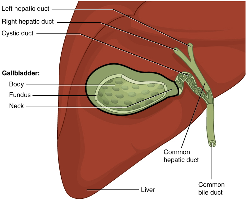
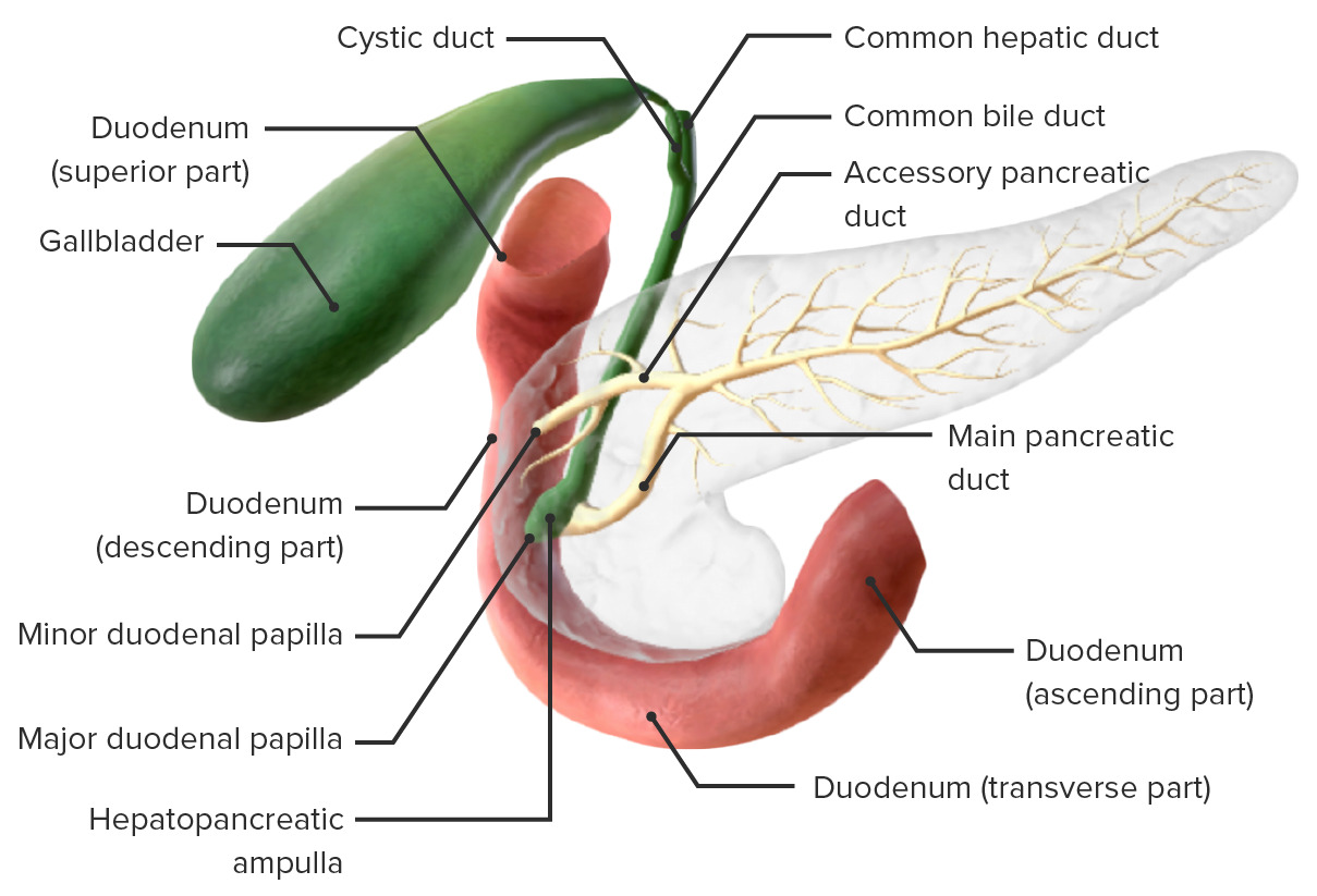
Location and basic anatomy of the gallbladder and major biliary ducts
Image: “The gallbladder stores and concentrates bile, and releases it into the two-way cystic duct when it is needed by the small intestine” by OpenStax College. License: CC BY 4.0
Gross Anatomy of the Biliary Tree
Bile, a digestive fluid produced and secreted by hepatocytesHepatocytesThe main structural component of the liver. They are specialized epithelial cells that are organized into interconnected plates called lobules.Liver: Anatomy in the liverLiverThe liver is the largest gland in the human body. The liver is found in the superior right quadrant of the abdomen and weighs approximately 1.5 kilograms. Its main functions are detoxification, metabolism, nutrient storage (e.g., iron and vitamins), synthesis of coagulation factors, formation of bile, filtration, and storage of blood. Liver: Anatomy, is transported to the gallbladder and small intestines by a series of branching bile ducts known collectively as the biliary tree.
Intrahepatic bile ducts
Ducts that originate within the liverLiverThe liver is the largest gland in the human body. The liver is found in the superior right quadrant of the abdomen and weighs approximately 1.5 kilograms. Its main functions are detoxification, metabolism, nutrient storage (e.g., iron and vitamins), synthesis of coagulation factors, formation of bile, filtration, and storage of blood. Liver: Anatomyitself are known as intrahepatic bile ducts and can be divided into:
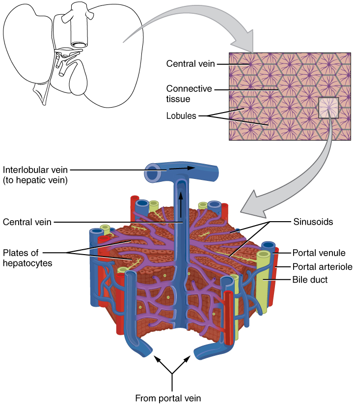
Bile canaliculi:
Narrow channelsChannelsThe Cell: Cell Membrane located between sheets of hepatocytesHepatocytesThe main structural component of the liver. They are specialized epithelial cells that are organized into interconnected plates called lobules.Liver: Anatomy within hepatic lobulesLobulesBreasts: Anatomy (hexagon-shaped functional units in the liverLiverThe liver is the largest gland in the human body. The liver is found in the superior right quadrant of the abdomen and weighs approximately 1.5 kilograms. Its main functions are detoxification, metabolism, nutrient storage (e.g., iron and vitamins), synthesis of coagulation factors, formation of bile, filtration, and storage of blood. Liver: Anatomy)
Drain into bile ductules at the exterior of the hepatic lobule
Bile ductules (also called interlobular bile ducts):
Part of the portal triads located at the “corners” of each hepatic lobule, which contain:
Hepatic arteriolesArteriolesThe smallest divisions of the arteries located between the muscular arteries and the capillaries.Arteries: Histology
Portal venulesVenulesThe minute vessels that collect blood from the capillary plexuses and join together to form veins.Veins: Histology
Bile ductules
Ultimately drain into the right or left hepatic ducts
Hepatic ducts:
Intrahepatic and extrahepatic segments
Right hepatic duct: drains the right lobe of the liverLiverThe liver is the largest gland in the human body. The liver is found in the superior right quadrant of the abdomen and weighs approximately 1.5 kilograms. Its main functions are detoxification, metabolism, nutrient storage (e.g., iron and vitamins), synthesis of coagulation factors, formation of bile, filtration, and storage of blood. Liver: Anatomy
Left hepatic duct: drains the left lobe of the liverLiverThe liver is the largest gland in the human body. The liver is found in the superior right quadrant of the abdomen and weighs approximately 1.5 kilograms. Its main functions are detoxification, metabolism, nutrient storage (e.g., iron and vitamins), synthesis of coagulation factors, formation of bile, filtration, and storage of blood. Liver: Anatomy
Microscopic anatomy of the liver: Bile is produced in the hepatocytes and secreted into the bile canaliculi. These canaliculi drain into the bile ductules (located next to the portal arterioles and venules). The bile ductules ultimately drain into the right and left hepatic ducts.
Image: “The liver receives oxygenated blood from the hepatic artery and nutrient-rich deoxygenated blood from the hepatic portal vein” by OpenStax College. License: CC BY 4.0
Extrahepatic bile ducts
Extrahepatic bile ducts are located outside the liverLiverThe liver is the largest gland in the human body. The liver is found in the superior right quadrant of the abdomen and weighs approximately 1.5 kilograms. Its main functions are detoxification, metabolism, nutrient storage (e.g., iron and vitamins), synthesis of coagulation factors, formation of bile, filtration, and storage of blood. Liver: Anatomy and are continuous with the intrahepatic bile ducts. There are many normal anatomic variants. These ducts include:
Extrahepatic segments of theright and left hepatic ducts
Common hepatic duct: formed via the combination of the right and left hepatic ducts
Formed via the combination of the common hepatic duct and the cysticCysticFibrocystic Change duct
Size:
Approximately 6 mm in diameter
Approximately 6–8 cm in length
Runs posterior to the duodenumDuodenumThe shortest and widest portion of the small intestine adjacent to the pylorus of the stomach. It is named for having the length equal to about the width of 12 fingers.Small Intestine: Anatomy
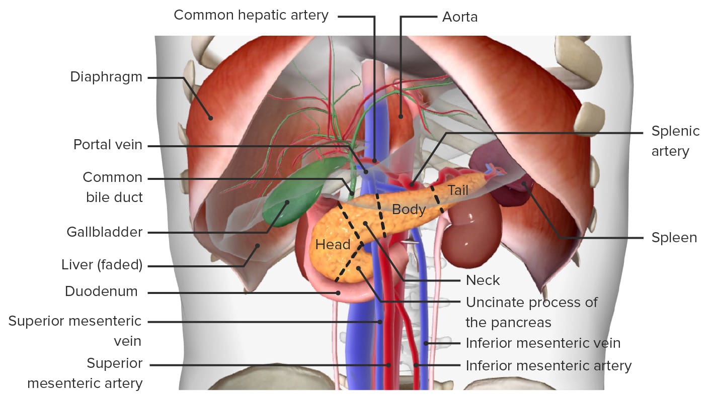
Runs through the head of the pancreasPancreasThe pancreas lies mostly posterior to the stomach and extends across the posterior abdominal wall from the duodenum on the right to the spleen on the left. This organ has both exocrine and endocrine tissue. Pancreas: Anatomy, where it joins with the main pancreatic ductMain pancreatic ductPancreas: Anatomy to form a swellingSwellingInflammation called the hepatopancreatic ampulla (also known as the ampulla of Vater)
Located within the head of the pancreasPancreasThe pancreas lies mostly posterior to the stomach and extends across the posterior abdominal wall from the duodenum on the right to the spleen on the left. This organ has both exocrine and endocrine tissue. Pancreas: Anatomy, just prior to its insertion into the small intestineSmall intestineThe small intestine is the longest part of the GI tract, extending from the pyloric orifice of the stomach to the ileocecal junction. The small intestine is the major organ responsible for chemical digestion and absorption of nutrients. It is divided into 3 segments: the duodenum, the jejunum, and the ileum. Small Intestine: Anatomy
Empties into the descending part of the duodenumDuodenumThe shortest and widest portion of the small intestine adjacent to the pylorus of the stomach. It is named for having the length equal to about the width of 12 fingers.Small Intestine: Anatomy via an opening called the major duodenal papilla
Contains the hepatopancreatic sphincter (also known as the sphincter of Oddi): controls release of bile and pancreatic juicePancreatic JuiceThe fluid containing digestive enzymes secreted by the pancreas in response to food in the duodenum.Pancreas: Anatomy
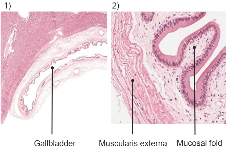
There are 3 primary layers in the wall of the gallbladder (from internal to external):
Mucosa:
Contains ridges/folds that flatten when gallbladder fills/distends
Simple columnar epithelial cells:
Line the lumen of the gallbladder
Contain Na+-ATP pumps to help concentrate bile
Connected to one another via tight junctionsTight junctionsCell-cell junctions that seal adjacent epithelial cells together, preventing the passage of most dissolved molecules from one side of the epithelial sheet to the other.The Cell: Cell Junctions
Intercellular space: clear spaces between epithelial cells where water is being absorbed (unable to move back into the lumen due to tight junctionsTight junctionsCell-cell junctions that seal adjacent epithelial cells together, preventing the passage of most dissolved molecules from one side of the epithelial sheet to the other.The Cell: Cell Junctions)
Collagenous connective tissueConnective tissueConnective tissues originate from embryonic mesenchyme and are present throughout the body except inside the brain and spinal cord. The main function of connective tissues is to provide structural support to organs. Connective tissues consist of cells and an extracellular matrix.Connective Tissue: Histology
Connective tissueConnective tissueConnective tissues originate from embryonic mesenchyme and are present throughout the body except inside the brain and spinal cord. The main function of connective tissues is to provide structural support to organs. Connective tissues consist of cells and an extracellular matrix.Connective Tissue: Histology layer
Merges with the Glisson capsuleCapsuleAn envelope of loose gel surrounding a bacterial cell which is associated with the virulence of pathogenic bacteria. Some capsules have a well-defined border, whereas others form a slime layer that trails off into the medium. Most capsules consist of relatively simple polysaccharides but there are some bacteria whose capsules are made of polypeptides.Bacteroides of the liverLiverThe liver is the largest gland in the human body. The liver is found in the superior right quadrant of the abdomen and weighs approximately 1.5 kilograms. Its main functions are detoxification, metabolism, nutrient storage (e.g., iron and vitamins), synthesis of coagulation factors, formation of bile, filtration, and storage of blood. Liver: Anatomy
Differences between gallbladder wall and other intestinal lumens:
No muscularis mucosa (within the mucosal layer)
No submucosal layer
Muscularis externa is much thinner
Bile ducts have the same structure as the gallbladder (except bile canaliculi lumen, which are lined by the apical poles of hepatocytesHepatocytesThe main structural component of the liver. They are specialized epithelial cells that are organized into interconnected plates called lobules.Liver: Anatomy, no epitheliumEpitheliumThe epithelium is a complex of specialized cellular organizations arranged into sheets and lining cavities and covering the surfaces of the body. The cells exhibit polarity, having an apical and a basal pole. Structures important for the epithelial integrity and function involve the basement membrane, the semipermeable sheet on which the cells rest, and interdigitations, as well as cellular junctions. Surface Epithelium: Histology)
1) Histologic slide depicting the 3 layers of the gallbladder, in addition to the lumen and neighboring hepatic tissue 2) Histologic slide depicting the mucosal folds and ridges, as well as the thin layer of smooth muscle found in the gallbladder
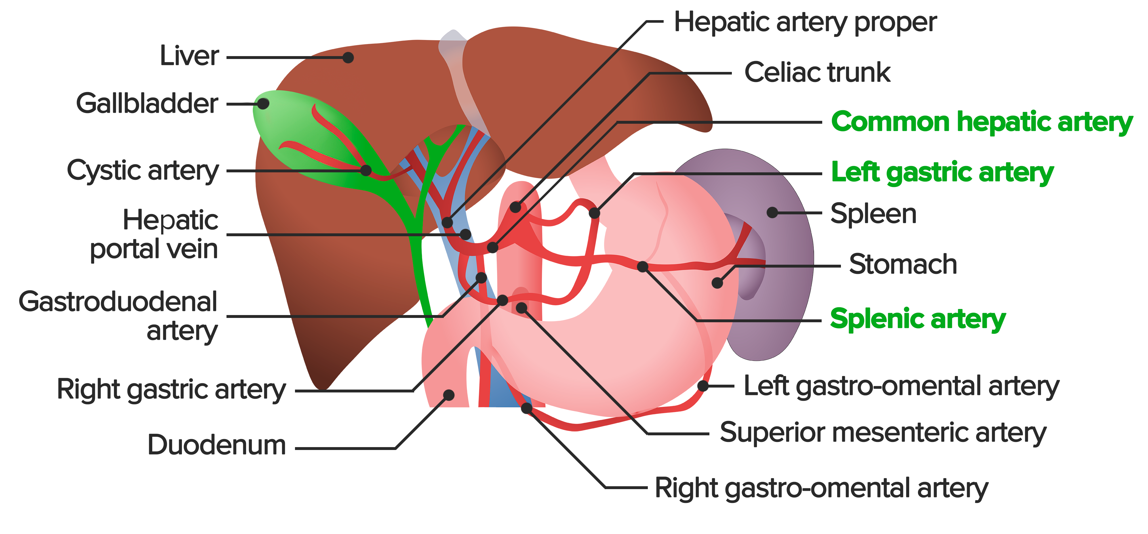
Gallbladder: CysticCysticFibrocystic Change artery, which most commonly arises from the right hepatic arteryHepatic arteryA branch of the celiac artery that distributes to the stomach, pancreas, duodenum, liver, gallbladder, and greater omentum.Liver: Anatomy
Hepatic ducts: branches of the hepatic arteryHepatic arteryA branch of the celiac artery that distributes to the stomach, pancreas, duodenum, liver, gallbladder, and greater omentum.Liver: Anatomy
NeckNeckThe part of a human or animal body connecting the head to the rest of the body.Peritonsillar Abscess and cysticCysticFibrocystic Change duct: cysticCysticFibrocystic ChangeveinsVeinsVeins are tubular collections of cells, which transport deoxygenated blood and waste from the capillary beds back to the heart. Veins are classified into 3 types: small veins/venules, medium veins, and large veins. Each type contains 3 primary layers: tunica intima, tunica media, and tunica adventitia. Veins: Histology → portal veinPortal veinA short thick vein formed by union of the superior mesenteric vein and the splenic vein.Liver: Anatomy
Hepatic and common bile duct: posterior superior pancreaticoduodenal vein → portal veinPortal veinA short thick vein formed by union of the superior mesenteric vein and the splenic vein.Liver: Anatomy
Overview of the abdominal arterial blood supply: The celiac trunk is the 1st major branch of the abdominal aorta. The trunk supplies the liver, stomach, spleen, pancreas and parts of the esophagus and duodenum with oxygenated blood. The celiac trunk gives off the left gastric artery, splenic artery, and the common hepatic artery. The common hepatic artery divides into the hepatic artery proper, the gastroduodenal artery, and the right gastric artery, all of which can be seen here.
Image by Lecturio.
Innervation
The gallbladder and biliary tree are primarily innervated by 3 nerves/complexes:
Celiac nerve plexus:
Sympathetic fibers (triggerTriggerThe type of signal that initiates the inspiratory phase by the ventilatorInvasive Mechanical Ventilation bile storage)
Relaxation of the gallbladder
Contraction of sphincter muscles
Visceral afferentAfferentNeurons which conduct nerve impulses to the central nervous system.Nervous System: Histology fibers → painPainAn unpleasant sensation induced by noxious stimuli which are detected by nerve endings of nociceptive neurons.Pain: Types and Pathways sensation
Vagus nerveVagus nerveThe 10th cranial nerve. The vagus is a mixed nerve which contains somatic afferents (from skin in back of the ear and the external auditory meatus), visceral afferents (from the pharynx, larynx, thorax, and abdomen), parasympathetic efferents (to the thorax and abdomen), and efferents to striated muscle (of the larynx and pharynx).Pharynx: Anatomy: parasympathetic fibers (triggerTriggerThe type of signal that initiates the inspiratory phase by the ventilatorInvasive Mechanical Ventilation bile release)
Contraction of the gallbladder
Relaxation of sphincter muscles
Right phrenic nervePhrenic nerveThe motor nerve of the diaphragm. The phrenic nerve fibers originate in the cervical spinal column (mostly C4) and travel through the cervical plexus to the diaphragm.Diaphragm: Anatomy: sensorySensoryNeurons which conduct nerve impulses to the central nervous system.Nervous System: Histology somatic afferentAfferentNeurons which conduct nerve impulses to the central nervous system.Nervous System: Histology fibers
Water follows the Na+ into the lamina propriaLamina propriaWhipple’s Disease (is unable to move back into the gallbladder lumen because of the tight occluding junctions at the surface)
Results in concentration of bile within the lumen
Release of bile: muscular layer contracts to release bile into the intestinal lumen when needed
Bile
Bile is a dark green-yellowish brown fluid produced by the liverLiverThe liver is the largest gland in the human body. The liver is found in the superior right quadrant of the abdomen and weighs approximately 1.5 kilograms. Its main functions are detoxification, metabolism, nutrient storage (e.g., iron and vitamins), synthesis of coagulation factors, formation of bile, filtration, and storage of blood. Liver: Anatomy.
Composition:
97% water
0.7% bile saltsBile saltsSteroid acids and salts. The primary bile acids are derived from cholesterol in the liver and usually conjugated with glycine or taurine. The secondary bile acids are further modified by bacteria in the intestine. They play an important role in the digestion and absorption of fat. They have also been used pharmacologically, especially in the treatment of gallstones.Cholelithiasis: critical for emulsificationEmulsificationGastrointestinal Secretions of fatsFatsThe glyceryl esters of a fatty acid, or of a mixture of fatty acids. They are generally odorless, colorless, and tasteless if pure, but they may be flavored according to origin. Fats are insoluble in water, soluble in most organic solvents. They occur in animal and vegetable tissue and are generally obtained by boiling or by extraction under pressure. They are important in the diet (dietary fats) as a source of energy.Energy Homeostasis
0.5% fatsFatsThe glyceryl esters of a fatty acid, or of a mixture of fatty acids. They are generally odorless, colorless, and tasteless if pure, but they may be flavored according to origin. Fats are insoluble in water, soluble in most organic solvents. They occur in animal and vegetable tissue and are generally obtained by boiling or by extraction under pressure. They are important in the diet (dietary fats) as a source of energy.Energy Homeostasis, including:
CholesterolCholesterolThe principal sterol of all higher animals, distributed in body tissues, especially the brain and spinal cord, and in animal fats and oils.Cholesterol Metabolism: for excretion
PhospholipidsPhospholipidsLipids containing one or more phosphate groups, particularly those derived from either glycerol (phosphoglycerides) or sphingosine (sphingolipids). They are polar lipids that are of great importance for the structure and function of cell membranes and are the most abundant of membrane lipids, although not stored in large amounts in the system.Lipid Metabolism: critical for emulsificationEmulsificationGastrointestinal Secretions
Primary pigment responsible for the color of bile (and ultimately stool)
Waste product from the breakdown of old/damaged RBCsRBCsErythrocytes, or red blood cells (RBCs), are the most abundant cells in the blood. While erythrocytes in the fetus are initially produced in the yolk sac then the liver, the bone marrow eventually becomes the main site of production.Erythrocytes: Histology in the spleenSpleenThe spleen is the largest lymphoid organ in the body, located in the LUQ of the abdomen, superior to the left kidney and posterior to the stomach at the level of the 9th-11th ribs just below the diaphragm. The spleen is highly vascular and acts as an important blood filter, cleansing the blood of pathogens and damaged erythrocytes. Spleen: Anatomy
Functions:
Emulsion and absorptionAbsorptionAbsorption involves the uptake of nutrient molecules and their transfer from the lumen of the GI tract across the enterocytes and into the interstitial space, where they can be taken up in the venous or lymphatic circulation.Digestion and Absorption of lipidsLipidsLipids are a diverse group of hydrophobic organic molecules, which include fats, oils, sterols, and waxes.Fatty Acids and Lipids
AbsorptionAbsorptionAbsorption involves the uptake of nutrient molecules and their transfer from the lumen of the GI tract across the enterocytes and into the interstitial space, where they can be taken up in the venous or lymphatic circulation.Digestion and Absorption of the fat-soluble substances, such as vitamins A, D, E, and K
Excretion of bilirubinBilirubinA bile pigment that is a degradation product of heme.Heme Metabolism and cholesterolCholesterolThe principal sterol of all higher animals, distributed in body tissues, especially the brain and spinal cord, and in animal fats and oils.Cholesterol Metabolism
Bile saltsBile saltsSteroid acids and salts. The primary bile acids are derived from cholesterol in the liver and usually conjugated with glycine or taurine. The secondary bile acids are further modified by bacteria in the intestine. They play an important role in the digestion and absorption of fat. They have also been used pharmacologically, especially in the treatment of gallstones.Cholelithiasis stimulate bowel movement.
Helps neutralize the hydrochloric acidHydrochloric acidA strong corrosive acid that is commonly used as a laboratory reagent. It is formed by dissolving hydrogen chloride in water. Gastric acid is the hydrochloric acid component of gastric juice.Caustic Ingestion (Cleaning Products) entering the intestine from the stomachStomachThe stomach is a muscular sac in the upper left portion of the abdomen that plays a critical role in digestion. The stomach develops from the foregut and connects the esophagus with the duodenum. Structurally, the stomach is C-shaped and forms a greater and lesser curvature and is divided grossly into regions: the cardia, fundus, body, and pylorus. Stomach: Anatomy (pHpHThe quantitative measurement of the acidity or basicity of a solution.Acid-Base Balance of bile: 7.6–8.6)
Antiseptic action against microorganisms present in food
Basic physiology:
CholecystokininCholecystokininA peptide, of about 33 amino acids, secreted by the upper intestinal mucosa and also found in the central nervous system. It causes gallbladder contraction, release of pancreatic exocrine (or digestive) enzymes, and affects other gastrointestinal functions. Cholecystokinin may be the mediator of satiety.Gastrointestinal Secretions:
GI hormone that is released when fatty chymeChymeSmall Intestine: Anatomy enters the duodenumDuodenumThe shortest and widest portion of the small intestine adjacent to the pylorus of the stomach. It is named for having the length equal to about the width of 12 fingers.Small Intestine: Anatomy
Causes contraction of the gallbladder and relaxation of the sphincter of Oddi
HepatocytesHepatocytesThe main structural component of the liver. They are specialized epithelial cells that are organized into interconnected plates called lobules.Liver: Anatomy produce about 500–1000 mL of bile per day
Bile acidsBile acidsSteroid acids and salts. The primary bile acids are derived from cholesterol in the liver and usually conjugated with glycine or taurine. The secondary bile acids are further modified by bacteria in the intestine. They play an important role in the digestion and absorption of fat. They have also been used pharmacologically, especially in the treatment of gallstones.Short Bowel Syndrome are reabsorbed in the ileumIleumThe distal and narrowest portion of the small intestine, between the jejunum and the ileocecal valve of the large intestine.Small Intestine: Anatomy → portal veinPortal veinA short thick vein formed by union of the superior mesenteric vein and the splenic vein.Liver: Anatomy → liverLiverThe liver is the largest gland in the human body. The liver is found in the superior right quadrant of the abdomen and weighs approximately 1.5 kilograms. Its main functions are detoxification, metabolism, nutrient storage (e.g., iron and vitamins), synthesis of coagulation factors, formation of bile, filtration, and storage of blood. Liver: Anatomy (enterohepatic circulationEnterohepatic CirculationRecycling through liver by excretion in bile, reabsorption from intestines (intestinal reabsorption) into portal circulation, passage back into liver, and re-excretion in bile.Pharmacokinetics and Pharmacodynamics)
CholelithiasisCholelithiasisCholelithiasis (gallstones) is the presence of stones in the gallbladder. Most gallstones are cholesterol stones, while the rest are composed of bilirubin (pigment stones) and other mixed components. Patients are commonly asymptomatic but may present with biliary colic (intermittent pain in the right upper quadrant). Cholelithiasis: presence of stones in the gallbladder. The stones are predominantly of the cholesterolCholesterolThe principal sterol of all higher animals, distributed in body tissues, especially the brain and spinal cord, and in animal fats and oils.Cholesterol Metabolism type, while the rest are composed of bilirubinBilirubinA bile pigment that is a degradation product of heme.Heme Metabolism (pigment stones) and other mixed components. Individuals are commonly asymptomatic but may present with biliary colic (intermittent painPainAn unpleasant sensation induced by noxious stimuli which are detected by nerve endings of nociceptive neurons.Pain: Types and Pathways in the RUQ, usually after a fatty meal); the painPainAn unpleasant sensation induced by noxious stimuli which are detected by nerve endings of nociceptive neurons.Pain: Types and Pathways is caused by gallbladder contractions around the stones. The diagnosis is established by ultrasonography.
Porcelain gallbladderPorcelain GallbladderCholelithiasis: chronic gallstonesGallstonesCholelithiasis (gallstones) is the presence of stones in the gallbladder. Most gallstones are cholesterol stones, while the rest are composed of bilirubin (pigment stones) and other mixed components. Patients are commonly asymptomatic but may present with biliary colic (intermittent pain in the right upper quadrant).Cholelithiasis irritate the gallbladder wall, leading to extensive calciumCalciumA basic element found in nearly all tissues. It is a member of the alkaline earth family of metals with the atomic symbol ca, atomic number 20, and atomic weight 40. Calcium is the most abundant mineral in the body and combines with phosphorus to form calcium phosphate in the bones and teeth. It is essential for the normal functioning of nerves and muscles and plays a role in blood coagulation (as factor IV) and in many enzymatic processes.Electrolytes deposition and brittle, hard walls. Porcelain gallbladderPorcelain GallbladderCholelithiasis carries an increased risk of gallbladder adenocarcinoma.
CholecystitisCholecystitisCholecystitis is the inflammation of the gallbladder (GB) usually caused by the obstruction of the cystic duct (acute cholecystitis). Mechanical irritation by gallstones can also produce chronic GB inflammation. Cholecystitis is one of the most common complications of cholelithiasis but inflammation without gallstones can occur in a minority of patients. Cholecystitis: inflammationInflammationInflammation is a complex set of responses to infection and injury involving leukocytes as the principal cellular mediators in the body’s defense against pathogenic organisms. Inflammation is also seen as a response to tissue injury in the process of wound healing. The 5 cardinal signs of inflammation are pain, heat, redness, swelling, and loss of function. Inflammation of the gallbladder usually resulting from obstruction of the cysticCysticFibrocystic Change duct by a gallstone. CholecystitisCholecystitisCholecystitis is the inflammation of the gallbladder (GB) usually caused by the obstruction of the cystic duct (acute cholecystitis). Mechanical irritation by gallstones can also produce chronic GB inflammation. Cholecystitis is one of the most common complications of cholelithiasis but inflammation without gallstones can occur in a minority of patients. Cholecystitis presents with RUQ abdominal painAbdominal PainAcute Abdomen, feverFeverFever is defined as a measured body temperature of at least 38°C (100.4°F). Fever is caused by circulating endogenous and/or exogenous pyrogens that increase levels of prostaglandin E2 in the hypothalamus. Fever is commonly associated with chills, rigors, sweating, and flushing of the skin. Fever, and leukocytosisLeukocytosisA transient increase in the number of leukocytes in a body fluid.West Nile Virus. The diagnosis is usually made clinically and confirmed via ultrasonography. Management is usually surgical (cholecystectomyCholecystectomyCholecystectomy is a surgical procedure performed with the goal of resecting and extracting the gallbladder. It is one of the most common abdominal surgeries performed in the Western world. Cholecystectomy is performed for symptomatic cholelithiasis, cholecystitis, gallbladder polyps > 0.5 cm, porcelain gallbladder, choledocholithiasis and gallstone pancreatitis, and rarely, for gallbladder cancer. Cholecystectomy).
Acute cholangitisAcute CholangitisAcute cholangitis is a life-threatening condition characterized by fever, jaundice, and abdominal pain which develops as a result of stasis and infection of the biliary tract. Septic shock, liver abscess, and multi-organ dysfunction are potential serious complications. Acute Cholangitis: life-threatening condition that develops as a result of stasis and infection of the biliary tract. Acute cholangitisAcute CholangitisAcute cholangitis is a life-threatening condition characterized by fever, jaundice, and abdominal pain which develops as a result of stasis and infection of the biliary tract. Septic shock, liver abscess, and multi-organ dysfunction are potential serious complications. Acute Cholangitis is characterized by feverFeverFever is defined as a measured body temperature of at least 38°C (100.4°F). Fever is caused by circulating endogenous and/or exogenous pyrogens that increase levels of prostaglandin E2 in the hypothalamus. Fever is commonly associated with chills, rigors, sweating, and flushing of the skin. Fever, jaundiceJaundiceJaundice is the abnormal yellowing of the skin and/or sclera caused by the accumulation of bilirubin. Hyperbilirubinemia is caused by either an increase in bilirubin production or a decrease in the hepatic uptake, conjugation, or excretion of bilirubin. Jaundice, and abdominal painAbdominal PainAcute Abdomen. Septic shockSeptic shockSepsis associated with hypotension or hypoperfusion despite adequate fluid resuscitation. Perfusion abnormalities may include, but are not limited to lactic acidosis; oliguria; or acute alteration in mental status.Sepsis and Septic Shock, liverLiverThe liver is the largest gland in the human body. The liver is found in the superior right quadrant of the abdomen and weighs approximately 1.5 kilograms. Its main functions are detoxification, metabolism, nutrient storage (e.g., iron and vitamins), synthesis of coagulation factors, formation of bile, filtration, and storage of blood. Liver: AnatomyabscessAbscessAccumulation of purulent material in tissues, organs, or circumscribed spaces, usually associated with signs of infection.Chronic Granulomatous Disease, and multiorgan dysfunction are potential serious complications.
Primary sclerosing cholangitisPrimary Sclerosing CholangitisPrimary sclerosing cholangitis (PSC) is an inflammatory disease that causes fibrosis and strictures of the bile ducts. The exact etiology is unknown, but there is a strong association with IBD. Patients typically present with an insidious onset of fatigue, pruritus, and jaundice, which can progress to cirrhosis and complications related to biliary obstruction. Primary Sclerosing Cholangitis: chronic inflammatory condition that is characterized by fibrosisFibrosisAny pathological condition where fibrous connective tissue invades any organ, usually as a consequence of inflammation or other injury.Bronchiolitis Obliterans and strictureStricturePrimary Sclerosing Cholangitis of the biliary ductal system. Presentation is with an insidious onset of fatigueFatigueThe state of weariness following a period of exertion, mental or physical, characterized by a decreased capacity for work and reduced efficiency to respond to stimuli.Fibromyalgia, pruritusPruritusAn intense itching sensation that produces the urge to rub or scratch the skin to obtain relief.Atopic Dermatitis (Eczema), and jaundiceJaundiceJaundice is the abnormal yellowing of the skin and/or sclera caused by the accumulation of bilirubin. Hyperbilirubinemia is caused by either an increase in bilirubin production or a decrease in the hepatic uptake, conjugation, or excretion of bilirubin. Jaundice, which can progress to cirrhosisCirrhosisCirrhosis is a late stage of hepatic parenchymal necrosis and scarring (fibrosis) most commonly due to hepatitis C infection and alcoholic liver disease. Patients may present with jaundice, ascites, and hepatosplenomegaly. Cirrhosis can also cause complications such as hepatic encephalopathy, portal hypertension, portal vein thrombosis, and hepatorenal syndrome. Cirrhosis and complications related to biliary obstruction. The exact etiology is unknown, but there is a strong association with inflammatory bowel disease.
Primary biliary cirrhosisBiliary cirrhosisFibrosis of the hepatic parenchyma due to obstruction of bile flow (cholestasis) in the intrahepatic or extrahepatic bile ducts. Primary biliary cholangitis involves the destruction of small intrahepatic bile ducts and decreased bile secretion. Secondary biliary cholangitis is produced by prolonged obstruction of large intrahepatic or extrahepatic bile ducts from a variety of causes.Cystic Fibrosis: chronic disease resulting in autoimmune destruction of the intrahepatic bile ducts. The typical presentation is that of a middle-aged woman presenting with pruritusPruritusAn intense itching sensation that produces the urge to rub or scratch the skin to obtain relief.Atopic Dermatitis (Eczema), fatigueFatigueThe state of weariness following a period of exertion, mental or physical, characterized by a decreased capacity for work and reduced efficiency to respond to stimuli.Fibromyalgia, and RUQ abdominal painAbdominal PainAcute Abdomen. Slow damage to the liverLiverThe liver is the largest gland in the human body. The liver is found in the superior right quadrant of the abdomen and weighs approximately 1.5 kilograms. Its main functions are detoxification, metabolism, nutrient storage (e.g., iron and vitamins), synthesis of coagulation factors, formation of bile, filtration, and storage of blood. Liver: Anatomy tissue can lead to scarringScarringInflammation, fibrosisFibrosisAny pathological condition where fibrous connective tissue invades any organ, usually as a consequence of inflammation or other injury.Bronchiolitis Obliterans, and eventually cirrhosisCirrhosisCirrhosis is a late stage of hepatic parenchymal necrosis and scarring (fibrosis) most commonly due to hepatitis C infection and alcoholic liver disease. Patients may present with jaundice, ascites, and hepatosplenomegaly. Cirrhosis can also cause complications such as hepatic encephalopathy, portal hypertension, portal vein thrombosis, and hepatorenal syndrome. Cirrhosis.
CholangiocarcinomaCholangiocarcinomaA malignant tumor arising from the epithelium of the bile ducts.Rare Malignant Liver Tumors: type of cancer that forms in the bile ducts. The risk factors include primary sclerosing cholangitisPrimary Sclerosing CholangitisPrimary sclerosing cholangitis (PSC) is an inflammatory disease that causes fibrosis and strictures of the bile ducts. The exact etiology is unknown, but there is a strong association with IBD. Patients typically present with an insidious onset of fatigue, pruritus, and jaundice, which can progress to cirrhosis and complications related to biliary obstruction. Primary Sclerosing Cholangitis, hepatolithiasisHepatolithiasisRare Malignant Liver Tumors, cysticCysticFibrocystic ChangefibrosisFibrosisAny pathological condition where fibrous connective tissue invades any organ, usually as a consequence of inflammation or other injury.Bronchiolitis Obliterans, cirrhosisCirrhosisCirrhosis is a late stage of hepatic parenchymal necrosis and scarring (fibrosis) most commonly due to hepatitis C infection and alcoholic liver disease. Patients may present with jaundice, ascites, and hepatosplenomegaly. Cirrhosis can also cause complications such as hepatic encephalopathy, portal hypertension, portal vein thrombosis, and hepatorenal syndrome. Cirrhosis, hepatitis BHepatitis BHepatitis B virus (HBV) is a partially double-stranded DNA virus, which belongs to the Orthohepadnavirus genus and the Hepadnaviridae family. Most individuals with acute HBV infection are asymptomatic or have mild, self-limiting symptoms. Chronic infection can be asymptomatic or create hepatic inflammation, leading to liver cirrhosis and hepatocellular carcinoma (HCC). Hepatitis B Virus and C, and certain liverLiverThe liver is the largest gland in the human body. The liver is found in the superior right quadrant of the abdomen and weighs approximately 1.5 kilograms. Its main functions are detoxification, metabolism, nutrient storage (e.g., iron and vitamins), synthesis of coagulation factors, formation of bile, filtration, and storage of blood. Liver: Anatomy flukes. CholangiocarcinomaCholangiocarcinomaA malignant tumor arising from the epithelium of the bile ducts.Rare Malignant Liver Tumors presents with jaundiceJaundiceJaundice is the abnormal yellowing of the skin and/or sclera caused by the accumulation of bilirubin. Hyperbilirubinemia is caused by either an increase in bilirubin production or a decrease in the hepatic uptake, conjugation, or excretion of bilirubin. Jaundice, pruritusPruritusAn intense itching sensation that produces the urge to rub or scratch the skin to obtain relief.Atopic Dermatitis (Eczema), abdominal painAbdominal PainAcute Abdomen, clay-colored stools, and fatigueFatigueThe state of weariness following a period of exertion, mental or physical, characterized by a decreased capacity for work and reduced efficiency to respond to stimuli.Fibromyalgia.
JaundiceJaundiceJaundice is the abnormal yellowing of the skin and/or sclera caused by the accumulation of bilirubin. Hyperbilirubinemia is caused by either an increase in bilirubin production or a decrease in the hepatic uptake, conjugation, or excretion of bilirubin. Jaundice: abnormal yellowing of the skinSkinThe skin, also referred to as the integumentary system, is the largest organ of the body. The skin is primarily composed of the epidermis (outer layer) and dermis (deep layer). The epidermis is primarily composed of keratinocytes that undergo rapid turnover, while the dermis contains dense layers of connective tissue.Skin: Structure and Functions and/or scleraScleraThe white, opaque, fibrous, outer tunic of the eyeball, covering it entirely excepting the segment covered anteriorly by the cornea. It is essentially avascular but contains apertures for vessels, lymphatics, and nerves.Eye: Anatomy caused by the accumulation of bilirubinBilirubinA bile pigment that is a degradation product of heme.Heme Metabolism. BilirubinBilirubinA bile pigment that is a degradation product of heme.Heme Metabolism is excreted in the bile, so abnormal biliary excretion can lead to jaundiceJaundiceJaundice is the abnormal yellowing of the skin and/or sclera caused by the accumulation of bilirubin. Hyperbilirubinemia is caused by either an increase in bilirubin production or a decrease in the hepatic uptake, conjugation, or excretion of bilirubin. Jaundice. JaundiceJaundiceJaundice is the abnormal yellowing of the skin and/or sclera caused by the accumulation of bilirubin. Hyperbilirubinemia is caused by either an increase in bilirubin production or a decrease in the hepatic uptake, conjugation, or excretion of bilirubin. Jaundice can be clinically appreciated when the serum bilirubinBilirubinA bile pigment that is a degradation product of heme.Heme Metabolism levels rise to > 2–3 mg/dL.