Achieve Mastery of Medical Concepts

Study for medical school and boards with Lecturio

Methemoglobinemia

Methemoglobinemia is a condition characterized by elevated levels of methemoglobin in the blood. Methemoglobin is the oxidized form of hemoglobin, where the heme iron Iron A metallic element with atomic symbol fe, atomic number 26, and atomic weight 55. 85. It is an essential constituent of hemoglobins; cytochromes; and iron-binding proteins. It plays a role in cellular redox reactions and in the transport of oxygen. Trace Elements has been converted from the usual ferrous (Fe2+) to the ferric (Fe3+) form. The Fe3+ form of iron Iron A metallic element with atomic symbol fe, atomic number 26, and atomic weight 55. 85. It is an essential constituent of hemoglobins; cytochromes; and iron-binding proteins. It plays a role in cellular redox reactions and in the transport of oxygen. Trace Elements cannot bind BIND Hyperbilirubinemia of the Newborn O2, and, thus, leads to tissue hypoxia Hypoxia Sub-optimal oxygen levels in the ambient air of living organisms. Ischemic Cell Damage. Methemoglobinemia results from congenital Congenital Chorioretinitis defects or can occur after exposure to oxidizing agents. Symptoms depend on methemoglobin levels and vary from simple cyanosis Cyanosis A bluish or purplish discoloration of the skin and mucous membranes due to an increase in the amount of deoxygenated hemoglobin in the blood or a structural defect in the hemoglobin molecule. Pulmonary Examination to neurologic and cardiac presentations. The condition can be fatal in the case of significant elevation of methemoglobin (> 70%). Diagnosis is established by measuring methemoglobin levels in blood using co-oximetry. Treatment of methemoglobinemia is with methylene blue or ascorbic acid, as they hasten the conversion of methemoglobin to hemoglobin.

Last updated: Oct 24, 2022

Editorial responsibility: Stanley Oiseth, Lindsay Jones, Evelin Maza

Overview

Definition

Methemoglobinemia occurs when RBCs RBCs Erythrocytes, or red blood cells (RBCs), are the most abundant cells in the blood. While erythrocytes in the fetus are initially produced in the yolk sac then the liver, the bone marrow eventually becomes the main site of production. Erythrocytes: Histology contain elevated methemoglobin levels (normal range in adults is 0%–3%).

  • Leads to functional anemia Anemia Anemia is a condition in which individuals have low Hb levels, which can arise from various causes. Anemia is accompanied by a reduced number of RBCs and may manifest with fatigue, shortness of breath, pallor, and weakness. Subtypes are classified by the size of RBCs, chronicity, and etiology. Anemia: Overview and Types
  • Cyanosis Cyanosis A bluish or purplish discoloration of the skin and mucous membranes due to an increase in the amount of deoxygenated hemoglobin in the blood or a structural defect in the hemoglobin molecule. Pulmonary Examination occurs at levels > 10%–15%.
  • Fatal at levels > 70%

Methemoglobin is a form of hemoglobin in which ferrous (Fe2+) heme iron Iron A metallic element with atomic symbol fe, atomic number 26, and atomic weight 55. 85. It is an essential constituent of hemoglobins; cytochromes; and iron-binding proteins. It plays a role in cellular redox reactions and in the transport of oxygen. Trace Elements is oxidized to the ferric (Fe3+) state, which is unable to bind BIND Hyperbilirubinemia of the Newborn O2.

Etiology

Congenital Congenital Chorioretinitis:

  • Due to abnormal forms of hemoglobin, i.e., hemoglobin M (more resistant to reduction)
  • Due to autosomal recessive Autosomal recessive Autosomal inheritance, both dominant and recessive, refers to the transmission of genes from the 22 autosomal chromosomes. Autosomal recessive diseases are only expressed when 2 copies of the recessive allele are inherited. Autosomal Recessive and Autosomal Dominant Inheritance defects in cytochrome b5 reductase (Cyb5R), the main enzyme responsible for the reduction of methemoglobin to hemoglobin:
    • Type I (majority of cases): enzyme deficiency limited to erythrocytes Erythrocytes Erythrocytes, or red blood cells (RBCs), are the most abundant cells in the blood. While erythrocytes in the fetus are initially produced in the yolk sac then the liver, the bone marrow eventually becomes the main site of production. Erythrocytes: Histology cyanosis Cyanosis A bluish or purplish discoloration of the skin and mucous membranes due to an increase in the amount of deoxygenated hemoglobin in the blood or a structural defect in the hemoglobin molecule. Pulmonary Examination in infants
    • Type II: widespread enzyme deficiency → severe neurologic defects
    • Type III: hematopoietic system enzyme deficiency → cyanosis Cyanosis A bluish or purplish discoloration of the skin and mucous membranes due to an increase in the amount of deoxygenated hemoglobin in the blood or a structural defect in the hemoglobin molecule. Pulmonary Examination
    • Type IV: enzyme deficiency limited to erythrocytes Erythrocytes Erythrocytes, or red blood cells (RBCs), are the most abundant cells in the blood. While erythrocytes in the fetus are initially produced in the yolk sac then the liver, the bone marrow eventually becomes the main site of production. Erythrocytes: Histology → chronic cyanosis Cyanosis A bluish or purplish discoloration of the skin and mucous membranes due to an increase in the amount of deoxygenated hemoglobin in the blood or a structural defect in the hemoglobin molecule. Pulmonary Examination

Acquired due to exposure to oxidizing agents:

  • Nitrates Nitrates Nitrates are a class of medications that cause systemic vasodilation (veins > arteries) by smooth muscle relaxation. Nitrates are primarily indicated for the treatment of angina, where preferential venodilation causes pooling of blood, decreased preload, and ultimately decreased myocardial O2 demand. Nitrates/nitrites (nitrate-containing foods, NO, well water)
  • Antimalarial drugs Antimalarial drugs Malaria, a vector-borne parasitic disease caused by Plasmodium spp., is transmitted via injection of sporozoites or immature forms of the parasite into a person’s bloodstream. Sporozoites then infect the hepatocytes and differentiate into schizonts, which subsequently rupture, and merozoites invade red blood cells. Antimalarial Drugs
  • Sulfonamides Sulfonamides A group of compounds that contain the structure so2nh2. Sulfonamides and Trimethoprim
  • Dapsone Dapsone A sulfone active against a wide range of bacteria but mainly employed for its actions against Mycobacterium leprae. Its mechanism of action is probably similar to that of the sulfonamides which involves inhibition of folic acid synthesis in susceptible organisms. It is also used with pyrimethamine in the treatment of malaria. Antimycobacterial Drugs
  • Topical anesthetics Anesthetics Agents that are capable of inducing a total or partial loss of sensation, especially tactile sensation and pain. They may act to induce general anesthesia, in which an unconscious state is achieved, or may act locally to induce numbness or lack of sensation at a targeted site. Anesthesiology: History and Basic Concepts
  • Antifreeze and other environmental substances (i.e., naphthalene)

Epidemiology

  • Acquired methemoglobinemia is more common.
  • Congenital Congenital Chorioretinitis form is endemic in Native American tribes (Navajo and Athabaskan Alaskans)
  • Infants < 4 months of age or premature infants Premature infants A human infant born before 37 weeks of gestation. Sudden Infant Death Syndrome (SIDS) more susceptible to acquired forms due to immature erythrocyte protective mechanism
  • Common in children receiving dapsone Dapsone A sulfone active against a wide range of bacteria but mainly employed for its actions against Mycobacterium leprae. Its mechanism of action is probably similar to that of the sulfonamides which involves inhibition of folic acid synthesis in susceptible organisms. It is also used with pyrimethamine in the treatment of malaria. Antimycobacterial Drugs for chemoprophylaxis Chemoprophylaxis Meningitis in Children
  • No gender Gender Gender Dysphoria predilection

Pathophysiology

Normal hemoglobin

  • 4 polypeptide globin chains 
  • 4 heme molecules containing iron Iron A metallic element with atomic symbol fe, atomic number 26, and atomic weight 55. 85. It is an essential constituent of hemoglobins; cytochromes; and iron-binding proteins. It plays a role in cellular redox reactions and in the transport of oxygen. Trace Elements
  • Each heme iron Iron A metallic element with atomic symbol fe, atomic number 26, and atomic weight 55. 85. It is an essential constituent of hemoglobins; cytochromes; and iron-binding proteins. It plays a role in cellular redox reactions and in the transport of oxygen. Trace Elements is in the Fe2+ state and can easily bind BIND Hyperbilirubinemia of the Newborn O2.
Hemoglobin structure

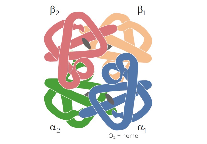
Hemoglobin structure: 4 globin chains (β1, β2, ɑ1, and ɑ2) and 4 heme molecules (with iron in the ferrous state) for binding with O2

Image by Lecturio.

Methemoglobin

  • 1 or more heme Fe converted to the Fe3+ state
  • Fe3+ cannot bind BIND Hyperbilirubinemia of the Newborn O2.
  • In normal circumstances, Fe3+ levels < 1%
  • Effect:
    • Remaining Fe2+ increase their affinity for O2:
    • Functional anemia Anemia Anemia is a condition in which individuals have low Hb levels, which can arise from various causes. Anemia is accompanied by a reduced number of RBCs and may manifest with fatigue, shortness of breath, pallor, and weakness. Subtypes are classified by the size of RBCs, chronicity, and etiology. Anemia: Overview and Types: functional hemoglobin < measured hemoglobin

Regulation of methemoglobin

Methemoglobin levels are limited through different mechanisms:

  • Cyb5R:
    • Physiologic pathway
    • Reduction of methemoglobin via nicotinamide adenine dinucleotide Nicotinamide adenine dinucleotide A coenzyme composed of ribosylnicotinamide 5′-diphosphate coupled to adenosine 5′-phosphate by pyrophosphate linkage. It is found widely in nature and is involved in numerous enzymatic reactions in which it serves as an electron carrier by being alternately oxidized (NAD+) and reduced (NADH). Pentose Phosphate Pathway (NADH)-dependent reaction
    • Cyb5R uses NADH to reduce methemoglobin.
    • Responsible for the reduction of 95%–99% of methemoglobin
  • Utilization of nicotinamide adenine dinucleotide Nicotinamide adenine dinucleotide A coenzyme composed of ribosylnicotinamide 5′-diphosphate coupled to adenosine 5′-phosphate by pyrophosphate linkage. It is found widely in nature and is involved in numerous enzymatic reactions in which it serves as an electron carrier by being alternately oxidized (NAD+) and reduced (NADH). Pentose Phosphate Pathway phosphate Phosphate Inorganic salts of phosphoric acid. Electrolytes ( NADPH NADPH Nicotinamide adenine dinucleotide phosphate. A coenzyme composed of ribosylnicotinamide 5′-phosphate (nmn) coupled by pyrophosphate linkage to the 5′-phosphate adenosine 2. Pentose Phosphate Pathway) methemoglobin reductase: 
    • Alternative pathway Alternative pathway Complement activation initiated by the interaction of microbial antigens with complement C3b. When complement factor B binds to the membrane-bound C3b, complement factor d cleaves it to form alternative C3 convertase (c3bbb) which, stabilized by complement factor p, is able to cleave multiple complement C3 to form alternative C5 convertase (c3bbb3b) leading to cleavage of complement C5 and the assembly of complement membrane attack complex. Innate Immunity: Barriers, Complement, and Cytokines
    • NADPH NADPH Nicotinamide adenine dinucleotide phosphate. A coenzyme composed of ribosylnicotinamide 5′-phosphate (nmn) coupled by pyrophosphate linkage to the 5′-phosphate adenosine 2. Pentose Phosphate Pathway derived from the hexose-monophosphate shunt pathway via the action of glucose-6-phosphate dehydrogenase Glucose-6-phosphate dehydrogenase Pentose Phosphate Pathway ( G6PD G6PD Pentose Phosphate Pathway
    • Under physiologic conditions, the role is insignificant because an intrinsic electron acceptor is required (and there is none in the RBC).
    • Effect on methemoglobin reduction becomes significant in the presence of certain compounds/extrinsic electron acceptors:
      • Methylene blue (MB): used as therapy
      • Riboflavin
  • Glutathione and ascorbic acid promote methemoglobin reduction through different pathways.
Pathophysiology of methemoglobin

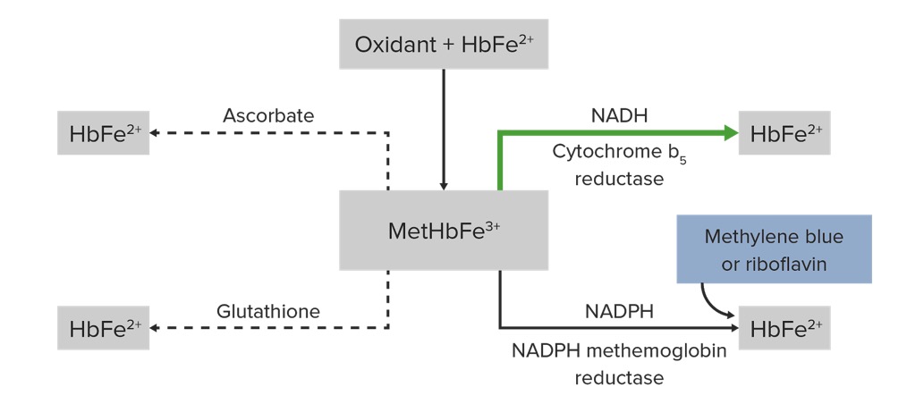
Pathophysiology and management of methemoglobin (HbFe²⁺: hemoglobin, MetHbFe³⁺: methemoglobin, NADPH: nicotinamide adenine dinucleotide phosphate, NADH: nicotinamide adenine dinucleotide):
Oxidation of heme iron from ferrous state to ferric state produces MetHbFe³⁺. Cytochrome b5 reductase reduces MetHbFe³⁺ to HbFe²⁺. Methylene blue acts as an electron acceptor for NADPH reductase and also facilitates the reduction of methemoglobin. Other compounds that can promote MetHbFe³⁺ reduction include glutathione and ascorbic acid.
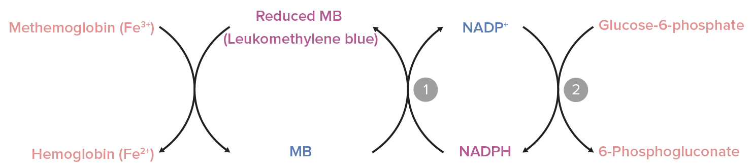
Image by Lecturio.

Clinical Presentation

Symptoms depend on the etiology, chronicity, and level of methemoglobin.

Acquired

  • Methemoglobin > 10%–15%: cyanosis Cyanosis A bluish or purplish discoloration of the skin and mucous membranes due to an increase in the amount of deoxygenated hemoglobin in the blood or a structural defect in the hemoglobin molecule. Pulmonary Examination or bluish/gray skin Skin The skin, also referred to as the integumentary system, is the largest organ of the body. The skin is primarily composed of the epidermis (outer layer) and dermis (deep layer). The epidermis is primarily composed of keratinocytes that undergo rapid turnover, while the dermis contains dense layers of connective tissue. Skin: Structure and Functions discoloration
  • Methemoglobin ≥ 20%–50%:
    • Headache Headache The symptom of pain in the cranial region. It may be an isolated benign occurrence or manifestation of a wide variety of headache disorders. Brain Abscess
    • Dyspnea Dyspnea Dyspnea is the subjective sensation of breathing discomfort. Dyspnea is a normal manifestation of heavy physical or psychological exertion, but also may be caused by underlying conditions (both pulmonary and extrapulmonary). Dyspnea
    • Lightheadedness Lightheadedness Hypotension, even syncope Syncope Syncope is a short-term loss of consciousness and loss of postural stability followed by spontaneous return of consciousness to the previous neurologic baseline without the need for resuscitation. The condition is caused by transient interruption of cerebral blood flow that may be benign or related to a underlying life-threatening condition. Syncope
    • Weakness
    • Confusion
    • Palpitations Palpitations Ebstein’s Anomaly, chest pain Pain An unpleasant sensation induced by noxious stimuli which are detected by nerve endings of nociceptive neurons. Pain: Types and Pathways
  • Methemoglobin > 50%–70%:
    • Cardiovascular system: arrhythmias
    • CNS: altered mental status Altered Mental Status Sepsis in Children; seizures Seizures A seizure is abnormal electrical activity of the neurons in the cerebral cortex that can manifest in numerous ways depending on the region of the brain affected. Seizures consist of a sudden imbalance that occurs between the excitatory and inhibitory signals in cortical neurons, creating a net excitation. The 2 major classes of seizures are focal and generalized. Seizures, delirium Delirium Delirium is a medical condition characterized by acute disturbances in attention and awareness. Symptoms may fluctuate during the course of a day and involve memory deficits and disorientation. Delirium, coma Coma Coma is defined as a deep state of unarousable unresponsiveness, characterized by a score of 3 points on the GCS. A comatose state can be caused by a multitude of conditions, making the precise epidemiology and prognosis of coma difficult to determine. Coma
    • Metabolic effects: significant acidosis Acidosis A pathologic condition of acid accumulation or depletion of base in the body. The two main types are respiratory acidosis and metabolic acidosis, due to metabolic acid build up. Respiratory Acidosis
  • Methemoglobin > 70%: death
Cyanosis in methemoglobinemia

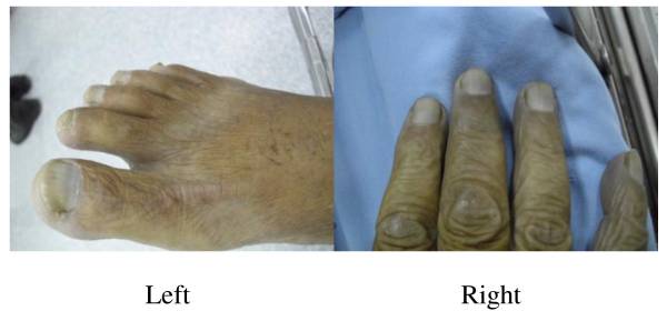
Cyanosis in methemoglobinemia:
The individual’s foot and hand show a bluish color and cyanotic nail beds.

Image: “Successful treatment of methemoglobinemia in an elderly couple with severe cyanosis” by Su YF et al. License: CC BY 2.0

Congenital Congenital Chorioretinitis

  • Infants or children with cyanosis Cyanosis A bluish or purplish discoloration of the skin and mucous membranes due to an increase in the amount of deoxygenated hemoglobin in the blood or a structural defect in the hemoglobin molecule. Pulmonary Examination (from chronic ↑ methemoglobin)
  • In cases of type II Cyb5R deficiency: developmental and neurologic abnormalities (i.e., seizures Seizures A seizure is abnormal electrical activity of the neurons in the cerebral cortex that can manifest in numerous ways depending on the region of the brain affected. Seizures consist of a sudden imbalance that occurs between the excitatory and inhibitory signals in cortical neurons, creating a net excitation. The 2 major classes of seizures are focal and generalized. Seizures, microcephaly Microcephaly A congenital abnormality in which the cerebrum is underdeveloped, the fontanels close prematurely, and, as a result, the head is small. (desk reference for neuroscience, 2nd ed. ). Fetal Alcohol Spectrum Disorder) noted

Diagnosis

Diagnostic approach

History:

  • History of exposure to oxidizing substances
  • Family history Family History Adult Health Maintenance and ethnicity
  • Medications
  • Proximity to contaminated wells
  • Symptoms suggestive of the intake of toxic substances (i.e., vomiting Vomiting The forcible expulsion of the contents of the stomach through the mouth. Hypokalemia, diarrhea Diarrhea Diarrhea is defined as ≥ 3 watery or loose stools in a 24-hour period. There are a multitude of etiologies, which can be classified based on the underlying mechanism of disease. The duration of symptoms (acute or chronic) and characteristics of the stools (e.g., watery, bloody, steatorrheic, mucoid) can help guide further diagnostic evaluation. Diarrhea)

Physical exam findings:

  • Sudden onset of cyanosis Cyanosis A bluish or purplish discoloration of the skin and mucous membranes due to an increase in the amount of deoxygenated hemoglobin in the blood or a structural defect in the hemoglobin molecule. Pulmonary Examination and hypoxia Hypoxia Sub-optimal oxygen levels in the ambient air of living organisms. Ischemic Cell Damage 
  • Altered mental status Altered Mental Status Sepsis in Children
  • Pulse oximetry O2 saturation of < 90% (usually around 85%), and does not improve with supplemental O2
  • Dark chocolate-colored blood during phlebotomy Phlebotomy The techniques used to draw blood from a vein for diagnostic purposes or for treatment of certain blood disorders such as erythrocytosis, hemochromatosis, polycythemia vera, and porphyria cutanea tarda. Hereditary Hemochromatosis
Color code suggestive of methemoglobin methemoglobinemia

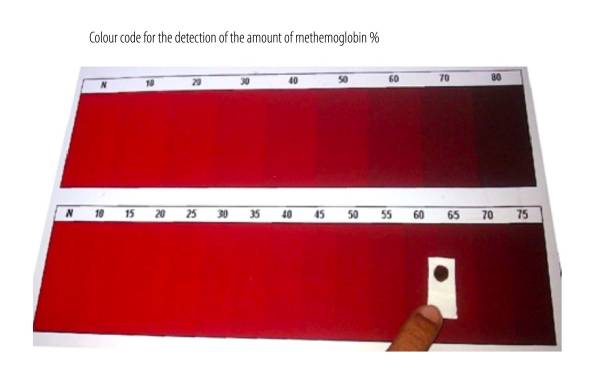
Color code suggestive of methemoglobin:
Blood with methemoglobin concentration > 15% of total hemoglobin levels appears brownish, as seen in the image (right side).

Image: “Semiquantitative estimation of methemoglobin levels in blood” by Shihana F et al. License: CC BY 3.0

Detecting methemoglobinemia

  • Blood gases:
    • Methemoglobin level > 5% (detected using most blood-gas analyzers)
    • + Saturation gap (difference between O2 saturation on pulse oximetry and arterial blood gas Arterial blood gas Respiratory Alkalosis
    • Metabolic acidosis Acidosis A pathologic condition of acid accumulation or depletion of base in the body. The two main types are respiratory acidosis and metabolic acidosis, due to metabolic acid build up. Respiratory Acidosis
  • Pulse oximetry is inaccurate:
    • Routine pulse oximeter measures light absorbance at 2 wavelengths (660 nm and 940 nm) to differentiate oxyhemoglobin from deoxyhemoglobin.
    • In methemoglobinemia:
      • Methemoglobin increases the absorption Absorption Absorption involves the uptake of nutrient molecules and their transfer from the lumen of the GI tract across the enterocytes and into the interstitial space, where they can be taken up in the venous or lymphatic circulation. Digestion and Absorption of light at both wavelengths (more at 940 nm), leading to errors in measurement.
      • O2 saturation by pulse oximetry plateaus at about 85%. 
  • Co-oximetry: 
    • Provides a method of measuring methemoglobin levels
    • Utilizes multiple wavelengths of light to detect methemoglobin absorption Absorption Absorption involves the uptake of nutrient molecules and their transfer from the lumen of the GI tract across the enterocytes and into the interstitial space, where they can be taken up in the venous or lymphatic circulation. Digestion and Absorption
  • Evelin-Malloy (direct) assay:
    • Most accurate
    • Not readily available
Methemoglobin measurement methemoglobinemia

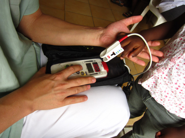
Methemoglobin measurement:
Use of co-oximetry (multiple-wavelength oximetry). Pictured above is a Rad-57 pulse oximeter applied to the finger of a child. Instead of just 2 wavelengths, co-oximetry utilizes 8 wavelengths.

Image: “Methemoglobin measurement with the pulse oximeter” by Thomas Hänscheid et al. License: CC BY 4.0

Additional tests

  • ECG ECG An electrocardiogram (ECG) is a graphic representation of the electrical activity of the heart plotted against time. Adhesive electrodes are affixed to the skin surface allowing measurement of cardiac impulses from many angles. The ECG provides 3-dimensional information about the conduction system of the heart, the myocardium, and other cardiac structures. Electrocardiogram (ECG): bradyarrhythmia Bradyarrhythmia Bradyarrhythmia is a rhythm in which the heart rate is less than 60/min. Bradyarrhythmia can be physiologic, without symptoms or hemodynamic change. Pathologic bradyarrhythmia results in reduced cardiac output and hemodynamic instability causing syncope, dizziness, or dyspnea. Bradyarrhythmias or ventricular dysrhythmia
  • Work-up for hemolysis:
    • CBC: generally with normal hemoglobin in conditions of acute onset
    • Reticulocyte Reticulocyte Immature erythrocytes. In humans, these are erythroid cells that have just undergone extrusion of their cell nucleus. They still contain some organelles that gradually decrease in number as the cells mature. Ribosomes are last to disappear. Certain staining techniques cause components of the ribosomes to precipitate into characteristic ‘reticulum’ (not the same as the endoplasmic reticulum), hence the name reticulocytes. Erythrocytes: Histology count
    • Peripheral blood smear Peripheral Blood Smear Anemia: Overview and Types
    • LDH LDH Osteosarcoma
    • Bilirubin Bilirubin A bile pigment that is a degradation product of heme. Heme Metabolism
    • Haptoglobin
  • Genetic testing Genetic Testing Detection of a mutation; genotype; karyotype; or specific alleles associated with genetic traits, heritable diseases, or predisposition to a disease, or that may lead to the disease in descendants. It includes prenatal genetic testing. Myotonic Dystrophies
  • Hemoglobin electrophoresis Electrophoresis An electrochemical process in which macromolecules or colloidal particles with a net electric charge migrate in a solution under the influence of an electric current. Blotting Techniques: detection of hemoglobin M
  • G6PD G6PD Pentose Phosphate Pathway activity: helps determine treatment

Management

Management of methemoglobinemia depends on the severity and chronicity.

Methemoglobin levels > 30% and associated with tissue hypoxia Hypoxia Sub-optimal oxygen levels in the ambient air of living organisms. Ischemic Cell Damage is a medical emergency:

  • Supportive management
  • Immediate discontinuation of any suspected oxidizing agent
  • MB:
    • MB (electron acceptor for NADPH NADPH Nicotinamide adenine dinucleotide phosphate. A coenzyme composed of ribosylnicotinamide 5′-phosphate (nmn) coupled by pyrophosphate linkage to the 5′-phosphate adenosine 2. Pentose Phosphate Pathway) → reduced to leukomethylene blue → reduction of methemoglobin
    • IV infusion
    • Rapid onset of action showing response within 1 hour
    • Does not work in G6PD G6PD Pentose Phosphate Pathway deficiency (as MB requires NADPH NADPH Nicotinamide adenine dinucleotide phosphate. A coenzyme composed of ribosylnicotinamide 5′-phosphate (nmn) coupled by pyrophosphate linkage to the 5′-phosphate adenosine 2. Pentose Phosphate Pathway generated by G6PD G6PD Pentose Phosphate Pathway)
    • Cannot be administered to patients Patients Individuals participating in the health care system for the purpose of receiving therapeutic, diagnostic, or preventive procedures. Clinician–Patient Relationship on serotonergic agents (↑ risk of serotonin Serotonin A biochemical messenger and regulator, synthesized from the essential amino acid l-tryptophan. In humans it is found primarily in the central nervous system, gastrointestinal tract, and blood platelets. Serotonin mediates several important physiological functions including neurotransmission, gastrointestinal motility, hemostasis, and cardiovascular integrity. Receptors and Neurotransmitters of the CNS syndrome)
  • High-dose ascorbic acid ( vitamin C Vitamin C A six carbon compound related to glucose. It is found naturally in citrus fruits and many vegetables. Ascorbic acid is an essential nutrient in human diets, and necessary to maintain connective tissue and bone. Its biologically active form, vitamin C, functions as a reducing agent and coenzyme in several metabolic pathways. Vitamin C is considered an antioxidant. Water-soluble Vitamins and their Deficiencies):
    • Reducing agent 
    • IV infusion
    • Symptom improvement in 1–3 days
    • Used when MB is unavailable or contraindicated
    • Useful in G6PD G6PD Pentose Phosphate Pathway deficiency 
  • Consider the following in severe cases:
    • Exchange transfusion
    • Hyperbaric O2

In congenital Congenital Chorioretinitis methemoglobinemia:

  • Therapy:
  • Genetic counseling Genetic Counseling An educational process that provides information and advice to individuals or families about a genetic condition that may affect them. The purpose is to help individuals make informed decisions about marriage, reproduction, and other health management issues based on information about the genetic disease, the available diagnostic tests, and management programs. Psychosocial support is usually offered. Myotonic Dystrophies for patients Patients Individuals participating in the health care system for the purpose of receiving therapeutic, diagnostic, or preventive procedures. Clinician–Patient Relationship with congenital Congenital Chorioretinitis disease and 1st-degree relatives

Differential Diagnosis

  • CO poisoning CO poisoning Carbon monoxide (CO) is an odorless, colorless, tasteless, nonirritating gas formed by hydrocarbon combustion (e.g., fires, car exhaust, gas heaters). Carbon monoxide has a higher affinity to hemoglobin than oxygen, forming carboxyhemoglobin (COHb). Increased levels of COHb lead to tissue hypoxia and brain damage. Carbon Monoxide Poisoning: an odorless, tasteless, colorless, nonirritating gas that is formed by hydrocarbon combustion (fires, car exhaust, gas heaters). Carbon monoxide Carbon monoxide Carbon monoxide (CO). A poisonous colorless, odorless, tasteless gas. It combines with hemoglobin to form carboxyhemoglobin, which has no oxygen carrying capacity. The resultant oxygen deprivation causes headache, dizziness, decreased pulse and respiratory rates, unconsciousness, and death. Carbon Monoxide Poisoning has a higher affinity to hemoglobin than O2, forming carboxyhemoglobin Carboxyhemoglobin Carbon Monoxide Poisoning (COHb) and resulting in impaired O2 transport and utilization. Features of CO poisoning CO poisoning Carbon monoxide (CO) is an odorless, colorless, tasteless, nonirritating gas formed by hydrocarbon combustion (e.g., fires, car exhaust, gas heaters). Carbon monoxide has a higher affinity to hemoglobin than oxygen, forming carboxyhemoglobin (COHb). Increased levels of COHb lead to tissue hypoxia and brain damage. Carbon Monoxide Poisoning include confusion, headache Headache The symptom of pain in the cranial region. It may be an isolated benign occurrence or manifestation of a wide variety of headache disorders. Brain Abscess, dizziness Dizziness An imprecise term which may refer to a sense of spatial disorientation, motion of the environment, or lightheadedness. Lateral Medullary Syndrome (Wallenberg Syndrome), decreased visual acuity Visual Acuity Clarity or sharpness of ocular vision or the ability of the eye to see fine details. Visual acuity depends on the functions of retina, neuronal transmission, and the interpretative ability of the brain. Normal visual acuity is expressed as 20/20 indicating that one can see at 20 feet what should normally be seen at that distance. Visual acuity can also be influenced by brightness, color, and contrast. Ophthalmic Exam, rose cheeks Cheeks The part of the face that is below the eye and to the side of the nose and mouth. Melasma, tachycardia Tachycardia Abnormally rapid heartbeat, usually with a heart rate above 100 beats per minute for adults. Tachycardia accompanied by disturbance in the cardiac depolarization (cardiac arrhythmia) is called tachyarrhythmia. Sepsis in Children, syncope Syncope Syncope is a short-term loss of consciousness and loss of postural stability followed by spontaneous return of consciousness to the previous neurologic baseline without the need for resuscitation. The condition is caused by transient interruption of cerebral blood flow that may be benign or related to a underlying life-threatening condition. Syncope, coma Coma Coma is defined as a deep state of unarousable unresponsiveness, characterized by a score of 3 points on the GCS. A comatose state can be caused by a multitude of conditions, making the precise epidemiology and prognosis of coma difficult to determine. Coma, and death. Blood is notably cherry-red, and pulse oximetry is inaccurate. Treatment involves the use of 100% O2. In case of treatment failure, hyperbaric O2 is considered.
  • Cyanide Cyanide Inorganic salts of hydrogen cyanide containing the -cn radical. The concept also includes isocyanides. It is distinguished from nitriles, which denotes organic compounds containing the -cn radical. Cyanide Poisoning poisoning: Hydrogen cyanide Cyanide Inorganic salts of hydrogen cyanide containing the -cn radical. The concept also includes isocyanides. It is distinguished from nitriles, which denotes organic compounds containing the -cn radical. Cyanide Poisoning (HCN) is a colorless, extremely poisonous, and flammable liquid used in multiple industries and in the manufacture of plastics and household paints. Lethal complications of cyanide Cyanide Inorganic salts of hydrogen cyanide containing the -cn radical. The concept also includes isocyanides. It is distinguished from nitriles, which denotes organic compounds containing the -cn radical. Cyanide Poisoning poisoning occur in closed-space fires when plastics burn. Exposure is via inhalation, dermal, or intestinal routes. Symptoms occur within minutes to seconds and involve cardiovascular, respiratory, and neurological changes. Treatment is with sodium thiosulfate Sodium Thiosulfate Cyanide Poisoning, nitrites, and hydroxocobalamin.

References

  1. Denshaw-Burke M., Delgiacco, E. (2020). Methemoglobinemia. Medscape. Retrieved April 9, 2021, from https://reference.medscape.com/article/204178-overview
  2. Ludlow, J.T., Wilkerson, R.G., Nappe, T.M. (2020). Methemoglobinemia. StatPearls. Treasure Island (FL): StatPearls Publishing. https://www.ncbi.nlm.nih.gov/books/NBK537317/
  3. Prchal, J.T. (2021). Methemoglobinemia. UpToDate. Retrieved April 9, 2021, from https://www.uptodate.com/contents/clinical-features-diagnosis-and-treatment-of-methemoglobinemia

Create your free account or log in to continue reading!

Sign up now and get free access to Lecturio with concept pages, medical videos, and questions for your medical education.

User Reviews

Details