Advertisement

Advertisement

Advertisement

Advertisement

Gastroschisis

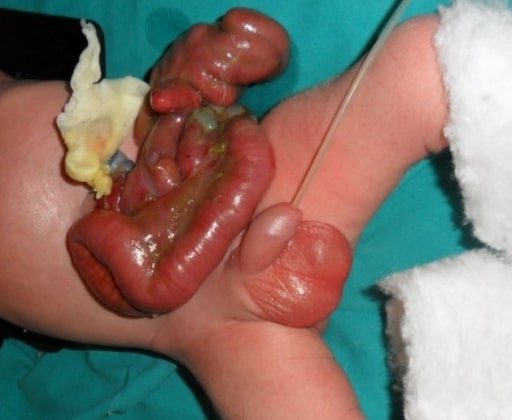
Gastroschisis is a congenital Congenital Chorioretinitis abdominal wall Abdominal wall The outer margins of the abdomen, extending from the osteocartilaginous thoracic cage to the pelvis. Though its major part is muscular, the abdominal wall consists of at least seven layers: the skin, subcutaneous fat, deep fascia; abdominal muscles, transversalis fascia, extraperitoneal fat, and the parietal peritoneum. Surgical Anatomy of the Abdomen defect characterized by the complete lack of closure of the abdominal musculature. A portion of intestine does not return to the abdominal cavity, thereby remaining in its early embryonic herniated state but with no coverings. Diagnosis is made by prenatal ultrasonography. Findings include a paraumbilical wall defect with uncovered herniated intestines. Fetal surveillance Surveillance Developmental Milestones and Normal Growth and bowel monitoring are done to measure fetal growth and amniotic fluid Amniotic fluid A clear, yellowish liquid that envelopes the fetus inside the sac of amnion. In the first trimester, it is likely a transudate of maternal or fetal plasma. In the second trimester, amniotic fluid derives primarily from fetal lung and kidney. Cells or substances in this fluid can be removed for prenatal diagnostic tests (amniocentesis). Placenta, Umbilical Cord, and Amniotic Cavity volume, as well as observe any bowel changes. The mode and timing of delivery depend on these factors. Neonatal care includes protection of the exposed bowel, fluid resuscitation Resuscitation The restoration to life or consciousness of one apparently dead. . Neonatal Respiratory Distress Syndrome, and airway Airway ABCDE Assessment maintenance. Primary closure can be performed within hours of birth. The presence of a large gastroschisis or thickened and dilated bowel warrants delayed closure, with the use a bowel covering or “silo,” and serial bowel reduction.

Last updated: Sep 22, 2022

Editorial responsibility: Stanley Oiseth, Lindsay Jones, Evelin Maza

Advertisement

Advertisement

Advertisement

Advertisement

Advertisement

Advertisement

Overview

Definition

Gastroschisis is a full-thickness defect of the anterior abdominal wall Abdominal wall The outer margins of the abdomen, extending from the osteocartilaginous thoracic cage to the pelvis. Though its major part is muscular, the abdominal wall consists of at least seven layers: the skin, subcutaneous fat, deep fascia; abdominal muscles, transversalis fascia, extraperitoneal fat, and the parietal peritoneum. Surgical Anatomy of the Abdomen through which the intestine and/or other abdominal viscera freely protrude.

  • No membrane or sac covers the bowel/intestine.
  • The defect is often to the right of the umbilical insertion site.

Embryology

  • Gastrointestinal (GI) system development from 3 germinal layers:
    • Mesoderm Mesoderm The middle germ layer of an embryo derived from three paired mesenchymal aggregates along the neural tube. Gastrulation and Neurulation: connective tissue Connective tissue Connective tissues originate from embryonic mesenchyme and are present throughout the body except inside the brain and spinal cord. The main function of connective tissues is to provide structural support to organs. Connective tissues consist of cells and an extracellular matrix. Connective Tissue: Histology (wall of gut tube, blood vessels and smooth muscle)
    • Endoderm Endoderm The inner of the three germ layers of an embryo. Gastrulation and Neurulation: epithelial lining
    • Ectoderm Ectoderm The outer of the three germ layers of an embryo. Gastrulation and Neurulation: epidermis Epidermis The external, nonvascular layer of the skin. It is made up, from within outward, of five layers of epithelium: (1) basal layer (stratum basale epidermidis); (2) spinous layer (stratum spinosum epidermidis); (3) granular layer (stratum granulosum epidermidis); (4) clear layer (stratum lucidum epidermidis); and (5) horny layer (stratum corneum epidermidis). Skin: Structure and Functions and neural crest Neural crest The two longitudinal ridges along the primitive streak appearing near the end of gastrulation during development of nervous system (neurulation). The ridges are formed by folding of neural plate. Between the ridges is a neural groove which deepens as the fold become elevated. When the folds meet at midline, the groove becomes a closed tube, the neural tube. Hirschsprung Disease ( neurons Neurons The basic cellular units of nervous tissue. Each neuron consists of a body, an axon, and dendrites. Their purpose is to receive, conduct, and transmit impulses in the nervous system. Nervous System: Histology of the GI tract)
  • GI tract divisions:
  • 6th week of gestation:
    • Rapid growth of the GI tract
    • Midgut Midgut Development of the Abdominal Organs herniates through the umbilical ring, developing entirely outside the peritoneal cavity Peritoneal Cavity The space enclosed by the peritoneum. It is divided into two portions, the greater sac and the lesser sac or omental bursa, which lies behind the stomach. The two sacs are connected by the foramen of winslow, or epiploic foramen. Peritoneum: Anatomy.
  • 10th week of gestation:
Normal process of herniation during embryologic development

Diagram showing the normal process of intestinal rotation and herniation during embryologic development

A: the midgut (multi-colored loop) before herniation.
B1-B3: as it grows rapidly, the midgut herniates through the umbilical ring, and starts rotation.
C: The midgut returns to the abdominal cavity.

Image by Lecturio.

Epidemiology

  • Prevalence Prevalence The total number of cases of a given disease in a specified population at a designated time. It is differentiated from incidence, which refers to the number of new cases in the population at a given time. Measures of Disease Frequency: 3–4 per 10,000 births
  • Similar incidence Incidence The number of new cases of a given disease during a given period in a specified population. It also is used for the rate at which new events occur in a defined population. It is differentiated from prevalence, which refers to all cases in the population at a given time. Measures of Disease Frequency in male and female newborns
  • In 10% of cases, gastroschisis is associated with anomalies outside the GI tract. 
  • Higher risk of preterm delivery in pregnancies with gastroschisis (28%) compared with those without gastroschisis (6%)
  • Maternal risk factors:
    • Young age (< 20 years)
    • Low body mass index Body mass index An indicator of body density as determined by the relationship of body weight to body height. Bmi=weight (kg)/height squared (m2). Bmi correlates with body fat (adipose tissue). Their relationship varies with age and gender. For adults, bmi falls into these categories: below 18. 5 (underweight); 18. 5-24. 9 (normal); 25. 0-29. 9 (overweight); 30. 0 and above (obese). Obesity ( BMI BMI An indicator of body density as determined by the relationship of body weight to body height. Bmi=weight (kg)/height squared (m2). Bmi correlates with body fat (adipose tissue). Their relationship varies with age and gender. For adults, bmi falls into these categories: below 18. 5 (underweight); 18. 5-24. 9 (normal); 25. 0-29. 9 (overweight); 30. 0 and above (obese). Obesity)
    • Exposure to cigarette smoke
    • Intake of aspirin Aspirin The prototypical analgesic used in the treatment of mild to moderate pain. It has anti-inflammatory and antipyretic properties and acts as an inhibitor of cyclooxygenase which results in the inhibition of the biosynthesis of prostaglandins. Aspirin also inhibits platelet aggregation and is used in the prevention of arterial and venous thrombosis. Nonsteroidal Antiinflammatory Drugs (NSAIDs) and ibuprofen Ibuprofen A nonsteroidal anti-inflammatory agent with analgesic properties used in the treatment of rheumatism and arthritis. Nonsteroidal Antiinflammatory Drugs (NSAIDs)
    • Intake of decongestants (pseudoephedrine, phenylpropanolamine)

Pathophysiology

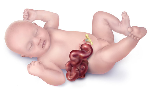
Embryologic hypotheses leading to herniation Herniation Omphalocele of the bowel: 
  • Defective formation
    • Failure of mesoderm Mesoderm The middle germ layer of an embryo derived from three paired mesenchymal aggregates along the neural tube. Gastrulation and Neurulation to form in the body wall
    • Abnormal folding of body wall resulting in a ventral body wall defect
  • Disruption of the abdominal wall Abdominal wall The outer margins of the abdomen, extending from the osteocartilaginous thoracic cage to the pelvis. Though its major part is muscular, the abdominal wall consists of at least seven layers: the skin, subcutaneous fat, deep fascia; abdominal muscles, transversalis fascia, extraperitoneal fat, and the parietal peritoneum. Surgical Anatomy of the Abdomen
    • Rupture of the amnion Amnion The innermost membranous sac that surrounds and protects the developing embryo which is bathed in the amniotic fluid. Amnion cells are secretory epithelial cells and contribute to the amniotic fluid. Placenta, Umbilical Cord, and Amniotic Cavity (sac protecting the embryo Embryo The entity of a developing mammal, generally from the cleavage of a zygote to the end of embryonic differentiation of basic structures. For the human embryo, this represents the first two months of intrauterine development preceding the stages of the fetus. Fertilization and First Week) around the umbilical ring 
    • Abnormal involution of the right umbilical vein umbilical vein Venous vessels in the umbilical cord. They carry oxygenated, nutrient-rich blood from the mother to the fetus via the placenta. In humans, there is normally one umbilical vein. Prenatal and Postnatal Physiology of the Neonate leading to weakening of the body wall
    • Disruption of the right vitelline ( yolk sac Yolk Sac The first of four extra-embryonic membranes to form during embryogenesis. In reptiles and birds, it arises from endoderm and mesoderm to incorporate the egg yolk into the digestive tract for nourishing the embryo. In placental mammals, its nutritional function is vestigial; however, it is the source of intestinal mucosa; blood cells; and germ cells. It is sometimes called the vitelline sac, which should not be confused with the vitelline membrane of the egg. Embryoblast and Trophoblast Development) artery leading to infarction at the base of the cord and subsequent body wall damage

Diagnosis and Monitoring

Diagnostic tests Diagnostic tests Diagnostic tests are important aspects in making a diagnosis. Some of the most important epidemiological values of diagnostic tests include sensitivity and specificity, false positives and false negatives, positive and negative predictive values, likelihood ratios, and pre-test and post-test probabilities. Epidemiological Values of Diagnostic Tests

  • Ultrasonography
    • Prenatal diagnosis made as early as 14 weeks, but usually around 20 weeks’ gestation
    • Finding(s): 
      • Paraumbilical abdominal wall Abdominal wall The outer margins of the abdomen, extending from the osteocartilaginous thoracic cage to the pelvis. Though its major part is muscular, the abdominal wall consists of at least seven layers: the skin, subcutaneous fat, deep fascia; abdominal muscles, transversalis fascia, extraperitoneal fat, and the parietal peritoneum. Surgical Anatomy of the Abdomen defect (on the right, never through the umbilicus)
      • Herniated intestines without a covering sac
      • Intestines freely floating in amniotic fluid Amniotic fluid A clear, yellowish liquid that envelopes the fetus inside the sac of amnion. In the first trimester, it is likely a transudate of maternal or fetal plasma. In the second trimester, amniotic fluid derives primarily from fetal lung and kidney. Cells or substances in this fluid can be removed for prenatal diagnostic tests (amniocentesis). Placenta, Umbilical Cord, and Amniotic Cavity
      • Oligohydramnios Oligohydramnios Oligohydramnios refers to amniotic fluid volume less than expected for the current gestational age. Oligohydramnios is diagnosed by ultrasound and defined as an amniotic fluid index (AFI) of ‰¤ 5 cm or a single deep pocket (SDP) of < 2 cm in the 2nd or 3rd trimester. Oligohydramnios (most common amniotic fluid Amniotic fluid A clear, yellowish liquid that envelopes the fetus inside the sac of amnion. In the first trimester, it is likely a transudate of maternal or fetal plasma. In the second trimester, amniotic fluid derives primarily from fetal lung and kidney. Cells or substances in this fluid can be removed for prenatal diagnostic tests (amniocentesis). Placenta, Umbilical Cord, and Amniotic Cavity finding)
      • Occasionally other visceral organs also herniated (e.g., liver Liver The liver is the largest gland in the human body. The liver is found in the superior right quadrant of the abdomen and weighs approximately 1.5 kilograms. Its main functions are detoxification, metabolism, nutrient storage (e.g., iron and vitamins), synthesis of coagulation factors, formation of bile, filtration, and storage of blood. Liver: Anatomy)
  • Laboratory test: elevated maternal serum ɑ-fetoprotein ( AFP AFP The first alpha-globulins to appear in mammalian sera during fetal development and the dominant serum proteins in early embryonic life. Hepatocellular Carcinoma (HCC) and Liver Metastases)
    • Elevated AFP AFP The first alpha-globulins to appear in mammalian sera during fetal development and the dominant serum proteins in early embryonic life. Hepatocellular Carcinoma (HCC) and Liver Metastases is suggestive of but non-specific for gastroschisis (seen also in spina bifida and anencephaly Anencephaly A malformation of the nervous system caused by failure of the anterior neuropore to close. Infants are born with intact spinal cords, cerebellums, and brainstems, but lack formation of neural structures above this level. The skull is only partially formed but the eyes are usually normal. This condition may be associated with folate deficiency. Affected infants are only capable of primitive (brain stem) reflexes and usually do not survive for more than two weeks. Neural Tube Defects).
    • Lab testing alone is insufficient for a diagnosis of gastroschisis.

Monitoring

  • Fetal monitoring Fetal monitoring The primary goals of antepartum testing and monitoring are to assess fetal well-being, identify treatable situations that may cause complications, and evaluate for chromosomal abnormalities. These tests are divided into screening tests (which include cell-free DNA testing, serum analyte testing, and nuchal translucency measurements), and diagnostic tests, which provide a definitive diagnosis of aneuploidy and include chorionic villus sampling (CVS) and amniocentesis. Antepartum Testing and Monitoring
    • Assess fetal growth including amniotic fluid Amniotic fluid A clear, yellowish liquid that envelopes the fetus inside the sac of amnion. In the first trimester, it is likely a transudate of maternal or fetal plasma. In the second trimester, amniotic fluid derives primarily from fetal lung and kidney. Cells or substances in this fluid can be removed for prenatal diagnostic tests (amniocentesis). Placenta, Umbilical Cord, and Amniotic Cavity volume
    • Typically started at 24 weeks
    • Look for intrauterine growth restriction (IUGR): associated with increased risk of fetal demise
  • Fetal bowel
    • Serial sonographic evaluation of the stomach Stomach The stomach is a muscular sac in the upper left portion of the abdomen that plays a critical role in digestion. The stomach develops from the foregut and connects the esophagus with the duodenum. Structurally, the stomach is C-shaped and forms a greater and lesser curvature and is divided grossly into regions: the cardia, fundus, body, and pylorus. Stomach: Anatomy and bowel
    • Check for substantial dilation (> 25 mm MM Multiple myeloma (MM) is a malignant condition of plasma cells (activated B lymphocytes) primarily seen in the elderly. Monoclonal proliferation of plasma cells results in cytokine-driven osteoclastic activity and excessive secretion of IgG antibodies. Multiple Myeloma) or acute thickening and edema Edema Edema is a condition in which excess serous fluid accumulates in the body cavity or interstitial space of connective tissues. Edema is a symptom observed in several medical conditions. It can be categorized into 2 types, namely, peripheral (in the extremities) and internal (in an organ or body cavity). Edema
    • Prenatal complications associated with bowel dilation Bowel dilation Large Bowel Obstruction:
  • Antepartum fetal surveillance Surveillance Developmental Milestones and Normal Growth
    • Biophysical profile Biophysical Profile Obstetric Imaging with nonstress test and amniotic fluid Amniotic fluid A clear, yellowish liquid that envelopes the fetus inside the sac of amnion. In the first trimester, it is likely a transudate of maternal or fetal plasma. In the second trimester, amniotic fluid derives primarily from fetal lung and kidney. Cells or substances in this fluid can be removed for prenatal diagnostic tests (amniocentesis). Placenta, Umbilical Cord, and Amniotic Cavity index
    • Usually started around 32 weeks
  • Fetal genetic studies
    • Considered in cases with extraintestinal structural anomalies (to determine associated chromosomal abnormalities)
    • Information regarding prognosis Prognosis A prediction of the probable outcome of a disease based on a individual’s condition and the usual course of the disease as seen in similar situations. Non-Hodgkin Lymphomas, delivery management, and neonatal care needed

Clinical Presentation

Newborn Newborn An infant during the first 28 days after birth. Physical Examination of the Newborn findings

  • Full-thickness paraumbilical abdominal wall Abdominal wall The outer margins of the abdomen, extending from the osteocartilaginous thoracic cage to the pelvis. Though its major part is muscular, the abdominal wall consists of at least seven layers: the skin, subcutaneous fat, deep fascia; abdominal muscles, transversalis fascia, extraperitoneal fat, and the parietal peritoneum. Surgical Anatomy of the Abdomen defect, usually to the right of the umbilical cord Umbilical cord The flexible rope-like structure that connects a developing fetus to the placenta in mammals. The cord contains blood vessels which carry oxygen and nutrients from the mother to the fetus and waste products away from the fetus. Placenta, Umbilical Cord, and Amniotic Cavity insertion site
  • Eviscerated bowel not covered by a sac 
  • Bowel appearance affected by:
    • Exposure to amniotic fluid Amniotic fluid A clear, yellowish liquid that envelopes the fetus inside the sac of amnion. In the first trimester, it is likely a transudate of maternal or fetal plasma. In the second trimester, amniotic fluid derives primarily from fetal lung and kidney. Cells or substances in this fluid can be removed for prenatal diagnostic tests (amniocentesis). Placenta, Umbilical Cord, and Amniotic Cavity
    • Amount of time of bowel is exposed 
    • Longer exposure to amniotic fluid Amniotic fluid A clear, yellowish liquid that envelopes the fetus inside the sac of amnion. In the first trimester, it is likely a transudate of maternal or fetal plasma. In the second trimester, amniotic fluid derives primarily from fetal lung and kidney. Cells or substances in this fluid can be removed for prenatal diagnostic tests (amniocentesis). Placenta, Umbilical Cord, and Amniotic Cavity → bowel is matted, thick, and covered with inflammatory rind
  • Abdominal defect is usually < 4 cm.

Categories

  • Simple gastroschisis: 
    • 75% of cases
    • Intact, uncompromised, continuous bowel
  • Complex gastroschisis: 
    • Associated with intestinal stenosis Stenosis Hypoplastic Left Heart Syndrome (HLHS), atresia Atresia Hypoplastic Left Heart Syndrome (HLHS), perforation Perforation A pathological hole in an organ, blood vessel or other soft part of the body, occurring in the absence of external force. Esophagitis, necrosis Necrosis The death of cells in an organ or tissue due to disease, injury or failure of the blood supply. Ischemic Cell Damage or volvulus Volvulus A volvulus is the twisting or axial rotation of a portion of the bowel around its mesentery. The most common site of volvulus in adults is the colon; most frequently the sigmoid volvulus. Patients typically present with symptoms of bowel obstruction such as abdominal pain, distension, vomiting, and constipation/obstipation. Volvulus
    • Related to vascular compromise caused by the herniated bowel
    • More GI, infectious, and respiratory diseases
    • Higher risk for in-hospital mortality Mortality All deaths reported in a given population. Measures of Health Status and complications ( bowel obstruction Bowel obstruction Any impairment, arrest, or reversal of the normal flow of intestinal contents toward the anal canal. Ascaris/Ascariasis, short bowel syndrome Short bowel syndrome Short bowel syndrome is a malabsorptive condition most commonly associated with extensive intestinal resection for etiologies such as Crohn’s disease, bowel obstruction, trauma, radiation therapy, and vascular insufficiency. The short length of bowel results in insufficient surface area for fluid and electrolyte absorption. Short Bowel Syndrome, and need for tube feedings on discharge)

Management and Prognosis

Delivery

  • Requires coordination Coordination Cerebellar Disorders of care among maternal-fetal medicine specialist, neonatologist, and pediatric surgeon
  • Delivery is recommended in a center with optimal neonatal care resources. 
  • Timing:
    • Affected by multiple factors, including:
      • Gestational age Gestational age The age of the conceptus, beginning from the time of fertilization. In clinical obstetrics, the gestational age is often estimated as the time from the last day of the last menstruation which is about 2 weeks before ovulation and fertilization. Pregnancy: Diagnosis, Physiology, and Care (lung maturity)
      • Ultrasound findings (fetal growth and bowel status)
      • Fetal testing results
    • Spontaneous labor (for pregnancies with gastroschisis) occurs at a mean Mean Mean is the sum of all measurements in a data set divided by the number of measurements in that data set. Measures of Central Tendency and Dispersion gestational age Gestational age The age of the conceptus, beginning from the time of fertilization. In clinical obstetrics, the gestational age is often estimated as the time from the last day of the last menstruation which is about 2 weeks before ovulation and fertilization. Pregnancy: Diagnosis, Physiology, and Care of 36 weeks.
  • Mode of delivery:
    • Uncomplicated gastroschisis: not a contraindication to vaginal delivery
    • Marked liver Liver The liver is the largest gland in the human body. The liver is found in the superior right quadrant of the abdomen and weighs approximately 1.5 kilograms. Its main functions are detoxification, metabolism, nutrient storage (e.g., iron and vitamins), synthesis of coagulation factors, formation of bile, filtration, and storage of blood. Liver: Anatomy herniation Herniation Omphalocele: Cesarean section should be considered.

Neonatal care

  • Reduce intestinal injury and reduce fluid loss:
    • Protect exposed bowel by wrapping bowel with sterile Sterile Basic Procedures saline dressings covered with plastic wrap.
    • In some centers, lower half of the neonate Neonate An infant during the first 28 days after birth. Physical Examination of the Newborn is placed into a plastic bag (allows protection and visualization of bowel perfusion).
  • Fluid and electrolyte resuscitation Resuscitation The restoration to life or consciousness of one apparently dead. . Neonatal Respiratory Distress Syndrome (expected fluid losses are 23 times that of a healthy neonate Neonate An infant during the first 28 days after birth. Physical Examination of the Newborn)
  • Prophylactic broad-spectrum Broad-Spectrum Fluoroquinolones antibiotics (cover for maternal vaginal flora)
  • Orogastric tube insertion to decompress the stomach Stomach The stomach is a muscular sac in the upper left portion of the abdomen that plays a critical role in digestion. The stomach develops from the foregut and connects the esophagus with the duodenum. Structurally, the stomach is C-shaped and forms a greater and lesser curvature and is divided grossly into regions: the cardia, fundus, body, and pylorus. Stomach: Anatomy
  • Airway Airway ABCDE Assessment maintenance and respiratory support as needed

Surgery

  • Primary closure
    • Surgery performed within hours of birth
    • To minimize intestinal injury during reduction, abdominal defect is increased by 1–2 cm. 
    • Successful in 70% of cases
  • Delayed closure
    • In cases of thickened and distended bowel loops
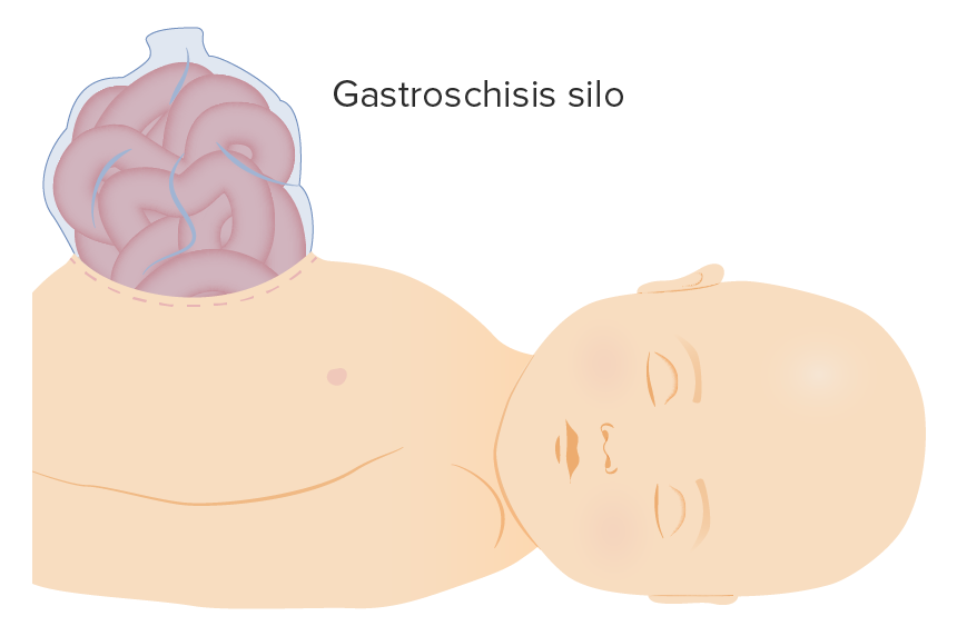
    • Eviscerated bowel is contained within a sterile Sterile Basic Procedures covering (silastic silo).
    • The bag and bowel are suspended over the infant, allowing decompression of bowel loops by gravity.
    • Serial reduction of bowel contents performed
Gastroschisis silo illustration

Gastroschisis silo

Image by Lecturio.

Prognosis Prognosis A prediction of the probable outcome of a disease based on a individual’s condition and the usual course of the disease as seen in similar situations. Non-Hodgkin Lymphomas

  • Gastroschisis: most favorable outcome compared with other abdominal wall Abdominal wall The outer margins of the abdomen, extending from the osteocartilaginous thoracic cage to the pelvis. Though its major part is muscular, the abdominal wall consists of at least seven layers: the skin, subcutaneous fat, deep fascia; abdominal muscles, transversalis fascia, extraperitoneal fat, and the parietal peritoneum. Surgical Anatomy of the Abdomen defects
  • Survival rate of neonates born in North America: 98%
  • Sepsis Sepsis Systemic inflammatory response syndrome with a proven or suspected infectious etiology. When sepsis is associated with organ dysfunction distant from the site of infection, it is called severe sepsis. When sepsis is accompanied by hypotension despite adequate fluid infusion, it is called septic shock. Sepsis and Septic Shock: only significant predictor of mortality Mortality All deaths reported in a given population. Measures of Health Status

Differential Diagnosis

  • Omphalocele Omphalocele Omphalocele is a congenital anterior abdominal wall defect in which the intestines are covered by peritoneum and amniotic membranes. The condition results from the failure of the midgut to return to the abdominal cavity by 10 weeks’ gestation. Omphalocele: similar to gastroschisis, but due to a failure of the bowel to return to the ventral cavity after normal herniation Herniation Omphalocele. The abdominal viscera (including the liver Liver The liver is the largest gland in the human body. The liver is found in the superior right quadrant of the abdomen and weighs approximately 1.5 kilograms. Its main functions are detoxification, metabolism, nutrient storage (e.g., iron and vitamins), synthesis of coagulation factors, formation of bile, filtration, and storage of blood. Liver: Anatomy) remain herniated through the umbilicus, covered by amnion Amnion The innermost membranous sac that surrounds and protects the developing embryo which is bathed in the amniotic fluid. Amnion cells are secretory epithelial cells and contribute to the amniotic fluid. Placenta, Umbilical Cord, and Amniotic Cavity and peritoneum Peritoneum The peritoneum is a serous membrane lining the abdominopelvic cavity. This lining is formed by connective tissue and originates from the mesoderm. The membrane lines both the abdominal walls (as parietal peritoneum) and all of the visceral organs (as visceral peritoneum). Peritoneum: Anatomy. Unlike gastroschisis, approximately half of cases of omphalocele Omphalocele Omphalocele is a congenital anterior abdominal wall defect in which the intestines are covered by peritoneum and amniotic membranes. The condition results from the failure of the midgut to return to the abdominal cavity by 10 weeks’ gestation. Omphalocele are associated with other birth defects and chromosomal abnormalities.
  • Pentalogy of Cantrell: a rare syndrome consisting of multiple congenital Congenital Chorioretinitis abnormalities, including a lower sternal defect, anterior diaphragm Diaphragm The diaphragm is a large, dome-shaped muscle that separates the thoracic cavity from the abdominal cavity. The diaphragm consists of muscle fibers and a large central tendon, which is divided into right and left parts. As the primary muscle of inspiration, the diaphragm contributes 75% of the total inspiratory muscle force. Diaphragm: Anatomy defect, pericardial defect, abdominal wall Abdominal wall The outer margins of the abdomen, extending from the osteocartilaginous thoracic cage to the pelvis. Though its major part is muscular, the abdominal wall consists of at least seven layers: the skin, subcutaneous fat, deep fascia; abdominal muscles, transversalis fascia, extraperitoneal fat, and the parietal peritoneum. Surgical Anatomy of the Abdomen defect, and congenital Congenital Chorioretinitis heart anomalies. Diagnosis is made prenatally by ultrasonography. Treatment requires complex medical and surgical care.
  • Bladder Bladder A musculomembranous sac along the urinary tract. Urine flows from the kidneys into the bladder via the ureters, and is held there until urination. Pyelonephritis and Perinephric Abscess exstrophy-epispadias complex: a spectrum of congenital Congenital Chorioretinitis defects: epispadias Epispadias A birth defect due to malformation of the urethra in which the urethral opening is above its normal location. In the male, the malformed urethra generally opens on the top or the side of the penis, but the urethra can also be open the entire length of the penis. In the female, the malformed urethral opening is often between the clitoris and the labia, or in the abdomen. Penile Anomalies and Conditions (failed closure of urethra Urethra A tube that transports urine from the urinary bladder to the outside of the body in both the sexes. It also has a reproductive function in the male by providing a passage for sperm. Urinary Tract: Anatomy), bladder Bladder A musculomembranous sac along the urinary tract. Urine flows from the kidneys into the bladder via the ureters, and is held there until urination. Pyelonephritis and Perinephric Abscess exstrophy, and cloacal exstrophy Cloacal Exstrophy Omphalocele (most severe defect, which includes anal atresia Atresia Hypoplastic Left Heart Syndrome (HLHS) and an omphalocele Omphalocele Omphalocele is a congenital anterior abdominal wall defect in which the intestines are covered by peritoneum and amniotic membranes. The condition results from the failure of the midgut to return to the abdominal cavity by 10 weeks’ gestation. Omphalocele). Treatment is surgical, with goals to close abdominal wall Abdominal wall The outer margins of the abdomen, extending from the osteocartilaginous thoracic cage to the pelvis. Though its major part is muscular, the abdominal wall consists of at least seven layers: the skin, subcutaneous fat, deep fascia; abdominal muscles, transversalis fascia, extraperitoneal fat, and the parietal peritoneum. Surgical Anatomy of the Abdomen defect, achieve urinary continence, and reconstruct the genito-urinary area.
  • Umbilical hernia Umbilical Hernia A hernia due to an imperfect closure or weakness of the umbilical ring. It appears as a skin-covered protrusion at the umbilicus during crying, coughing, or straining. The hernia generally consists of omentum or small intestine. The vast majority of umbilical hernias are congenital but can be acquired due to severe abdominal distention. Abdominal Hernias: presents as a protrusion in the neonate Neonate An infant during the first 28 days after birth. Physical Examination of the Newborn’s umbilical area. There is incomplete closure of the fascia Fascia Layers of connective tissue of variable thickness. The superficial fascia is found immediately below the skin; the deep fascia invests muscles, nerves, and other organs. Cellulitis of the umbilical ring but with intact skin Skin The skin, also referred to as the integumentary system, is the largest organ of the body. The skin is primarily composed of the epidermis (outer layer) and dermis (deep layer). The epidermis is primarily composed of keratinocytes that undergo rapid turnover, while the dermis contains dense layers of connective tissue. Skin: Structure and Functions covering the ring. It is important to determine the reducibility, as incarcerated hernia Incarcerated Hernia Abdominal Hernias is a complication. Surgery is indicated for defects > 1.5 cm in those > 2 years of age and for complications, including strangulation Strangulation Inguinal Canal: Anatomy and Hernias, incarceration Incarceration Inguinal Canal: Anatomy and Hernias, or rupture.

References

  1. Bhatia, A., Shatanof, R., Bordoni, B. (2020) Embryology, Gastrointestinal. StatPearls. https://www.ncbi.nlm.nih.gov/books/NBK537172/ 
  2. Bradnock, T. et al. (2011). Gastroschisis: one year outcomes from national cohort study. British Medical Journal. doi: 10.1136/bmj.d6749
  3. Feldkamp, M., Carey, J., Sadler, T. (2007). Development of Gastroschisis: Review of hypothesis, and implications for research. American Journal of Medical Genetics. https://doi.org/10.1002/ajmg.a.31578
  4. Ebert, A. et al. (2009). The Exstrophy-epispadias complex. Orphanet Journal of rare diseases. 4:23. doi: 10.1186/1750-1172-4-23
  5. Glasser, J., Windle, M., Carter, B. (2019). Pediatric Omphalocele and Gastroschisis (Abdominal wall defects). Medscape. Retrieved 4 Oct 2020, from https://emedicine.medscape.com/article/975583-overview#a5
  6. Rentea, R., Gupta, V. (2020). Gastroschisis. StatPearls. https://www.ncbi.nlm.nih.gov/books/NBK557894/
  7. Stephenson, C. et al. (2020). Gastroschisis. UpToDate. Retrieved 4 Oct 2020, from https://www.uptodate.com/contents/gastroschisis?search=gastroschisis
  8. Troullioud Lucas, A., Jaafar, S., Mendez, M. (2020). Pediatric Umbilical Hernia. StatPearls. https://www.ncbi.nlm.nih.gov/books/NBK459294/-

Back to School, Back to Success

📚
Get 50% off
all Premium plans!

Create your free account or log in to continue reading!

Sign up now and get free access to Lecturio with concept pages, medical videos, and questions for your medical education.

User Reviews

Details