Fat necrosisNecrosisThe death of cells in an organ or tissue due to disease, injury or failure of the blood supply.Ischemic Cell Damage of the breast is an inflammatory, benignBenignFibroadenoma condition resulting from injury to the breast tissue. Forms of injury include blunt traumatic injury as well as trauma from surgical procedures, biopsies, and radiationRadiationEmission or propagation of acoustic waves (sound), electromagnetic energy waves (such as light; radio waves; gamma rays; or x-rays), or a stream of subatomic particles (such as electrons; neutrons; protons; or alpha particles).Osteosarcoma therapy. Fat necrosisNecrosisThe death of cells in an organ or tissue due to disease, injury or failure of the blood supply.Ischemic Cell Damage of the breast is characterized by the presence of an ill-defined breast massMassThree-dimensional lesion that occupies a space within the breastImaging of the Breast that is usually accompanied by overlying skinSkinThe skin, also referred to as the integumentary system, is the largest organ of the body. The skin is primarily composed of the epidermis (outer layer) and dermis (deep layer). The epidermis is primarily composed of keratinocytes that undergo rapid turnover, while the dermis contains dense layers of connective tissue.Skin: Structure and Functions changes. Oil cystsCystsAny fluid-filled closed cavity or sac that is lined by an epithelium. Cysts can be of normal, abnormal, non-neoplastic, or neoplastic tissues.Fibrocystic Change may also form as fibrosisFibrosisAny pathological condition where fibrous connective tissue invades any organ, usually as a consequence of inflammation or other injury.Bronchiolitis Obliterans and calcification trap oil from degenerating fat cells. Fat necrosisNecrosisThe death of cells in an organ or tissue due to disease, injury or failure of the blood supply.Ischemic Cell Damage of the breast may be clinically and radiographically difficult to distinguish from a malignant massMassThree-dimensional lesion that occupies a space within the breastImaging of the Breast. Diagnosis relies on a history consistent with trauma, breast imagingBreast ImagingFemale breasts, made of glandular, adipose, and connective tissue, are hormone-sensitive organs that undergo changes along with the menstrual cycle and during pregnancy. Breasts may be affected by various diseases, in which different imaging methods are important to arrive at the correct diagnosis and management. Mammography is used for breast cancer screening and diagnostic evaluation of various breast-related symptoms.Imaging of the Breast, and, less commonly, a core needle biopsyCore Needle BiopsyFibrocystic Change for definitive diagnosis. Treatment is usually not required. The primary clinical significance of this condition is its possible confusion with breast cancerBreast cancerBreast cancer is a disease characterized by malignant transformation of the epithelial cells of the breast. Breast cancer is the most common form of cancer and 2nd most common cause of cancer-related death among women. Breast Cancer on exam and imaging.
Fat necrosisNecrosisThe death of cells in an organ or tissue due to disease, injury or failure of the blood supply.Ischemic Cell Damageis a benignBenignFibroadenoma breast lesion that results from injury to the breast tissue.
Epidemiology[6]
IncidenceIncidenceThe number of new cases of a given disease during a given period in a specified population. It also is used for the rate at which new events occur in a defined population. It is differentiated from prevalence, which refers to all cases in the population at a given time.Measures of Disease Frequency: 0.6%
Up to 50% of patientsPatientsIndividuals participating in the health care system for the purpose of receiving therapeutic, diagnostic, or preventive procedures.Clinician–Patient Relationship may not report/recall trauma.
RadiationRadiationEmission or propagation of acoustic waves (sound), electromagnetic energy waves (such as light; radio waves; gamma rays; or x-rays), or a stream of subatomic particles (such as electrons; neutrons; protons; or alpha particles).Osteosarcoma therapy
MastitisMastitisMastitis is inflammation of the breast tissue with or without infection. The most common form of mastitis is associated with lactation in the first few weeks after birth. Non-lactational mastitis includes periductal mastitis and idiopathic granulomatous mastitis (IGM).Mastitis/breast infectionsInfectionsInvasion of the host organism by microorganisms or their toxins or by parasites that can cause pathological conditions or diseases.Chronic Granulomatous Disease
Risk factors[6]
Large or pendulous breastsBreastsThe breasts are found on the anterior thoracic wall and consist of mammary glands surrounded by connective tissue. The mammary glands are modified apocrine sweat glands that produce milk, which serves as nutrition for infants. Breasts are rudimentary and usually nonfunctioning in men. Breasts: Anatomy
Older age
SmokingSmokingWillful or deliberate act of inhaling and exhaling smoke from burning substances or agents held by hand.Interstitial Lung Diseases
ObesityObesityObesity is a condition associated with excess body weight, specifically with the deposition of excessive adipose tissue. Obesity is considered a global epidemic. Major influences come from the western diet and sedentary lifestyles, but the exact mechanisms likely include a mixture of genetic and environmental factors. Obesity
Treatment for breast cancerBreast cancerBreast cancer is a disease characterized by malignant transformation of the epithelial cells of the breast. Breast cancer is the most common form of cancer and 2nd most common cause of cancer-related death among women. Breast Cancer
Pathophysiology and Clinical Presentation
Pathophysiology[5,6]
Mechanisms of injury:
LacerationLacerationTorn, ragged, mangled wounds.Blunt Chest Trauma of breast tissue blood supply during procedures → ischemiaIschemiaA hypoperfusion of the blood through an organ or tissue caused by a pathologic constriction or obstruction of its blood vessels, or an absence of blood circulation.Ischemic Cell Damage → necrosisNecrosisThe death of cells in an organ or tissue due to disease, injury or failure of the blood supply.Ischemic Cell Damage
Traumatic hemorrhage within breast adipose tissueAdipose tissueAdipose tissue is a specialized type of connective tissue that has both structural and highly complex metabolic functions, including energy storage, glucose homeostasis, and a multitude of endocrine capabilities. There are three types of adipose tissue, white adipose tissue, brown adipose tissue, and beige or “brite” adipose tissue, which is a transitional form.Adipose Tissue: Histology
Fatty acidsAcidsChemical compounds which yield hydrogen ions or protons when dissolved in water, whose hydrogen can be replaced by metals or basic radicals, or which react with bases to form salts and water (neutralization). An extension of the term includes substances dissolved in media other than water.Acid-Base Balance are released from triglyceridesTriglyceridesFatty Acids and Lipids by the blood or tissue lipaseLipaseAn enzyme of the hydrolase class that catalyzes the reaction of triacylglycerol and water to yield diacylglycerol and a fatty acid anion. It is produced by glands on the tongue and by the pancreas and initiates the digestion of dietary fats.Malabsorption and Maldigestion.
Fatty acidsAcidsChemical compounds which yield hydrogen ions or protons when dissolved in water, whose hydrogen can be replaced by metals or basic radicals, or which react with bases to form salts and water (neutralization). An extension of the term includes substances dissolved in media other than water.Acid-Base Balance form a complex with calciumCalciumA basic element found in nearly all tissues. It is a member of the alkaline earth family of metals with the atomic symbol ca, atomic number 20, and atomic weight 40. Calcium is the most abundant mineral in the body and combines with phosphorus to form calcium phosphate in the bones and teeth. It is essential for the normal functioning of nerves and muscles and plays a role in blood coagulation (as factor IV) and in many enzymatic processes.Electrolytes (calcification).
Reactive inflammationInflammationInflammation is a complex set of responses to infection and injury involving leukocytes as the principal cellular mediators in the body’s defense against pathogenic organisms. Inflammation is also seen as a response to tissue injury in the process of wound healing. The 5 cardinal signs of inflammation are pain, heat, redness, swelling, and loss of function. Inflammation around saponified tissue results in fibrosisFibrosisAny pathological condition where fibrous connective tissue invades any organ, usually as a consequence of inflammation or other injury.Bronchiolitis Obliterans and scarringScarringInflammation.
Calcification and fibrosisFibrosisAny pathological condition where fibrous connective tissue invades any organ, usually as a consequence of inflammation or other injury.Bronchiolitis Obliterans can form around the degenerated fat → oil cystsCystsAny fluid-filled closed cavity or sac that is lined by an epithelium. Cysts can be of normal, abnormal, non-neoplastic, or neoplastic tissues.Fibrocystic Change
Clinical presentation[5,6]
Firm, irregular breast massMassThree-dimensional lesion that occupies a space within the breastImaging of the Breast (mimics breast cancerBreast cancerBreast cancer is a disease characterized by malignant transformation of the epithelial cells of the breast. Breast cancer is the most common form of cancer and 2nd most common cause of cancer-related death among women. Breast Cancer)
May be tender, painful, or painless
Usually located in the periareolar area, but may occur anywhere on the breast
May be accompanied by erythemaErythemaRedness of the skin produced by congestion of the capillaries. This condition may result from a variety of disease processes.Chalazion and/or ecchymosisEcchymosisExtravasation of blood into the skin, resulting in a nonelevated, rounded or irregular, blue or purplish patch, larger than a petechia.Orbital Fractures
SkinSkinThe skin, also referred to as the integumentary system, is the largest organ of the body. The skin is primarily composed of the epidermis (outer layer) and dermis (deep layer). The epidermis is primarily composed of keratinocytes that undergo rapid turnover, while the dermis contains dense layers of connective tissue.Skin: Structure and Functions or nipple retractionNipple RetractionMastitis
Fat necrosis of the breast with an area of skin necrosis secondary to injection of methylene blue dye
Image: “Skin and fat necrosis of the right breast” by St Georges Hospital, London, UK. License: CC BY 2.0
Breast surgery/biopsyBiopsyRemoval and pathologic examination of specimens from the living body.Ewing Sarcoma
InfectionsInfectionsInvasion of the host organism by microorganisms or their toxins or by parasites that can cause pathological conditions or diseases.Chronic Granulomatous Disease/mastitisMastitisMastitis is inflammation of the breast tissue with or without infection. The most common form of mastitis is associated with lactation in the first few weeks after birth. Non-lactational mastitis includes periductal mastitis and idiopathic granulomatous mastitis (IGM).Mastitis
Breast/chest radiationRadiationEmission or propagation of acoustic waves (sound), electromagnetic energy waves (such as light; radio waves; gamma rays; or x-rays), or a stream of subatomic particles (such as electrons; neutrons; protons; or alpha particles).Osteosarcoma
Physical exam[6]
Thorough breast exam:
Firm irregular massMassThree-dimensional lesion that occupies a space within the breastImaging of the Breast, fixed to dermisDermisA layer of vascularized connective tissue underneath the epidermis. The surface of the dermis contains innervated papillae. Embedded in or beneath the dermis are sweat glands; hair follicles; and sebaceous glands.Skin: Structure and Functions
Nipple retractionNipple RetractionMastitis/skinSkinThe skin, also referred to as the integumentary system, is the largest organ of the body. The skin is primarily composed of the epidermis (outer layer) and dermis (deep layer). The epidermis is primarily composed of keratinocytes that undergo rapid turnover, while the dermis contains dense layers of connective tissue.Skin: Structure and Functions tethering
Axillary lymphLymphThe interstitial fluid that is in the lymphatic system.Secondary Lymphatic Organs node palpationPalpationApplication of fingers with light pressure to the surface of the body to determine consistency of parts beneath in physical diagnosis; includes palpation for determining the outlines of organs.Dermatologic Examination: LymphadenopathyLymphadenopathyLymphadenopathy is lymph node enlargement (> 1 cm) and is benign and self-limited in most patients. Etiologies include malignancy, infection, and autoimmune disorders, as well as iatrogenic causes such as the use of certain medications. Generalized lymphadenopathy often indicates underlying systemic disease. Lymphadenopathy may point toward breast cancerBreast cancerBreast cancer is a disease characterized by malignant transformation of the epithelial cells of the breast. Breast cancer is the most common form of cancer and 2nd most common cause of cancer-related death among women. Breast Cancer.
Imaging[6]
Choosing the initial imaging method:[1,2]
All patientsPatientsIndividuals participating in the health care system for the purpose of receiving therapeutic, diagnostic, or preventive procedures.Clinician–Patient Relationship with a palpable breast massMassThree-dimensional lesion that occupies a space within the breastImaging of the Breast on exam should undergo imaging.[1]
1st test for patientsPatientsIndividuals participating in the health care system for the purpose of receiving therapeutic, diagnostic, or preventive procedures.Clinician–Patient Relationship ≥ 40 years of age (options):
Digital breast tomosynthesisDigital breast tomosynthesisPremature separation of the normally implanted placenta from the uterus. Signs of varying degree of severity include uterine bleeding, uterine muscle hypertonia, and fetal distress or fetal death.Breast Cancer Screening (DBT; commonly known as “3D mammographyMammographyRadiographic examination of the breast.Breast Cancer Screening”)
1st test for patientsPatientsIndividuals participating in the health care system for the purpose of receiving therapeutic, diagnostic, or preventive procedures.Clinician–Patient Relationship 30–39 years of age (options):
1st test for patientsPatientsIndividuals participating in the health care system for the purpose of receiving therapeutic, diagnostic, or preventive procedures.Clinician–Patient Relationship < 30 years of age: ultrasonography
Findings differ based on the degree of fibrosisFibrosisAny pathological condition where fibrous connective tissue invades any organ, usually as a consequence of inflammation or other injury.Bronchiolitis Obliterans.
May appear as a smooth-bordered lucent massMassThree-dimensional lesion that occupies a space within the breastImaging of the Breast
Increased echogenicity of subcutaneous tissueSubcutaneous tissueLoose connective tissue lying under the dermis, which binds skin loosely to subjacent tissues. It may contain a pad of adipocytes, which vary in number according to the area of the body and vary in size according to the nutritional state.Soft Tissue Abscess
Oil cystsCystsAny fluid-filled closed cavity or sac that is lined by an epithelium. Cysts can be of normal, abnormal, non-neoplastic, or neoplastic tissues.Fibrocystic Change:
May be helpful in cases with significant fibrosisFibrosisAny pathological condition where fibrous connective tissue invades any organ, usually as a consequence of inflammation or other injury.Bronchiolitis Obliterans
Differentiates fat necrosisNecrosisThe death of cells in an organ or tissue due to disease, injury or failure of the blood supply.Ischemic Cell Damage from carcinoma
Fat necrosisNecrosisThe death of cells in an organ or tissue due to disease, injury or failure of the blood supply.Ischemic Cell Damage usually appears identical to adjacent fat on MRI.
Findings on breast imagingBreast ImagingFemale breasts, made of glandular, adipose, and connective tissue, are hormone-sensitive organs that undergo changes along with the menstrual cycle and during pregnancy. Breasts may be affected by various diseases, in which different imaging methods are important to arrive at the correct diagnosis and management. Mammography is used for breast cancer screening and diagnostic evaluation of various breast-related symptoms.Imaging of the Breast studies are classified according to the Breast Imaging Reporting and Data SystemThe Breast Imaging Reporting And Data SystemBreast Cancer Screening (BI-RADs):[2]
Helps to determine the risk of malignancyMalignancyHemothorax based on the findings
Provides a category from 0 to 6 that is associated with specific management recommendations.
Table: Breast ImagingBreast ImagingFemale breasts, made of glandular, adipose, and connective tissue, are hormone-sensitive organs that undergo changes along with the menstrual cycle and during pregnancy. Breasts may be affected by various diseases, in which different imaging methods are important to arrive at the correct diagnosis and management. Mammography is used for breast cancer screening and diagnostic evaluation of various breast-related symptoms.Imaging of the Breast Reporting And Data System (BI-RADS) and follow-up of asymptomatic patientsPatientsIndividuals participating in the health care system for the purpose of receiving therapeutic, diagnostic, or preventive procedures.Clinician–Patient Relationship[1,2]
Imaging approach to a palpable breast massMassThree-dimensional lesion that occupies a space within the breastImaging of the Breast
The following details are based on recommendations of the American College of Obstetricians and Gynecologists(ACOG)[1] and the American College of Radiology(ACR).[2]
Table: Imaging for patientsPatientsIndividuals participating in the health care system for the purpose of receiving therapeutic, diagnostic, or preventive procedures.Clinician–Patient Relationship with a palpable breast massMassThree-dimensional lesion that occupies a space within the breastImaging of the Breast (< 30 years of age and ≥ 30 years of age)[1,2]
DBT and ultrasonography are also options for patientsPatientsIndividuals participating in the health care system for the purpose of receiving therapeutic, diagnostic, or preventive procedures.Clinician–Patient Relationship 30–39 years of age.[2]
Low clinical suspicion: short-term follow-up with ultrasonography and/or diagnostic mammographyMammographyRadiographic examination of the breast.Breast Cancer Screening
High clinical suspicion: consider biopsyBiopsyRemoval and pathologic examination of specimens from the living body.Ewing Sarcoma[1]
BI-RADS 4
BiopsyBiopsyRemoval and pathologic examination of specimens from the living body.Ewing Sarcoma
BI-RADS 5
BiopsyBiopsyRemoval and pathologic examination of specimens from the living body.Ewing Sarcoma
BI-RADS: Breast Imaging Reporting and Data System
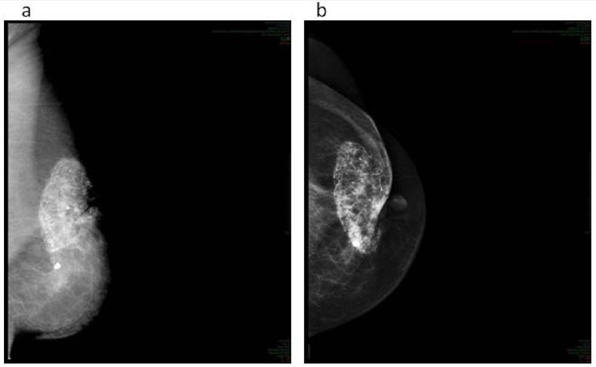
Mammography demonstrating fat necrosis
Image: “G3 fat necrosis” by Department of Radiation Oncology, Laboratory of Medical Physics and Expert Systems, Regina Elena National Cancer Institute, Rome, Italy. License: CC BY 2.0
Typically the biopsyBiopsyRemoval and pathologic examination of specimens from the living body.Ewing Sarcoma method of choice
Uses a 9- to 14-gauge biopsyBiopsyRemoval and pathologic examination of specimens from the living body.Ewing Sarcoma needle to obtain a tissue sample
Done under imaging guidance (typically ultrasonographically or stereotactically)
Fine-needle aspiration (FNA):
Performed under palpationPalpationApplication of fingers with light pressure to the surface of the body to determine consistency of parts beneath in physical diagnosis; includes palpation for determining the outlines of organs.Dermatologic Examination or US guidance
Surgical biopsyBiopsyRemoval and pathologic examination of specimens from the living body.Ewing Sarcoma:
Typically 2nd-line
Indications: if CNB is not feasible or was inconclusive or if CNB results are discordant with imaging
Types:
Excisional biopsyBiopsyRemoval and pathologic examination of specimens from the living body.Ewing Sarcoma: surgically removes the entire lesion
Incisional biopsyBiopsyRemoval and pathologic examination of specimens from the living body.Ewing Sarcoma: surgically removes a portion of the lesion, typically used to confirm the diagnosis with a large massMassThree-dimensional lesion that occupies a space within the breastImaging of the Breast
Findings consistent with fat necrosisNecrosisThe death of cells in an organ or tissue due to disease, injury or failure of the blood supply.Ischemic Cell Damage:[5]
Initially an intense neutrophilic infiltration, followed by lymphocytic infiltration
Foamy macrophagesMacrophagesThe relatively long-lived phagocytic cell of mammalian tissues that are derived from blood monocytes. Main types are peritoneal macrophages; alveolar macrophages; histiocytes; kupffer cells of the liver; and osteoclasts. They may further differentiate within chronic inflammatory lesions to epithelioid cells or may fuse to form foreign body giant cells or langhans giant cells.Innate Immunity: Phagocytes and Antigen Presentation and multinucleated giant cellsGiant cellsMultinucleated masses produced by the fusion of many cells; often associated with viral infections. In aids, they are induced when the envelope glycoprotein of the HIV virus binds to the CD4 antigen of uninfected neighboring T4 cells. The resulting syncytium leads to cell death and thus may account for the cytopathic effect of the virus.Giant Cell Arteritis
Management of fat necrosisNecrosisThe death of cells in an organ or tissue due to disease, injury or failure of the blood supply.Ischemic Cell Damage should be individualized, but it can generally be managed conservatively.[3]
Natural history: Lesions may enlarge, remain unchanged, or regress.
Surgical management usually not required, but may be chosen if the massMassThree-dimensional lesion that occupies a space within the breastImaging of the Breast:
Does not resolve
Causes painPainAn unpleasant sensation induced by noxious stimuli which are detected by nerve endings of nociceptive neurons.Pain: Types and Pathways
Aspiration of oil cystsCystsAny fluid-filled closed cavity or sac that is lined by an epithelium. Cysts can be of normal, abnormal, non-neoplastic, or neoplastic tissues.Fibrocystic Change with a needle if the cystsCystsAny fluid-filled closed cavity or sac that is lined by an epithelium. Cysts can be of normal, abnormal, non-neoplastic, or neoplastic tissues.Fibrocystic Change cause discomfort
Differential Diagnosis
Breast cancerBreast cancerBreast cancer is a disease characterized by malignant transformation of the epithelial cells of the breast. Breast cancer is the most common form of cancer and 2nd most common cause of cancer-related death among women. Breast Cancer: the most important diagnosis to rule out when a patient presents with a breast massMassThree-dimensional lesion that occupies a space within the breastImaging of the Breast or evidence of calcifications and fibrosisFibrosisAny pathological condition where fibrous connective tissue invades any organ, usually as a consequence of inflammation or other injury.Bronchiolitis Obliterans on imaging, as fat necrosisNecrosisThe death of cells in an organ or tissue due to disease, injury or failure of the blood supply.Ischemic Cell Damage may present in a very similar way. If the diagnosis cannot be made based on imaging alone, core needle biopsyCore Needle BiopsyFibrocystic Change is required. Management may involve surgery, chemotherapyChemotherapyOsteosarcoma, radiationRadiationEmission or propagation of acoustic waves (sound), electromagnetic energy waves (such as light; radio waves; gamma rays; or x-rays), or a stream of subatomic particles (such as electrons; neutrons; protons; or alpha particles).Osteosarcoma, and hormonal treatment.
FibrocysticFibrocysticFibrocystic Change changes of the breast: a non-specific term referring to several types of benignBenignFibroadenoma breast conditions that usually occur as a result of cyclic hormonal stimulation from estrogenEstrogenCompounds that interact with estrogen receptors in target tissues to bring about the effects similar to those of estradiol. Estrogens stimulate the female reproductive organs, and the development of secondary female sex characteristics. Estrogenic chemicals include natural, synthetic, steroidal, or non-steroidal compounds.Ovaries: Anatomy and progesteroneProgesteroneThe major progestational steroid that is secreted primarily by the corpus luteum and the placenta. Progesterone acts on the uterus, the mammary glands and the brain. It is required in embryo implantation; pregnancy maintenance, and the development of mammary tissue for milk production. Progesterone, converted from pregnenolone, also serves as an intermediate in the biosynthesis of gonadal steroid hormones and adrenal corticosteroids.Gonadal Hormones. The most common types of changes are non-proliferativeNon-ProliferativeFibrocystic Change lesions including cystsCystsAny fluid-filled closed cavity or sac that is lined by an epithelium. Cysts can be of normal, abnormal, non-neoplastic, or neoplastic tissues.Fibrocystic Change within the ducts and fibrosisFibrosisAny pathological condition where fibrous connective tissue invades any organ, usually as a consequence of inflammation or other injury.Bronchiolitis Obliterans resulting from chronic inflammationChronic InflammationInflammation after these cystsCystsAny fluid-filled closed cavity or sac that is lined by an epithelium. Cysts can be of normal, abnormal, non-neoplastic, or neoplastic tissues.Fibrocystic Change rupture. Diagnosis is made with mammogramMammogramFibrocystic Change and ultrasound imaging. Treatment is supportive.
MastitisMastitisMastitis is inflammation of the breast tissue with or without infection. The most common form of mastitis is associated with lactation in the first few weeks after birth. Non-lactational mastitis includes periductal mastitis and idiopathic granulomatous mastitis (IGM).Mastitis and/or breast abscessBreast AbscessBenign Breast Conditions: inflammationInflammationInflammation is a complex set of responses to infection and injury involving leukocytes as the principal cellular mediators in the body’s defense against pathogenic organisms. Inflammation is also seen as a response to tissue injury in the process of wound healing. The 5 cardinal signs of inflammation are pain, heat, redness, swelling, and loss of function. Inflammation of the breast tissue, most commonly due to infection with skinSkinThe skin, also referred to as the integumentary system, is the largest organ of the body. The skin is primarily composed of the epidermis (outer layer) and dermis (deep layer). The epidermis is primarily composed of keratinocytes that undergo rapid turnover, while the dermis contains dense layers of connective tissue.Skin: Structure and Functions or oral flora introduced during breastfeedingBreastfeedingBreastfeeding is often the primary source of nutrition for the newborn. During pregnancy, hormonal stimulation causes the number and size of mammary glands in the breast to significantly increase. After delivery, prolactin stimulates milk production, while oxytocin stimulates milk expulsion through the lactiferous ducts, where it is sucked out through the nipple by the infant. Breastfeeding. A purulent abscessAbscessAccumulation of purulent material in tissues, organs, or circumscribed spaces, usually associated with signs of infection.Chronic Granulomatous Disease may form. Occasionally, non-lactational mastitisNon-Lactational MastitisMastitis and abscesses are also possible. Cases usually present with a feverFeverFever is defined as a measured body temperature of at least 38°C (100.4°F). Fever is caused by circulating endogenous and/or exogenous pyrogens that increase levels of prostaglandin E2 in the hypothalamus. Fever is commonly associated with chills, rigors, sweating, and flushing of the skin. Fever and painPainAn unpleasant sensation induced by noxious stimuli which are detected by nerve endings of nociceptive neurons.Pain: Types and Pathways, erythemaErythemaRedness of the skin produced by congestion of the capillaries. This condition may result from a variety of disease processes.Chalazion, and edemaEdemaEdema is a condition in which excess serous fluid accumulates in the body cavity or interstitial space of connective tissues. Edema is a symptom observed in several medical conditions. It can be categorized into 2 types, namely, peripheral (in the extremities) and internal (in an organ or body cavity). Edema of the breast, with or without a tender fluctuantFluctuantDermatologic ExaminationmassMassThree-dimensional lesion that occupies a space within the breastImaging of the Breast (abscessAbscessAccumulation of purulent material in tissues, organs, or circumscribed spaces, usually associated with signs of infection.Chronic Granulomatous Disease). Management involves antibiotics, continued expression of breast milk if lactating, and incision and drainageIncision And DrainageChalazion of an abscessAbscessAccumulation of purulent material in tissues, organs, or circumscribed spaces, usually associated with signs of infection.Chronic Granulomatous Disease.
FibroadenomaFibroadenomaFibroadenomas are the most common benign tumor of the female breast and the most common breast tumor in adolescent and young women. The tumors are well-circumscribed, mobile, and unencapsulated, with a rubbery or firm consistency.Fibroadenoma: a benignBenignFibroadenoma solid breast massMassThree-dimensional lesion that occupies a space within the breastImaging of the Breast composed of fibrousFibrousFibrocystic Change and glandular tissue, which presents as a small, well-defined, mobile massMassThree-dimensional lesion that occupies a space within the breastImaging of the Breast with a rubbery or firm consistencyConsistencyDermatologic Examination. The exact etiology is unknown. Diagnosis is confirmed with a core needle biopsyCore Needle BiopsyFibrocystic Change. Management is either excision or observation.
Phyllodes tumorTumorInflammation: a fibroepithelial tumorTumorInflammation similar to fibroadenomas, usually characterized by rapid growth. These tumors may behave like benignBenignFibroadenoma fibroadenomas or may become malignant and metastasize. Phyllodes tumors are associated with Li-Fraumeni syndrome. Diagnosis is by core needle biopsyCore Needle BiopsyFibrocystic Change and management involves complete resection, with adjuvantAdjuvantSubstances that augment, stimulate, activate, potentiate, or modulate the immune response at either the cellular or humoral level. The classical agents (freund’s adjuvant, bcg, corynebacterium parvum, et al.) contain bacterial antigens. Some are endogenous (e.g., histamine, interferon, transfer factor, tuftsin, interleukin-1). Their mode of action is either non-specific, resulting in increased immune responsiveness to a wide variety of antigens, or antigen-specific, i.e., affecting a restricted type of immune response to a narrow group of antigens. The therapeutic efficacy of many biological response modifiers is related to their antigen-specific immunoadjuvanticity.VaccinationradiationRadiationEmission or propagation of acoustic waves (sound), electromagnetic energy waves (such as light; radio waves; gamma rays; or x-rays), or a stream of subatomic particles (such as electrons; neutrons; protons; or alpha particles).Osteosarcoma in malignant cases.
Billing and Coding
Diagnosis Codes:
This code is used to document a diagnosis of fat necrosisNecrosisThe death of cells in an organ or tissue due to disease, injury or failure of the blood supply.Ischemic Cell Damage of the breast, a benignBenignFibroadenoma inflammatory condition that occurs when an area of fatty breast tissue is damaged, often after surgery, radiationRadiationEmission or propagation of acoustic waves (sound), electromagnetic energy waves (such as light; radio waves; gamma rays; or x-rays), or a stream of subatomic particles (such as electrons; neutrons; protons; or alpha particles).Osteosarcoma, or trauma.
Domain
Code
Description
ICD-10-CM
N64.1
Fat necrosisNecrosisThe death of cells in an organ or tissue due to disease, injury or failure of the blood supply.Ischemic Cell Damage of breast
SNOMED CT
41659005
Fat necrosisNecrosisThe death of cells in an organ or tissue due to disease, injury or failure of the blood supply.Ischemic Cell Damage of breast (disorder)
Evaluation & Workup:
These CPT codes are for the imaging and biopsyBiopsyRemoval and pathologic examination of specimens from the living body.Ewing Sarcoma procedures used to evaluate a breast lump. Because fat necrosisNecrosisThe death of cells in an organ or tissue due to disease, injury or failure of the blood supply.Ischemic Cell Damage can mimic the appearance of breast cancerBreast cancerBreast cancer is a disease characterized by malignant transformation of the epithelial cells of the breast. Breast cancer is the most common form of cancer and 2nd most common cause of cancer-related death among women. Breast Cancer on a mammogramMammogramFibrocystic Change or ultrasound, a biopsyBiopsyRemoval and pathologic examination of specimens from the living body.Ewing Sarcoma is often required to confirm the benignBenignFibroadenoma diagnosis.
Ultrasound, breast, unilateral, real time with image documentationDocumentationSystematic organization, storage, retrieval, and dissemination of specialized information, especially of a scientific or technical nature. It often involves authenticating or validating information.Advance Directives
CPT
19083
BiopsyBiopsyRemoval and pathologic examination of specimens from the living body.Ewing Sarcoma, breast, with placement of breast localization device(s), percutaneous; using stereotactic guidance
Procedures/Interventions:
This code is used for a lumpectomyLumpectomyFat Necrosis of the Breast, or excisional biopsyBiopsyRemoval and pathologic examination of specimens from the living body.Ewing Sarcoma, which may be performed to remove the area of fat necrosisNecrosisThe death of cells in an organ or tissue due to disease, injury or failure of the blood supply.Ischemic Cell Damage if it is painful, large, or if the diagnosis cannot be definitively confirmed without removing the entire lesion.
Domain
Code
Description
CPT
19120
Excision of cyst, fibroadenomaFibroadenomaFibroadenomas are the most common benign tumor of the female breast and the most common breast tumor in adolescent and young women. The tumors are well-circumscribed, mobile, and unencapsulated, with a rubbery or firm consistency.Fibroadenoma, or other benignBenignFibroadenoma or malignant tumorTumorInflammation, aberrant breast tissue, duct lesion, nippleNippleThe conic organs which usually give outlet to milk from the mammary glands.Examination of the Breast or areolar lesion
References
American College of Obstetricians and Gynecologists Committee on Practice Bulletins‒Gynecology. (2016). Practice bulletin no. 164: diagnosis and management of benign breast disorders. Obstetrics and Gynecology, 127(6), e141–e156. https://doi.org/10.1097/AOG.0000000000001482
Haran, O., Legarda, C., Gofstein, D., Adelson, D., Singolda, R., Madah, E., Arad, E., Grush, A. E., Barnea, Y. (2022). Treatment algorithm of postsurgical fat necrosis of the breast—revisited (abstract). Seminars in Plastic Surgery, 36(2), 94–100. https://doi.org/10.1055/s-0042-1750435