Overview
Definition and epidemiology
Tricuspid regurgitation (TR) is the backflow of blood through the tricuspid valve into the right atrium during ventricular systole.
- Incidence for physiologically significant TR worldwide is < 1%.
- 70% of normal adults will have a minimal degree of TR.
- Similar incidence rates in men and women
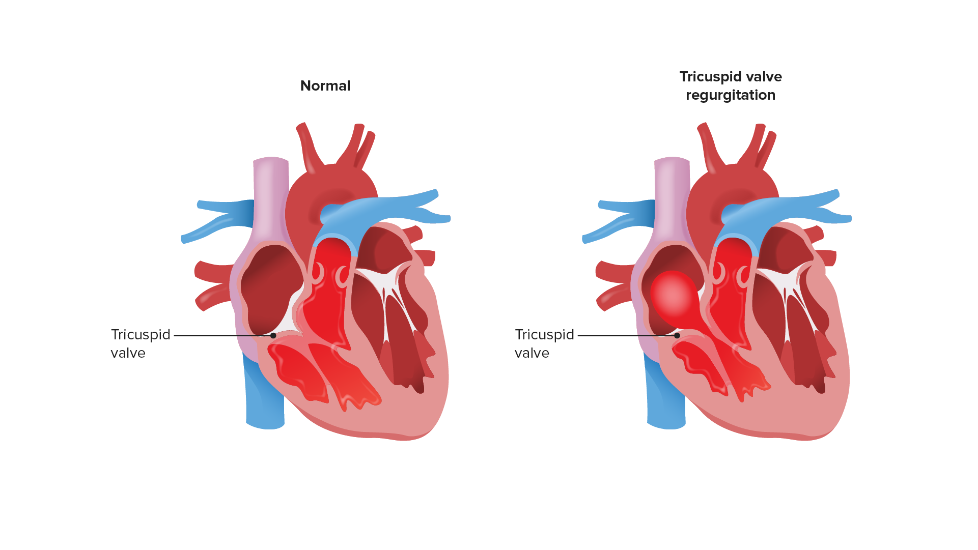
Image shows reflux of blood into the right atrium during systole. This backflow increases right atrial pressures.
Image by Lecturio.Etiology
- Most commonly due to dilation of the valve annulus from right ventricular dilation:
- Pulmonary hypertension
- Mitral or pulmonary stenosis
- Ischemic or idiopathic dilated cardiomyopathy
- Left ventricular heart failure
- Structural anomalies:
- Tricuspid valve prolapse
- Papillary muscle dysfunction
- Myocardial infarction
- Fibrosis
- Infiltrative diseases
- Ebstein anomaly
- Inflammatory or systemic disease:
- Rheumatic heart disease
- Infective endocarditis
- Connective-tissue diseases
- Marfan syndrome
- Ehlers-Danlos syndrome
- Osteogenesis imperfecta
- Systemic lupus erythematosus
- Iatrogenic:
- Pacemaker leads
- Endomyocardial biopsy
- Medications:
- Phentermine
- Fenfluramine
Pathophysiology
- Backflow of blood into the right atrium during systole → increased right atrial pressure → increased venous pressure → systemic venous congestion (can lead to right ventricular systolic dysfunction and low cardiac output)
- Inspiration widens the right ventricle → enlarges the tricuspid valve annulus → worsens regurgitation
Related videos
Clinical Presentation
Symptoms
- Mild or moderate TR is usually asymptomatic.
- Severe TR is associated with right heart failure symptoms:
- Dyspnea on exertion
- Orthopnea
- Paroxysmal nocturnal dyspnea
- Abdominal distension
- Peripheral edema
- Fatigue
Physical exam
- Blowing holosytolic murmur at the lower left sternal border:
- Carvallo’s sign: augmented with inspiration
- Reduced when standing or during Valsalva maneuver
- S3 and/or S4 gallop that increases with inspiration
- Signs of systemic venous congestion:
- Jugular venous distention
- Hepatomegaly
- Hepatojugular reflux
- Abdominal distension and ascites
- Peripheral edema
- If febrile, consider infective endocarditis.
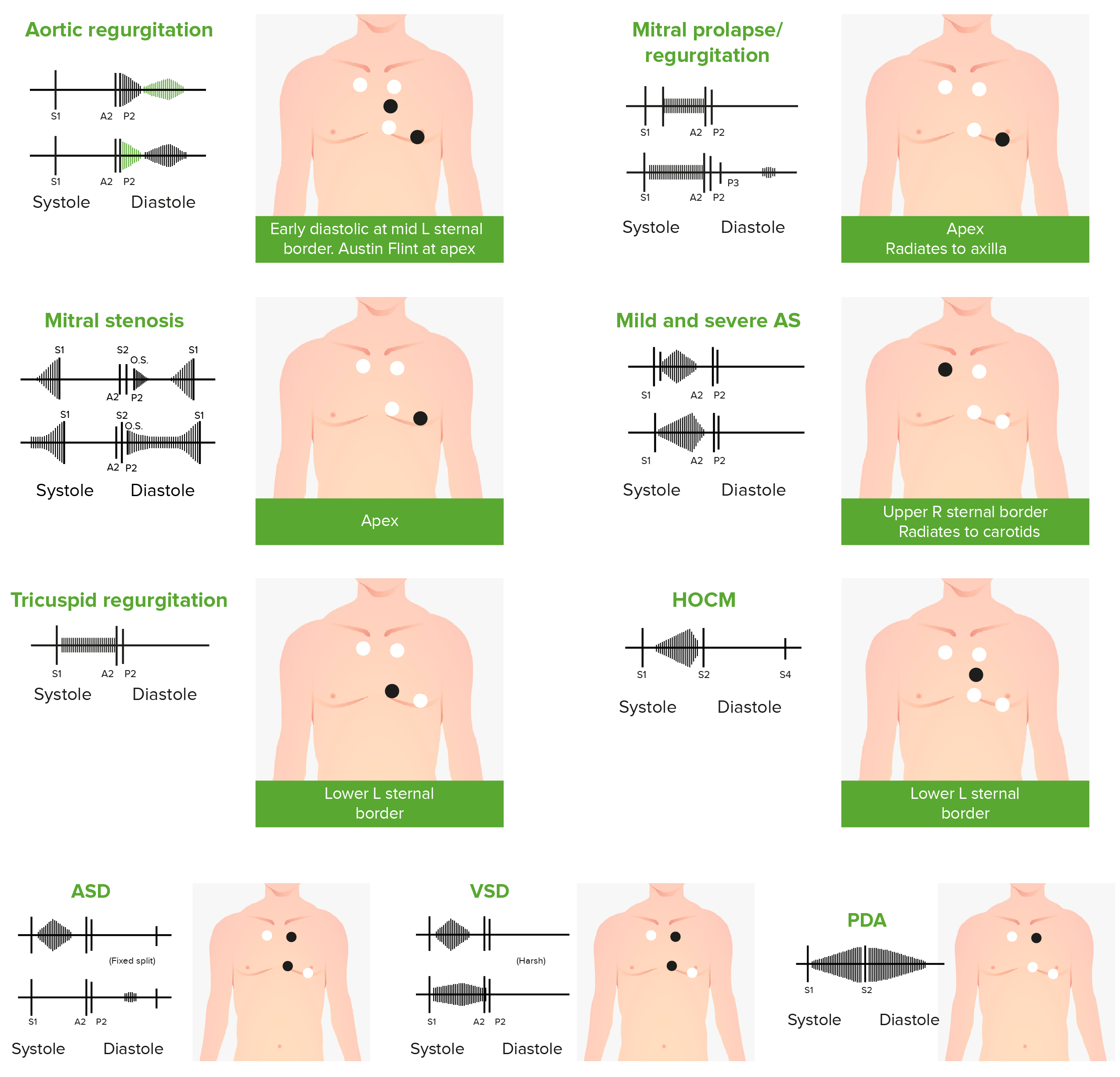
Phonocardiograms of abnormal heart sounds caused by the following cardiac defects:
aortic regurgitation, mitral valve prolapse, mitral stenosis (MS), aortic stenosis (AS), tricuspid regurgitation, hypertrophic obstructive cardiomyopathy (HOCM), atrial septal defect (ASD), ventricular septal defect (VSD), and patent ductus arteriosus (PDA)
Diagnosis and Management
Diagnosis
- Echocardiography (modality of choice)
- Right atrial and right ventricular dilation
- Tricuspid annulus dilation
- Prolapsing, displaced, or scarred leaflets
- Vegetations → endocarditis
- Doppler:
- Establish the severity.
- Evaluate for pulmonary hypertension.
- Cardiac magnetic resonance imaging
- Can help with the diagnosis if echo imaging is suboptimal or inconclusive
- Gives a quantitative assessment of:
- Amount of blood regurgitating back into the right atrium
- Ratio of TR volume to stroke volume
- Right ventricular volumes and ejection fraction
- Cardiac catheterization: not used for the diagnosis of TR, but may assist with finding underlying causes
- Measure pulmonary pressures → pulmonary hypertension
- Evaluate for coronary artery disease and left-sided heart disease.
- Electrocardiogram: no characteristic findings, but may point to an underlying cause
- Q-waves in inferior leads → right ventricular myocardial infarction
- Right ventricular hypertrophy with right axis deviation → pulmonary hypertension
- Right bundle branch block with preexcitation → Ebstein’s anomaly
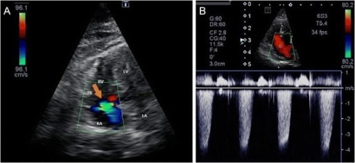
Color Doppler image of tricuspid regurgitation (a) and continuous Doppler (CW) image (b) from the left apical 4-chamber view optimized for the right ventricle
Image: “Tricuspid regurgitation” by US National Library of Medicine. License: CC BY 4.0
LA: left atrium, LV: left ventricle, RA: right atrium, RV: right ventricle, TR: tricuspid regurgitation (orange arrow)
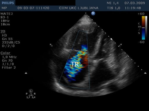
Transthoracic echocardiography from apical 4-chamber view shows massive tricuspid regurgitation (TR).
Image: “Transthoracic echocardiography” by Institute of Pathophysiology, Faculty of Medicine, University of Ljubljana, Slovenia. License: CC BY 2.0
Management
- Systemic volume overload treatment:
- Sodium restriction
- Fluid restriction
- Diuretics
- Treat underlying causes, such as left heart failure and pulmonary hypertension.
- Surgery (repair or replacement) for moderate-to-severe TR is considered if:
- Left-sided valve surgery is also needed
- Endocarditis
Differential Diagnosis
- Congestive heart failure: results when the heart cannot maintain a normal cardiac output. Etiologies can include ischemic, structural, inflammatory, and valvular disease. Symptoms depend on the side of involvement, but include dyspnea, orthopnea, and edema. Diagnosis is made by echocardiogram, and treatment involves diuretics and salt/fluid restriction. Congestive heart failure can occur in conjunction with tricuspid regurgitation, and will be established by echocardiogram.
- Cirrhosis: chronic disease of the liver marked by fibrosis of the parenchyma and impaired function. Symptoms include jaundice, ascites, hepatosplenomegaly, and edema. Diagnosis is made based on liver function test anomalies and an ultrasound showing distorted hepatic architecture with portal hypertension. Treatment includes management of the underlying cause, diuretics, and salt restriction. Echo findings will help distinguish this condition from tricuspid regurgitation.
- Mitral regurgitation: valve disorder where blood refluxes from the left ventricle to the left atrium during systole. Signs and symptoms are based on severity and can include exertional dyspnea, fatigue, or edema. Exam will be notable for a systolic murmur at the cardiac apex, and echocardiogram can establish the diagnosis and differentiate the condition from tricuspid regurgitation. Treatment includes sodium restriction, diuretics, and surgery for severe cases.
- Mitral stenosis: narrowing of the mitral valve, which results in obstruction of blood flow from the left atrium to the left ventricle. Rheumatic heart disease is the most common cause. Patients may be asymptomatic or present with dyspnea. Exam may reveal a low-pitched, rumbling, diastolic murmur at the cardiac apex. Diagnosis is made by echocardiography, and will differentiate this condition from tricuspid regurgitation.
- Tricuspid stenosis: narrowing of the tricuspid valve, which prevents normal blood flow from the right atrium to the right ventricle. Patients may be asymptomatic or present with signs and symptoms of systemic venous congestion. A mid-diastolic murmur at the left lower sternal border distinguishes this condition from tricuspid regurgitation. Echocardiogram will establish the diagnosis. Management includes sodium restriction, diuretics, and surgery for severe cases.
References
- Mancini, M.C. (2018). Tricuspid regurgitation. In O’Brien, T.X. (Ed.), Medscape. Retrieved October 21, 2020, from https://emedicine.medscape.com/article/158484-overview
- Kasper, D.L., Fauci, A. S., Longo, D.L., Bruanwald, E., Hauser, S. L., Jameson, J.L., (2007). Harrison’s principles of internal medicine (16th edition.). New York: McGraw Hill Education.
- Otto, C.M. (2020). Etiology, clinical features, and evaluation of tricuspid regurgitation. In Yeon, S.B. (Ed.), UpToDate. Retrieved October 23, 2020, from https://www.uptodate.com/contents/etiology-clinical-features-and-evaluation-of-tricuspid-regurgitation
- Otto, C.M. (2020). Management and prognosis of tricuspid regurgitation. In Yeon, S.B. (Ed.), UpToDate. Retrieved October 23, 2020, from https://www.uptodate.com/contents/management-and-prognosis-of-tricuspid-regurgitation