Achieve Mastery of Medical Concepts

Study for medical school and boards with Lecturio

Tricuspid Stenosis

Tricuspid stenosis Stenosis Hypoplastic Left Heart Syndrome (HLHS) (TS) is a valvular defect that obstructs blood flow Blood flow Blood flow refers to the movement of a certain volume of blood through the vasculature over a given unit of time (e.g., mL per minute). Vascular Resistance, Flow, and Mean Arterial Pressure from the right atrium to the right ventricle during diastole Diastole Post-systolic relaxation of the heart, especially the heart ventricles. Cardiac Cycle. This condition most commonly results from rheumatic heart disease Rheumatic Heart Disease Cardiac manifestation of systemic rheumatological conditions, such as rheumatic fever. Rheumatic heart disease can involve any part the heart, most often the heart valves and the endocardium. Rheumatic Fever or a congenital Congenital Chorioretinitis defect, and is usually found in conjunction with other valvular disease. A mid-diastolic murmur is best heard at the lower left sternal border. Mild TS may be asymptomatic or present with systemic venous congestion due to increased right atrial and venous pressures. Echocardiography Echocardiography Ultrasonic recording of the size, motion, and composition of the heart and surrounding tissues. The standard approach is transthoracic. Tricuspid Valve Atresia (TVA) can establish the diagnosis. Treatment focuses on heart failure Heart Failure A heterogeneous condition in which the heart is unable to pump out sufficient blood to meet the metabolic need of the body. Heart failure can be caused by structural defects, functional abnormalities (ventricular dysfunction), or a sudden overload beyond its capacity. Chronic heart failure is more common than acute heart failure which results from sudden insult to cardiac function, such as myocardial infarction. Total Anomalous Pulmonary Venous Return (TAPVR) management, and surgery is reserved for severe disease.

Last updated: Jul 10, 2023

Editorial responsibility: Stanley Oiseth, Lindsay Jones, Evelin Maza

Overview

Definition and epidemiology

Tricuspid stenosis Stenosis Hypoplastic Left Heart Syndrome (HLHS) (TS) is the narrowing of the tricuspid valve Tricuspid valve The valve consisting of three cusps situated between the right atrium and right ventricle of the heart. Heart: Anatomy, which obstructs the flow Flow Blood flows through the heart, arteries, capillaries, and veins in a closed, continuous circuit. Flow is the movement of volume per unit of time. Flow is affected by the pressure gradient and the resistance fluid encounters between 2 points. Vascular resistance is the opposition to flow, which is caused primarily by blood friction against vessel walls. Vascular Resistance, Flow, and Mean Arterial Pressure of blood from the right atrium to the right ventricle, resulting in a transvalvular gradient.

  • Rare
    • < 1% of the population in the United States
    • Approximately 3% worldwide (more prevalent in areas with a high incidence Incidence The number of new cases of a given disease during a given period in a specified population. It also is used for the rate at which new events occur in a defined population. It is differentiated from prevalence, which refers to all cases in the population at a given time. Measures of Disease Frequency of rheumatic fever Rheumatic fever Acute rheumatic fever (ARF) is an autoimmune inflammatory process that usually follows Streptococcal pharyngitis. Acute rheumatic fever usually occurs 2-4 weeks after an untreated infection and affects the heart, skin, joints, and nervous system. Rheumatic Fever)
  • More common in women
  • Usually not seen as an isolated condition; most commonly associated with mitral stenosis Stenosis Hypoplastic Left Heart Syndrome (HLHS) and tricuspid regurgitation Regurgitation Gastroesophageal Reflux Disease (GERD)

Etiology

  • Inflammatory or systemic disease
    • Rheumatic fever Rheumatic fever Acute rheumatic fever (ARF) is an autoimmune inflammatory process that usually follows Streptococcal pharyngitis. Acute rheumatic fever usually occurs 2-4 weeks after an untreated infection and affects the heart, skin, joints, and nervous system. Rheumatic Fever (most common cause) 
    • Infective endocarditis Infective endocarditis Infective endocarditis (IE) is caused by infection or inflammation of the inner lining of the heart (endocardium), most commonly affecting the heart valves. Endocarditis
    • Systemic lupus erythematosus Systemic lupus erythematosus Systemic lupus erythematosus (SLE) is a chronic autoimmune, inflammatory condition that causes immune-complex deposition in organs, resulting in systemic manifestations. Women, particularly those of African American descent, are more commonly affected. Systemic Lupus Erythematosus (Libman-Sacks)
  • Congenital Congenital Chorioretinitis
  • Malignancy Malignancy Hemothorax
    • Carcinoid syndrome Carcinoid syndrome A symptom complex associated with carcinoid tumor and characterized by attacks of severe flushing of the skin, diarrheal watery stools, bronchoconstriction, sudden drops in blood pressure, edema, and ascites. The carcinoid tumors are usually located in the gastrointestinal tract and metastasize to the liver. Symptoms are caused by tumor secretion of serotonin, prostaglandins, and other biologically active substances. Cardiac manifestations constitute carcinoid heart disease. Carcinoid Tumors and Syndrome
    • Tumors of the right heart may cause a functional TS (valve is not affected, but blood flow Blood flow Blood flow refers to the movement of a certain volume of blood through the vasculature over a given unit of time (e.g., mL per minute). Vascular Resistance, Flow, and Mean Arterial Pressure is obstructed by the tumor Tumor Inflammation).
  • Iatrogenic Iatrogenic Any adverse condition in a patient occurring as the result of treatment by a physician, surgeon, or other health professional, especially infections acquired by a patient during the course of treatment. Anterior Cord Syndrome
    • Injury from cardiac procedures leading to fibrosis Fibrosis Any pathological condition where fibrous connective tissue invades any organ, usually as a consequence of inflammation or other injury. Bronchiolitis Obliterans of the valve:
    • Radiation Radiation Emission or propagation of acoustic waves (sound), electromagnetic energy waves (such as light; radio waves; gamma rays; or x-rays), or a stream of subatomic particles (such as electrons; neutrons; protons; or alpha particles). Osteosarcoma therapy

Pathophysiology

Tricuspid stenosis

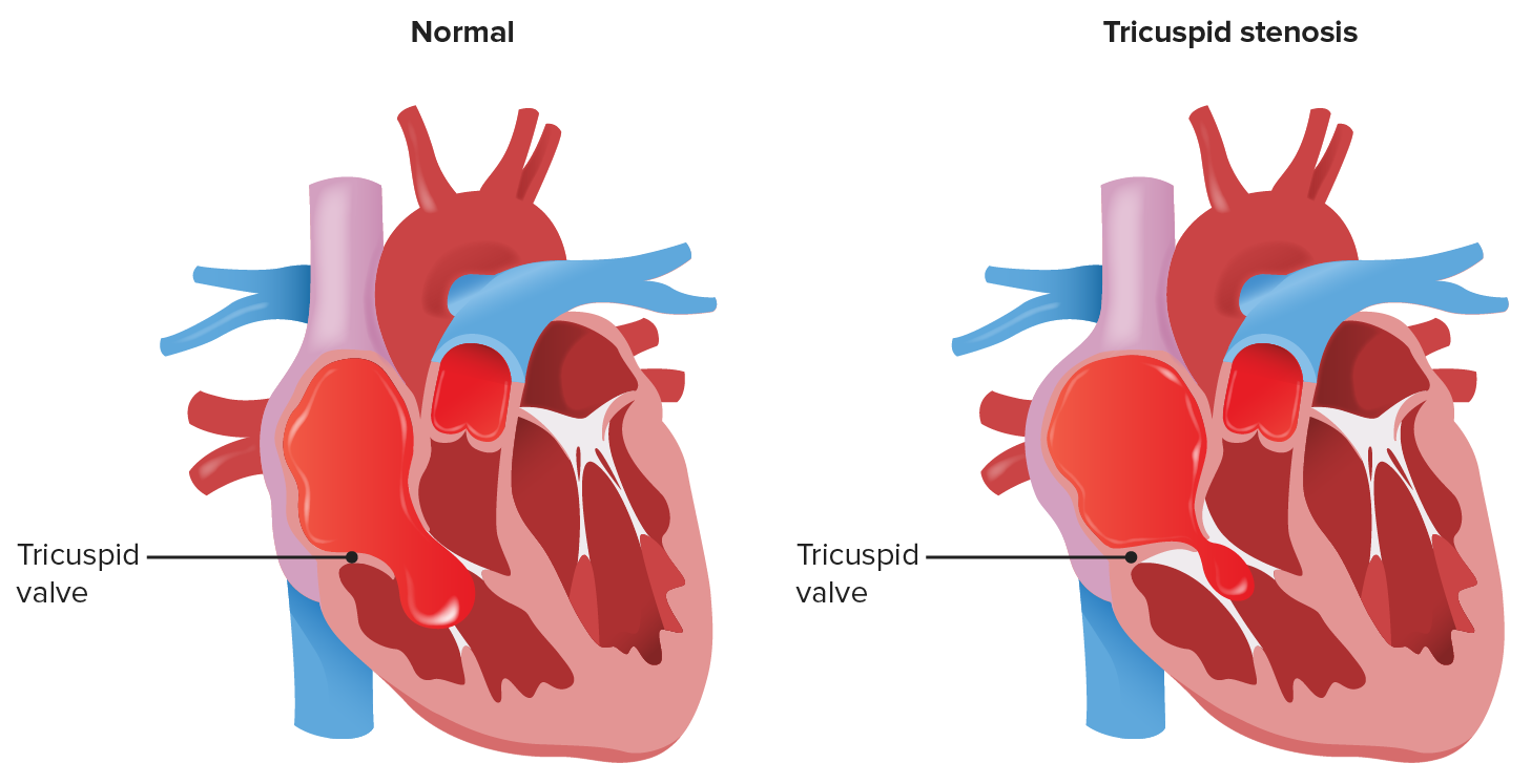
Diseased valve is obstructing blood flow out of the right atrium.

Image by Lecturio.

Clinical Presentation

Symptoms

  • Mild or no dyspnea Dyspnea Dyspnea is the subjective sensation of breathing discomfort. Dyspnea is a normal manifestation of heavy physical or psychological exertion, but also may be caused by underlying conditions (both pulmonary and extrapulmonary). Dyspnea
  • Fatigue Fatigue The state of weariness following a period of exertion, mental or physical, characterized by a decreased capacity for work and reduced efficiency to respond to stimuli. Fibromyalgia due to reduced cardiac output Cardiac output The volume of blood passing through the heart per unit of time. It is usually expressed as liters (volume) per minute so as not to be confused with stroke volume (volume per beat). Cardiac Mechanics 
  • Abdominal discomfort or distension
  • Peripheral edema Peripheral edema Peripheral edema is the swelling of the lower extremities, namely, legs, feet, and ankles. Edema

Physical exam

  • Mid-diastolic murmur heard best at the left lower sternal border or over the xiphoid.
    • Carvallo’s sign:
      • Accentuation of the murmur during inspiration Inspiration Ventilation: Mechanics of Breathing, leg Leg The lower leg, or just “leg” in anatomical terms, is the part of the lower limb between the knee and the ankle joint. The bony structure is composed of the tibia and fibula bones, and the muscles of the leg are grouped into the anterior, lateral, and posterior compartments by extensions of fascia. Leg: Anatomy raise, squatting, and exercise
      • Due to increased blood flow Blood flow Blood flow refers to the movement of a certain volume of blood through the vasculature over a given unit of time (e.g., mL per minute). Vascular Resistance, Flow, and Mean Arterial Pressure across the valve
  • May hear an opening snap Opening snap Mitral Stenosis and widely split S1 S1 Heart Sounds
  • Evidence of systemic venous congestion:
    • Jugular venous distension Jugular Venous Distension Cardiovascular Examination
    • Hepatomegaly
    • Hepatojugular reflux
    • Abdominal distension from ascites Ascites Ascites is the pathologic accumulation of fluid within the peritoneal cavity that occurs due to an osmotic and/or hydrostatic pressure imbalance secondary to portal hypertension (cirrhosis, heart failure) or non-portal hypertension (hypoalbuminemia, malignancy, infection). Ascites
    • Peripheral edema Peripheral edema Peripheral edema is the swelling of the lower extremities, namely, legs, feet, and ankles. Edema

Related videos

Diagnosis and Management

Diagnosis

  • Echocardiography Echocardiography Ultrasonic recording of the size, motion, and composition of the heart and surrounding tissues. The standard approach is transthoracic. Tricuspid Valve Atresia (TVA) (diagnostic modality of choice)
    • Thickening and/or distortion Distortion Defense Mechanisms of the tricuspid valve Tricuspid valve The valve consisting of three cusps situated between the right atrium and right ventricle of the heart. Heart: Anatomy
    • Limited leaflet mobility
    • Right atrium and inferior vena cava Inferior vena cava The venous trunk which receives blood from the lower extremities and from the pelvic and abdominal organs. Mediastinum and Great Vessels: Anatomy enlargement
    • Associated valvular and structural abnormalities
    • Doppler Doppler Ultrasonography applying the doppler effect, with frequency-shifted ultrasound reflections produced by moving targets (usually red blood cells) in the bloodstream along the ultrasound axis in direct proportion to the velocity of movement of the targets, to determine both direction and velocity of blood flow. Ultrasound (Sonography) for estimation of the transvalvular gradient
  • Cardiac catheterization Cardiac Catheterization Procedures in which placement of cardiac catheters is performed for therapeutic or diagnostic procedures. Cardiac Surgery
    • Not required, but useful if there is diagnostic uncertainty from the clinical presentation and echo
    • Measures right atrial and ventricular pressures to calculate the pressure gradient Pressure gradient Vascular Resistance, Flow, and Mean Arterial Pressure
    • Evaluates cardiac output Cardiac output The volume of blood passing through the heart per unit of time. It is usually expressed as liters (volume) per minute so as not to be confused with stroke volume (volume per beat). Cardiac Mechanics and valve area
  • Electrocardiogram Electrocardiogram An electrocardiogram (ECG) is a graphic representation of the electrical activity of the heart plotted against time. Adhesive electrodes are affixed to the skin surface allowing measurement of cardiac impulses from many angles. The ECG provides 3-dimensional information about the conduction system of the heart, the myocardium, and other cardiac structures. Electrocardiogram (ECG)

Management

  • Systemic venous congestion treatment:
  • Treat underlying causes ( endocarditis Endocarditis Endocarditis is an inflammatory disease involving the inner lining (endometrium) of the heart, most commonly affecting the cardiac valves. Both infectious and noninfectious etiologies lead to vegetations on the valve leaflets. Patients may present with nonspecific symptoms such as fever and fatigue. Endocarditis, cardiac tumors).
  • Invasive interventions (reserved for severe, symptomatic stenosis Stenosis Hypoplastic Left Heart Syndrome (HLHS)):
    • Balloon valvotomy
      • Percutaneous procedure to separate and stretch the valve leaflets to increase the valve opening
      • Preferred for high-risk patients Patients Individuals participating in the health care system for the purpose of receiving therapeutic, diagnostic, or preventive procedures. Clinician–Patient Relationship
      • May worsen concurrent tricuspid regurgitation Regurgitation Gastroesophageal Reflux Disease (GERD)
      • Vegetations, thrombi, and tumors are contraindications Contraindications A condition or factor associated with a recipient that makes the use of a drug, procedure, or physical agent improper or inadvisable. Contraindications may be absolute (life threatening) or relative (higher risk of complications in which benefits may outweigh risks). Noninvasive Ventilation due to embolism risk.
    • Surgery
      • Valve repair
      • Valve replacement
Malignant rheumatic heart disease presenting as quadrivalvular stenosis

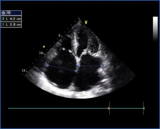
Transthoracic echocardiogram of apical 4-chamber view showing thickened and doming mitral valve and tricuspid valve in rheumatic heart disease

Image: “Transthoracic echocardiogram” by US National Library of Medicine. License: CC BY 2.0

Differential Diagnosis

  • Heart failure Heart Failure A heterogeneous condition in which the heart is unable to pump out sufficient blood to meet the metabolic need of the body. Heart failure can be caused by structural defects, functional abnormalities (ventricular dysfunction), or a sudden overload beyond its capacity. Chronic heart failure is more common than acute heart failure which results from sudden insult to cardiac function, such as myocardial infarction. Total Anomalous Pulmonary Venous Return (TAPVR): results when the heart cannot maintain a normal cardiac output Cardiac output The volume of blood passing through the heart per unit of time. It is usually expressed as liters (volume) per minute so as not to be confused with stroke volume (volume per beat). Cardiac Mechanics. Etiologies can include ischemic, structural, inflammatory, and valvular disease. Symptoms depend on the side of involvement but include dyspnea Dyspnea Dyspnea is the subjective sensation of breathing discomfort. Dyspnea is a normal manifestation of heavy physical or psychological exertion, but also may be caused by underlying conditions (both pulmonary and extrapulmonary). Dyspnea, orthopnea Orthopnea Pulmonary Edema, and edema Edema Edema is a condition in which excess serous fluid accumulates in the body cavity or interstitial space of connective tissues. Edema is a symptom observed in several medical conditions. It can be categorized into 2 types, namely, peripheral (in the extremities) and internal (in an organ or body cavity). Edema. Diagnosis is made by echocardiogram Echocardiogram Transposition of the Great Vessels, and treatment involves diuretics Diuretics Agents that promote the excretion of urine through their effects on kidney function. Heart Failure and Angina Medication and salt/fluid restriction. This condition can occur in conjunction with tricuspid stenosis Stenosis Hypoplastic Left Heart Syndrome (HLHS) and will be established by echocardiogram Echocardiogram Transposition of the Great Vessels.
  • Cirrhosis Cirrhosis Cirrhosis is a late stage of hepatic parenchymal necrosis and scarring (fibrosis) most commonly due to hepatitis C infection and alcoholic liver disease. Patients may present with jaundice, ascites, and hepatosplenomegaly. Cirrhosis can also cause complications such as hepatic encephalopathy, portal hypertension, portal vein thrombosis, and hepatorenal syndrome. Cirrhosis: chronic disease of the liver Liver The liver is the largest gland in the human body. The liver is found in the superior right quadrant of the abdomen and weighs approximately 1.5 kilograms. Its main functions are detoxification, metabolism, nutrient storage (e.g., iron and vitamins), synthesis of coagulation factors, formation of bile, filtration, and storage of blood. Liver: Anatomy marked by fibrosis Fibrosis Any pathological condition where fibrous connective tissue invades any organ, usually as a consequence of inflammation or other injury. Bronchiolitis Obliterans of the parenchyma and impaired function. Symptoms include jaundice Jaundice Jaundice is the abnormal yellowing of the skin and/or sclera caused by the accumulation of bilirubin. Hyperbilirubinemia is caused by either an increase in bilirubin production or a decrease in the hepatic uptake, conjugation, or excretion of bilirubin. Jaundice, ascites Ascites Ascites is the pathologic accumulation of fluid within the peritoneal cavity that occurs due to an osmotic and/or hydrostatic pressure imbalance secondary to portal hypertension (cirrhosis, heart failure) or non-portal hypertension (hypoalbuminemia, malignancy, infection). Ascites, hepatosplenomegaly Hepatosplenomegaly Cytomegalovirus, and edema Edema Edema is a condition in which excess serous fluid accumulates in the body cavity or interstitial space of connective tissues. Edema is a symptom observed in several medical conditions. It can be categorized into 2 types, namely, peripheral (in the extremities) and internal (in an organ or body cavity). Edema. Diagnosis is made based on liver Liver The liver is the largest gland in the human body. The liver is found in the superior right quadrant of the abdomen and weighs approximately 1.5 kilograms. Its main functions are detoxification, metabolism, nutrient storage (e.g., iron and vitamins), synthesis of coagulation factors, formation of bile, filtration, and storage of blood. Liver: Anatomy function test abnormalities and an ultrasound showing distorted hepatic architecture with portal hypertension Portal hypertension Portal hypertension is increased pressure in the portal venous system. This increased pressure can lead to splanchnic vasodilation, collateral blood flow through portosystemic anastomoses, and increased hydrostatic pressure. There are a number of etiologies, including cirrhosis, right-sided congestive heart failure, schistosomiasis, portal vein thrombosis, hepatitis, and Budd-Chiari syndrome. Portal Hypertension. Treatment includes management of the underlying cause, diuretics Diuretics Agents that promote the excretion of urine through their effects on kidney function. Heart Failure and Angina Medication, and salt  restriction. Echo findings will help differentiate this condition from tricuspid stenosis Stenosis Hypoplastic Left Heart Syndrome (HLHS).
  • Mitral regurgitation Regurgitation Gastroesophageal Reflux Disease (GERD): valve disorder where blood refluxes from the left ventricle to the left atrium during systole Systole Period of contraction of the heart, especially of the heart ventricles. Cardiac Cycle. Signs and symptoms are based on severity and can include exertional dyspnea Dyspnea Dyspnea is the subjective sensation of breathing discomfort. Dyspnea is a normal manifestation of heavy physical or psychological exertion, but also may be caused by underlying conditions (both pulmonary and extrapulmonary). Dyspnea, fatigue Fatigue The state of weariness following a period of exertion, mental or physical, characterized by a decreased capacity for work and reduced efficiency to respond to stimuli. Fibromyalgia, or edema Edema Edema is a condition in which excess serous fluid accumulates in the body cavity or interstitial space of connective tissues. Edema is a symptom observed in several medical conditions. It can be categorized into 2 types, namely, peripheral (in the extremities) and internal (in an organ or body cavity). Edema. Exam will be notable for a systolic murmur at the cardiac apex, and echocardiogram Echocardiogram Transposition of the Great Vessels can establish the diagnosis and differentiate it from tricuspid stenosis Stenosis Hypoplastic Left Heart Syndrome (HLHS). Treatment includes sodium Sodium A member of the alkali group of metals. It has the atomic symbol na, atomic number 11, and atomic weight 23. Hyponatremia restriction, diuretics Diuretics Agents that promote the excretion of urine through their effects on kidney function. Heart Failure and Angina Medication, and surgery for severe cases.
  • Mitral stenosis Stenosis Hypoplastic Left Heart Syndrome (HLHS): narrowing of the mitral valve Mitral valve The valve between the left atrium and left ventricle of the heart. Heart: Anatomy, which results in obstruction of blood flow Blood flow Blood flow refers to the movement of a certain volume of blood through the vasculature over a given unit of time (e.g., mL per minute). Vascular Resistance, Flow, and Mean Arterial Pressure from the left atrium to the left ventricle. Rheumatic heart disease Rheumatic Heart Disease Cardiac manifestation of systemic rheumatological conditions, such as rheumatic fever. Rheumatic heart disease can involve any part the heart, most often the heart valves and the endocardium. Rheumatic Fever is the most common cause. Patients Patients Individuals participating in the health care system for the purpose of receiving therapeutic, diagnostic, or preventive procedures. Clinician–Patient Relationship may be asymptomatic or present with dyspnea Dyspnea Dyspnea is the subjective sensation of breathing discomfort. Dyspnea is a normal manifestation of heavy physical or psychological exertion, but also may be caused by underlying conditions (both pulmonary and extrapulmonary). Dyspnea. Exam may reveal a low-pitched, rumbling, diastolic murmur at the cardiac apex. Diagnosis is made by echocardiography Echocardiography Ultrasonic recording of the size, motion, and composition of the heart and surrounding tissues. The standard approach is transthoracic. Tricuspid Valve Atresia (TVA) and will differentiate this condition from tricuspid stenosis Stenosis Hypoplastic Left Heart Syndrome (HLHS).
  • Tricuspid regurgitation Regurgitation Gastroesophageal Reflux Disease (GERD): valve disorder allowing blood to reflux into the right atrium from the right ventricle during systole Systole Period of contraction of the heart, especially of the heart ventricles. Cardiac Cycle. Patients Patients Individuals participating in the health care system for the purpose of receiving therapeutic, diagnostic, or preventive procedures. Clinician–Patient Relationship may be asymptomatic or present with signs and symptoms of systemic venous congestion. A holosystolic murmur Holosystolic Murmur Tricuspid Valve Atresia (TVA) at the left lower sternal border distinguishes this condition from tricuspid stenosis Stenosis Hypoplastic Left Heart Syndrome (HLHS). Echocardiogram Echocardiogram Transposition of the Great Vessels will establish the diagnosis. Management involves treating the underlying cause, sodium Sodium A member of the alkali group of metals. It has the atomic symbol na, atomic number 11, and atomic weight 23. Hyponatremia restriction, diuretics Diuretics Agents that promote the excretion of urine through their effects on kidney function. Heart Failure and Angina Medication, and surgery for severe cases.

References

  1. Mancini, M.C. (2016). Tricuspid stenosis. In Lange, R.A. (Ed.), Medscape. Retrieved October 21, 2020, from https://emedicine.medscape.com/article/158604-overview
  2. Kasper, D.L., Fauci, A. S., Longo, D.L., Bruanwald, E., Hauser, S. L., Jameson, J.L., (2007). Harrison’s principles of internal medicine (16th edition.). New York: McGraw Hill Education.
  3. Peters, F. (2020). Tricuspid Stenosis. In Yeon, S. B. (Ed.), UpToDate. Retrieved October 23, 2020, from https://www.uptodate.com/contents/tricuspid-stenosis
  4. Connolly, M. (2020). Clinical manifestations and diagnosis of Ebstein anomaly. Retrieved April 20, 2022, from: https://www.uptodate.com/contents/clinical-manifestations-and-diagnosis-of-ebstein-anomaly

Create your free account or log in to continue reading!

Sign up now and get free access to Lecturio with concept pages, medical videos, and questions for your medical education.

User Reviews

Details