Overview
Epidemiology
- Most common cardiac valve anomaly
- Affects 3%–7% of the global population
- Women > men
- Most common between 15–30 years of age
- Complications are more often seen in men.
Etiology
- Primary/idiopathic (most common)
- Secondary:
- Connective tissue disorders:
- Marfan’s syndrome
- Ehlers-Danlos syndrome
- Osteogenesis imperfecta
- Pseudoxanthoma elasticum
- Cardiac abnormalities:
- Atrial septal defect
- Ebstein’s anomaly
- Hypertrophic cardiomyopathy
- Connective tissue disorders:
Pathophysiology
Mitral valve prolapse (MVP) is the bulging of the mitral valve (MV) leaflets into the atrium by ≥ 2 mm above the plane of the MV annulus during ventricular systole:
- Most commonly characterized by myxomatous degeneration (Barlow’s syndrome): accumulation of glycosaminoglycans (GAGs) and alterations in collagen structure → expansion of the spongiosa layer of the MV leaflets and chordae tendineae leading to:
- Thickened, stretchy leaflets with excess tissue
- Elongated chordae tendineae (rupture less common)
- MV annulus enlargement
- Endothelial disruption of the MV leaflets → possible sites for endocarditis and thrombus formation
- Fibroelastic deficiency is another potential cause (usually older patients): ↓ collagen, elastin, and proteoglycans leads to:
- Thin leaflets, no excess tissue
- Some annulus dilation
- Thin, elongated chordae → ↑ risk of rupture
- MVP may progress to mitral regurgitation (MR):
- Billowing of leaflets into the atrium during systole → mild MR
- Ruptured chordae tendineae → flail leaflets → severe MR
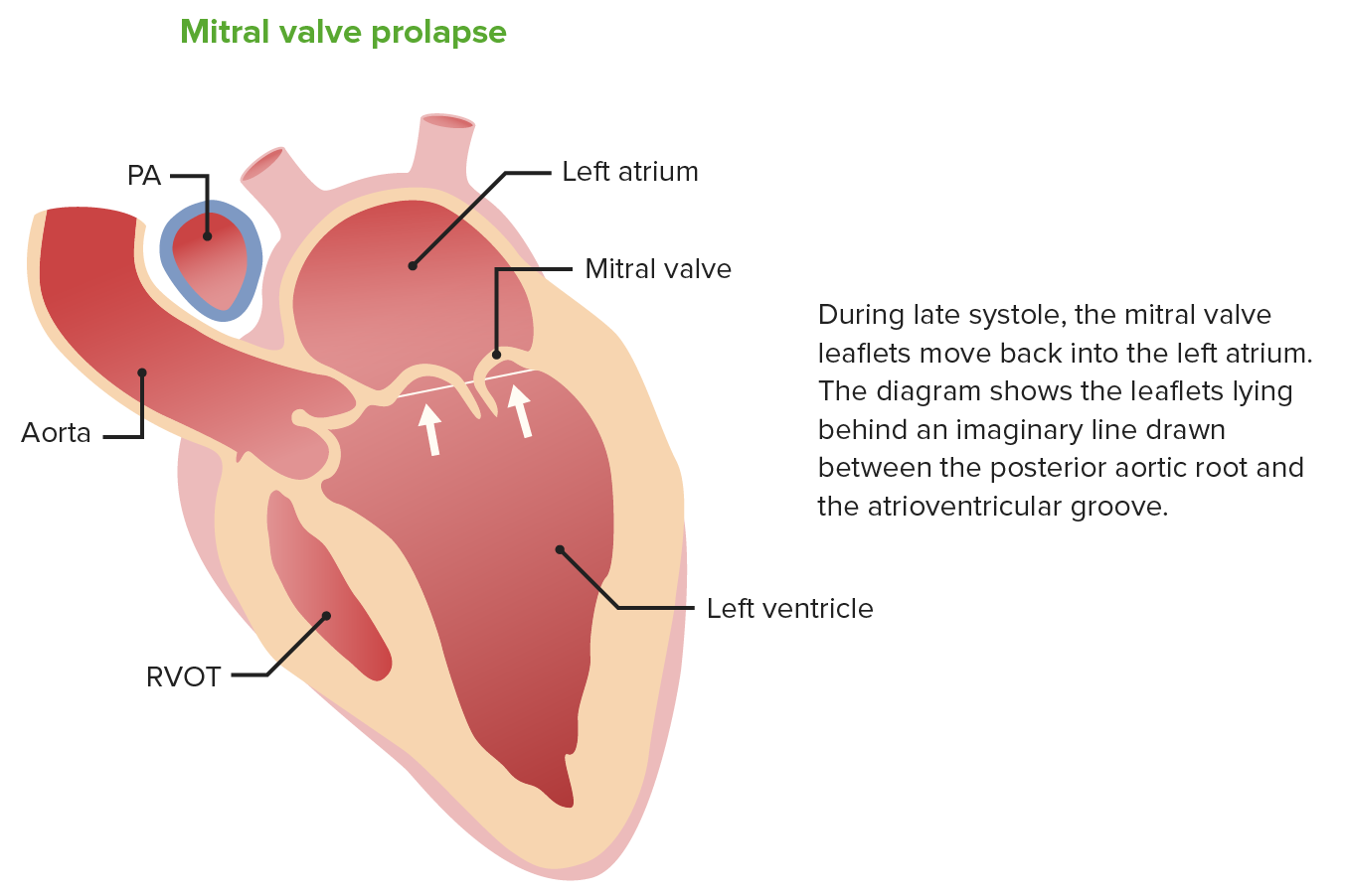
Billowing of the mitral valve leaflets into the left atrium during systole in mitral valve prolapse
Image by Lecturio.
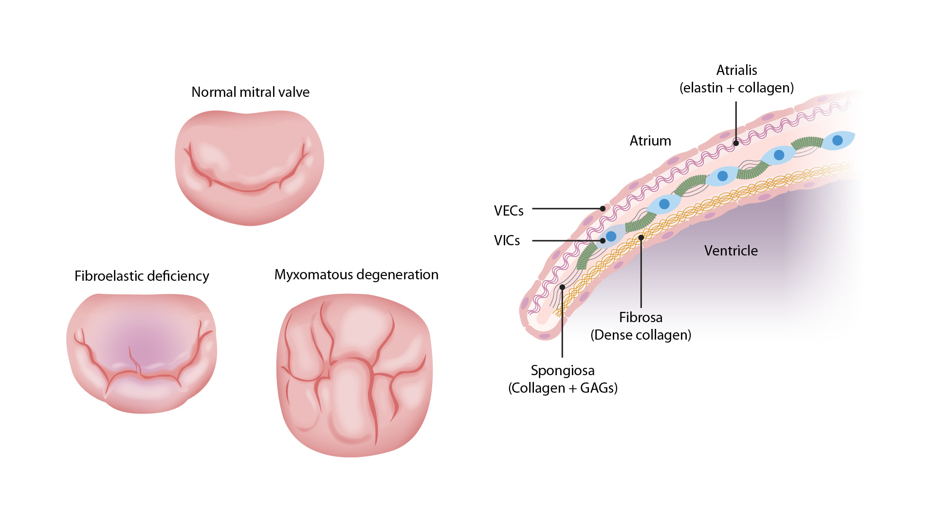
Left: Images comparing the different pathologies seen in MVP. The top valve is a normal MV with the anterior (upper) and posterior (lower) leaflets. The bottom right valve shows thickened, redundant leaflet tissue of myxomatous degeneration, which can billow into the atrium during systole. The bottom left valve has the thinned leaflets of fibroelastic deficiency. The chordae tendinae are also thin and can easily rupture, allowing the leaflet to flail into the atrium.
Right: Image demonstrating a normal MV. There are 3 layers: the atrialis, the spongiosa, and the fibrosa. Myxomatous degeneration in MVP requires proliferation of the spongiosa layer with increased GAGs. There will also be altered collagen composition and elastin fragmentation, which can disrupt the valvular endothelial cells (VECs), which can become sites for endocarditis and thrombus formation.VICs = valvular interstitial cells
Image by Lecturio.
Related videos
Clinical Presentation
Clinical manifestations
- Most cases are asymptomatic (90% of cases).
- Autonomic dysfunction (also known as “MVP syndrome”) may sometimes be seen, but it is unclear whether these symptoms are directly attributable to MVP:
- Anxiety and panic attacks
- Dyspnea
- Palpitations
- Exercise intolerance
- Atypical chest pain
- Orthostasis
- Neuropsychiatric symptoms
- Syncope
- Patients may develop progressive symptoms, typically related to MR:
- Dyspnea
- Orthopnea
- Paroxysmal nocturnal dyspnea
- Chest pain
- Symptoms of an arrhythmia:
- Palpitations
- Light-headedness
- Syncope
- Sudden death (rare)
Physical exam
- Mid-to-late systolic click (snapping of the mitral chordae during systole):
- Heard earlier with decreased venous return:
- Standing
- Valsalva maneuver
- Delayed with increased venous return:
- Squatting
- Hand grip
- Isometric exercise
- Heard earlier with decreased venous return:
- Mid-to-late systolic murmur over the apex following the click: may radiate to the axilla and back, or be heard over the precordial area, depending on the direction of the regurgitant jet
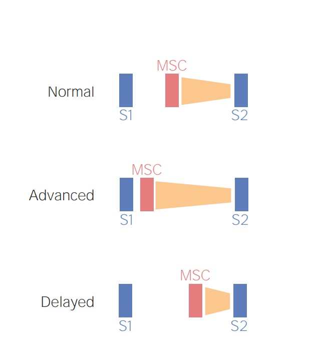
Schematic diagram depicting the preload-dependent changes in the mid-systolic click (MSC) and late-systolic murmur (orange). Increased venous return (e.g., squatting), causes a delay in the MSC-murmur complex while a reduced venous return (e.g., standing) would bring the MSC-murmur complex closer to S1.
Image by Lecturio.
Related videos
Diagnosis and Complications
Diagnosis
- Transthoracic echocardiography (TTE) is the preferred initial study for MVP:
- Systolic displacement of the valve leaflets by ≥ 2 mm above the mitral annulus plane into the LA
- Abnormal leaflet length and thickening
- Chordal elongation
- Enlarged annular diameter
- Doppler is used to diagnose and assess the severity of MR.
- Transesophageal echocardiography (TEE):
- Most often done prior to valve repair surgery
- Gives a more accurate view of the affected leaflets
- Cardiac magnetic resonance imaging (cMRI)
- Emerging tool that is under investigation for valvular disease
- Can be used for diagnosis and to assess for MR
- Electrocardiogram (ECG):
- Frequently normal
- Occasional T-wave inversion in inferior leads (II, III, aVF) or premature atrial or ventricular beats
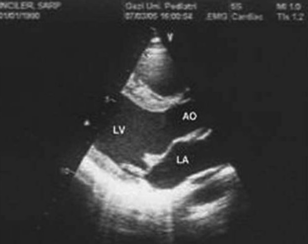
Mitral valve prolapse in cross-sectional echocardiographic examination
AO: aorta, LA: left atrium, LV: left ventricle
Image: “Mitral valve prolapse” by Kecioren Training and Research Hospital, Ankara, Turkey. License: CC BY 2.5, edited by Lecturio.A cMRI showing the bileaflet prolapse of the MV at the end of systole (see arrows)
Image: “Bileaflet prolapse” by the Manchester Heart Centre, Manchester Royal Infirmary, Oxford Road, Manchester M13 9WL, UK. License: CC BY 2.0.
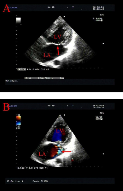
Transthoracic echocardiography of MV prolapse.
Image: “Transthoracic echocardiography of mitral valve prolapse” by Shi-Min Yuan, MMed, PhD. License: CC BY 4.0.
A: prolapse of the anterior MV leaflet behind the annular plane (arrow)
B: mitral insufficiency with a regurgitant flow along the posterior wall of the LA (arrow)
LA: left atrium; LV: left ventricle
Complications
- MVP is the most common etiology of MR:
- Results from progressive prolapse
- Most patients will only have trace or mild MR.
- The development of severe MR may lead to heart failure.
- Arrhythmias:
- Atrial fibrillation (due to MR)
- Paroxysmal supraventricular tachycardia
- Ventricular ectopy
- Infective endocarditis
- Transient ischemic attacks and cerebral vascular accidents:
- Likely related to the risk of atrial fibrillation
- Endothelial disruption of the valve leaflets from myxomatous changes may increase the risk of thrombus formation.
- Sudden cardiac death (rare):
- Usually due to ventricular fibrillation
- Relationship to MVP is uncertain.
Management and Prognosis
Management
- No treatment is required in asymptomatic patients; exercise is encouraged.
- Medications:
- Beta blockers:
- For symptomatic relief of chest pain or palpitations
- Reduce the risk of tachyarrhythmias
- Antibiotic prophylaxis for infective endocarditis:
- No longer recommended
- Can be used for those at high risk of complications from endocarditis
- Anticoagulation:
- Patients with atrial fibrillation
- History of thromboembolism
- Beta blockers:
- Surgery (repair or replacement) is reserved for severe, symptomatic MR.
Prognosis
- Usually benign
- Risk of serious complications: 1% per year
- Risk of sudden cardiac death: approximately 1% per year; more common with:
- Severe symptoms
- Depressed left ventricle (LV) systolic function
- Flail mitral leaflet (from chordal rupture)
Differential Diagnosis
- MR: valve disorder where blood refluxes from the LV to the LA during systole. MVP is the most common etiology. Signs and symptoms are based on severity and can include exertional dyspnea, fatigue, or edema. The exam will be notable for a systolic murmur at the cardiac apex, and an echocardiogram can establish the diagnosis and differentiate MR from MVP. Treatment includes sodium restriction, diuretics, and surgery for severe cases.
- Mitral stenosis: narrowing of the MV, which results in obstruction of blood flow from the LA to the LV. Rheumatic heart disease is the most common cause. Patients may be asymptomatic or may present with dyspnea. The exam may reveal a low-pitched, rumbling, diastolic murmur at the cardiac apex, which will differentiate mitral stenosis from MVP. Diagnosis is made by echocardiography, and treatment includes diuretics, beta blockers, and surgery for severe disease.
- Aortic stenosis: narrowing of the aortic valve, which obstructs blood flow from the LV to the aorta. Patients may develop the classic triad of syncope, angina, and exertional dyspnea as aortic stenosis progresses. The exam will reveal a systolic crescendo-decrescendo murmur at the upper right sternal border, which will differentiate aortic stenosis from MVP. The diagnosis is confirmed by echocardiography, and valvuloplasty or valve replacement is performed for severe disease.
- Tricuspid stenosis: narrowing of the tricuspid valve, which prevents normal blood flow from the right atrium (RA) to the right ventricle (RV). Patients may be asymptomatic or may present with signs and symptoms of systemic venous congestion. A mid-diastolic murmur at the left lower sternal border distinguishes tricuspid stenosis from MVP. Echocardiography will establish the diagnosis. Management includes sodium restriction, diuretics, and surgery for severe cases.
- Tricuspid regurgitation: valve disorder allowing blood to reflux into the RA from the RV during systole. Patients may be asymptomatic or may present with signs and symptoms of systemic venous congestion. A holosystolic murmur at the left lower sternal border distinguishes tricuspid regurgitation from MVP. Echocardiography will establish the diagnosis. Management involves treating the underlying cause, sodium restriction, diuretics, and surgery for severe cases.
References
- Jelani, Q., Schussheim, A.E., & Thakkar, B.V. (2016). Mitral valve prolapse. In Lange, R.A. (Ed.), Medscape. Retrieved November 15, 2020, from https://emedicine.medscape.com/article/155494-overview#a1
- Pislaru, S., & Enriquez-Sarano, M. (2017). Definition and diagnosis of mitral valve prolapse. In Yeon, S.B. (Ed.), Uptodate. Retrieved November 15, 2020, from https://www.uptodate.com/contents/definition-and-diagnosis-of-mitral-valve-prolapse
- Sorrentino, M.J. (2016). Mitral valve prolapse syndrome. In Yeon, S.B. (Ed.), Uptodate. Retrieved November 15, 2020, from https://www.uptodate.com/contents/mitral-valve-prolapse-syndrome
- Pislaru, S., & Enriquez-Sarano, M. (2018). Nonarrhythmic complications of mitral valve prolapse. In Yeon, S.B. (Ed.), Uptodate. Retrieved November 15, 2020, from https://www.uptodate.com/contents/nonarrhythmic-complications-of-mitral-valve-prolapse
- Sorrentino, M.J. (2019). Arrhythmic complications of mitral valve prolapse. In Yeon, S.B. (Ed.), Uptodate. Retrieved November 15, 2020, from https://www.uptodate.com/contents/arrhythmic-complications-of-mitral-valve-prolapse
- Armstrong, G.P. (2020). Mitral valve prolapse. [online] MSD Manual Professional Version. Retrieved November 15, 2020, from https://www.msdmanuals.com/professional/cardiovascular-disorders/valvular-disorders/mitral-valve-prolapse-mvp