Achieve Mastery of Medical Concepts

Study for medical school and boards with Lecturio

True Hermaphroditism

True hermaphroditism, or ovotesticular disorder of sexual development (ODSD), is characterized by the presence of an ovotesticular gonad that contains both ovarian and testicular elements. Individuals are usually born with ambiguous genitalia Ambiguous Genitalia Primary Amenorrhea, but the diagnosis is rarely confirmed before puberty Puberty Puberty is a complex series of physical, psychosocial, and cognitive transitions usually experienced by adolescents (11-19 years of age). Puberty is marked by a growth in stature and the development of secondary sexual characteristics, achievement of fertility, and changes in most body systems. Puberty. The most common karyotype Karyotype The full set of chromosomes presented as a systematized array of metaphase chromosomes from a photomicrograph of a single cell nucleus arranged in pairs in descending order of size and according to the position of the centromere. Congenital Malformations of the Female Reproductive System is 46,XX, and less often, 46,XY can be identified. Gonadal biopsy Biopsy Removal and pathologic examination of specimens from the living body. Ewing Sarcoma with histologic examination confirms the diagnosis. Management needs to consider the individual’s preferences wherever possible. Treatment options range from hormone replacement to surgical removal of part of the ovotestis gonad to improve fertility and to decrease the risk of malignancies.

Last updated: Jan 17, 2023

Editorial responsibility: Stanley Oiseth, Lindsay Jones, Evelin Maza

Overview

Epidemiology

  • Incidence Incidence The number of new cases of a given disease during a given period in a specified population. It also is used for the rate at which new events occur in a defined population. It is differentiated from prevalence, which refers to all cases in the population at a given time. Measures of Disease Frequency of true hermaphroditism, or ovotesticular disorder of sexual development (ODSD): rare 
  • More common in Southern Africa than anywhere else in the world
  • Estimated frequency of the presence of ovotestis is 1 in 83,000 births.
  • Responsible for approximately 10% of cases of ambiguous genitalia Ambiguous Genitalia Primary Amenorrhea
  • Only 20% of cases are diagnosed by 5 years of age.
  • Only 75% of cases are diagnosed by 20 years of age.

Genetics Genetics Genetics is the study of genes and their functions and behaviors. Basic Terms of Genetics

  • Karyotype Karyotype The full set of chromosomes presented as a systematized array of metaphase chromosomes from a photomicrograph of a single cell nucleus arranged in pairs in descending order of size and according to the position of the centromere. Congenital Malformations of the Female Reproductive System: typically normal
    • 46,XX (80% of cases)
    • 46,XY (approximately 10%)
    • 46,XX and XY mosaicism Mosaicism The occurrence in an individual of two or more cell populations of different chromosomal constitutions, derived from a single zygote, as opposed to chimerism in which the different cell populations are derived from more than one zygote. Chromosome Testing (< 1%)
  • Possible etiologies include:
    • Translocation of SRY ( sex Sex The totality of characteristics of reproductive structure, functions, phenotype, and genotype, differentiating the male from the female organism. Gender Dysphoria determining the region on the Y chromosome Y chromosome The male sex chromosome, being the differential sex chromosome carried by half the male gametes and none of the female gametes in humans and in some other male-heterogametic species in which the homologue of the X chromosome has been retained. Basic Terms of Genetics) to the X chromosome X chromosome The female sex chromosome, being the differential sex chromosome carried by half the male gametes and all female gametes in human and other male-heterogametic species. Basic Terms of Genetics or any other non-sex chromosome Chromosome In a prokaryotic cell or in the nucleus of a eukaryotic cell, a structure consisting of or containing DNA which carries the genetic information essential to the cell. Basic Terms of GeneticsSRY needed for gonadal development and testis differentiation
    • NR5A1 gene mutation Gene Mutation Myotonic Dystrophies, which is important for gonadal development and testicular differentiation

Pathophysiology

Ovotesticular disorder of sexual development (ODSD) is characterized by the presence of gonads Gonads The gamete-producing glands, ovary or testis. Hormones: Overview and Types that contain ovarian tissue with primordial follicles and testicular tissue with seminiferous tubules Seminiferous Tubules The convoluted tubules in the testis where sperm are produced (spermatogenesis) and conveyed to the rete testis. Spermatogenic tubules are composed of developing germ cells and the supporting sertoli cells. Testicles: Anatomy in the same individual.

An affected person may have:

  • An ovotestis gonad:
    • Gonad in which testicular and ovarian elements are combined
    • Present in approximately 60% of patients Patients Individuals participating in the health care system for the purpose of receiving therapeutic, diagnostic, or preventive procedures. Clinician–Patient Relationship with ODSD
    • May be bilateral (50%) or unilateral (20%) 
    • 50% intra-abdominal, 25% inguinal, and 25% labioscrotal 
    • Ovarian part of the ovotestis usually functional and responds to estrogens → resulting in excess estrogen Estrogen Compounds that interact with estrogen receptors in target tissues to bring about the effects similar to those of estradiol. Estrogens stimulate the female reproductive organs, and the development of secondary female sex characteristics. Estrogenic chemicals include natural, synthetic, steroidal, or non-steroidal compounds. Ovaries: Anatomy → inhibiting spermatozoa development → rendering the testicular part of the ovotestis non-functional
  • A normal ovary on 1 side (typically the left side) and an ovotestis gonad on the other
  • A normal testis on 1 side (typically the right side) and an ovotestis gonad on the other
  • A normal testis on 1 side and a normal ovary on the other (30%)

The type of internal genitalia present depends on the adjacent gonad:
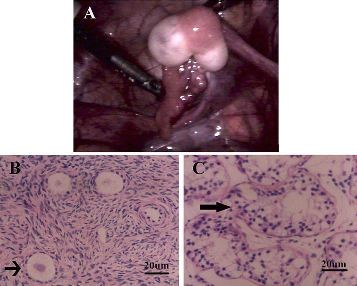
  • Fallopian tubes Fallopian tubes The uterus, cervix, and fallopian tubes are part of the internal female reproductive system. The fallopian tubes receive an ovum after ovulation and help move it and/or a fertilized embryo toward the uterus via ciliated cells lining the tubes and peristaltic movements of its smooth muscle. Uterus, Cervix, and Fallopian Tubes: Anatomy develop beside an ovary.
  • Vas deferens Vas Deferens The excretory duct of the testes that carries spermatozoa. It rises from the scrotum and joins the seminal vesicles to form the ejaculatory duct. Testicles: Anatomy and epididymis Epididymis The convoluted cordlike structure attached to the posterior of the testis. Epididymis consists of the head (caput), the body (corpus), and the tail (cauda). A network of ducts leaving the testis joins into a common epididymal tubule proper which provides the transport, storage, and maturation of spermatozoa. Testicles: Anatomy develop beside a testicle.
  • In the presence of ovotestis, fallopian tubes Fallopian tubes The uterus, cervix, and fallopian tubes are part of the internal female reproductive system. The fallopian tubes receive an ovum after ovulation and help move it and/or a fertilized embryo toward the uterus via ciliated cells lining the tubes and peristaltic movements of its smooth muscle. Uterus, Cervix, and Fallopian Tubes: Anatomy develop in 60%–70% of cases.
  • A uterus Uterus The uterus, cervix, and fallopian tubes are part of the internal female reproductive system. The uterus has a thick wall made of smooth muscle (the myometrium) and an inner mucosal layer (the endometrium). The most inferior portion of the uterus is the cervix, which connects the uterine cavity to the vagina. Uterus, Cervix, and Fallopian Tubes: Anatomy can develop in the case of an ovotestis with a contralateral ovary.
Ovotestis gonads

Ovotestis gonads

A: Gross appearance of a bilobed ovotestis with a fallopian tube: The tan-yellow tissue in the center of the ovotestis revealed ovarian tissue and the white tissue at each pole showed testicular tissue on histologic examination.
B: Microscopic examination of the tan-yellow tissue showed typical ovarian stroma with primordial and primary follicles (arrow), and examination of the white tissue.
C: Image shows unremarkable testicular tissue with well-developed seminiferous tubules (arrow), with no definite spermatids.
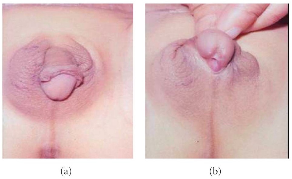
Image: “Pathological analysis” by the Institute of Laboratory Medicine, Jinling Hospital, Nanjing University School of Medicine, Nanjing 210002, PR China. License: CC BY 2.0.

Related videos

Clinical Presentation

Female individuals (46,XX)

  • Infancy:
  • Adulthood:
    • Cervical atresia Atresia Hypoplastic Left Heart Syndrome (HLHS) producing an obstructed genital tract:
      • Cryptomenorrhea ( menstruation Menstruation The periodic shedding of the endometrium and associated menstrual bleeding in the menstrual cycle of humans and primates. Menstruation is due to the decline in circulating progesterone, and occurs at the late luteal phase when luteolysis of the corpus luteum takes place. Menstrual Cycle occurs but not visible due to obstruction of the outflow tract)
      • Hematometra (blood collects or retains in the uterus Uterus The uterus, cervix, and fallopian tubes are part of the internal female reproductive system. The uterus has a thick wall made of smooth muscle (the myometrium) and an inner mucosal layer (the endometrium). The most inferior portion of the uterus is the cervix, which connects the uterine cavity to the vagina. Uterus, Cervix, and Fallopian Tubes: Anatomy)
      • Endometriosis Endometriosis Endometriosis is a common disease in which patients have endometrial tissue implanted outside of the uterus. Endometrial implants can occur anywhere in the pelvis, including the ovaries, the broad and uterosacral ligaments, the pelvic peritoneum, and the urinary and gastrointestinal tracts. Endometriosis (tissue that normally lines the inside of the uterus Uterus The uterus, cervix, and fallopian tubes are part of the internal female reproductive system. The uterus has a thick wall made of smooth muscle (the myometrium) and an inner mucosal layer (the endometrium). The most inferior portion of the uterus is the cervix, which connects the uterine cavity to the vagina. Uterus, Cervix, and Fallopian Tubes: Anatomy is outside of the uterus Uterus The uterus, cervix, and fallopian tubes are part of the internal female reproductive system. The uterus has a thick wall made of smooth muscle (the myometrium) and an inner mucosal layer (the endometrium). The most inferior portion of the uterus is the cervix, which connects the uterine cavity to the vagina. Uterus, Cervix, and Fallopian Tubes: Anatomy)
    • Amenorrhea Amenorrhea Absence of menstruation. Congenital Malformations of the Female Reproductive System secondary to outflow obstruction or uterus Uterus The uterus, cervix, and fallopian tubes are part of the internal female reproductive system. The uterus has a thick wall made of smooth muscle (the myometrium) and an inner mucosal layer (the endometrium). The most inferior portion of the uterus is the cervix, which connects the uterine cavity to the vagina. Uterus, Cervix, and Fallopian Tubes: Anatomy did not form
    • Lower abdominal pain Abdominal Pain Acute Abdomen due to menstrual flow Flow Blood flows through the heart, arteries, capillaries, and veins in a closed, continuous circuit. Flow is the movement of volume per unit of time. Flow is affected by the pressure gradient and the resistance fluid encounters between 2 points. Vascular resistance is the opposition to flow, which is caused primarily by blood friction against vessel walls. Vascular Resistance, Flow, and Mean Arterial Pressure obstruction
    • Uterus Uterus The uterus, cervix, and fallopian tubes are part of the internal female reproductive system. The uterus has a thick wall made of smooth muscle (the myometrium) and an inner mucosal layer (the endometrium). The most inferior portion of the uterus is the cervix, which connects the uterine cavity to the vagina. Uterus, Cervix, and Fallopian Tubes: Anatomy may be abnormally structured.
    • Fertility possible but may have infertility Infertility Infertility is the inability to conceive in the context of regular intercourse. The most common causes of infertility in women are related to ovulatory dysfunction or tubal obstruction, whereas, in men, abnormal sperm is a common cause. Infertility as well

Male individuals (46,XY)

  • Infancy: 
    • May have normal external genitalia
    • Hypospadias Hypospadias A birth defect due to malformation of the urethra in which the urethral opening is below its normal location. In the male, the malformed urethra generally opens on the ventral surface of the penis or on the perineum. In the female, the malformed urethral opening is in the vagina. Penile Anomalies and Conditions ( congenital Congenital Chorioretinitis defect in which the urethra Urethra A tube that transports urine from the urinary bladder to the outside of the body in both the sexes. It also has a reproductive function in the male by providing a passage for sperm. Urinary Tract: Anatomy opening is on the underside of the penis Penis The penis is the male organ of copulation and micturition. The organ is composed of a root, body, and glans. The root is attached to the pubic bone by the crura penis. The body consists of the 2 parallel corpora cavernosa and the corpus spongiosum. The glans is ensheathed by the prepuce or foreskin. Penis: Anatomy instead of the tip)
    • Cryptorchidism Cryptorchidism Cryptorchidism is one of the most common congenital anomalies in young boys. Typically, this asymptomatic condition presents during a routine well-child examination where 1 or both testicles are not palpable in the scrotum. Cryptorchidism (undescended testes Testes Gonadal Hormones)
  • Adulthood:
    • Gynecomastia Gynecomastia Gynecomastia is a benign proliferation of male breast glandular ductal tissue, usually bilateral, caused by increased estrogen activity, decreased testosterone activity, or medications. The condition is common and physiological in neonates, adolescent boys, and elderly men. Gynecomastia
    • Recurrent groin Groin The external junctural region between the lower part of the abdomen and the thigh. Male Genitourinary Examination or scrotal pain Pain An unpleasant sensation induced by noxious stimuli which are detected by nerve endings of nociceptive neurons. Pain: Types and Pathways
    • Testicular enlargement
    • Infertility Infertility Infertility is the inability to conceive in the context of regular intercourse. The most common causes of infertility in women are related to ovulatory dysfunction or tubal obstruction, whereas, in men, abnormal sperm is a common cause. Infertility (defective spermatogenesis Spermatogenesis The process of germ cell development in the male from the primordial germ cells, through spermatogonia; spermatocytes; spermatids; to the mature haploid spermatozoa. Gametogenesis)

Associated conditions

  • Emotional insecurities and social stigma
  • Malignant tumors of the gonads Gonads The gamete-producing glands, ovary or testis. Hormones: Overview and Types develop in approximately 6% of cases (typically gonadoblastoma or dysgerminoma)
  • Inguinal hernias Inguinal Hernias An abdominal hernia with an external bulge in the groin region. It can be classified by the location of herniation. Indirect inguinal hernias occur through the internal inguinal ring. Direct inguinal hernias occur through defects in the abdominal wall (transversalis fascia) in Hesselbach’s triangle. The former type is commonly seen in children and young adults; the latter in adults. Inguinal Canal: Anatomy and Hernias 
  • Gonadal torsion
Case of true hermaphroditism

External genitalia in hermaphroditism
An individual with hermaphroditism with a well-developed phallus and right scrotal ovotestis gonad (a), and penoscrotal hypospadias (b)

Image: “Case of True Hermaphrodite” by Shilpa Sharma and Devendra K. Gupta. License: CC BY 4.0.

Diagnosis

  • History and physical examination
  • Measurement of baseline electrolytes Electrolytes Electrolytes are mineral salts that dissolve in water and dissociate into charged particles called ions, which can be either be positively (cations) or negatively (anions) charged. Electrolytes are distributed in the extracellular and intracellular compartments in different concentrations. Electrolytes are essential for various basic life-sustaining functions. Electrolytes and adrenal steroids Steroids A group of polycyclic compounds closely related biochemically to terpenes. They include cholesterol, numerous hormones, precursors of certain vitamins, bile acids, alcohols (sterols), and certain natural drugs and poisons. Steroids have a common nucleus, a fused, reduced 17-carbon atom ring system, cyclopentanoperhydrophenanthrene. Most steroids also have two methyl groups and an aliphatic side-chain attached to the nucleus. Benign Liver Tumors → exclude congenital Congenital Chorioretinitis adrenal hyperplasia Hyperplasia An increase in the number of cells in a tissue or organ without tumor formation. It differs from hypertrophy, which is an increase in bulk without an increase in the number of cells. Cellular Adaptation (potentially life threatening!)
  • Labs: follicle-stimulating hormone ( FSH FSH A major gonadotropin secreted by the adenohypophysis. Follicle-stimulating hormone stimulates gametogenesis and the supporting cells such as the ovarian granulosa cells, the testicular sertoli cells, and leydig cells. Fsh consists of two noncovalently linked subunits, alpha and beta. Within a species, the alpha subunit is common in the three pituitary glycoprotein hormones (TSH, LH, and FSH), but the beta subunit is unique and confers its biological specificity. Menstrual Cycle), luteinizing hormone ( LH LH A major gonadotropin secreted by the adenohypophysis. Luteinizing hormone regulates steroid production by the interstitial cells of the testis and the ovary. The preovulatory luteinizing hormone surge in females induces ovulation, and subsequent luteinization of the follicle. Luteinizing hormone consists of two noncovalently linked subunits, alpha and beta. Within a species, the alpha subunit is common in the three pituitary glycoprotein hormones (TSH, LH, and FSH), but the beta subunit is unique and confers its biological specificity. Menstrual Cycle), anti-Mullerian hormone Anti-Mullerian hormone A glycoprotein that causes regression of mullerian ducts. It is produced by sertoli cells of the testes. In the absence of this hormone, the mullerian ducts develop into structures of the female reproductive tract. In males, defects of this hormone result in persistent mullerian duct, a form of male pseudohermaphroditism. Primary Amenorrhea ( AMH AMH A glycoprotein that causes regression of mullerian ducts. It is produced by sertoli cells of the testes. In the absence of this hormone, the mullerian ducts develop into structures of the female reproductive tract. In males, defects of this hormone result in persistent mullerian duct, a form of male pseudohermaphroditism. Primary Amenorrhea), testosterone Testosterone A potent androgenic steroid and major product secreted by the leydig cells of the testis. Its production is stimulated by luteinizing hormone from the pituitary gland. In turn, testosterone exerts feedback control of the pituitary LH and FSH secretion. Depending on the tissues, testosterone can be further converted to dihydrotestosterone or estradiol. Androgens and Antiandrogens levels:
    • Assessment of testicular function: Administration of a human chorionic gonadotropin (hCG) stimulation test is accompanied by a rise in the baseline testosterone Testosterone A potent androgenic steroid and major product secreted by the leydig cells of the testis. Its production is stimulated by luteinizing hormone from the pituitary gland. In turn, testosterone exerts feedback control of the pituitary LH and FSH secretion. Depending on the tissues, testosterone can be further converted to dihydrotestosterone or estradiol. Androgens and Antiandrogens levels.
    • Assessment of ovarian function: 
      • Measurement of inhibin A Inhibin A Glycoproteins that inhibit pituitary follicle stimulating hormone secretion. Inhibins are secreted by the sertoli cells of the testes, the granulosa cells of the ovarian follicles, the placenta, and other tissues. Inhibins and activins are modulators of follicle stimulating hormone secretions; both groups belong to the TGF-beta superfamily, as the transforming growth factor beta. Inhibins consist of a disulfide-linked heterodimer with a unique alpha linked to either a beta a or a beta B subunit to form inhibin a or inhibin b, respectively. Menstrual Cycle level
      • Administration of a human menopausal gonadotropins (hMG) stimulation test is accompanied by a rise in estrogen Estrogen Compounds that interact with estrogen receptors in target tissues to bring about the effects similar to those of estradiol. Estrogens stimulate the female reproductive organs, and the development of secondary female sex characteristics. Estrogenic chemicals include natural, synthetic, steroidal, or non-steroidal compounds. Ovaries: Anatomy levels.
  • Imaging: ultrasound or magnetic resonance imaging (MRI) of the abdomen and pelvis Pelvis The pelvis consists of the bony pelvic girdle, the muscular and ligamentous pelvic floor, and the pelvic cavity, which contains viscera, vessels, and multiple nerves and muscles. The pelvic girdle, composed of 2 “hip” bones and the sacrum, is a ring-like bony structure of the axial skeleton that links the vertebral column with the lower extremities. Pelvis: Anatomy to assess the presence and structure of gonads Gonads The gamete-producing glands, ovary or testis. Hormones: Overview and Types, uterus Uterus The uterus, cervix, and fallopian tubes are part of the internal female reproductive system. The uterus has a thick wall made of smooth muscle (the myometrium) and an inner mucosal layer (the endometrium). The most inferior portion of the uterus is the cervix, which connects the uterine cavity to the vagina. Uterus, Cervix, and Fallopian Tubes: Anatomy, and/or vagina Vagina The vagina is the female genital canal, extending from the vulva externally to the cervix uteri internally. The structures have sexual, reproductive, and urinary functions and a rich blood supply, mainly arising from the internal iliac artery. Vagina, Vulva, and Pelvic Floor: Anatomy
  • Karyotyping Karyotyping Mapping of the karyotype of a cell. Chromosome Testing: should be done as soon as possible:
  • Gonadal biopsy Biopsy Removal and pathologic examination of specimens from the living body. Ewing Sarcoma: the gold standard to confirm the diagnosis of ODSD via histologic examination

Management

Medical management

  • Gender assignment Gender assignment Gender Dysphoria typically still recommended during the neonatal period
  • Hormone replacement therapy Hormone Replacement Therapy Hormone replacement therapy (HRT) is used to treat symptoms associated with female menopause and in combination to suppress ovulation. Risks and side effects include uterine bleeding, predisposition to cancer, breast tenderness, hyperpigmentation, migraine headaches, hypertension, bloating, and mood changes. Noncontraceptive Estrogen and Progestins to initiate puberty Puberty Puberty is a complex series of physical, psychosocial, and cognitive transitions usually experienced by adolescents (11-19 years of age). Puberty is marked by a growth in stature and the development of secondary sexual characteristics, achievement of fertility, and changes in most body systems. Puberty if delayed
  • Counseling services with expertise in disorders of sexual development

Surgical management

  • Removal of the abnormal gonad (testis, ovary, or ovotestis) is often indicated once the diagnosis is confirmed:
    • Removal of testicular tissue improves fertility for 46,XX individuals with functional ovarian tissue.
    • Removal of ovotestis gonad in individuals with a Y chromosome Y chromosome The male sex chromosome, being the differential sex chromosome carried by half the male gametes and none of the female gametes in humans and in some other male-heterogametic species in which the homologue of the X chromosome has been retained. Basic Terms of Genetics due to the risk of malignant transformation Transformation Change brought about to an organism’s genetic composition by unidirectional transfer (transfection; transduction, genetic; conjugation, genetic, etc.) and incorporation of foreign DNA into prokaryotic or eukaryotic cells by recombination of part or all of that DNA into the cell’s genome. Bacteriology
  • If not removed, gonads Gonads The gamete-producing glands, ovary or testis. Hormones: Overview and Types with Y chromosomal material positioned intra-abdominally should be brought down to the scrotum Scrotum A cutaneous pouch of skin containing the testicles and spermatic cords. Testicles: Anatomy and/or placed in a fixed position that can be easily monitored.
  • Surgical reconstruction of genitalia often delayed beyond infancy:
    • Chromosomal, behavioral, and hormonal factors are considered.
    • The child should be involved in the decision-making process!

Differential Diagnosis

  • Aromatase Aromatase An enzyme that catalyzes the desaturation (aromatization) of the ring a of C19 androgens and converts them to C18 estrogens. In this process, the 19-methyl is removed. This enzyme is membrane-bound, located in the endoplasmic reticulum of estrogen-producing cells of ovaries, placenta, testes, adipose, and brain tissues. Aromatase is encoded by the cyp19 gene, and functions in complex with NADPH-ferrihemoprotein reductase in the cytochrome p450 system. Adipose Tissue: Histology deficiency: a rare genetic condition with autosomal recessive inheritance Autosomal recessive inheritance Autosomal Recessive and Autosomal Dominant Inheritance that is characterized by congenital Congenital Chorioretinitis estrogen Estrogen Compounds that interact with estrogen receptors in target tissues to bring about the effects similar to those of estradiol. Estrogens stimulate the female reproductive organs, and the development of secondary female sex characteristics. Estrogenic chemicals include natural, synthetic, steroidal, or non-steroidal compounds. Ovaries: Anatomy deprivation with increased levels of testosterone Testosterone A potent androgenic steroid and major product secreted by the leydig cells of the testis. Its production is stimulated by luteinizing hormone from the pituitary gland. In turn, testosterone exerts feedback control of the pituitary LH and FSH secretion. Depending on the tissues, testosterone can be further converted to dihydrotestosterone or estradiol. Androgens and Antiandrogens due to decreased levels of the aromatase Aromatase An enzyme that catalyzes the desaturation (aromatization) of the ring a of C19 androgens and converts them to C18 estrogens. In this process, the 19-methyl is removed. This enzyme is membrane-bound, located in the endoplasmic reticulum of estrogen-producing cells of ovaries, placenta, testes, adipose, and brain tissues. Aromatase is encoded by the cyp19 gene, and functions in complex with NADPH-ferrihemoprotein reductase in the cytochrome p450 system. Adipose Tissue: Histology enzyme. Aromatase Aromatase An enzyme that catalyzes the desaturation (aromatization) of the ring a of C19 androgens and converts them to C18 estrogens. In this process, the 19-methyl is removed. This enzyme is membrane-bound, located in the endoplasmic reticulum of estrogen-producing cells of ovaries, placenta, testes, adipose, and brain tissues. Aromatase is encoded by the cyp19 gene, and functions in complex with NADPH-ferrihemoprotein reductase in the cytochrome p450 system. Adipose Tissue: Histology deficiency is characterized by ambiguous genitalia Ambiguous Genitalia Primary Amenorrhea in girls, virilization, changes in stature, and other clinical manifestations. Girls have female reproductive organs internally and boys have normal gonadal and external genitalia development. Treatment involves hormone replacement therapy Hormone Replacement Therapy Hormone replacement therapy (HRT) is used to treat symptoms associated with female menopause and in combination to suppress ovulation. Risks and side effects include uterine bleeding, predisposition to cancer, breast tenderness, hyperpigmentation, migraine headaches, hypertension, bloating, and mood changes. Noncontraceptive Estrogen and Progestins.  
  • Congenital Congenital Chorioretinitis adrenal hyperplasia Hyperplasia An increase in the number of cells in a tissue or organ without tumor formation. It differs from hypertrophy, which is an increase in bulk without an increase in the number of cells. Cellular Adaptation ( CAH CAH Congenital adrenal hyperplasia (CAH) consists of a group of autosomal recessive disorders that cause a deficiency of an enzyme needed in cortisol, aldosterone, and androgen synthesis. The most common subform of cah is 21-hydroxylase deficiency, followed by 11β-hydroxylase deficiency. Congenital Adrenal Hyperplasia): consists of a group of autosomal recessive Autosomal recessive Autosomal inheritance, both dominant and recessive, refers to the transmission of genes from the 22 autosomal chromosomes. Autosomal recessive diseases are only expressed when 2 copies of the recessive allele are inherited. Autosomal Recessive and Autosomal Dominant Inheritance disorders that cause a deficiency of an enzyme used in cortisol Cortisol Glucocorticoids and/or aldosterone Aldosterone A hormone secreted by the adrenal cortex that regulates electrolyte and water balance by increasing the renal retention of sodium and the excretion of potassium. Hyperkalemia synthesis Synthesis Polymerase Chain Reaction (PCR), resulting in increased formation of androgens Androgens Androgens are naturally occurring steroid hormones responsible for development and maintenance of the male sex characteristics, including penile, scrotal, and clitoral growth, development of sexual hair, deepening of the voice, and musculoskeletal growth. Androgens and Antiandrogens with virilization of the external female genitals. Congenital Congenital Chorioretinitis adrenal hyperplasia Hyperplasia An increase in the number of cells in a tissue or organ without tumor formation. It differs from hypertrophy, which is an increase in bulk without an increase in the number of cells. Cellular Adaptation is the most common cause of ambiguous genitalia Ambiguous Genitalia Primary Amenorrhea in 46,XX genotypic females. All forms of CAH CAH Congenital adrenal hyperplasia (CAH) consists of a group of autosomal recessive disorders that cause a deficiency of an enzyme needed in cortisol, aldosterone, and androgen synthesis. The most common subform of cah is 21-hydroxylase deficiency, followed by 11β-hydroxylase deficiency. Congenital Adrenal Hyperplasia are characterized by low levels of cortisol Cortisol Glucocorticoids, high levels of adrenocorticotropic hormone Adrenocorticotropic hormone An anterior pituitary hormone that stimulates the adrenal cortex and its production of corticosteroids. Acth is a 39-amino acid polypeptide of which the n-terminal 24-amino acid segment is identical in all species and contains the adrenocorticotropic activity. Upon further tissue-specific processing, acth can yield alpha-msh and corticotropin-like intermediate lobe peptide (clip). Adrenal Hormones (ACTH), and adrenal hyperplasia Hyperplasia An increase in the number of cells in a tissue or organ without tumor formation. It differs from hypertrophy, which is an increase in bulk without an increase in the number of cells. Cellular Adaptation. Surgical correction of ambiguous genitalia Ambiguous Genitalia Primary Amenorrhea and lifelong glucocorticoid replacement therapy are needed. 
  • Mixed gonadal dysgenesis Gonadal dysgenesis A number of syndromes with defective gonadal developments such as streak gonads and dysgenetic testes or ovaries. The spectrum of gonadal and sexual abnormalities is reflected in their varied sex chromosome (sex chromosomes) constitution as shown by the karyotypes of 45, X monosomy (Turner syndrome); 46, XX (gonadal dysgenesis, 46xx); 46, XY (gonadal dysgenesis, 46, xy); and sex chromosome mosaicism. Their phenotypes range from female, through ambiguous, to male. This concept includes gonadal agenesis. Wilms Tumor: a condition in which there is asymmetric gonadal development, with typically the right gonad developing into a testis and the left gonad being an ovary or streak gonad and corresponding ipsilateral development of respective internal and external reproductive anatomy. External genitalia may appear asymmetric. Mixed gonadal dysgenesis Gonadal dysgenesis A number of syndromes with defective gonadal developments such as streak gonads and dysgenetic testes or ovaries. The spectrum of gonadal and sexual abnormalities is reflected in their varied sex chromosome (sex chromosomes) constitution as shown by the karyotypes of 45, X monosomy (Turner syndrome); 46, XX (gonadal dysgenesis, 46xx); 46, XY (gonadal dysgenesis, 46, xy); and sex chromosome mosaicism. Their phenotypes range from female, through ambiguous, to male. This concept includes gonadal agenesis. Wilms Tumor is associated with Turner syndrome Turner syndrome Turner syndrome is a genetic condition affecting women, in which 1 X chromosome is partly or completely missing. The classic result is the karyotype 45,XO with a female phenotype. Turner syndrome is associated with decreased sex hormone levels and is the most common cause of primary amenorrhea. Turner Syndrome mosaicism Mosaicism The occurrence in an individual of two or more cell populations of different chromosomal constitutions, derived from a single zygote, as opposed to chimerism in which the different cell populations are derived from more than one zygote. Chromosome Testing with an 45,X0/46,XY karyotype Karyotype The full set of chromosomes presented as a systematized array of metaphase chromosomes from a photomicrograph of a single cell nucleus arranged in pairs in descending order of size and according to the position of the centromere. Congenital Malformations of the Female Reproductive System. Additional features of Turner syndrome Turner syndrome Turner syndrome is a genetic condition affecting women, in which 1 X chromosome is partly or completely missing. The classic result is the karyotype 45,XO with a female phenotype. Turner syndrome is associated with decreased sex hormone levels and is the most common cause of primary amenorrhea. Turner Syndrome may be present. Karyotyping Karyotyping Mapping of the karyotype of a cell. Chromosome Testing and biopsy Biopsy Removal and pathologic examination of specimens from the living body. Ewing Sarcoma of gonadal tissue help make the diagnosis.

References

  1. Chan, Y., & Levitsky, L. L. (2020). Causes of differences of sex development. UpToDate. Retrieved January 4, 2021, from https://www.uptodate.com/contents/causes-of-differences-of-sex-development
  2. Chan, Y., & Levitsky, L. L. (2020). Evaluation of the infant with atypical genitalia (disorder of sex development). UpToDate. Retrieved January 4, 2021, from https://www.uptodate.com/contents/evaluation-of-the-infant-with-atypical-genitalia-disorder-of-sex-development
  3. Houk, C.P., Baskin, L.S., & Levitsky, L.L. (2020). Management of the infant with atypical genitalia (disorder of sex development). UpToDate. Retrieved January 4, 2021, from https://www.uptodate.com/contents/management-of-the-infant-with-atypical-genitalia-disorder-of-sex-development
  4. Mao, Y., Chen, S., Wang, R., Wang, X., Qin, D., & Tang, Y. (2017). Evaluation and treatment for ovotesticular disorder of sex development (OT-DSD) – Experience based on a Chinese series. BMC urology, 17(1), 21. https://doi.org/10.1186/s12894-017-0212-8

Create your free account or log in to continue reading!

Sign up now and get free access to Lecturio with concept pages, medical videos, and questions for your medical education.

User Reviews

Details