The spinal cord is the major conduction pathway connecting the brainBrainThe part of central nervous system that is contained within the skull (cranium). Arising from the neural tube, the embryonic brain is comprised of three major parts including prosencephalon (the forebrain); mesencephalon (the midbrain); and rhombencephalon (the hindbrain). The developed brain consists of cerebrum; cerebellum; and other structures in the brain stem.Nervous System: Anatomy, Structure, and Classification to the body; it is part of the CNS. The spinal cord is divided into cervical, thoracic, lumbar, and sacral regions, though because the spinal cord is shorter than the vertebral columnVertebral columnThe human spine, or vertebral column, is the most important anatomical and functional axis of the human body. It consists of 7 cervical vertebrae, 12 thoracic vertebrae, and 5 lumbar vertebrae and is limited cranially by the skull and caudally by the sacrum. Vertebral Column: Anatomy, these regions do not line up with their corresponding vertebral levels. In cross section, the spinal cord is divided into an H-shaped area of gray matterGray matterRegion of central nervous system that appears darker in color than the other type, white matter. It is composed of neuronal cell bodies; neuropil; glial cells and capillaries but few myelinated nerve fibers.Cerebral Cortex: Anatomy (consisting of synapsing neuronal cell bodies) and a surrounding area of white matterWhite MatterThe region of central nervous system that appears lighter in color than the other type, gray matter. It mainly consists of myelinated nerve fibers and contains few neuronal cell bodies or dendrites.Brown-Séquard Syndrome (consisting of ascending and descending tracts of myelinatedMyelinatedInternuclear OphthalmoplegiaaxonsAxonsNerve fibers that are capable of rapidly conducting impulses away from the neuron cell body.Nervous System: Histology). Like the brainBrainThe part of central nervous system that is contained within the skull (cranium). Arising from the neural tube, the embryonic brain is comprised of three major parts including prosencephalon (the forebrain); mesencephalon (the midbrain); and rhombencephalon (the hindbrain). The developed brain consists of cerebrum; cerebellum; and other structures in the brain stem.Nervous System: Anatomy, Structure, and Classification, the spinal cord is surrounded by 3 layers of connective tissueConnective tissueConnective tissues originate from embryonic mesenchyme and are present throughout the body except inside the brain and spinal cord. The main function of connective tissues is to provide structural support to organs. Connective tissues consist of cells and an extracellular matrix.Connective Tissue: Histology, collectively known as the meningesMeningesThe brain and the spinal cord are enveloped by 3 overlapping layers of connective tissue called the meninges. The layers are, from the most external layer to the most internal layer, the dura mater, arachnoid mater, and pia mater. Between these layers are 3 potential spaces called the epidural, subdural, and subarachnoid spaces. Meninges: Anatomy; these layers are the dura materDura materThe outermost of the three meninges, a fibrous membrane of connective tissue that covers the brain and the spinal cord.Meninges: Anatomy, arachnoid materArachnoid materA delicate membrane enveloping the brain and spinal cord. It lies between the pia mater and the dura mater. It is separated from the pia mater by the subarachnoid cavity which is filled with cerebrospinal fluid.Meninges: Anatomy, and pia materPia materThe innermost layer of the three meninges covering the brain and spinal cord. It is the fine vascular membrane that lies under the arachnoid and the dura mater.Meninges: Anatomy. The spinal cord is supplied by 1 anterior and 2 posterior spinal arteriesArteriesArteries are tubular collections of cells that transport oxygenated blood and nutrients from the heart to the tissues of the body. The blood passes through the arteries in order of decreasing luminal diameter, starting in the largest artery (the aorta) and ending in the small arterioles. Arteries are classified into 3 types: large elastic arteries, medium muscular arteries, and small arteries and arterioles. Arteries: Histology.
Summary of neurulationNeurulationAn early embryonic developmental process of chordates that is characterized by morphogenic movements of ectoderm resulting in the formation of the neural plate; the neural crest; and the neural tube. Improper closure of the neural groove results in congenital neural tube defects.Gastrulation and Neurulation
NeurulationNeurulationAn early embryonic developmental process of chordates that is characterized by morphogenic movements of ectoderm resulting in the formation of the neural plate; the neural crest; and the neural tube. Improper closure of the neural groove results in congenital neural tube defects.Gastrulation and Neurulation is the process by which ectodermEctodermThe outer of the three germ layers of an embryo.Gastrulation and Neurulationin the trilaminar embryoEmbryoThe entity of a developing mammal, generally from the cleavage of a zygote to the end of embryonic differentiation of basic structures. For the human embryo, this represents the first two months of intrauterine development preceding the stages of the fetus.Fertilization and First Week develops into the neural tubeNeural tubeA tube of ectodermal tissue in an embryo that will give rise to the central nervous system, including the spinal cord and the brain. Lumen within the neural tube is called neural canal which gives rise to the central canal of the spinal cord and the ventricles of the brain.Gastrulation and Neurulation. This process occurs as the cells destined to become the spinal cord progress through the following structures:
Neural plateNeural plateThe region in the dorsal ectoderm of a chordate embryo that gives rise to the future central nervous system. Tissue in the neural plate is called the neuroectoderm, often used as a synonym of neural plate.Gastrulation and Neurulation: a thickening of the ectodermEctodermThe outer of the three germ layers of an embryo.Gastrulation and Neurulation along the midline
Neural grooveNeural grooveGastrulation and Neurulation: a depression forms in the center of the neural plateNeural plateThe region in the dorsal ectoderm of a chordate embryo that gives rise to the future central nervous system. Tissue in the neural plate is called the neuroectoderm, often used as a synonym of neural plate.Gastrulation and Neurulation
Adrenal medullaAdrenal MedullaThe inner portion of the adrenal gland. Derived from ectoderm, adrenal medulla consists mainly of chromaffin cells that produces and stores a number of neurotransmitters, mainly adrenaline (epinephrine) and norepinephrine. The activity of the adrenal medulla is regulated by the sympathetic nervous system.Adrenal Glands: Anatomy (part of the sympathetic nervous systemNervous systemThe nervous system is a small and complex system that consists of an intricate network of neural cells (or neurons) and even more glial cells (for support and insulation). It is divided according to its anatomical components as well as its functional characteristics. The brain and spinal cord are referred to as the central nervous system, and the branches of nerves from these structures are referred to as the peripheral nervous system.Nervous System: Anatomy, Structure, and Classification)
Enteric nerve plexuses
Neural tubeNeural tubeA tube of ectodermal tissue in an embryo that will give rise to the central nervous system, including the spinal cord and the brain. Lumen within the neural tube is called neural canal which gives rise to the central canal of the spinal cord and the ventricles of the brain.Gastrulation and Neurulation:
Neural crest cellsNeural crest cellsGastrulation and Neurulation separate and are located between the neural tubeNeural tubeA tube of ectodermal tissue in an embryo that will give rise to the central nervous system, including the spinal cord and the brain. Lumen within the neural tube is called neural canal which gives rise to the central canal of the spinal cord and the ventricles of the brain.Gastrulation and Neurulation and the ectodermEctodermThe outer of the three germ layers of an embryo.Gastrulation and Neurulation.
Cranial portion of the neural tubeNeural tubeA tube of ectodermal tissue in an embryo that will give rise to the central nervous system, including the spinal cord and the brain. Lumen within the neural tube is called neural canal which gives rise to the central canal of the spinal cord and the ventricles of the brain.Gastrulation and Neurulation: enlarges to become the brainBrainThe part of central nervous system that is contained within the skull (cranium). Arising from the neural tube, the embryonic brain is comprised of three major parts including prosencephalon (the forebrain); mesencephalon (the midbrain); and rhombencephalon (the hindbrain). The developed brain consists of cerebrum; cerebellum; and other structures in the brain stem.Nervous System: Anatomy, Structure, and Classification
Caudal portion of the neural tubeNeural tubeA tube of ectodermal tissue in an embryo that will give rise to the central nervous system, including the spinal cord and the brain. Lumen within the neural tube is called neural canal which gives rise to the central canal of the spinal cord and the ventricles of the brain.Gastrulation and Neurulation: remains tubular, becomes the spinal cord
Development requires folateFolateFolate and vitamin B12 are 2 of the most clinically important water-soluble vitamins. Deficiencies can present with megaloblastic anemia, GI symptoms, neuropsychiatric symptoms, and adverse pregnancy complications, including neural tube defects. Folate and Vitamin B12; folate deficiencyFolate deficiencyA nutritional condition produced by a deficiency of folic acid in the diet. Many plant and animal tissues contain folic acid, abundant in green leafy vegetables, yeast, liver, and mushrooms but destroyed by long-term cooking. Alcohol interferes with its intermediate metabolism and absorption. Folic acid deficiency may develop in long-term anticonvulsant therapy or with use of oral contraceptives. This deficiency causes anemia, macrocytic anemia, and megaloblastic anemia. It is indistinguishable from vitamin B12 deficiency in peripheral blood and bone marrow findings, but the neurologic lesions seen in B12 deficiency do not occur.Megaloblastic Anemia → neural tubeNeural tubeA tube of ectodermal tissue in an embryo that will give rise to the central nervous system, including the spinal cord and the brain. Lumen within the neural tube is called neural canal which gives rise to the central canal of the spinal cord and the ventricles of the brain.Gastrulation and Neurulation defects
Differentiation of the spinal cord
The neural tubeNeural tubeA tube of ectodermal tissue in an embryo that will give rise to the central nervous system, including the spinal cord and the brain. Lumen within the neural tube is called neural canal which gives rise to the central canal of the spinal cord and the ventricles of the brain.Gastrulation and Neurulation differentiates into 3 layers.
Layers:
Ependymal zone:
Made up of neuroepithelial cells
Ultimately lines the spinal canalSpinal CanalThe cavity within the spinal column through which the spinal cord passes.Spinal Cord Injuries and produces CSF
Mantle zone:
Made up of neuroblast cells
Ultimately becomes the gray matterGray matterRegion of central nervous system that appears darker in color than the other type, white matter. It is composed of neuronal cell bodies; neuropil; glial cells and capillaries but few myelinated nerve fibers.Cerebral Cortex: Anatomy
Marginal layer:
Made up of neuronsNeuronsThe basic cellular units of nervous tissue. Each neuron consists of a body, an axon, and dendrites. Their purpose is to receive, conduct, and transmit impulses in the nervous system.Nervous System: Histology
Ultimately becomes the white matterWhite MatterThe region of central nervous system that appears lighter in color than the other type, gray matter. It mainly consists of myelinated nerve fibers and contains few neuronal cell bodies or dendrites.Brown-Séquard Syndrome
Forms on the anterior/ventral side of the spinal cord
Ultimately becomes the motorMotorNeurons which send impulses peripherally to activate muscles or secretory cells.Nervous System: HistologyneuronsNeuronsThe basic cellular units of nervous tissue. Each neuron consists of a body, an axon, and dendrites. Their purpose is to receive, conduct, and transmit impulses in the nervous system.Nervous System: Histology of the anterior and lateral horns
Forms on the posterior/dorsal side of the spinal cord
Ultimately becomes the sensorySensoryNeurons which conduct nerve impulses to the central nervous system.Nervous System: HistologyneuronsNeuronsThe basic cellular units of nervous tissue. Each neuron consists of a body, an axon, and dendrites. Their purpose is to receive, conduct, and transmit impulses in the nervous system.Nervous System: Histology of the posterior hornPosterior hornOne of three central columns of the spinal cord. It is composed of gray matter spinal laminae i-vi.Brown-Séquard Syndrome
Extends from the foramen magnum in the occipitalOccipitalPart of the back and base of the cranium that encloses the foramen magnum.Skull: AnatomyboneBoneBone is a compact type of hardened connective tissue composed of bone cells, membranes, an extracellular mineralized matrix, and central bone marrow. The 2 primary types of bone are compact and spongy. Bones: Structure and Types to the level of the L1 vertebra
Size (adults):
Length: 42–45 cm
Width: approximately 1.8 cm
Divided into 4 regions:
Cervical
Thoracic
Lumbar
Sacral
Divided into 31 segments:
Cord gives rise to 31 pairs of spinal nerves that exit through the intervertebral foramina.
A single segment is the area supplying a pair of spinal nerves.
Filum terminale: thin strand of connective tissueConnective tissueConnective tissues originate from embryonic mesenchyme and are present throughout the body except inside the brain and spinal cord. The main function of connective tissues is to provide structural support to organs. Connective tissues consist of cells and an extracellular matrix.Connective Tissue: Histology running in the center of the cauda equinaCauda EquinaThe lower part of the spinal cord consisting of the lumbar, sacral, and coccygeal nerve roots.Spinal Cord Injuries (extensionExtensionExamination of the Upper Limbs of the pia materPia materThe innermost layer of the three meninges covering the brain and spinal cord. It is the fine vascular membrane that lies under the arachnoid and the dura mater.Meninges: Anatomy)
Cauda equinaCauda EquinaThe lower part of the spinal cord consisting of the lumbar, sacral, and coccygeal nerve roots.Spinal Cord Injuries:
Enlargements: the spinal cord is enlarged in 2 regions:
Cervical enlargement: extends from C4 through T1
Lumbosacral enlargement: extends from T11 through S1S1Heart Sounds
Spinal nerves:
Each nerve consists of:
Pair (left and right) of ventral/motorMotorNeurons which send impulses peripherally to activate muscles or secretory cells.Nervous System: Histology spinal nerve roots
Pair (left and right) of dorsal/sensorySensoryNeurons which conduct nerve impulses to the central nervous system.Nervous System: Histology spinal nerve roots
The ventral and dorsal roots combine with each other laterally to form a spinal nerve.
The spinal nerve passes through the intervertebral foramenIntervertebral ForamenSpinal Stenosis as it exits the vertebral columnVertebral columnThe human spine, or vertebral column, is the most important anatomical and functional axis of the human body. It consists of 7 cervical vertebrae, 12 thoracic vertebrae, and 5 lumbar vertebrae and is limited cranially by the skull and caudally by the sacrum. Vertebral Column: Anatomy.
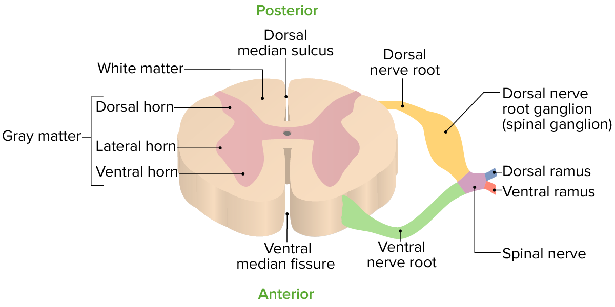
Cross-sectional view of an individual spinal segment
Image by Lecturio.
Cross-sectional anatomy
When viewed in cross section, the spinal cord is divided into gray matterGray matterRegion of central nervous system that appears darker in color than the other type, white matter. It is composed of neuronal cell bodies; neuropil; glial cells and capillaries but few myelinated nerve fibers.Cerebral Cortex: Anatomy and white matterWhite MatterThe region of central nervous system that appears lighter in color than the other type, gray matter. It mainly consists of myelinated nerve fibers and contains few neuronal cell bodies or dendrites.Brown-Séquard Syndrome.
Gray matterGray matterRegion of central nervous system that appears darker in color than the other type, white matter. It is composed of neuronal cell bodies; neuropil; glial cells and capillaries but few myelinated nerve fibers.Cerebral Cortex: Anatomy:
H- or butterfly-shaped area in the center of the cord
Consists of the neuronal cell bodies
Site of synaptic connections between neuronsNeuronsThe basic cellular units of nervous tissue. Each neuron consists of a body, an axon, and dendrites. Their purpose is to receive, conduct, and transmit impulses in the nervous system.Nervous System: Histology
Dorsal horns (posterior):
Gives rise to the dorsal roots at the dorsolateral surface of the cord
Consists of sensorySensoryNeurons which conduct nerve impulses to the central nervous system.Nervous System: HistologyneuronsNeuronsThe basic cellular units of nervous tissue. Each neuron consists of a body, an axon, and dendrites. Their purpose is to receive, conduct, and transmit impulses in the nervous system.Nervous System: Histology
Ventral horns (anterior):
Gives rise to the ventral roots at the ventrolateral surface of the cord
Consists of motorMotorNeurons which send impulses peripherally to activate muscles or secretory cells.Nervous System: HistologyneuronsNeuronsThe basic cellular units of nervous tissue. Each neuron consists of a body, an axon, and dendrites. Their purpose is to receive, conduct, and transmit impulses in the nervous system.Nervous System: Histology:
NeuronsNeuronsThe basic cellular units of nervous tissue. Each neuron consists of a body, an axon, and dendrites. Their purpose is to receive, conduct, and transmit impulses in the nervous system.Nervous System: Histology that innervate proximal muscles are medial
NeuronsNeuronsThe basic cellular units of nervous tissue. Each neuron consists of a body, an axon, and dendrites. Their purpose is to receive, conduct, and transmit impulses in the nervous system.Nervous System: Histology that innervate distal muscles are lateral
Lateral horns (anterolateral):
Also called the intermediolateral columns
Found only in the thoracic and lumbar regions
Contains neuronsNeuronsThe basic cellular units of nervous tissue. Each neuron consists of a body, an axon, and dendrites. Their purpose is to receive, conduct, and transmit impulses in the nervous system.Nervous System: Histology of the sympathetic nervous systemNervous systemThe nervous system is a small and complex system that consists of an intricate network of neural cells (or neurons) and even more glial cells (for support and insulation). It is divided according to its anatomical components as well as its functional characteristics. The brain and spinal cord are referred to as the central nervous system, and the branches of nerves from these structures are referred to as the peripheral nervous system.Nervous System: Anatomy, Structure, and Classification
Send out axonsAxonsNerve fibers that are capable of rapidly conducting impulses away from the neuron cell body.Nervous System: Histology via the ventral roots
Gray commissure:
Central area where the right and left halves cross over
Contains the central canal (collapsed in most areas in the adult)
White matterWhite MatterThe region of central nervous system that appears lighter in color than the other type, gray matter. It mainly consists of myelinated nerve fibers and contains few neuronal cell bodies or dendrites.Brown-Séquard Syndrome:
Area surrounding the gray matterGray matterRegion of central nervous system that appears darker in color than the other type, white matter. It is composed of neuronal cell bodies; neuropil; glial cells and capillaries but few myelinated nerve fibers.Cerebral Cortex: Anatomy
Composed of bundles of axonsAxonsNerve fibers that are capable of rapidly conducting impulses away from the neuron cell body.Nervous System: Histology called tracts
Organized into:
Columns (funiculi):
Dorsal (posterior) column
Lateral column
Ventral (anterior) column
Columns are subdivided into fasciculi or tracts.
Spinal meningesMeningesThe brain and the spinal cord are enveloped by 3 overlapping layers of connective tissue called the meninges. The layers are, from the most external layer to the most internal layer, the dura mater, arachnoid mater, and pia mater. Between these layers are 3 potential spaces called the epidural, subdural, and subarachnoid spaces. Meninges: Anatomy
The meningesMeningesThe brain and the spinal cord are enveloped by 3 overlapping layers of connective tissue called the meninges. The layers are, from the most external layer to the most internal layer, the dura mater, arachnoid mater, and pia mater. Between these layers are 3 potential spaces called the epidural, subdural, and subarachnoid spaces. Meninges: Anatomy are the fibrousFibrousFibrocystic Change membranes that encase the spinal cord (and brainBrainThe part of central nervous system that is contained within the skull (cranium). Arising from the neural tube, the embryonic brain is comprised of three major parts including prosencephalon (the forebrain); mesencephalon (the midbrain); and rhombencephalon (the hindbrain). The developed brain consists of cerebrum; cerebellum; and other structures in the brain stem.Nervous System: Anatomy, Structure, and Classification). The 3 layers and 2 defined spaces between/around the layers are (from outside to inside):
Dura materDura materThe outermost of the three meninges, a fibrous membrane of connective tissue that covers the brain and the spinal cord.Meninges: Anatomy:
Outermost membrane of the spinal cord
Forms a long tubular sheath around the spinal cord within the vertebral canal called the dural sheath
The space outside the dura materDura materThe outermost of the three meninges, a fibrous membrane of connective tissue that covers the brain and the spinal cord.Meninges: Anatomy, between dura materDura materThe outermost of the three meninges, a fibrous membrane of connective tissue that covers the brain and the spinal cord.Meninges: Anatomy and periosteumPeriosteumThin outer membrane that surrounds a bone. It contains connective tissue, capillaries, nerves, and a number of cell types.Bones: Structure and Types of the vertebral boneBoneBone is a compact type of hardened connective tissue composed of bone cells, membranes, an extracellular mineralized matrix, and central bone marrow. The 2 primary types of bone are compact and spongy. Bones: Structure and Types
Arachnoid materArachnoid materA delicate membrane enveloping the brain and spinal cord. It lies between the pia mater and the dura mater. It is separated from the pia mater by the subarachnoid cavity which is filled with cerebrospinal fluid.Meninges: Anatomy:
Adheres to the dura materDura materThe outermost of the three meninges, a fibrous membrane of connective tissue that covers the brain and the spinal cord.Meninges: Anatomy
Subarachnoid spaceSubarachnoid spaceThe space between the arachnoid membrane and pia mater, filled with cerebrospinal fluid. It contains large blood vessels that supply the brain and spinal cord.Subarachnoid Hemorrhage:
Space between the arachnoid materArachnoid materA delicate membrane enveloping the brain and spinal cord. It lies between the pia mater and the dura mater. It is separated from the pia mater by the subarachnoid cavity which is filled with cerebrospinal fluid.Meninges: Anatomy and pia materPia materThe innermost layer of the three meninges covering the brain and spinal cord. It is the fine vascular membrane that lies under the arachnoid and the dura mater.Meninges: Anatomy
Contains: CSF and a web of collagenous and elasticElasticConnective Tissue: Histology tissue (connecting the pia materPia materThe innermost layer of the three meninges covering the brain and spinal cord. It is the fine vascular membrane that lies under the arachnoid and the dura mater.Meninges: Anatomy and arachnoid materArachnoid materA delicate membrane enveloping the brain and spinal cord. It lies between the pia mater and the dura mater. It is separated from the pia mater by the subarachnoid cavity which is filled with cerebrospinal fluid.Meninges: Anatomy)
Pia materPia materThe innermost layer of the three meninges covering the brain and spinal cord. It is the fine vascular membrane that lies under the arachnoid and the dura mater.Meninges: Anatomy:
Innermost membrane, in direct contact with the spinal cord
Thin and transparent
Closely follows all the surface features of the spinal cord
Directly covers the roots of the spinal nerves and the spinal blood vessels
Inferior to the conus medullarisConus MedullarisSpinal Cord Injuries, the pia materPia materThe innermost layer of the three meninges covering the brain and spinal cord. It is the fine vascular membrane that lies under the arachnoid and the dura mater.Meninges: Anatomy continues as the filum terminale.
The spinal cord is divided into 31 segments, each corresponding to a pair of spinal nerves.
The spinal cord is shorter than the bony vertebral columnVertebral columnThe human spine, or vertebral column, is the most important anatomical and functional axis of the human body. It consists of 7 cervical vertebrae, 12 thoracic vertebrae, and 5 lumbar vertebrae and is limited cranially by the skull and caudally by the sacrum. Vertebral Column: Anatomy; therefore, the spinal cord segments do not all match up with their similarly named vertebral level.
Cross-sectional view of the 31 spinal segments and their relationship to the bony vertebral column
Cervical cord segmentsCervical Cord SegmentsThe segment of the spinal cord within the cervical vertebrae.Spinal Cord Injuries C1–C7 give rise to nerve roots that exit abovetheir corresponding vertebrae.
C8 nerve root emerges between C7 and T1.
C1–C8 cord segments lie within the C1–C7 region of the vertebral columnVertebral columnThe human spine, or vertebral column, is the most important anatomical and functional axis of the human body. It consists of 7 cervical vertebrae, 12 thoracic vertebrae, and 5 lumbar vertebrae and is limited cranially by the skull and caudally by the sacrum. Vertebral Column: Anatomy.
Cervical spinal segments and nerves innervate:
DiaphragmDiaphragmThe diaphragm is a large, dome-shaped muscle that separates the thoracic cavity from the abdominal cavity. The diaphragm consists of muscle fibers and a large central tendon, which is divided into right and left parts. As the primary muscle of inspiration, the diaphragm contributes 75% of the total inspiratory muscle force.Diaphragm: Anatomy (C3–C5)
Upper limb sensorySensoryNeurons which conduct nerve impulses to the central nervous system.Nervous System: Histology and motorMotorNeurons which send impulses peripherally to activate muscles or secretory cells.Nervous System: Histology structures
T1–T12 cord segments lie within the T1–T8 region of the vertebral columnVertebral columnThe human spine, or vertebral column, is the most important anatomical and functional axis of the human body. It consists of 7 cervical vertebrae, 12 thoracic vertebrae, and 5 lumbar vertebrae and is limited cranially by the skull and caudally by the sacrum. Vertebral Column: Anatomy.
Thoracic spinal segments and nerves innervate:
Intercostal nerves
Thoracic and abdominal wallAbdominal wallThe outer margins of the abdomen, extending from the osteocartilaginous thoracic cage to the pelvis. Though its major part is muscular, the abdominal wall consists of at least seven layers: the skin, subcutaneous fat, deep fascia; abdominal muscles, transversalis fascia, extraperitoneal fat, and the parietal peritoneum.Surgical Anatomy of the Abdomen muscles and dermatomes
Sympathetic innervation of the thoracic, abdominal, and pelvic viscera
Lumbar, sacral, and coccygeal cord segments
5 lumbar segments:
Named L1–L5
Lie within the T9–T11 region of the vertebral columnVertebral columnThe human spine, or vertebral column, is the most important anatomical and functional axis of the human body. It consists of 7 cervical vertebrae, 12 thoracic vertebrae, and 5 lumbar vertebrae and is limited cranially by the skull and caudally by the sacrum. Vertebral Column: Anatomy
Lie within the T12–L1 region of the vertebral columnVertebral columnThe human spine, or vertebral column, is the most important anatomical and functional axis of the human body. It consists of 7 cervical vertebrae, 12 thoracic vertebrae, and 5 lumbar vertebrae and is limited cranially by the skull and caudally by the sacrum. Vertebral Column: Anatomy
1 coccygeal segment:
Named C0
Lies within the L1 region of the vertebral columnVertebral columnThe human spine, or vertebral column, is the most important anatomical and functional axis of the human body. It consists of 7 cervical vertebrae, 12 thoracic vertebrae, and 5 lumbar vertebrae and is limited cranially by the skull and caudally by the sacrum. Vertebral Column: Anatomy
Innervate lower limb sensorySensoryNeurons which conduct nerve impulses to the central nervous system.Nervous System: Histology and motorMotorNeurons which send impulses peripherally to activate muscles or secretory cells.Nervous System: Histology structures
In general, the major functions of the spinal cord include:
Conduction of nerve signals:
AfferentAfferentNeurons which conduct nerve impulses to the central nervous system.Nervous System: Histology/sensorySensoryNeurons which conduct nerve impulses to the central nervous system.Nervous System: Histology input from the periphery → brainBrainThe part of central nervous system that is contained within the skull (cranium). Arising from the neural tube, the embryonic brain is comprised of three major parts including prosencephalon (the forebrain); mesencephalon (the midbrain); and rhombencephalon (the hindbrain). The developed brain consists of cerebrum; cerebellum; and other structures in the brain stem.Nervous System: Anatomy, Structure, and Classification
EfferentEfferentNeurons which send impulses peripherally to activate muscles or secretory cells.Nervous System: Histology/motorMotorNeurons which send impulses peripherally to activate muscles or secretory cells.Nervous System: Histology/visceral signals from the brainBrainThe part of central nervous system that is contained within the skull (cranium). Arising from the neural tube, the embryonic brain is comprised of three major parts including prosencephalon (the forebrain); mesencephalon (the midbrain); and rhombencephalon (the hindbrain). The developed brain consists of cerebrum; cerebellum; and other structures in the brain stem.Nervous System: Anatomy, Structure, and Classification → periphery
Modulates reflexes
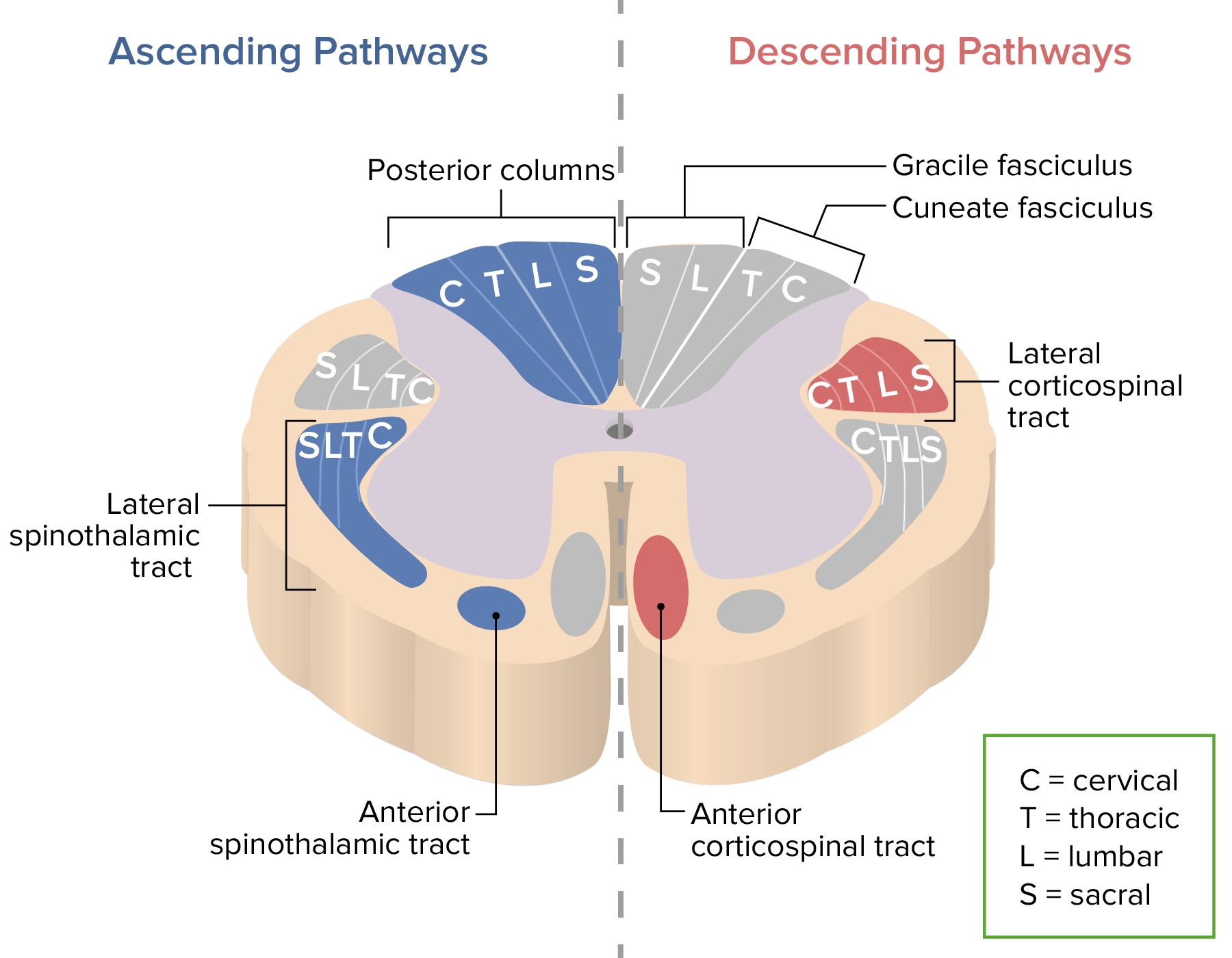
Overview of spinal tracts
Ascending tracts:
Carry sensorySensoryNeurons which conduct nerve impulses to the central nervous system.Nervous System: Histology information up the cord to the brainBrainThe part of central nervous system that is contained within the skull (cranium). Arising from the neural tube, the embryonic brain is comprised of three major parts including prosencephalon (the forebrain); mesencephalon (the midbrain); and rhombencephalon (the hindbrain). The developed brain consists of cerebrum; cerebellum; and other structures in the brain stem.Nervous System: Anatomy, Structure, and Classification
Pathway consists of 3 types of neuronsNeuronsThe basic cellular units of nervous tissue. Each neuron consists of a body, an axon, and dendrites. Their purpose is to receive, conduct, and transmit impulses in the nervous system.Nervous System: Histology:
1st-order neuronsNeuronsThe basic cellular units of nervous tissue. Each neuron consists of a body, an axon, and dendrites. Their purpose is to receive, conduct, and transmit impulses in the nervous system.Nervous System: Histology: detect the stimulus and transmit it to the spinal cord
2nd-order neuronsNeuronsThe basic cellular units of nervous tissue. Each neuron consists of a body, an axon, and dendrites. Their purpose is to receive, conduct, and transmit impulses in the nervous system.Nervous System: Histology: carry the signal up the spinal cord to the brainBrainThe part of central nervous system that is contained within the skull (cranium). Arising from the neural tube, the embryonic brain is comprised of three major parts including prosencephalon (the forebrain); mesencephalon (the midbrain); and rhombencephalon (the hindbrain). The developed brain consists of cerebrum; cerebellum; and other structures in the brain stem.Nervous System: Anatomy, Structure, and Classification stem
3rd-order neuronsNeuronsThe basic cellular units of nervous tissue. Each neuron consists of a body, an axon, and dendrites. Their purpose is to receive, conduct, and transmit impulses in the nervous system.Nervous System: Histology: carry the signal to the sensorySensoryNeurons which conduct nerve impulses to the central nervous system.Nervous System: Histology region of the cerebral cortexCerebral cortexThe cerebral cortex is the largest and most developed part of the human brain and CNS. Occupying the upper part of the cranial cavity, the cerebral cortex has 4 lobes and is divided into 2 hemispheres that are joined centrally by the corpus callosum. Cerebral Cortex: Anatomy
Descending tracts:
Carry motorMotorNeurons which send impulses peripherally to activate muscles or secretory cells.Nervous System: Histology and visceral impulses down the cord
Pathway consists of 2 types of neuronsNeuronsThe basic cellular units of nervous tissue. Each neuron consists of a body, an axon, and dendrites. Their purpose is to receive, conduct, and transmit impulses in the nervous system.Nervous System: Histology:
Upper motorMotorNeurons which send impulses peripherally to activate muscles or secretory cells.Nervous System: HistologyneuronsNeuronsThe basic cellular units of nervous tissue. Each neuron consists of a body, an axon, and dendrites. Their purpose is to receive, conduct, and transmit impulses in the nervous system.Nervous System: Histology (UMNs): soma in the brainBrainThe part of central nervous system that is contained within the skull (cranium). Arising from the neural tube, the embryonic brain is comprised of three major parts including prosencephalon (the forebrain); mesencephalon (the midbrain); and rhombencephalon (the hindbrain). The developed brain consists of cerebrum; cerebellum; and other structures in the brain stem.Nervous System: Anatomy, Structure, and Classification, synapses with the lower motor neuronLower Motor NeuronMotor Neuron Lesions
Lower motorMotorNeurons which send impulses peripherally to activate muscles or secretory cells.Nervous System: HistologyneuronsNeuronsThe basic cellular units of nervous tissue. Each neuron consists of a body, an axon, and dendrites. Their purpose is to receive, conduct, and transmit impulses in the nervous system.Nervous System: Histology (LMNs): carries the signal to the muscle or target organ
Decussation: refers to neuronsNeuronsThe basic cellular units of nervous tissue. Each neuron consists of a body, an axon, and dendrites. Their purpose is to receive, conduct, and transmit impulses in the nervous system.Nervous System: Histologycrossing overCrossing overThe reciprocal exchange of segments at corresponding positions along pairs of homologous chromosomes by symmetrical breakage and crosswise rejoining forming cross-over sites (holliday junctions) that are resolved during chromosome segregation. Crossing-over typically occurs during meiosis but it may also occur in the absence of meiosis, for example, with bacterial chromosomes, organelle chromosomes, or somatic cell nuclear chromosomes.Basic Terms of Genetics the midline (from right to left or vice versa) within the spinal cord or brainBrainThe part of central nervous system that is contained within the skull (cranium). Arising from the neural tube, the embryonic brain is comprised of three major parts including prosencephalon (the forebrain); mesencephalon (the midbrain); and rhombencephalon (the hindbrain). The developed brain consists of cerebrum; cerebellum; and other structures in the brain stem.Nervous System: Anatomy, Structure, and Classification stem
Naming conventions:
Tracts are named by combining 2 locations: origin (1st) → termination (2nd)
For example, the corticospinal tract originates in the cortex and travels down through the spinal cord.
Autonomic fibers:
Located in the lateral aspect of the spinal cord
Do not exist in well-defined tracts
SynapseSynapseThe junction between 2 neurons is called a synapse. The synapse allows a neuron to pass an electrical or chemical signal to another neuron or target effector cell. Synapses and Neurotransmission with cell bodies in the intermediolateral columns of gray matterGray matterRegion of central nervous system that appears darker in color than the other type, white matter. It is composed of neuronal cell bodies; neuropil; glial cells and capillaries but few myelinated nerve fibers.Cerebral Cortex: Anatomy
Major ascending (blue) and descending (red) tracts of the spinal cord: The letters C, T, L, and S denote where the fibers associated with each region are located.
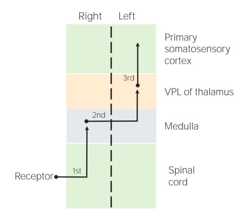
Ascending sensorySensoryNeurons which conduct nerve impulses to the central nervous system.Nervous System: Histology tracts in the posterior portion of the cord
No decussation
Fibers end in the ipsilateral medulla oblongataMedulla OblongataThe lower portion of the brain stem. It is inferior to the pons and anterior to the cerebellum. Medulla oblongata serves as a relay station between the brain and the spinal cord, and contains centers for regulating respiratory, vasomotor, cardiac, and reflex activities.Brain Stem: Anatomy → thalamusThalamusThe thalamus is a large, ovoid structure in the dorsal part of the diencephalon that is located between the cerebral cortex and midbrain. It consists of several interconnected nuclei of grey matter separated by the laminae of white matter. The thalamus is the main conductor of information that passes between the cerebral cortex and the periphery, spinal cord, or brain stem.Thalamus: Anatomy → somatosensory cortexSomatosensory cortexArea of the parietal lobe concerned with receiving sensations such as movement, pain, pressure, position, temperature, touch, and vibration. It lies posterior to the central sulcus.Cerebral Cortex: Anatomy
ProprioceptionProprioceptionSensory functions that transduce stimuli received by proprioceptive receptors in joints, tendons, muscles, and the inner ear into neural impulses to be transmitted to the central nervous system. Proprioception provides sense of stationary positions and movements of one’s body parts, and is important in maintaining kinesthesia and postural balance.Neurological Examination (conscious)
Visceral painVisceral painPain originating from internal organs (viscera) associated with autonomic phenomena (pallor; sweating; nausea; and vomiting). It often becomes a referred pain.Pain: Types and Pathways
Diagram depicting the locations of the 1st-, 2nd-, and 3rd-order sensory neurons in the dorsal columns
VPL = ventral posterolateral nucleus of the thalamus
Image by Lecturio.
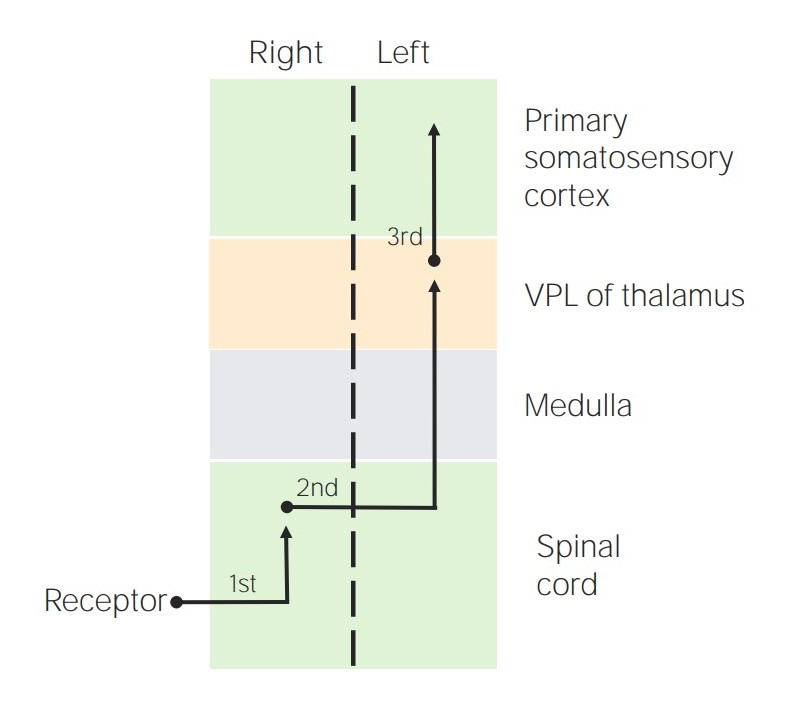
Spinothalamic tractsSpinothalamic tractsA bundle of nerve fibers connecting each posterior horn of the spinal cord to the opposite side of the thalamus, carrying information about pain, temperature, and touch. It is one of two major routes by which afferent spinal nerve fibers carrying sensations of somaesthesia are transmitted to the thalamus.Central Cord Syndrome:
Ascending sensorySensoryNeurons which conduct nerve impulses to the central nervous system.Nervous System: Histology tracts in the anterolateral portion of the cord
Decussate upon entering the spinal cord
Fibers end in the contralateral thalamusThalamusThe thalamus is a large, ovoid structure in the dorsal part of the diencephalon that is located between the cerebral cortex and midbrain. It consists of several interconnected nuclei of grey matter separated by the laminae of white matter. The thalamus is the main conductor of information that passes between the cerebral cortex and the periphery, spinal cord, or brain stem.Thalamus: Anatomy.
Transmit sensations of:
PainPainAn unpleasant sensation induced by noxious stimuli which are detected by nerve endings of nociceptive neurons.Pain: Types and Pathways
Temperature
Gross touch
Pressure
Itch and tickle
Diagram depicting the locations of the 1st-, 2nd-, and 3rd-order sensory neurons in the spinothalamic tracts
Image by Lecturio.
Spinocerebellar tracts:
Ascending sensorySensoryNeurons which conduct nerve impulses to the central nervous system.Nervous System: Histology tracts in the lateral portion of the cord
Have dorsal and ventral components
Decussation:
Dorsal fibers: no decussation
Ventral fibers: decussate upon entering the spinal cord; decussate again in the brainBrainThe part of central nervous system that is contained within the skull (cranium). Arising from the neural tube, the embryonic brain is comprised of three major parts including prosencephalon (the forebrain); mesencephalon (the midbrain); and rhombencephalon (the hindbrain). The developed brain consists of cerebrum; cerebellum; and other structures in the brain stem.Nervous System: Anatomy, Structure, and Classification stem
All fibers end in the ipsilateral cerebellumCerebellumThe cerebellum, Latin for “little brain,” is located in the posterior cranial fossa, dorsal to the pons and midbrain, and its principal role is in the coordination of movements. The cerebellum consists of 3 lobes on either side of its 2 hemispheres and is connected in the middle by the vermis. Cerebellum: Anatomy.
Transmits: proprioceptionProprioceptionSensory functions that transduce stimuli received by proprioceptive receptors in joints, tendons, muscles, and the inner ear into neural impulses to be transmitted to the central nervous system. Proprioception provides sense of stationary positions and movements of one’s body parts, and is important in maintaining kinesthesia and postural balance.Neurological Examination (unconsciousUnconsciousThose forces and content of the mind which are not ordinarily available to conscious awareness or to immediate recall.Psychotherapy)
Descending motorMotorNeurons which send impulses peripherally to activate muscles or secretory cells.Nervous System: Histology tracts in the anteromedial portion of spinal cord
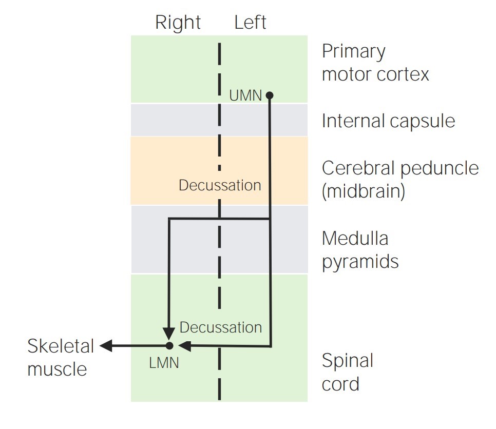
Previously called “pyramidal tracts”
Have lateral and ventral/anterior components
Decussation:
Lateral CST (90%): decussate in the medulla oblongataMedulla OblongataThe lower portion of the brain stem. It is inferior to the pons and anterior to the cerebellum. Medulla oblongata serves as a relay station between the brain and the spinal cord, and contains centers for regulating respiratory, vasomotor, cardiac, and reflex activities.Brain Stem: Anatomy
Ventral/anterior CST (10%): decussate just before exiting the spinal cord
Control limb (lateral CST) and axialAxialComputed Tomography (CT) (anterior CST) movements on the contralateral side
Diagram showcasing the pathway of the corticospinal tract
UMN = upper motor neuron
LMN = lower motor neuron
Image by Lecturio.
Extrapyramidal tracts:
Reticulospinal tract, which is involved in:
Controlling limb muscles related to posture and balance
PainPainAn unpleasant sensation induced by noxious stimuli which are detected by nerve endings of nociceptive neurons.Pain: Types and Pathways signaling
Vestibulospinal tract: receives impulses to maintain balance and posture (impulses are based on input received from the inner earInner earThe essential part of the hearing organ consists of two labyrinthine compartments: the bony labyrinthine and the membranous labyrinth.Ear: Anatomy)
Tectospinal tract:involved in reflex movements of the head
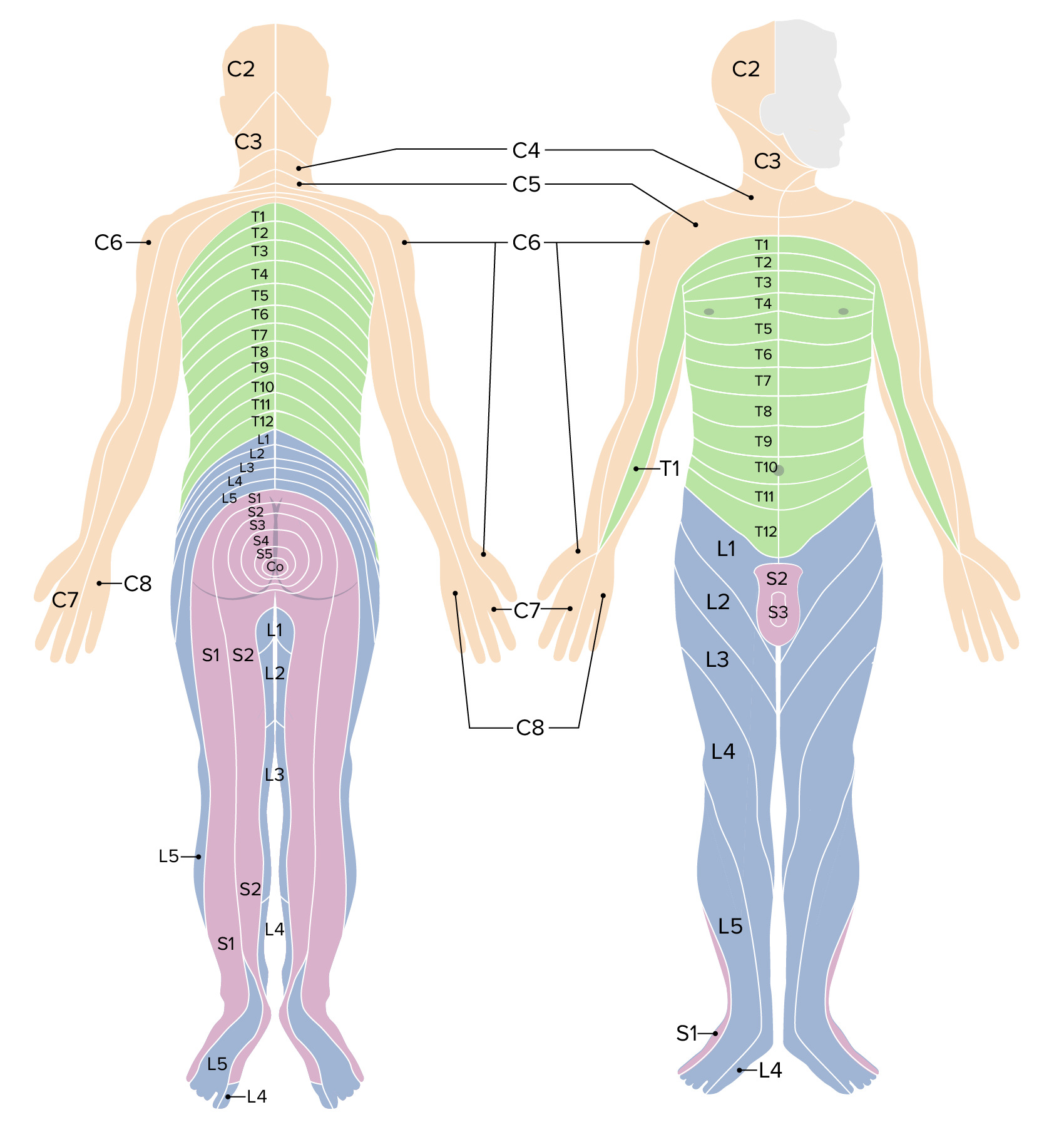
DermatomeDermatomeSpinal Disk Herniation: the sensorySensoryNeurons which conduct nerve impulses to the central nervous system.Nervous System: Histology region of skinSkinThe skin, also referred to as the integumentary system, is the largest organ of the body. The skin is primarily composed of the epidermis (outer layer) and dermis (deep layer). The epidermis is primarily composed of keratinocytes that undergo rapid turnover, while the dermis contains dense layers of connective tissue.Skin: Structure and Functions innervated by a single spinal nerve
Considerable overlap between adjacent dermatomes → lesions of a single nerve root cause a decrease but not a complete loss of sensation in a given dermatomeDermatomeSpinal Disk Herniation
Cervical dermatomes:
Head (C2–C3)
NeckNeckThe part of a human or animal body connecting the head to the rest of the body.Peritonsillar Abscess (C3–C4)
Upper extremities (C5–C8)
Thoracic dermatomes:
Inner arms (T1)
Chest wallChest wallThe chest wall consists of skin, fat, muscles, bones, and cartilage. The bony structure of the chest wall is composed of the ribs, sternum, and thoracic vertebrae. The chest wall serves as armor for the vital intrathoracic organs and provides the stability necessary for the movement of the shoulders and arms. Chest Wall: Anatomy (T1–T7)
Abdomen (T8–T12)
Back (T1–T12)
T4T4The major hormone derived from the thyroid gland. Thyroxine is synthesized via the iodination of tyrosines (monoiodotyrosine) and the coupling of iodotyrosines (diiodotyrosine) in the thyroglobulin. Thyroxine is released from thyroglobulin by proteolysis and secreted into the blood. Thyroxine is peripherally deiodinated to form triiodothyronine which exerts a broad spectrum of stimulatory effects on cell metabolism.Thyroid Hormones: nippleNippleThe conic organs which usually give outlet to milk from the mammary glands.Examination of the Breast
The spinal cord is supplied by 3 longitudinal arteriesArteriesArteries are tubular collections of cells that transport oxygenated blood and nutrients from the heart to the tissues of the body. The blood passes through the arteries in order of decreasing luminal diameter, starting in the largest artery (the aorta) and ending in the small arterioles. Arteries are classified into 3 types: large elastic arteries, medium muscular arteries, and small arteries and arterioles. Arteries: Histology, including 1 anterior and 2 posterior spinal arteriesArteriesArteries are tubular collections of cells that transport oxygenated blood and nutrients from the heart to the tissues of the body. The blood passes through the arteries in order of decreasing luminal diameter, starting in the largest artery (the aorta) and ending in the small arterioles. Arteries are classified into 3 types: large elastic arteries, medium muscular arteries, and small arteries and arterioles. Arteries: Histology.
The spinal arteriesArteriesArteries are tubular collections of cells that transport oxygenated blood and nutrients from the heart to the tissues of the body. The blood passes through the arteries in order of decreasing luminal diameter, starting in the largest artery (the aorta) and ending in the small arterioles. Arteries are classified into 3 types: large elastic arteries, medium muscular arteries, and small arteries and arterioles. Arteries: Histology arise from the vertebral arteriesArteriesArteries are tubular collections of cells that transport oxygenated blood and nutrients from the heart to the tissues of the body. The blood passes through the arteries in order of decreasing luminal diameter, starting in the largest artery (the aorta) and ending in the small arterioles. Arteries are classified into 3 types: large elastic arteries, medium muscular arteries, and small arteries and arterioles. Arteries: Histology and travel downward.
Sulcal arteriesArteriesArteries are tubular collections of cells that transport oxygenated blood and nutrients from the heart to the tissues of the body. The blood passes through the arteries in order of decreasing luminal diameter, starting in the largest artery (the aorta) and ending in the small arterioles. Arteries are classified into 3 types: large elastic arteries, medium muscular arteries, and small arteries and arterioles. Arteries: Histology branch off the anterior spinal arteryAnterior Spinal ArteryAnterior Cord Syndrome → enter the spinal cord through the fissureFissureA crack or split that extends into the dermisGeneralized and Localized Rashes
Posterior spinal arteriesArteriesArteries are tubular collections of cells that transport oxygenated blood and nutrients from the heart to the tissues of the body. The blood passes through the arteries in order of decreasing luminal diameter, starting in the largest artery (the aorta) and ending in the small arterioles. Arteries are classified into 3 types: large elastic arteries, medium muscular arteries, and small arteries and arterioles. Arteries: Histology:
Paired vessels (right and left) are located around 11:00 and 1:00 when viewing the spinal cord in cross section with the posterior aspect at the top.
Each supplies ½ of the posterior ⅓ of the cord.
Segmental medullary and radicular arteriesArteriesArteries are tubular collections of cells that transport oxygenated blood and nutrients from the heart to the tissues of the body. The blood passes through the arteries in order of decreasing luminal diameter, starting in the largest artery (the aorta) and ending in the small arterioles. Arteries are classified into 3 types: large elastic arteries, medium muscular arteries, and small arteries and arterioles. Arteries: Histology:
Arise from the aortaAortaThe main trunk of the systemic arteries.Mediastinum and Great Vessels: Anatomy, cervical, deep cervical, vertebral, intercostal, and lumbar arteriesArteriesArteries are tubular collections of cells that transport oxygenated blood and nutrients from the heart to the tissues of the body. The blood passes through the arteries in order of decreasing luminal diameter, starting in the largest artery (the aorta) and ending in the small arterioles. Arteries are classified into 3 types: large elastic arteries, medium muscular arteries, and small arteries and arterioles. Arteries: Histology
Run along the nerve roots
Join with the anterior and posterior spinal arteriesArteriesArteries are tubular collections of cells that transport oxygenated blood and nutrients from the heart to the tissues of the body. The blood passes through the arteries in order of decreasing luminal diameter, starting in the largest artery (the aorta) and ending in the small arterioles. Arteries are classified into 3 types: large elastic arteries, medium muscular arteries, and small arteries and arterioles. Arteries: Histology, providing collateral blood supply (especially to lower regions of the cord)
Great ventral radicular artery (Adamkiewicz artery):
Most common/consistent radicular artery
If present, enters the spinal cord around T5–L1 (usually T9–T12)
The spinal cord is drained via spinal veinsVeinsVeins are tubular collections of cells, which transport deoxygenated blood and waste from the capillary beds back to the heart. Veins are classified into 3 types: small veins/venules, medium veins, and large veins. Each type contains 3 primary layers: tunica intima, tunica media, and tunica adventitia. Veins: Histology:
3 anterior and 3 posterior spinal veinsVeinsVeins are tubular collections of cells, which transport deoxygenated blood and waste from the capillary beds back to the heart. Veins are classified into 3 types: small veins/venules, medium veins, and large veins. Each type contains 3 primary layers: tunica intima, tunica media, and tunica adventitia. Veins: Histology
Run longitudinally along the cord
Have multiple communications between one another
Similar distribution to the spinal arteriesArteriesArteries are tubular collections of cells that transport oxygenated blood and nutrients from the heart to the tissues of the body. The blood passes through the arteries in order of decreasing luminal diameter, starting in the largest artery (the aorta) and ending in the small arterioles. Arteries are classified into 3 types: large elastic arteries, medium muscular arteries, and small arteries and arterioles. Arteries: Histology
Join the internal vertebral (epidural) venous plexuses in the epidural spaceEpidural spaceSpace between the dura mater and the walls of the vertebral canal.Epidural Hemorrhage
Clinical Relevance
Spinal cord syndromes
Central cord syndromeCentral Cord SyndromeCentral cord syndrome (CCS) is a neurological syndrome caused by an injury to the center of the spinal cord, affecting the spinothalamic tracts ((STTs) sensory) and medial aspect of the corticospinal tracts ((CSTs) motor), most often due to trauma in patients with cervical spondylosis. Central Cord Syndrome: neurologic syndrome caused by an injury to the center of the spinal cord, affecting the spinothalamic tractsSpinothalamic tractsA bundle of nerve fibers connecting each posterior horn of the spinal cord to the opposite side of the thalamus, carrying information about pain, temperature, and touch. It is one of two major routes by which afferent spinal nerve fibers carrying sensations of somaesthesia are transmitted to the thalamus.Central Cord Syndrome (sensorySensoryNeurons which conduct nerve impulses to the central nervous system.Nervous System: Histology) and medial aspect of the CSTsCSTsCentral Cord Syndrome (motorMotorNeurons which send impulses peripherally to activate muscles or secretory cells.Nervous System: Histology).
Anterior cord syndromeAnterior cord syndromeAnterior cord syndrome (ACS) is an incomplete cord syndrome predominantly affecting the anterior (ventral) …” of the spinal cord while sparing the dorsal columns. Anterior cord syndrome can be caused by occlusion of the anterior spinal artery or by trauma, which results in disc herniation and bone fragments disrupting the spinal cord.Anterior Cord Syndrome: incomplete cord syndrome resulting from injury to the anterior (ventral) ⅔ of the spinal cord and sparing the dorsal columnsDorsal ColumnsPosterior Cord Syndrome. Clinical manifestations are loss of motorMotorNeurons which send impulses peripherally to activate muscles or secretory cells.Nervous System: Histology and sensorySensoryNeurons which conduct nerve impulses to the central nervous system.Nervous System: Histology function below the level of injury.
Posterior cord syndromePosterior cord syndromePosterior cord syndrome (PCS) is an incomplete spinal cord syndrome affecting the dorsal columns, the corticospinal tracts (CSTs), and descending autonomic tracts to the bladder. Posterior cord syndrome is rare but has a diverse range of etiologies, including demyelinating disorders, degenerative spinal conditions, neoplastic causes, vascular abnormalities, and hereditary neurodegenerative disorders.Posterior Cord Syndrome: incomplete spinal cord syndrome affecting the dorsal columnsDorsal ColumnsPosterior Cord Syndrome, the CSTsCSTsCentral Cord Syndrome (motorMotorNeurons which send impulses peripherally to activate muscles or secretory cells.Nervous System: Histology), and descending autonomic tracts to the bladderBladderA musculomembranous sac along the urinary tract. Urine flows from the kidneys into the bladder via the ureters, and is held there until urination.Pyelonephritis and Perinephric Abscess. Clinical symptoms include gait ataxiaGait ataxiaImpairment of the ability to coordinate the movements required for normal ambulation (walking) which may result from impairments of motor function or sensory feedback. This condition may be associated with brain diseases (including cerebellar diseases and basal ganglia diseases); spinal cord diseases; or peripheral nervous system diseases.Friedreich Ataxia, paresthesiasParesthesiasSubjective cutaneous sensations (e.g., cold, warmth, tingling, pressure, etc.) that are experienced spontaneously in the absence of stimulation.Posterior Cord Syndrome with loss of position and vibrationVibrationA continuing periodic change in displacement with respect to a fixed reference.Neurological Examination sense, and urinary incontinenceUrinary incontinenceUrinary incontinence (UI) is involuntary loss of bladder control or unintentional voiding, which represents a hygienic or social problem to the patient. Urinary incontinence is a symptom, a sign, and a disorder. The 5 types of UI include stress, urge, mixed, overflow, and functional.Urinary Incontinence.
Brown-Séquard syndromeBrown-Séquard syndromeBrown-Séquard syndrome (BSS) is a rare neurologic injury that causes hemisection of the spinal cord, resulting in weakness and paralysis of one side of the body and sensory loss on the opposite side.Brown-Séquard Syndrome: rare neurologic injury that results in hemisectionHemisectionBrown-Séquard Syndrome of the spinal cord, leading to ipsilateral loss of motorMotorNeurons which send impulses peripherally to activate muscles or secretory cells.Nervous System: Histology function and dorsal column sensations, and contralateral loss of spinothalamic sensations 1–2 levels below the level of cord damage.
Degenerative conditions
Amyotrophic lateral sclerosisSclerosisA pathological process consisting of hardening or fibrosis of an anatomical structure, often a vessel or a nerve.Wilms Tumor (ALSALSAmyotrophic lateral sclerosis (ALS), also known as Lou Gehrig’s disease, is a sporadic or inherited neurodegenerative disease of upper motor neurons (UMNs) and lower motor neurons (LMNs). ALS is the most common progressive motor neuron disease in North America, primarily affecting men and individuals of Caucasian ethnicity. Amyotrophic Lateral Sclerosis): also known as Lou Gehrig’s diseaseLou Gehrig’s diseaseAmyotrophic lateral sclerosis (ALSs), also known as Lou Gehrig’s disease, is a sporadic or inherited neurodegenerative disease of upper motor neurons (UMNs) and lower motor neurons (LMNs). Als is the most common progressive motor neuron disease in North America, primarily affecting men and individuals of caucasian ethnicity.Amyotrophic Lateral Sclerosis, is a sporadicSporadicSelective IgA Deficiency or inherited neurodegenerative disease of both UMNs and LMNs. Amyotrophic lateral sclerosisSclerosisA pathological process consisting of hardening or fibrosis of an anatomical structure, often a vessel or a nerve.Wilms Tumor is the most common progressive motorMotorNeurons which send impulses peripherally to activate muscles or secretory cells.Nervous System: Histology neuron disease in the United States. The diagnosis is made clinically, and management is supportive, progressing to end-of-life care.
Multiple sclerosisSclerosisA pathological process consisting of hardening or fibrosis of an anatomical structure, often a vessel or a nerve.Wilms Tumor: chronic inflammatory autoimmune disease leading to demyelinationDemyelinationMultiple Sclerosis of the central nervous systemCentral nervous systemThe main information-processing organs of the nervous system, consisting of the brain, spinal cord, and meninges.Nervous System: Anatomy, Structure, and Classification (CNS). Multiple sclerosisSclerosisA pathological process consisting of hardening or fibrosis of an anatomical structure, often a vessel or a nerve.Wilms Tumor is the most common demyelinating condition, with young women affected more predominantly. The clinical presentation varies widely depending on the site of lesions but typically involves neurologic symptoms affecting visionVisionOphthalmic Exam, motorMotorNeurons which send impulses peripherally to activate muscles or secretory cells.Nervous System: Histology functions, sensation, and autonomic function. The diagnosis is made via MRI imaging of the entire CNS (brainBrainThe part of central nervous system that is contained within the skull (cranium). Arising from the neural tube, the embryonic brain is comprised of three major parts including prosencephalon (the forebrain); mesencephalon (the midbrain); and rhombencephalon (the hindbrain). The developed brain consists of cerebrum; cerebellum; and other structures in the brain stem.Nervous System: Anatomy, Structure, and Classification and spineSpineThe human spine, or vertebral column, is the most important anatomical and functional axis of the human body. It consists of 7 cervical vertebrae, 12 thoracic vertebrae, and 5 lumbar vertebrae and is limited cranially by the skull and caudally by the sacrum.Vertebral Column: Anatomy) as well as CSF examination.
Neural tubeNeural tubeA tube of ectodermal tissue in an embryo that will give rise to the central nervous system, including the spinal cord and the brain. Lumen within the neural tube is called neural canal which gives rise to the central canal of the spinal cord and the ventricles of the brain.Gastrulation and Neurulation defects
Neural tubeNeural tubeA tube of ectodermal tissue in an embryo that will give rise to the central nervous system, including the spinal cord and the brain. Lumen within the neural tube is called neural canal which gives rise to the central canal of the spinal cord and the ventricles of the brain.Gastrulation and Neurulation defects (NTDsNTDsNeural tube defects (NTDs) are the 2nd-most common type of congenital birth defects. Neural tube defects can range from asymptomatic (closed ntd) to very severe malformations of the spine or brain (open ntd). Neural tube defects are caused by the failure of the neural tube to close properly during the 3rd and 4th week of embryological development.Neural Tube Defects): caused by the failure of the neural tubeNeural tubeA tube of ectodermal tissue in an embryo that will give rise to the central nervous system, including the spinal cord and the brain. Lumen within the neural tube is called neural canal which gives rise to the central canal of the spinal cord and the ventricles of the brain.Gastrulation and Neurulation to close properly during embryologic development, potentially resulting in protrusion of neural tissue. These defects may involve the spinal cord and/or craniumCraniumThe skull (cranium) is the skeletal structure of the head supporting the face and forming a protective cavity for the brain. The skull consists of 22 bones divided into the viscerocranium (facial skeleton) and the neurocranium.Skull: Anatomy and may be open (involving the meningesMeningesThe brain and the spinal cord are enveloped by 3 overlapping layers of connective tissue called the meninges. The layers are, from the most external layer to the most internal layer, the dura mater, arachnoid mater, and pia mater. Between these layers are 3 potential spaces called the epidural, subdural, and subarachnoid spaces. Meninges: Anatomy and/or neural tissue) or closed (involving the bony vertebral columnVertebral columnThe human spine, or vertebral column, is the most important anatomical and functional axis of the human body. It consists of 7 cervical vertebrae, 12 thoracic vertebrae, and 5 lumbar vertebrae and is limited cranially by the skull and caudally by the sacrum. Vertebral Column: Anatomy). Prenatal diagnosis by ultrasonography and maternal α-fetoprotein level is common. Management of an open NTD is mainly surgical.
Specific types of NTDsNTDsNeural tube defects (NTDs) are the 2nd-most common type of congenital birth defects. Neural tube defects can range from asymptomatic (closed ntd) to very severe malformations of the spine or brain (open ntd). Neural tube defects are caused by the failure of the neural tube to close properly during the 3rd and 4th week of embryological development.Neural Tube Defects related to the spinal cord:
MeningoceleMeningoceleA congenital or acquired protrusion of the meninges, unaccompanied by neural tissue, through a bony defect in the skull or vertebral column.Neural Tube Defects: only the meningesMeningesThe brain and the spinal cord are enveloped by 3 overlapping layers of connective tissue called the meninges. The layers are, from the most external layer to the most internal layer, the dura mater, arachnoid mater, and pia mater. Between these layers are 3 potential spaces called the epidural, subdural, and subarachnoid spaces. Meninges: Anatomy protrude
MeningomyeloceleMeningomyeloceleCongenital, or rarely acquired, herniation of meningeal and spinal cord tissue through a bony defect in the vertebral column. The majority of these defects occur in the lumbosacral region. Clinical features include paraplegia, loss of sensation in the lower body, and incontinence. This condition may be associated with the arnold-chiari malformation and hydrocephalus.Neural Tube Defects: both meningesMeningesThe brain and the spinal cord are enveloped by 3 overlapping layers of connective tissue called the meninges. The layers are, from the most external layer to the most internal layer, the dura mater, arachnoid mater, and pia mater. Between these layers are 3 potential spaces called the epidural, subdural, and subarachnoid spaces. Meninges: Anatomy and spinal cord protrude (most common NTD)
Procedures
Lumbar spinal puncture: withdrawal of CSF from the lumbar cistern, below the level of the spinal cord. Lumbar spinal puncture is an important diagnostic tool for evaluating a variety of central nervous systemCentral nervous systemThe main information-processing organs of the nervous system, consisting of the brain, spinal cord, and meninges.Nervous System: Anatomy, Structure, and Classification (CNS) disorders. Many diseases of the CNS may alter the cells in the CSF or change the concentration of its chemical constituents, aiding diagnosis.
Epidural and spinal anesthesiaSpinal anesthesiaProcedure in which an anesthetic is injected directly into the spinal cord.Anesthesiology: History and Basic Concepts: Injection of opioidOpioidCompounds with activity like opiate alkaloids, acting at opioid receptors. Properties include induction of analgesia or narcosis.Constipation medications into the epidural or subarachnoid spaceSubarachnoid spaceThe space between the arachnoid membrane and pia mater, filled with cerebrospinal fluid. It contains large blood vessels that supply the brain and spinal cord.Subarachnoid Hemorrhage can provide effective anesthesiaAnesthesiaA state characterized by loss of feeling or sensation. This depression of nerve function is usually the result of pharmacologic action and is induced to allow performance of surgery or other painful procedures.Anesthesiology: History and Basic Concepts for childbirth and surgical procedures in the lower abdomen (e.g., cesarean section). Epidural anesthesiaEpidural anesthesiaProcedure in which an anesthetic is injected into the epidural space.Anesthesiology: History and Basic Concepts involves placement of a catheter in the epidural spaceEpidural spaceSpace between the dura mater and the walls of the vertebral canal.Epidural Hemorrhage, allowing for continuous infusion of medication. Spinal anesthesiaSpinal anesthesiaProcedure in which an anesthetic is injected directly into the spinal cord.Anesthesiology: History and Basic Concepts is a single injection of opioidOpioidCompounds with activity like opiate alkaloids, acting at opioid receptors. Properties include induction of analgesia or narcosis.Constipation into the subarachnoid spaceSubarachnoid spaceThe space between the arachnoid membrane and pia mater, filled with cerebrospinal fluid. It contains large blood vessels that supply the brain and spinal cord.Subarachnoid Hemorrhage; the effects are much shorter, though anesthesiaAnesthesiaA state characterized by loss of feeling or sensation. This depression of nerve function is usually the result of pharmacologic action and is induced to allow performance of surgery or other painful procedures.Anesthesiology: History and Basic Concepts is superior to that achieved via an epidural catheter.
References
Blumenfeld, H. (2021). Neuroanatomy through clinical cases (3rd ed.). Oxford University Press.
Drake, R. L., Vogl, W. A., & Mitchell, A. W. M. (2023). Gray’s anatomy for students (5th ed.). Elsevier.
Fleetwood, I. G., & Novak, K. (2024). Spinal cord anatomy and localization of disorders. Neurosurgery Clinics of North America, 35(1), 1-14.
Gatterman, M. I. (2023). Foundations of chiropractic: Subluxation (3rd ed.). Mosby.
Jallo, J., & Vaccaro, A. R. (2022). Neurotrauma and critical care of the spine (3rd ed.). Thieme.
Moore, K. L., Dalley, A. F., & Agur, A. M. R. (2022). Clinically oriented anatomy (9th ed.). Wolters Kluwer.
Naidich, T. P., Castillo, M., Cha, S., & Smirniotopoulos, J. G. (2022). Imaging of the spine (2nd ed.). Elsevier.
Patestas, M. A., & Gartner, L. P. (2023). A textbook of neuroanatomy (3rd ed.). Wiley-Blackwell.
Standring, S. (2023). Gray’s anatomy: The anatomical basis of clinical practice (44th ed.). Elsevier.
Watson, C., Paxinos, G., & Kayalioglu, G. (2024). The spinal cord: A Christopher and Dana Reeve Foundation text and atlas (3rd ed.). Academic Press.
Yanagisawa, M., & Greenberg, J. (2025). Contemporary understanding of spinal cord neural circuits. Nature Reviews Neuroscience, 26(1), 42-55.
Create your free account or log in to continue reading!