Heart sounds are brief, transient sounds produced by valve opening and closure and by movement of blood in the heart. They are divided into systolic and diastolic sounds. In most cases, only the first (S1) and second (S2) heart sounds are heard. These are high-frequency sounds and arise from mitral and tricuspid valveTricuspid valveThe valve consisting of three cusps situated between the right atrium and right ventricle of the heart.Heart: Anatomy closure (S1), as well as aortic and pulmonary valvePulmonary valveA valve situated at the entrance to the pulmonary trunk from the right ventricle.Heart: Anatomy closure (S2). The third heart sound (S3) may be physiologic (e.g., athletes) or pathologic (e.g., congestive heart failureHeart FailureA heterogeneous condition in which the heart is unable to pump out sufficient blood to meet the metabolic need of the body. Heart failure can be caused by structural defects, functional abnormalities (ventricular dysfunction), or a sudden overload beyond its capacity. Chronic heart failure is more common than acute heart failure which results from sudden insult to cardiac function, such as myocardial infarction.Total Anomalous Pulmonary Venous Return (TAPVR)), and is related to abnormally rapid decelerationDecelerationA decrease in the rate of speed.Blunt Chest Trauma of early diastolic left ventricular inflow. The fourth heart sound (S4) is associated with contraction of the atria into partially-filled and non-compliant (stiff) ventricles. S4 is a pathologic sign in the young, but may be found in older individuals due to an age-related decrease in ventricular complianceComplianceDistensibility measure of a chamber such as the lungs (lung compliance) or bladder. Compliance is expressed as a change in volume per unit change in pressure.Veins: Histology. Additional sounds include murmurs (physiologic and pathologic), clicks, and snaps. These sounds are heard in individuals with structural abnormalities of the heart such as septal defects, valvular stenosisStenosisHypoplastic Left Heart Syndrome (HLHS), and mitral regurgitationRegurgitationGastroesophageal Reflux Disease (GERD).
The more turbulent the flowFlowBlood flows through the heart, arteries, capillaries, and veins in a closed, continuous circuit. Flow is the movement of volume per unit of time. Flow is affected by the pressure gradient and the resistance fluid encounters between 2 points. Vascular resistance is the opposition to flow, which is caused primarily by blood friction against vessel walls.Vascular Resistance, Flow, and Mean Arterial Pressure, the more audible the created vibrations.
On auscultation, 2 heart sounds heard from a normal heart are reflective of the cardiac cycleCardiac cycleThe cardiac cycle describes a complete contraction and relaxation of all 4 chambers of the heart during a standard heartbeat. The cardiac cycle includes 7 phases, which together describe the cycle of ventricular filling, isovolumetric contraction, ventricular ejection, and isovolumetric relaxation.Cardiac Cycle.
The cardiac cycleCardiac cycleThe cardiac cycle describes a complete contraction and relaxation of all 4 chambers of the heart during a standard heartbeat. The cardiac cycle includes 7 phases, which together describe the cycle of ventricular filling, isovolumetric contraction, ventricular ejection, and isovolumetric relaxation.Cardiac Cycle is a sequence of pressure changes in the heart, resulting in:
SystoleSystolePeriod of contraction of the heart, especially of the heart ventricles.Cardiac Cycle (ventricular contraction and ejection of blood) and
DiastoleDiastolePost-systolic relaxation of the heart, especially the heart ventricles.Cardiac Cycle (ventricular relaxation and filling)
S1 and S2 mark the beginning and end, respectively, of the cardiac cycleCardiac cycleThe cardiac cycle describes a complete contraction and relaxation of all 4 chambers of the heart during a standard heartbeat. The cardiac cycle includes 7 phases, which together describe the cycle of ventricular filling, isovolumetric contraction, ventricular ejection, and isovolumetric relaxation.Cardiac Cycle phases: systoleSystolePeriod of contraction of the heart, especially of the heart ventricles.Cardiac Cycle and diastoleDiastolePost-systolic relaxation of the heart, especially the heart ventricles.Cardiac Cycle; they are high-frequency sounds.
Colloquially referred to as the “lub-dub” sound of the heart
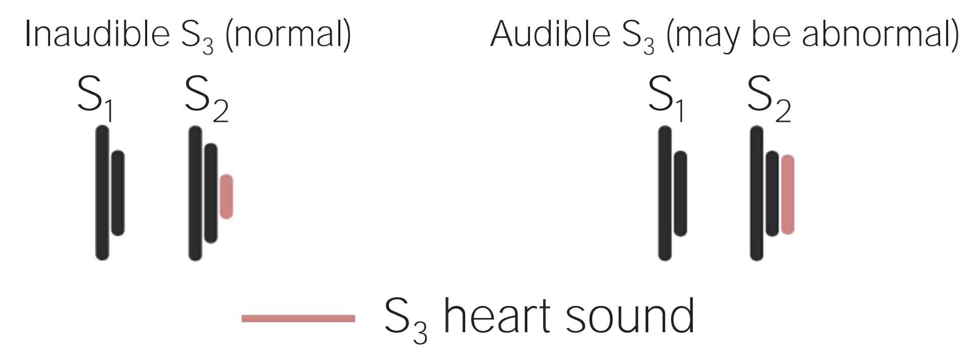
S3 and S4 are low-frequency sounds which may be heard in various conditions.
Rapid filling of ventricles
(early diastoleDiastolePost-systolic relaxation of the heart, especially the heart ventricles.Cardiac Cycle)
Normal in pregnant women, children, athletes
Ventricular dilation (e.g., congestive heart failureHeart FailureA heterogeneous condition in which the heart is unable to pump out sufficient blood to meet the metabolic need of the body. Heart failure can be caused by structural defects, functional abnormalities (ventricular dysfunction), or a sudden overload beyond its capacity. Chronic heart failure is more common than acute heart failure which results from sudden insult to cardiac function, such as myocardial infarction.Total Anomalous Pulmonary Venous Return (TAPVR))
S4
Late filling of ventricles by atrial contraction (late diastoleDiastolePost-systolic relaxation of the heart, especially the heart ventricles.Cardiac Cycle)
Noncompliant or stiff ventricles
Pathologic in children and young people
May be seen in older people with age-related stiff ventricles
The 4 cardiac valves are mitral and tricuspid (atrioventricular) valves, and aortic and pulmonary (semilunar) valves.
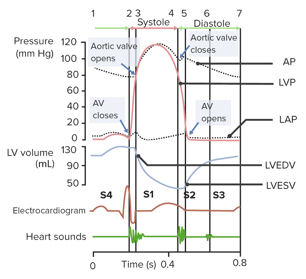
The cardiac cycle with heart sounds (phases denoted by top numbers): Phase 1 (atrial systole): seen as P wave on electrocardiogram (ECG); left atrium contracts (seen as the a wave in LAP or left atrial pressure). Phase 2 (isovolumetric contraction of the ventricle): left ventricular pressure (LVP) increases and mitral valve closes (S1). Phase 3 and 4 (ejection): the contraction of the ventricle continues until the aortic valve opens in response to the high LVP. There is a sharp increase, then decrease in LVP as blood is ejected. The residual volume in the ventricle is the left ventricular end-systolic volume (LVESV). Phase 5 (isovolumetric relaxation): beginning of ventricular diastole. LVP decreases. Aortic valve closes due to the decreasing left intraventricular pressure (S2). Phases 6 and 7 (ventricular filling): the mitral valve opens from the effects of decreased left intraventricular pressure as well as left atrial contraction, and ventricles fill up with blood, ending with an LV end-diastolic volume (LVEDV). S3 occurs in early diastole during the rapid filling phase. S4 occurs in late diastole.
Image by Lecturio.
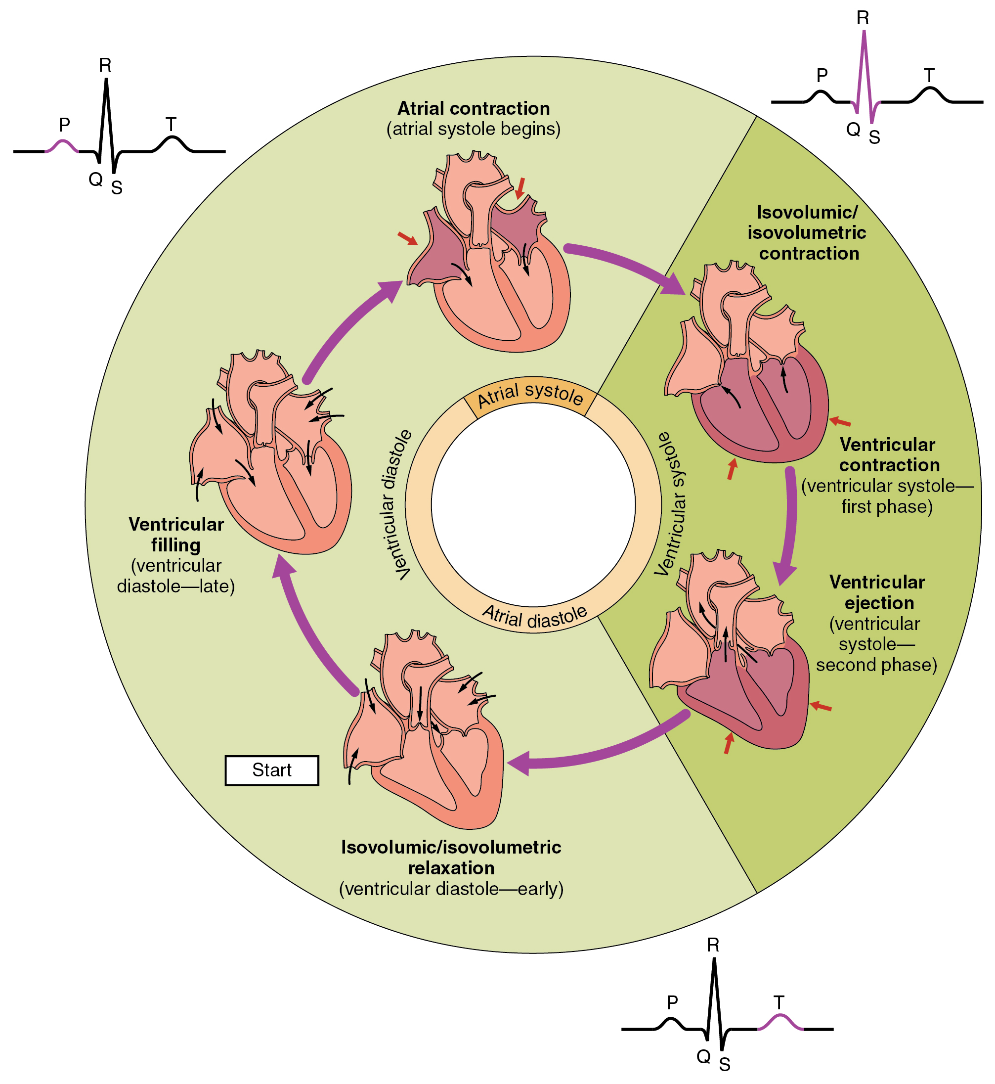
Phases of the cardiac cycle in correlation with an ECG (from left upper):
Phase 1 (atrial systole): seen as P wave on ECG; left atrium contracts.
Phase 2 (isovolumetric contraction): left ventricular (LV) pressure increases and mitral valve closes; LV depolarization is seen as a QRS complex on ECG.
Phases 3 and 4: isovolumetric contraction of the ventricle continues until the aortic valve opens in response to high LVP. Ventricles contract and blood is ejected. LV repolarization is seen as a T wave on ECG.
Phase 5 (isovolumetric relaxation): beginning of ventricular diastole. The aortic valve closes due to decreasing left intraventricular pressure. T wave resolution is seen on ECG.
Phases 6 and 7 (ventricular filling): mitral valve opens due to effects of decreased left intraventricular pressure and left atrial contraction, and ventricles fill with blood.
Image: “The cardiac cycle” by OpenStax College. License: CC BY 4.0
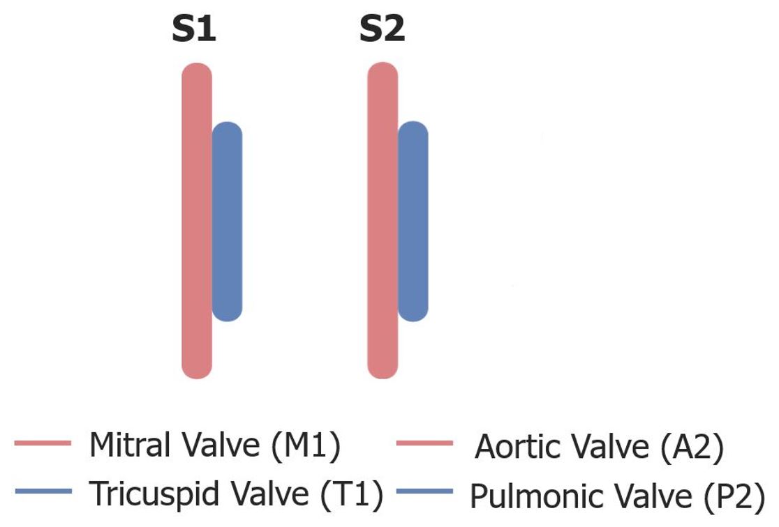
Mitral valveMitral valveThe valve between the left atrium and left ventricle of the heart.Heart: Anatomy (M1) closes before the tricuspid valveTricuspid valveThe valve consisting of three cusps situated between the right atrium and right ventricle of the heart.Heart: Anatomy (T1).
Indicates the start of systoleSystolePeriod of contraction of the heart, especially of the heart ventricles.Cardiac Cycle
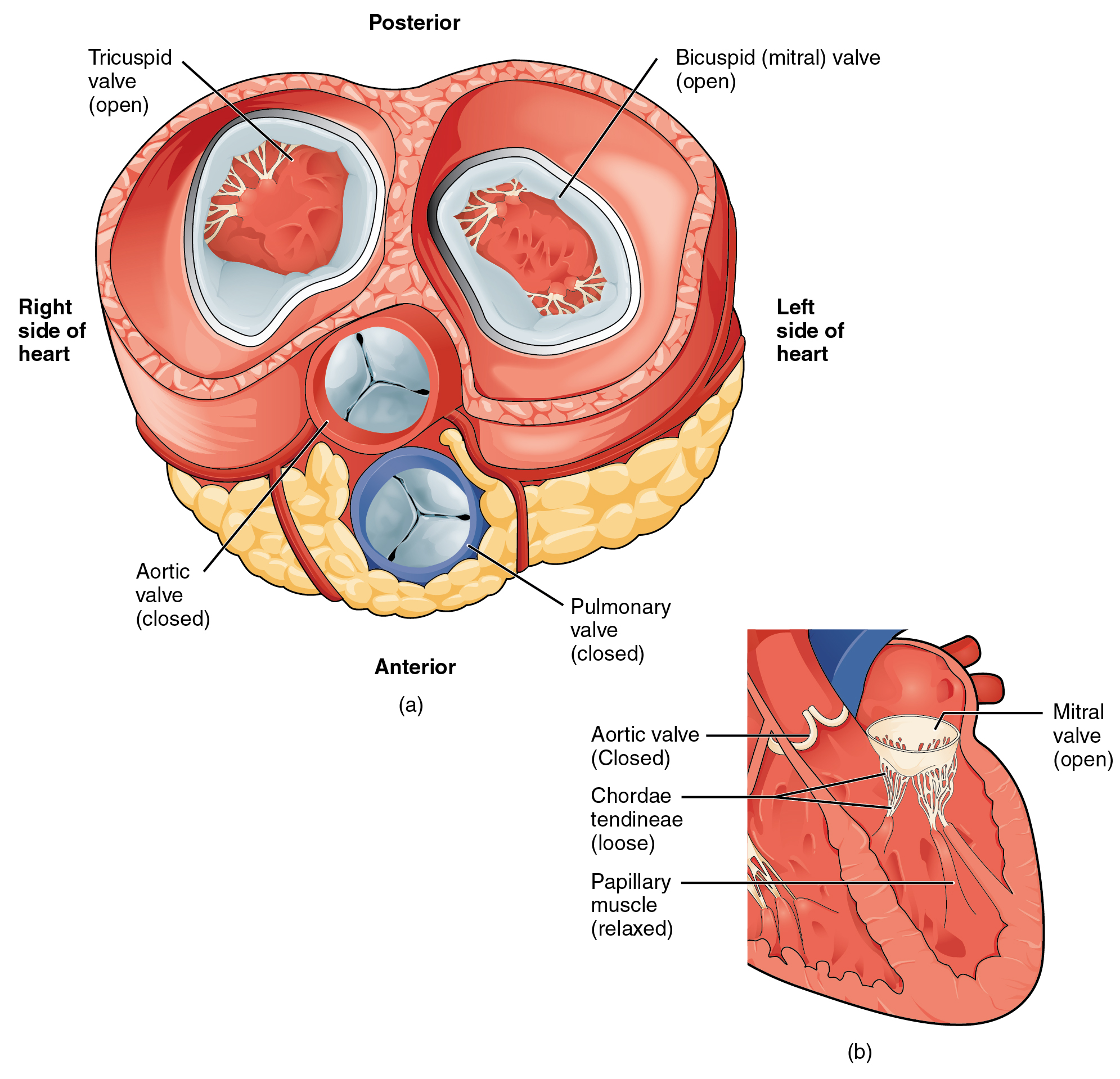
S1: Closure of the atrioventricular valves (tricuspid and mitral) at the beginning of systole. In the systole phase of the cardiac cycle, the right and left ventricles develop pressure, leading to ventricular contraction and ejection of blood into the pulmonary artery and aorta, respectively. Thus, the pulmonary and aortic valves are open. The closed atrioventricular valves prevent the backflow of blood into the atria during ventricular contraction.
Image: “2013 Blood Flow Contracted Ventricles” by OpenStax College. License: CC BY 3.0
S2
Closure of the semilunar valves (pulmonary and aortic)
Indicates the start of diastoleDiastolePost-systolic relaxation of the heart, especially the heart ventricles.Cardiac Cycle
The aortic valveAortic valveThe valve between the left ventricle and the ascending aorta which prevents backflow into the left ventricle.Heart: Anatomy (A2 component of S2) closes before the pulmonic valve (P2 component of S2).
Loudest at left upper sternal border
S2: Closure of the semilunar (aortic and pulmonary) valves at the beginning of diastole. When the intraventricular pressure falls below the atrial pressure, the mitral and tricuspid valves open, allowing for ventricular filling.
Image: “2012 Blood Flow Relaxed Ventricles” by OpenStax College. License: CC BY 3.0
Chronological order of the closing of the valves that make up S1 and S2
Image by Lecturio.
Audio:
Normal S1 and S2: In this audio clip, normal S1 and S2 heart sounds can be heard. S1 corresponds to the closure of the AV valves, marking the beginning of systoleSystolePeriod of contraction of the heart, especially of the heart ventricles.Cardiac Cycle. S2 corresponds to the closure of the semilunar valves, marking the beginning of diastoleDiastolePost-systolic relaxation of the heart, especially the heart ventricles.Cardiac Cycle.
S3 and S4 sounds are heard in certain clinical situations and are produced by turbulent blood entering the ventricle at different points during diastole. S3 and S4 are the so-called “extra heart sounds.” They are low-frequency sounds.
↑ Left ventricular fillingVentricular fillingCardiac Cycle pressure → ↑ cardiac outputCardiac outputThe volume of blood passing through the heart per unit of time. It is usually expressed as liters (volume) per minute so as not to be confused with stroke volume (volume per beat).Cardiac Mechanics
Best heard at the apex (left lateral decubitus position)
Can be normal in children, pregnant women, and athletes
Associated pathologic conditions:
Dilated cardiomyopathyDilated CardiomyopathyDilated cardiomyopathy (DCM) is the most common type of non-ischemic cardiomyopathy and a common cause of heart failure (HF). The cause may be idiopathic, familial, or secondary to a variety of underlying conditions. The disease is characterized by the enlargement of 1 or both ventricles and reduced systolic function. Dilated Cardiomyopathy
Congestive heart failureHeart FailureA heterogeneous condition in which the heart is unable to pump out sufficient blood to meet the metabolic need of the body. Heart failure can be caused by structural defects, functional abnormalities (ventricular dysfunction), or a sudden overload beyond its capacity. Chronic heart failure is more common than acute heart failure which results from sudden insult to cardiac function, such as myocardial infarction.Total Anomalous Pulmonary Venous Return (TAPVR)
Chronic aortic regurgitationRegurgitationGastroesophageal Reflux Disease (GERD) (ARARAortic regurgitation (AR) is a cardiac condition characterized by the backflow of blood from the aorta to the left ventricle during diastole. Aortic regurgitation is associated with an abnormal aortic valve and/or aortic root stemming from multiple causes, commonly rheumatic heart disease as well as congenital and degenerative valvular disorders. Aortic Regurgitation)
ThyrotoxicosisThyrotoxicosisA hypermetabolic syndrome caused by excess thyroid hormones which may come from endogenous or exogenous sources. The endogenous source of hormone may be thyroid hyperplasia; thyroid neoplasms; or hormone-producing extrathyroidal tissue. Thyrotoxicosis is characterized by nervousness; tachycardia; fatigue; weight loss; heat intolerance; and excessive sweating.Thyrotoxicosis and Hyperthyroidism
Timing and amplitude of S3 when inaudible and audible
Image by Lecturio.
Audio:
S3: In this audio clip, the S3 gallopS3 gallopHeart Failure can be heard (left decubitus, heard with the bell of the stethoscope). S3 occurs after S2 during the rapid filling phase of ventricular diastoleVentricular diastoleCardiac Cycle.
May be auscultated in older adults due to loss of ventricular complianceComplianceDistensibility measure of a chamber such as the lungs (lung compliance) or bladder. Compliance is expressed as a change in volume per unit change in pressure.Veins: Histology with age
Best heard at the apex (left lateral decubitus position)
Associated conditions:
Hypertrophic cardiomyopathyHypertrophic CardiomyopathyHypertrophic cardiomyopathy (HCM) is the most commonly inherited cardiomyopathy, which is characterized by an asymmetric increase in thickness (hypertrophy) of the left ventricular wall, diastolic dysfunction, and often left ventricular outflow tract obstruction. Hypertrophic Cardiomyopathy
AS
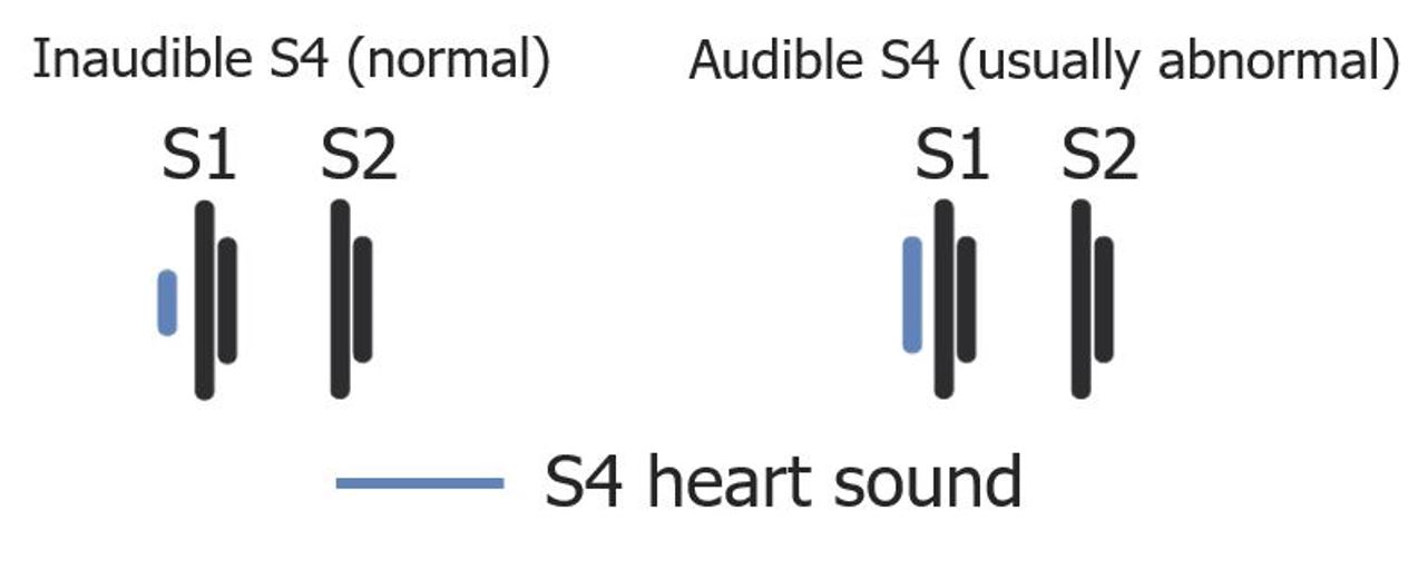
Timing and amplitude of S4 when inaudible and audible
Image by Lecturio.
Audio:
S4 gallop: In this audio clip, the S4 gallop can be heard (left decubitus, heard with the bell of the stethoscope). S4 occurs before S1 during the atrial filling phase. S4 is heard in conditions where there is stiffness or low complianceComplianceDistensibility measure of a chamber such as the lungs (lung compliance) or bladder. Compliance is expressed as a change in volume per unit change in pressure.Veins: Histology in the ventricle.
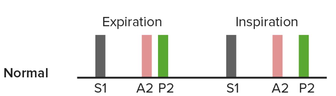
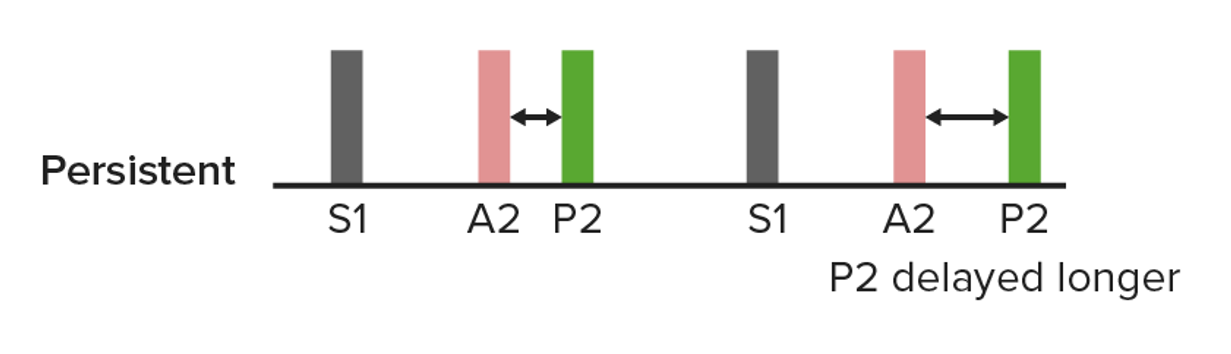
Diagram showing widening of the S2 components (A2 and P2) during increased preload conditions such as inspiration, which produces the physiologic splitting of S2
Diagram showing persistent S2 splitting in which closure of the pulmonic valve is further delayed by inspiration (right). This splitting can occur in a right bundle branch block.
Image by Lecturio.
Audio:
Persistent S2 splittingSplittingDefense Mechanisms: heard in right bundle branch blockRight bundle branch blockBundle Branch and Fascicular Blocks (listening with the diaphragmDiaphragmThe diaphragm is a large, dome-shaped muscle that separates the thoracic cavity from the abdominal cavity. The diaphragm consists of muscle fibers and a large central tendon, which is divided into right and left parts. As the primary muscle of inspiration, the diaphragm contributes 75% of the total inspiratory muscle force.Diaphragm: Anatomy of the stethoscope)
Associated condition: atrial septal defectAtrial Septal DefectAtrial septal defects (ASDs) are benign acyanotic congenital heart defects characterized by an opening in the interatrial septum that causes blood to flow from the left atrium (LA) to the right atrium (RA) (left-to-right shunt). Atrial Septal Defect (ASD) (ASDASDAutism spectrum disorder (ASD) is a neurodevelopmental disorder marked by poor social skills, restricted interests/social interactions, and repetitive/stereotyped behaviors. The condition is termed a “spectrum” because of the wide variability in the severity of symptoms exhibited.Autism Spectrum Disorder)
Diagram showing fixed splitting, in which closure of P2 is NOT delayed by inspiration (right)
Delayed aortic valveAortic valveThe valve between the left ventricle and the ascending aorta which prevents backflow into the left ventricle.Heart: Anatomy closure
Pulmonic valve closure (P2) occurs before delayed aortic valveAortic valveThe valve between the left ventricle and the ascending aorta which prevents backflow into the left ventricle.Heart: Anatomy closure (A2).
Associated conditions:
Delayed aortic valveAortic valveThe valve between the left ventricle and the ascending aorta which prevents backflow into the left ventricle.Heart: Anatomy closure due to obstruction:
Hypertrophic obstructive cardiomyopathyCardiomyopathyCardiomyopathy refers to a group of myocardial diseases associated with structural changes of the heart muscles (myocardium) and impaired systolic and/or diastolic function in the absence of other heart disorders (coronary artery disease, hypertension, valvular disease, and congenital heart disease). Cardiomyopathy: Overview and Types (HOCM)
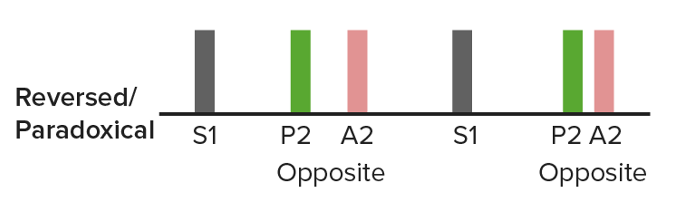
Diagram showing paradoxical splitting in which closure of the aortic valve is delayed:
The name “paradoxical” is because the split narrows the inspiration (right). The split can be heard in some individuals with a left bundle branch block.
Image by Lecturio.
Clicks and Snaps
Clicks
A high-pitched sound occurring at the point of maximal opening of the valves
Usually mid-to-late systolic: swellingSwellingInflammation out and sudden stop of the leaflets of a prolapsed mitral valveMitral valveThe valve between the left atrium and left ventricle of the heart.Heart: Anatomy
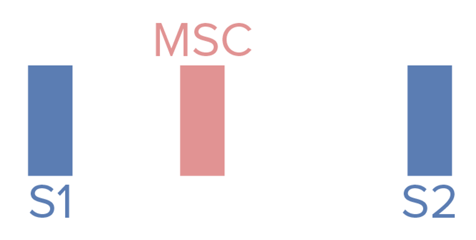
Schematic diagram depicting the mid-systolic click (MSC): MSC occurs after S1.
Image by Lecturio.
Audio:
Mid-systolic click(MSC): This audio clip is an example of an MSC heard in mitral valveMitral valveThe valve between the left atrium and left ventricle of the heart.Heart: Anatomy prolapse. An MSC is a crisp sound occurring between S1 and S2 (no murmur follows).
A high-frequency diastolic sound made by the opening of a stenotic mitral valveMitral valveThe valve between the left atrium and left ventricle of the heart.Heart: Anatomy (most common)
Leaflets swell out into the ventricle and suddenly stop, producing a mid-to-high–frequency sound.
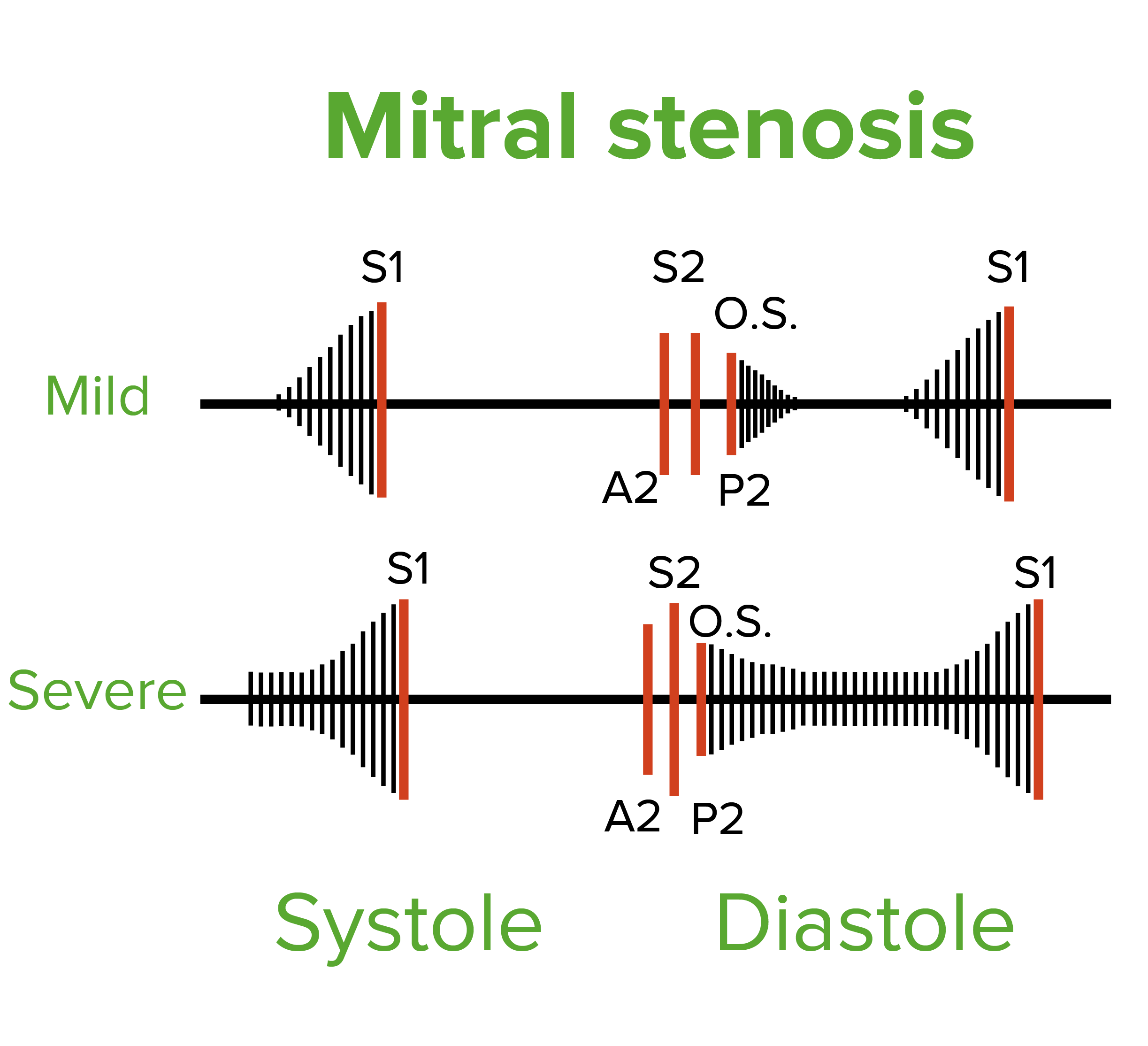
Diastolic filling and rumbling murmur in mild and severe mitral stenosis:
The mid-diastolic murmur starts after the opening snap (O.S.). The presystolic murmur is due to atrial contraction (absent in atrial fibrillation) and is best heard over the apex with the bell of a stethoscope.
FlowFlowBlood flows through the heart, arteries, capillaries, and veins in a closed, continuous circuit. Flow is the movement of volume per unit of time. Flow is affected by the pressure gradient and the resistance fluid encounters between 2 points. Vascular resistance is the opposition to flow, which is caused primarily by blood friction against vessel walls.Vascular Resistance, Flow, and Mean Arterial Pressure through a narrow opening (e.g., valvular stenosisStenosisHypoplastic Left Heart Syndrome (HLHS))
Backward flowFlowBlood flows through the heart, arteries, capillaries, and veins in a closed, continuous circuit. Flow is the movement of volume per unit of time. Flow is affected by the pressure gradient and the resistance fluid encounters between 2 points. Vascular resistance is the opposition to flow, which is caused primarily by blood friction against vessel walls.Vascular Resistance, Flow, and Mean Arterial Pressure through an incompetent valve (e.g., valvular regurgitationRegurgitationGastroesophageal Reflux Disease (GERD))
Not all murmurs indicate structural heart disease.
Determine characteristics by:
Timing (cardiac cycleCardiac cycleThe cardiac cycle describes a complete contraction and relaxation of all 4 chambers of the heart during a standard heartbeat. The cardiac cycle includes 7 phases, which together describe the cycle of ventricular filling, isovolumetric contraction, ventricular ejection, and isovolumetric relaxation.Cardiac Cycle)
Intensity
Pattern or configuration
Pitch and qualityQualityActivities and programs intended to assure or improve the quality of care in either a defined medical setting or a program. The concept includes the assessment or evaluation of the quality of care; identification of problems or shortcomings in the delivery of care; designing activities to overcome these deficiencies; and follow-up monitoring to ensure effectiveness of corrective steps.Quality Measurement and Improvement
Location or auscultation area
Maneuvers, position, and exercise
Classification according to cardiac cycleCardiac cycleThe cardiac cycle describes a complete contraction and relaxation of all 4 chambers of the heart during a standard heartbeat. The cardiac cycle includes 7 phases, which together describe the cycle of ventricular filling, isovolumetric contraction, ventricular ejection, and isovolumetric relaxation.Cardiac Cycle
Systolic (occurs at or after S1 and ends before or at S2)
Moderate-to-severe ARARAortic regurgitation (AR) is a cardiac condition characterized by the backflow of blood from the aorta to the left ventricle during diastole. Aortic regurgitation is associated with an abnormal aortic valve and/or aortic root stemming from multiple causes, commonly rheumatic heart disease as well as congenital and degenerative valvular disorders. Aortic Regurgitation (Austin Flint murmurAustin Flint murmurAortic Regurgitation)
Continuous (not confined to either systoleSystolePeriod of contraction of the heart, especially of the heart ventricles.Cardiac Cycle or diastoleDiastolePost-systolic relaxation of the heart, especially the heart ventricles.Cardiac Cycle):
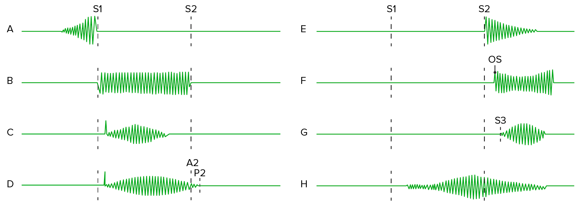
Patterns of heart murmurs (with examples): A: presystolic or late diastolic, crescendo murmur (tricuspid stenosis)
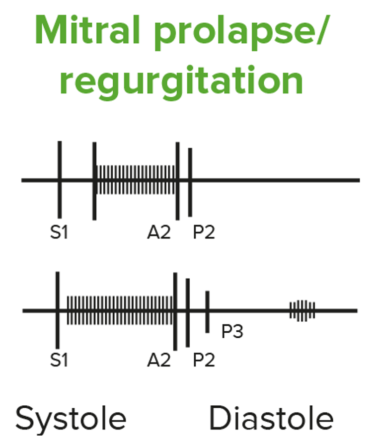
B: holosystolic murmur (mitral regurgitation)
C: midsystolic, crescendo-decrescendo murmur (aortic stenosis)
D: long systolic, crescendo-decrescendo murmur (pulmonic stenosis)
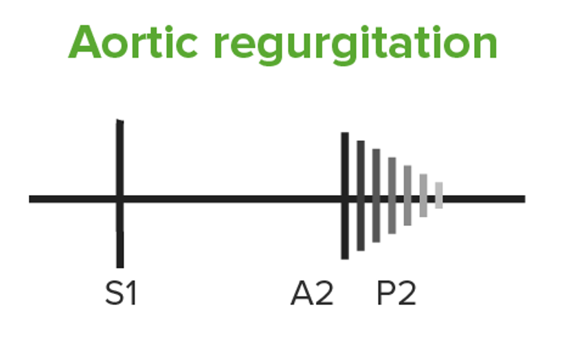
E: early diastolic, decrescendo murmur (aortic regurgitation)
F: mid-diastolic murmur (mitral stenosis)
G: short mid-diastolic murmur
H: continuous murmur (patent ductus arteriosus)
Image by Lecturio.
Diastolic murmur: Chronic aortic regurgitation (AR) results in an early diastolic murmur (high pitched). The murmur becomes holodiastolic in severe AR.
Using the Levine system, murmurs can be graded on a scaleScaleDermatologic Examination from I to VI, which reflects the intensity of the murmur.
I: very soft, may only be heard by experienced cardiologists
II: faint but readily audible
III: readily audible, louder than grade 2, no thrill
IV: loud and accompanied by a palpable thrill
V: loud enough to be heard with a stethoscope lightly touching the chest
VI: loud enough to be heard with a stethoscope off the chest
Classification according to pattern
Crescendo-decrescendo murmur:
Ascending then descending systolic ejection murmur
Diamond shaped
Example: AS
Crescendo:
Ascending intensity from faint to loud
Example: can be heard in mitral valveMitral valveThe valve between the left atrium and left ventricle of the heart.Heart: Anatomy prolapse
Decrescendo:
Descending intensity from loud to faint
Example: ARARAortic regurgitation (AR) is a cardiac condition characterized by the backflow of blood from the aorta to the left ventricle during diastole. Aortic regurgitation is associated with an abnormal aortic valve and/or aortic root stemming from multiple causes, commonly rheumatic heart disease as well as congenital and degenerative valvular disorders. Aortic Regurgitation
Crescendo-decrescendo murmurs: ascending then descending systolic murmur (diamond shaped) heard in aortic stenosis
Image by Lecturio.
Audio:
Crescendo-decrescendo murmur: In this audio clip, the sound of severe AS, a harsh, crescendo-decrescendo murmur occurring between S1 and S2, can be heard. The S2 heart sound is inaudible due to the severity of AS.
Classification according to pitch and qualityQualityActivities and programs intended to assure or improve the quality of care in either a defined medical setting or a program. The concept includes the assessment or evaluation of the quality of care; identification of problems or shortcomings in the delivery of care; designing activities to overcome these deficiencies; and follow-up monitoring to ensure effectiveness of corrective steps.Quality Measurement and Improvement
Pitch:
Frequency of the murmur
High pitch:
TR
MRMRCalculated as the ratio of the total number of people who die due to all causes over a specific time period to the total number of people in the selected population.Measures of Health Status
ARARAortic regurgitation (AR) is a cardiac condition characterized by the backflow of blood from the aorta to the left ventricle during diastole. Aortic regurgitation is associated with an abnormal aortic valve and/or aortic root stemming from multiple causes, commonly rheumatic heart disease as well as congenital and degenerative valvular disorders. Aortic Regurgitation
QualityQualityActivities and programs intended to assure or improve the quality of care in either a defined medical setting or a program. The concept includes the assessment or evaluation of the quality of care; identification of problems or shortcomings in the delivery of care; designing activities to overcome these deficiencies; and follow-up monitoring to ensure effectiveness of corrective steps.Quality Measurement and Improvement: can be blowing, harsh, rumbling, scratchy, vibratory, squeaky, or musical
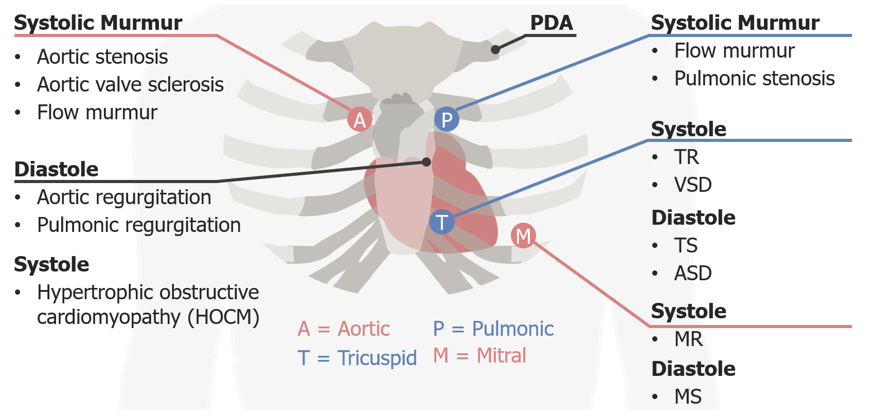
The 5 areas of auscultation can be recalled using the mnemonic, “All People Enjoy Time Magazine.”
Aortic area: right 2nd intercostal space adjacent to the sternumSternumA long, narrow, and flat bone commonly known as breastbone occurring in the midsection of the anterior thoracic segment or chest region, which stabilizes the rib cage and serves as the point of origin for several muscles that move the arms, head, and neck.Chest Wall: Anatomy
AS
Aortic valveAortic valveThe valve between the left ventricle and the ascending aorta which prevents backflow into the left ventricle.Heart: AnatomysclerosisSclerosisA pathological process consisting of hardening or fibrosis of an anatomical structure, often a vessel or a nerve.Wilms Tumor
Systolic flowFlowBlood flows through the heart, arteries, capillaries, and veins in a closed, continuous circuit. Flow is the movement of volume per unit of time. Flow is affected by the pressure gradient and the resistance fluid encounters between 2 points. Vascular resistance is the opposition to flow, which is caused primarily by blood friction against vessel walls.Vascular Resistance, Flow, and Mean Arterial Pressure murmurs
Pulmonic area: left 2nd intercostal space adjacent to the sternumSternumA long, narrow, and flat bone commonly known as breastbone occurring in the midsection of the anterior thoracic segment or chest region, which stabilizes the rib cage and serves as the point of origin for several muscles that move the arms, head, and neck.Chest Wall: Anatomy
Systolic flowFlowBlood flows through the heart, arteries, capillaries, and veins in a closed, continuous circuit. Flow is the movement of volume per unit of time. Flow is affected by the pressure gradient and the resistance fluid encounters between 2 points. Vascular resistance is the opposition to flow, which is caused primarily by blood friction against vessel walls.Vascular Resistance, Flow, and Mean Arterial Pressure murmurs
Atrial septal defectAtrial Septal DefectAtrial septal defects (ASDs) are benign acyanotic congenital heart defects characterized by an opening in the interatrial septum that causes blood to flow from the left atrium (LA) to the right atrium (RA) (left-to-right shunt). Atrial Septal Defect (ASD) (fixed splittingSplittingDefense Mechanisms of S2)
ErbRBChlamydia’s (auscultation) point (left sternal border): left 3rd intercostal space
Systolic murmurs: HOCM
Diastolic murmurs:
ARARAortic regurgitation (AR) is a cardiac condition characterized by the backflow of blood from the aorta to the left ventricle during diastole. Aortic regurgitation is associated with an abnormal aortic valve and/or aortic root stemming from multiple causes, commonly rheumatic heart disease as well as congenital and degenerative valvular disorders. Aortic Regurgitation
Tricuspid area: left 4th–5th intercostal space adjacent to the sternumSternumA long, narrow, and flat bone commonly known as breastbone occurring in the midsection of the anterior thoracic segment or chest region, which stabilizes the rib cage and serves as the point of origin for several muscles that move the arms, head, and neck.Chest Wall: Anatomy
Systolic murmurs:
TR
VSD
Diastolic murmurs:
TS
Mitral area (apex): left 4th intercostal space, midclavicular line
Systolic murmurs:
MRMRCalculated as the ratio of the total number of people who die due to all causes over a specific time period to the total number of people in the selected population.Measures of Health Status (holosystolic)
Mitral valveMitral valveThe valve between the left atrium and left ventricle of the heart.Heart: Anatomy prolapse
Diastolic murmurs: mitral stenosisStenosisHypoplastic Left Heart Syndrome (HLHS) (MSMSMultiple sclerosis (MS) is a chronic inflammatory autoimmune disease that leads to demyelination of the nerves in the CNS. Young women are more predominantly affected by this most common demyelinating condition.Multiple Sclerosis)
Auscultation areas and associated murmurs that are heard: aortic, pulmonic, Erb’s point, tricuspid, and mitral areas (APETM) TR: tricuspid regurgitation VSD: ventricular septal defect TS: tricuspid stenosis ASD: atrial septal defect MR: mitral regurgitation MS: mitral stenosis
Image by Lecturio.
Dynamic Auscultation
Cardiac physiologyCardiac PhysiologyA complex system of coordinated electrical circuitry within the heart governs cardiac muscle activity. The heart generates its own electrical impulses within its pacemaker cells. The signal then travels through specialized myocytes, which act as electrical wiring, distributing the signal throughout the heart.Cardiac Physiology and maneuvers
Squatting (seen in tetralogy of FallotTetralogy of FallotTetralogy of Fallot is the most common cyanotic congenital heart disease. The disease is the confluence of 4 pathologic cardiac features: overriding aorta, ventricular septal defect, right ventricular outflow obstruction, and right ventricular hypertrophy. Tetralogy of Fallot)
Passive legLegThe lower leg, or just “leg” in anatomical terms, is the part of the lower limb between the knee and the ankle joint. The bony structure is composed of the tibia and fibula bones, and the muscles of the leg are grouped into the anterior, lateral, and posterior compartments by extensions of fascia.Leg: Anatomy raise (increased venous return due to gravity)
AfterloadAfterloadAfterload is the resistance in the aorta that prevents blood from leaving the heart. Afterload represents the pressure the LV needs to overcome to eject blood into the aorta.Cardiac Mechanics:
Effective pressure against which the heart ejects blood during ventricular contraction
↑ AfterloadAfterloadAfterload is the resistance in the aorta that prevents blood from leaving the heart. Afterload represents the pressure the LV needs to overcome to eject blood into the aorta.Cardiac Mechanics: handgrip
Effects on the intensity of heart murmurs
RespirationRespirationThe act of breathing with the lungs, consisting of inhalation, or the taking into the lungs of the ambient air, and of exhalation, or the expelling of the modified air which contains more carbon dioxide than the air taken in.Nose Anatomy (External & Internal) (↑preloadPreloadCardiac Mechanics):
Mitral valveMitral valveThe valve between the left atrium and left ventricle of the heart.Heart: Anatomy prolapse (becomes longer and louder)
While squatting and with passive legLegThe lower leg, or just “leg” in anatomical terms, is the part of the lower limb between the knee and the ankle joint. The bony structure is composed of the tibia and fibula bones, and the muscles of the leg are grouped into the anterior, lateral, and posterior compartments by extensions of fascia.Leg: Anatomy raise (↑preloadPreloadCardiac Mechanics), most murmurs become louder, EXCEPT:
HOCM (becomes softer)
Mitral valveMitral valveThe valve between the left atrium and left ventricle of the heart.Heart: Anatomy prolapse (becomes shorter, except in severe MRMRCalculated as the ratio of the total number of people who die due to all causes over a specific time period to the total number of people in the selected population.Measures of Health Status)
With isotonicIsotonicSolutions having the same osmotic pressure as blood serum, or another solution with which they are compared.Renal Sodium and Water Regulation and isometric (sustained handgrip) exercise (↑ afterloadAfterloadAfterload is the resistance in the aorta that prevents blood from leaving the heart. Afterload represents the pressure the LV needs to overcome to eject blood into the aorta.Cardiac Mechanics), most murmurs increase, EXCEPT:
HOCM (decreases in intensity)
AS (decreases in intensity, helping to differentiate AS from MRMRCalculated as the ratio of the total number of people who die due to all causes over a specific time period to the total number of people in the selected population.Measures of Health Status)
Table: Maneuvers that change the intensity of murmurs
Passive legLegThe lower leg, or just “leg” in anatomical terms, is the part of the lower limb between the knee and the ankle joint. The bony structure is composed of the tibia and fibula bones, and the muscles of the leg are grouped into the anterior, lateral, and posterior compartments by extensions of fascia.Leg: Anatomy raise
Squatting
Most murmurs
HOCM
Mitral valveMitral valveThe valve between the left atrium and left ventricle of the heart.Heart: Anatomy prolapse
Mitral valveMitral valveThe valve between the left atrium and left ventricle of the heart.Heart: Anatomy prolapse
Most murmurs
Increased afterloadAfterloadAfterload is the resistance in the aorta that prevents blood from leaving the heart. Afterload represents the pressure the LV needs to overcome to eject blood into the aorta.Cardiac Mechanics
Handgrip
Most murmurs, especially ARARAortic regurgitation (AR) is a cardiac condition characterized by the backflow of blood from the aorta to the left ventricle during diastole. Aortic regurgitation is associated with an abnormal aortic valve and/or aortic root stemming from multiple causes, commonly rheumatic heart disease as well as congenital and degenerative valvular disorders. Aortic Regurgitation, MRMRCalculated as the ratio of the total number of people who die due to all causes over a specific time period to the total number of people in the selected population.Measures of Health Status, VSD
The table below lists cardiac abnormalities with their corresponding murmurs.
Table: Systolic murmurs
Type
Cardiac cycleCardiac cycleThe cardiac cycle describes a complete contraction and relaxation of all 4 chambers of the heart during a standard heartbeat. The cardiac cycle includes 7 phases, which together describe the cycle of ventricular filling, isovolumetric contraction, ventricular ejection, and isovolumetric relaxation.Cardiac Cycle
Radiates to axillaAxillaThe axilla is a pyramid-shaped space located between the upper thorax and the arm. The axilla has a base, an apex, and 4 walls (anterior, medial, lateral, posterior). The base of the pyramid is made up of the axillary skin. The apex is the axillary inlet, located between the 1st rib, superior border of the scapula, and clavicle. Axilla and Brachial Plexus: Anatomy
Cardiac cycleCardiac cycleThe cardiac cycle describes a complete contraction and relaxation of all 4 chambers of the heart during a standard heartbeat. The cardiac cycle includes 7 phases, which together describe the cycle of ventricular filling, isovolumetric contraction, ventricular ejection, and isovolumetric relaxation.Cardiac Cycle
Pattern
Location
Additional description
ARARAortic regurgitation (AR) is a cardiac condition characterized by the backflow of blood from the aorta to the left ventricle during diastole. Aortic regurgitation is associated with an abnormal aortic valve and/or aortic root stemming from multiple causes, commonly rheumatic heart disease as well as congenital and degenerative valvular disorders. Aortic Regurgitation
Diastolic
Decrescendo
Erb’s point
S3 in acute ARARAortic regurgitation (AR) is a cardiac condition characterized by the backflow of blood from the aorta to the left ventricle during diastole. Aortic regurgitation is associated with an abnormal aortic valve and/or aortic root stemming from multiple causes, commonly rheumatic heart disease as well as congenital and degenerative valvular disorders. Aortic Regurgitation
Frequently with MSMSMultiple sclerosis (MS) is a chronic inflammatory autoimmune disease that leads to demyelination of the nerves in the CNS. Young women are more predominantly affected by this most common demyelinating condition.Multiple Sclerosis (but softer and shorter than MSMSMultiple sclerosis (MS) is a chronic inflammatory autoimmune disease that leads to demyelination of the nerves in the CNS. Young women are more predominantly affected by this most common demyelinating condition.Multiple Sclerosis)
MRMRCalculated as the ratio of the total number of people who die due to all causes over a specific time period to the total number of people in the selected population.Measures of Health Status (Mitral Regurgitation)
P (mitral valveMitral valveThe valve between the left atrium and left ventricle of the heart.Heart: AnatomyProlapse)
MSMSMultiple sclerosis (MS) is a chronic inflammatory autoimmune disease that leads to demyelination of the nerves in the CNS. Young women are more predominantly affected by this most common demyelinating condition.Multiple Sclerosis (Mitral Stenosis)
ARARAortic regurgitation (AR) is a cardiac condition characterized by the backflow of blood from the aorta to the left ventricle during diastole. Aortic regurgitation is associated with an abnormal aortic valve and/or aortic root stemming from multiple causes, commonly rheumatic heart disease as well as congenital and degenerative valvular disorders. Aortic Regurgitation(Aortic Regurgitation)
TS (Tricuspid Stenosis)
References
Alpert, M.A. (1990). Systolic Murmurs. In Walker, H.K., et al. (Ed). Clinical Methods: The History, Physical, and Laboratory Examinations. (3rd ed.) https://www.ncbi.nlm.nih.gov/books/NBK345/
Williams, E.S. (1990). The Fourth Heart Sound. In Walker H.K., et al. (Ed.), Clinical Methods: The History, Physical, and Laboratory Examinations (3rd ed.) https://www.ncbi.nlm.nih.gov/books/NBK344/
Create your free account or log in to continue reading!