Ovarian torsion is a clinical emergency in which an ovary with or without the fallopian tubes twists along its axis, leading to partial or complete obstruction of its blood supply. It is also called an adnexal or tubo-ovarian torsion, especially if the fallopian tube is also involved. The ovaries twist along the suspensory ligament of the ovary and the utero-ovarian ligament, which support the ovaries and secure them to the inner pelvic walls and uterus, respectively. It most commonly occurs in women of reproductive age and when the ovary is larger than 5 cm. The torsion cuts off the ovarian blood supply, leading to pooling of blood, edema, and severe pain. As a clinical emergency, it needs to be treated promptly with surgical intervention in order to prevent necrosis of the ovaries and other complications.
Ovarian torsionOvarian torsionOvarian torsion is a clinical emergency in which the ovaries (with or without the fallopian tubes) twist along their axis, leading to partial or complete obstruction of their blood supply. Ovarian torsion is also called adnexal or tubo-ovarian torsion, especially if a fallopian tube is also involved. Ovarian Torsion is the twisting of the ovariesOvariesOvaries are the paired gonads of the female reproductive system that contain haploid gametes known as oocytes. The ovaries are located intraperitoneally in the pelvis, just posterior to the broad ligament, and are connected to the pelvic sidewall and to the uterus by ligaments. These organs function to secrete hormones (estrogen and progesterone) and to produce the female germ cells (oocytes).Ovaries: Anatomy along their axis. Ovarian torsionOvarian torsionOvarian torsion is a clinical emergency in which the ovaries (with or without the fallopian tubes) twist along their axis, leading to partial or complete obstruction of their blood supply. Ovarian torsion is also called adnexal or tubo-ovarian torsion, especially if a fallopian tube is also involved. Ovarian Torsion may or may not include the fallopian tubesFallopian tubesThe uterus, cervix, and fallopian tubes are part of the internal female reproductive system. The fallopian tubes receive an ovum after ovulation and help move it and/or a fertilized embryo toward the uterus via ciliated cells lining the tubes and peristaltic movements of its smooth muscle. Uterus, Cervix, and Fallopian Tubes: Anatomy and if it does, it is termed adnexal torsionAdnexal torsionOvarian torsion is a clinical emergency in which the ovaries (with or without the fallopian tubes) twist along their axis, leading to partial or complete obstruction of their blood supply. Ovarian torsion is also called adnexal or tubo-ovarian torsion, especially if a fallopian tube is also involved.Ovarian Torsion.
Anatomy[2]
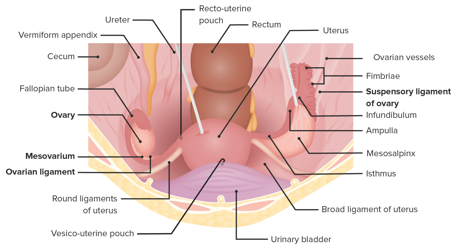
The ovariesOvariesOvaries are the paired gonads of the female reproductive system that contain haploid gametes known as oocytes. The ovaries are located intraperitoneally in the pelvis, just posterior to the broad ligament, and are connected to the pelvic sidewall and to the uterus by ligaments. These organs function to secrete hormones (estrogen and progesterone) and to produce the female germ cells (oocytes).Ovaries: Anatomy are paired glandular organs found within the lesser pelvisLesser pelvisThe part of the pelvis, inferior to the pelvic brim, that comprises both the pelvic cavity and the part of the perineum lying inferior to the pelvic diaphragm.Pelvis: Anatomy.
Attaches the ovary to the lateral wall of the inner surface of the pelvisPelvisThe pelvis consists of the bony pelvic girdle, the muscular and ligamentous pelvic floor, and the pelvic cavity, which contains viscera, vessels, and multiple nerves and muscles. The pelvic girdle, composed of 2 “hip” bones and the sacrum, is a ring-like bony structure of the axial skeleton that links the vertebral column with the lower extremities.Pelvis: Anatomy
Attaches the ovary medially to the uterusUterusThe uterus, cervix, and fallopian tubes are part of the internal female reproductive system. The uterus has a thick wall made of smooth muscle (the myometrium) and an inner mucosal layer (the endometrium). The most inferior portion of the uterus is the cervix, which connects the uterine cavity to the vagina.Uterus, Cervix, and Fallopian Tubes: Anatomy
Primarily responsible for preventing ovarian torsionOvarian torsionOvarian torsion is a clinical emergency in which the ovaries (with or without the fallopian tubes) twist along their axis, leading to partial or complete obstruction of their blood supply. Ovarian torsion is also called adnexal or tubo-ovarian torsion, especially if a fallopian tube is also involved. Ovarian Torsion
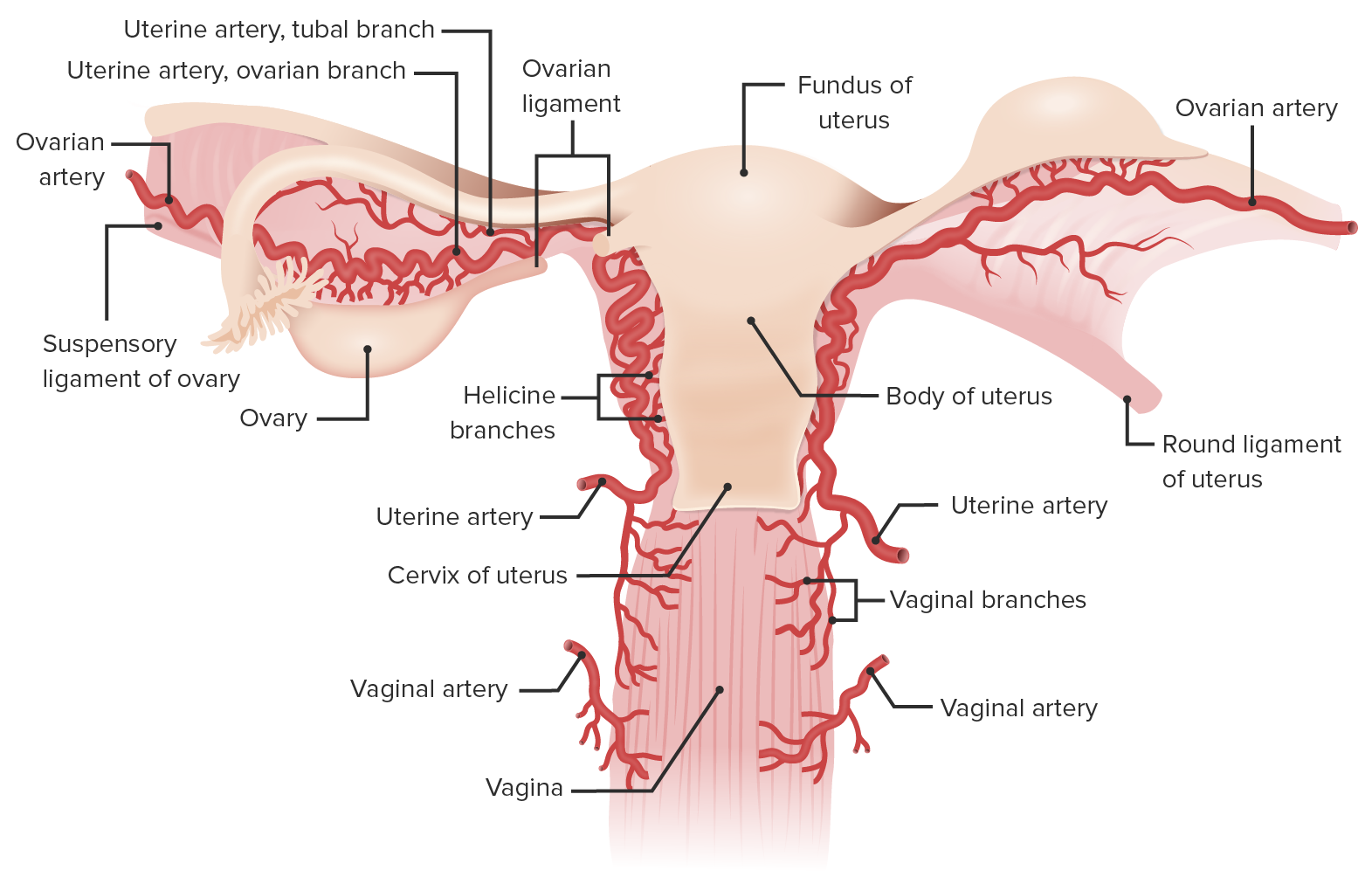
The ovariesOvariesOvaries are the paired gonads of the female reproductive system that contain haploid gametes known as oocytes. The ovaries are located intraperitoneally in the pelvis, just posterior to the broad ligament, and are connected to the pelvic sidewall and to the uterus by ligaments. These organs function to secrete hormones (estrogen and progesterone) and to produce the female germ cells (oocytes).Ovaries: Anatomy have a dual blood supply, receiving blood from both the ovarian and uterine blood vessels.
Posterior view of the uterus showing the blood supply of the uterus and ovaries:
Note the ovarian artery traveling along the ovarian suspensory ligament, supplying both the ovaries and the lateral portion of the fallopian tube. The ovarian artery continues in the mesosalpinx to anastomose with branches of the uterine artery. (Note: the round ligament comes off the anterior surface of the uterus.)
More common in women of reproductive age, due to the more common formation of physiologic cystsCystsAny fluid-filled closed cavity or sac that is lined by an epithelium. Cysts can be of normal, abnormal, non-neoplastic, or neoplastic tissues.Fibrocystic Change.
Can also occur in female fetuses, neonates, pre-pubertal, and postmenopausal women, though less common
More commonly affects women with ovariesOvariesOvaries are the paired gonads of the female reproductive system that contain haploid gametes known as oocytes. The ovaries are located intraperitoneally in the pelvis, just posterior to the broad ligament, and are connected to the pelvic sidewall and to the uterus by ligaments. These organs function to secrete hormones (estrogen and progesterone) and to produce the female germ cells (oocytes).Ovaries: Anatomy > 5 cm, which is more common in:
Ovarian cystsCystsAny fluid-filled closed cavity or sac that is lined by an epithelium. Cysts can be of normal, abnormal, non-neoplastic, or neoplastic tissues.Fibrocystic Change and tumors (found in > 85% of cases)
Women undergoing fertility treatments
Polycystic ovarian syndromePolycystic ovarian syndromePolycystic ovarian syndrome (PCOS) is the most common endocrine disorder of reproductive-age women, affecting nearly 5%-10% of women in the age group. It is characterized by hyperandrogenism, chronic anovulation leading to oligomenorrhea (or amenorrhea), and metabolic dysfunction.Polycystic Ovarian Syndrome (PCOSPCOSPolycystic ovarian syndrome (PCOS) is the most common endocrine disorder of reproductive-age women, affecting nearly 5%-10% of women in the age group. It is characterized by hyperandrogenism, chronic anovulation leading to oligomenorrhea (or amenorrhea), and metabolic dysfunction.Polycystic Ovarian Syndrome)
PregnancyPregnancyThe status during which female mammals carry their developing young (embryos or fetuses) in utero before birth, beginning from fertilization to birth.Pregnancy: Diagnosis, Physiology, and Care
Torsion of the left ovary is less frequent because it is supported by the sigmoidSigmoidA segment of the colon between the rectum and the descending colon.VolvuluscolonColonThe large intestines constitute the last portion of the digestive system. The large intestine consists of the cecum, appendix, colon (with ascending, transverse, descending, and sigmoid segments), rectum, and anal canal. The primary function of the colon is to remove water and compact the stool prior to expulsion from the body via the rectum and anal canal. Colon, Cecum, and Appendix: Anatomy.
Fixation of the ovariesOvariesOvaries are the paired gonads of the female reproductive system that contain haploid gametes known as oocytes. The ovaries are located intraperitoneally in the pelvis, just posterior to the broad ligament, and are connected to the pelvic sidewall and to the uterus by ligaments. These organs function to secrete hormones (estrogen and progesterone) and to produce the female germ cells (oocytes).Ovaries: Anatomy decreases the risk of torsion, and can be seen with:
Adhesions from prior surgeries
EndometriosisEndometriosisEndometriosis is a common disease in which patients have endometrial tissue implanted outside of the uterus. Endometrial implants can occur anywhere in the pelvis, including the ovaries, the broad and uterosacral ligaments, the pelvic peritoneum, and the urinary and gastrointestinal tracts.Endometriosis
Prior tubo-ovarian abscessAbscessAccumulation of purulent material in tissues, organs, or circumscribed spaces, usually associated with signs of infection.Chronic Granulomatous Disease
Ovarian or adnexal torsionAdnexal torsionOvarian torsion is a clinical emergency in which the ovaries (with or without the fallopian tubes) twist along their axis, leading to partial or complete obstruction of their blood supply. Ovarian torsion is also called adnexal or tubo-ovarian torsion, especially if a fallopian tube is also involved.Ovarian Torsion involves the following sequence of events:[1–3]
CompressionCompressionBlunt Chest Trauma/blockage of the venous and lymphatic drainage from the ovariesOvariesOvaries are the paired gonads of the female reproductive system that contain haploid gametes known as oocytes. The ovaries are located intraperitoneally in the pelvis, just posterior to the broad ligament, and are connected to the pelvic sidewall and to the uterus by ligaments. These organs function to secrete hormones (estrogen and progesterone) and to produce the female germ cells (oocytes).Ovaries: Anatomy
Local edemaEdemaEdema is a condition in which excess serous fluid accumulates in the body cavity or interstitial space of connective tissues. Edema is a symptom observed in several medical conditions. It can be categorized into 2 types, namely, peripheral (in the extremities) and internal (in an organ or body cavity). Edema of the ovary, fallopian tubesFallopian tubesThe uterus, cervix, and fallopian tubes are part of the internal female reproductive system. The fallopian tubes receive an ovum after ovulation and help move it and/or a fertilized embryo toward the uterus via ciliated cells lining the tubes and peristaltic movements of its smooth muscle. Uterus, Cervix, and Fallopian Tubes: Anatomy, and supportive ligaments
CompressionCompressionBlunt Chest Trauma/blockage of the arterial supply of the ovariesOvariesOvaries are the paired gonads of the female reproductive system that contain haploid gametes known as oocytes. The ovaries are located intraperitoneally in the pelvis, just posterior to the broad ligament, and are connected to the pelvic sidewall and to the uterus by ligaments. These organs function to secrete hormones (estrogen and progesterone) and to produce the female germ cells (oocytes).Ovaries: Anatomy
IschemiaIschemiaA hypoperfusion of the blood through an organ or tissue caused by a pathologic constriction or obstruction of its blood vessels, or an absence of blood circulation.Ischemic Cell Damage, followed by necrosisNecrosisThe death of cells in an organ or tissue due to disease, injury or failure of the blood supply.Ischemic Cell Damage of the ovarian tissue
Local hemorrhage from friable necrotic tissue
Clinical Presentation
The most common symptom is sudden, severe lower abdominal/pelvic painPainAn unpleasant sensation induced by noxious stimuli which are detected by nerve endings of nociceptive neurons.Pain: Types and Pathways.[4–7]
Usually unilateral, though may be diffuse
May radiate to the rest of the abdomen, back, and flank
Physical examination findings:
Signs of an acute abdomenAcute AbdomenAcute abdomen, which is in many cases a surgical emergency, is the sudden onset of abdominal pain that may be caused by inflammation, infection, perforation, ischemia, or obstruction. The location of the pain, its characteristics, and associated symptoms (e.g., jaundice) are important tools that help narrow the differential diagnosis.Acute Abdomen:
Unilateral tender adnexal massMassThree-dimensional lesion that occupies a space within the breastImaging of the Breast
Cervical motion tenderness
NauseaNauseaAn unpleasant sensation in the stomach usually accompanied by the urge to vomit. Common causes are early pregnancy, sea and motion sickness, emotional stress, intense pain, food poisoning, and various enteroviruses.Antiemetics and vomitingVomitingThe forcible expulsion of the contents of the stomach through the mouth.Hypokalemia:
Common
May be continuous or appear in waves
FeverFeverFever is defined as a measured body temperature of at least 38°C (100.4°F). Fever is caused by circulating endogenous and/or exogenous pyrogens that increase levels of prostaglandin E2 in the hypothalamus. Fever is commonly associated with chills, rigors, sweating, and flushing of the skin. Fever can be seen in cases in which the ovary is undergoing necrosisNecrosisThe death of cells in an organ or tissue due to disease, injury or failure of the blood supply.Ischemic Cell Damage or rupture.
Abnormal vaginal bleeding and discharge might be present if associated with rupture or an abscessAbscessAccumulation of purulent material in tissues, organs, or circumscribed spaces, usually associated with signs of infection.Chronic Granulomatous Disease.
Diagnosis and Management
Adnexal torsionAdnexal torsionOvarian torsion is a clinical emergency in which the ovaries (with or without the fallopian tubes) twist along their axis, leading to partial or complete obstruction of their blood supply. Ovarian torsion is also called adnexal or tubo-ovarian torsion, especially if a fallopian tube is also involved.Ovarian Torsion is suspected based on typical symptoms, supported by imaging with transvaginal ultrasonography or color DopplerDopplerUltrasonography applying the doppler effect, with frequency-shifted ultrasound reflections produced by moving targets (usually red blood cells) in the bloodstream along the ultrasound axis in direct proportion to the velocity of movement of the targets, to determine both direction and velocity of blood flow.Ultrasound (Sonography) ultrasonography, and confirmed during immediate exploratory surgery.
Laboratory tests[4,5]
CBC to look for anemiaAnemiaAnemia is a condition in which individuals have low Hb levels, which can arise from various causes. Anemia is accompanied by a reduced number of RBCs and may manifest with fatigue, shortness of breath, pallor, and weakness. Subtypes are classified by the size of RBCs, chronicity, and etiology. Anemia: Overview and Types and leukocytosisLeukocytosisA transient increase in the number of leukocytes in a body fluid.West Nile Virus:
AnemiaAnemiaAnemia is a condition in which individuals have low Hb levels, which can arise from various causes. Anemia is accompanied by a reduced number of RBCs and may manifest with fatigue, shortness of breath, pallor, and weakness. Subtypes are classified by the size of RBCs, chronicity, and etiology. Anemia: Overview and Types and leukocytosisLeukocytosisA transient increase in the number of leukocytes in a body fluid.West Nile Virus are seen in cases of hemorrhage.
LeukocytosisLeukocytosisA transient increase in the number of leukocytes in a body fluid.West Nile Virus is seen in cases of rupture.
Urine pregnancyPregnancyThe status during which female mammals carry their developing young (embryos or fetuses) in utero before birth, beginning from fertilization to birth.Pregnancy: Diagnosis, Physiology, and Care test is important to rule out ectopic pregnancyEctopic pregnancyEctopic pregnancy refers to the implantation of a fertilized egg (embryo) outside the uterine cavity. The main cause is disruption of the normal anatomy of the fallopian tube. Ectopic Pregnancy.
Imaging[4,5,7–9]
Ultrasonography with pelvic DopplerDopplerUltrasonography applying the doppler effect, with frequency-shifted ultrasound reflections produced by moving targets (usually red blood cells) in the bloodstream along the ultrasound axis in direct proportion to the velocity of movement of the targets, to determine both direction and velocity of blood flow.Ultrasound (Sonography); often nondiagnostic, but may show:
An asymmetrical, enlarged ovary
An adnexal massMassThree-dimensional lesion that occupies a space within the breastImaging of the Breast, which increases the risk for torsion
Decreased venous blood flowBlood flowBlood flow refers to the movement of a certain volume of blood through the vasculature over a given unit of time (e.g., mL per minute).Vascular Resistance, Flow, and Mean Arterial Pressure on DopplerDopplerUltrasonography applying the doppler effect, with frequency-shifted ultrasound reflections produced by moving targets (usually red blood cells) in the bloodstream along the ultrasound axis in direct proportion to the velocity of movement of the targets, to determine both direction and velocity of blood flow.Ultrasound (Sonography) points towards ovarian torsionOvarian torsionOvarian torsion is a clinical emergency in which the ovaries (with or without the fallopian tubes) twist along their axis, leading to partial or complete obstruction of their blood supply. Ovarian torsion is also called adnexal or tubo-ovarian torsion, especially if a fallopian tube is also involved. Ovarian Torsion.
Whirlpool signWhirlpool signTwisting of the superior mesenteric vein and the mesentery around the superior mesenteric artery.Intestinal Malrotation: twisting of the vascular pedicle; seen as a round hyperechoicHyperechoicA structure that produces a high-amplitude echo (lighter grays and white)Ultrasound (Sonography)massMassThree-dimensional lesion that occupies a space within the breastImaging of the Breast with hypoechoicHypoechoicA structure that produces a low-amplitude echo (darker grays)Ultrasound (Sonography) stripes
Free fluid might be present in the pelvic cavity.
Note: Normal DopplerDopplerUltrasonography applying the doppler effect, with frequency-shifted ultrasound reflections produced by moving targets (usually red blood cells) in the bloodstream along the ultrasound axis in direct proportion to the velocity of movement of the targets, to determine both direction and velocity of blood flow.Ultrasound (Sonography)flowFlowBlood flows through the heart, arteries, capillaries, and veins in a closed, continuous circuit. Flow is the movement of volume per unit of time. Flow is affected by the pressure gradient and the resistance fluid encounters between 2 points. Vascular resistance is the opposition to flow, which is caused primarily by blood friction against vessel walls.Vascular Resistance, Flow, and Mean Arterial Pressure does not exclude torsion.
MRI: not routinely used, but may be considered if US findings are inconclusive
CT can rule out other abdominal conditions, such as appendicitisAppendicitisAppendicitis is the acute inflammation of the vermiform appendix and the most common abdominal surgical emergency globally. The condition has a lifetime risk of 8%. Characteristic features include periumbilical abdominal pain that migrates to the right lower quadrant, fever, anorexia, nausea, and vomiting.Appendicitis.
Surgical confirmation[4]
LaparoscopyLaparoscopyLaparoscopy is surgical exploration and interventions performed through small incisions with a camera and long instruments. Laparotomy and Laparoscopy/laparotomyLaparotomyIncision into the side of the abdomen between the ribs and pelvis.Laparotomy and Laparoscopy is the gold standard for the diagnosis of ovarian torsionOvarian torsionOvarian torsion is a clinical emergency in which the ovaries (with or without the fallopian tubes) twist along their axis, leading to partial or complete obstruction of their blood supply. Ovarian torsion is also called adnexal or tubo-ovarian torsion, especially if a fallopian tube is also involved. Ovarian Torsion, as the twisted ovary can be directly visualized by the surgeon.
Management[4–7,10]
Management may vary by location. The following information is based on recommendations by US and UK medical societies:
Prompt and early surgical resolution of the torsion must be attempted in order to save the ovary.
Preferred approach: laparoscopyLaparoscopyLaparoscopy is surgical exploration and interventions performed through small incisions with a camera and long instruments. Laparotomy and Laparoscopy
The viability of the ovary is evaluated by the surgeon.
NecrosisNecrosisThe death of cells in an organ or tissue due to disease, injury or failure of the blood supply.Ischemic Cell Damage is rare when torsion is appropriately treated → most ovariesOvariesOvaries are the paired gonads of the female reproductive system that contain haploid gametes known as oocytes. The ovaries are located intraperitoneally in the pelvis, just posterior to the broad ligament, and are connected to the pelvic sidewall and to the uterus by ligaments. These organs function to secrete hormones (estrogen and progesterone) and to produce the female germ cells (oocytes).Ovaries: Anatomy can simply be “untwisted” and saved
Necrotic tissue should be removed.
Cystectomy is usually indicated if a benignBenignFibroadenoma cyst is present.
In post-menopausal women, salpingo-oophorectomy is preferred.
In adolescents, oophorectomy should be avoided unless absolutely necessary owing to severe necrosisNecrosisThe death of cells in an organ or tissue due to disease, injury or failure of the blood supply.Ischemic Cell Damage.
If malignancyMalignancyHemothorax is suspected, consultation with a gynecologic oncologist is recommended.
Ectopic pregnancyEctopic pregnancyEctopic pregnancy refers to the implantation of a fertilized egg (embryo) outside the uterine cavity. The main cause is disruption of the normal anatomy of the fallopian tube. Ectopic Pregnancy:implantationImplantationEndometrial implantation of embryo, mammalian at the blastocyst stage.Fertilization and First Week of the fertilized embryoEmbryoThe entity of a developing mammal, generally from the cleavage of a zygote to the end of embryonic differentiation of basic structures. For the human embryo, this represents the first two months of intrauterine development preceding the stages of the fetus.Fertilization and First Week outside the uterine cavity, usually in a fallopian tubeFallopian TubeA pair of highly specialized canals extending from the uterus to its corresponding ovary. They provide the means for ovum transport from the ovaries and they are the site of the ovum’s final maturation and fertilization. The fallopian tube consists of an interstitium, an isthmus, an ampulla, an infundibulum, and fimbriae. Its wall consists of three layers: serous, muscular, and an internal mucosal layer lined with both ciliated and secretory cells.Uterus, Cervix, and Fallopian Tubes: Anatomy. Ectopic pregnancyEctopic pregnancyEctopic pregnancy refers to the implantation of a fertilized egg (embryo) outside the uterine cavity. The main cause is disruption of the normal anatomy of the fallopian tube. Ectopic Pregnancy presents with sudden and severe abdominal painAbdominal PainAcute Abdomen. When rupture occurs, ectopic pregnancyEctopic pregnancyEctopic pregnancy refers to the implantation of a fertilized egg (embryo) outside the uterine cavity. The main cause is disruption of the normal anatomy of the fallopian tube. Ectopic Pregnancy can present with feverFeverFever is defined as a measured body temperature of at least 38°C (100.4°F). Fever is caused by circulating endogenous and/or exogenous pyrogens that increase levels of prostaglandin E2 in the hypothalamus. Fever is commonly associated with chills, rigors, sweating, and flushing of the skin. Fever, peritonitisPeritonitisInflammation of the peritoneum lining the abdominal cavity as the result of infectious, autoimmune, or chemical processes. Primary peritonitis is due to infection of the peritoneal cavity via hematogenous or lymphatic spread and without intra-abdominal source. Secondary peritonitis arises from the abdominal cavity itself through rupture or abscess of intra-abdominal organs.Penetrating Abdominal Injury, and/or hemorrhagic shockHemorrhagic shockAcute hemorrhage or excessive fluid loss resulting in hypovolemia.Hemothorax. Usually, there is a history of a missed menstrual period. Findings include a positive pregnancyPregnancyThe status during which female mammals carry their developing young (embryos or fetuses) in utero before birth, beginning from fertilization to birth.Pregnancy: Diagnosis, Physiology, and Care test and painPainAn unpleasant sensation induced by noxious stimuli which are detected by nerve endings of nociceptive neurons.Pain: Types and Pathways with cervical manipulation (pelvic exam). Diagnosis is confirmed with pelvic ultrasonography and an abnormal trend on serial hCG levels.
Ruptured ovarian cyst: an ovarian cyst is a fluid-filled sac within an ovary or on its surface, which can form as a result of ovulationOvulationThe discharge of an ovum from a rupturing follicle in the ovary.Menstrual Cycle. Rupture of a cyst can cause severe painPainAn unpleasant sensation induced by noxious stimuli which are detected by nerve endings of nociceptive neurons.Pain: Types and Pathways and internal bleeding. Diagnostic findings may include an adnexal massMassThree-dimensional lesion that occupies a space within the breastImaging of the Breast and free fluid within the pelvisPelvisThe pelvis consists of the bony pelvic girdle, the muscular and ligamentous pelvic floor, and the pelvic cavity, which contains viscera, vessels, and multiple nerves and muscles. The pelvic girdle, composed of 2 “hip” bones and the sacrum, is a ring-like bony structure of the axial skeleton that links the vertebral column with the lower extremities.Pelvis: Anatomy on ultrasonography, along with a negative pregnancyPregnancyThe status during which female mammals carry their developing young (embryos or fetuses) in utero before birth, beginning from fertilization to birth.Pregnancy: Diagnosis, Physiology, and Care test. Management includes watchful waiting for uncomplicated cystsCystsAny fluid-filled closed cavity or sac that is lined by an epithelium. Cysts can be of normal, abnormal, non-neoplastic, or neoplastic tissues.Fibrocystic Change and surgery for cystsCystsAny fluid-filled closed cavity or sac that is lined by an epithelium. Cysts can be of normal, abnormal, non-neoplastic, or neoplastic tissues.Fibrocystic Change associated with hemorrhage.
Pelvic inflammatory diseasePelvic inflammatory diseasePelvic inflammatory disease (PID) is defined as a polymicrobial infection of the upper female reproductive system. The disease can affect the uterus, fallopian tubes, ovaries, and adjacent structures. Pelvic inflammatory disease is closely linked with sexually transmitted diseases, most commonly caused by Chlamydia trachomatis, Neisseria gonorrhoeae, and Gardnerella vaginalis. Pelvic Inflammatory Disease (PIDPIDPelvic inflammatory disease (PID) is defined as a polymicrobial infection of the upper female reproductive system. The disease can affect the uterus, fallopian tubes, ovaries, and adjacent structures. Pelvic inflammatory disease is closely linked with sexually transmitted diseases, most commonly caused by Chlamydia trachomatis, Neisseria gonorrhoeae, and gardnerella vaginalis.Pelvic Inflammatory Disease):STISTISexually transmitted infections (STIs) are infections that spread either by vaginal intercourse, anal sex, or oral sex. Symptoms and signs may include vaginal discharge, penile discharge, dysuria, skin lesions (e.g., warts, ulcers) on or around the genitals, and pelvic pain. Some infections can lead to infertility and chronic debilitating disease.Sexually Transmitted Infections (STIs) involving internal reproductive organs, usually in a young adult woman. This disease presents with lower abdominal painAbdominal PainAcute Abdomen (mostly bilateral), exquisite pelvic organ tenderness on pelvic exam, feverFeverFever is defined as a measured body temperature of at least 38°C (100.4°F). Fever is caused by circulating endogenous and/or exogenous pyrogens that increase levels of prostaglandin E2 in the hypothalamus. Fever is commonly associated with chills, rigors, sweating, and flushing of the skin. Fever, and vaginal discharge. Diagnosis is established with a pelvic exam and ultrasonography.
AppendicitisAppendicitisAppendicitis is the acute inflammation of the vermiform appendix and the most common abdominal surgical emergency globally. The condition has a lifetime risk of 8%. Characteristic features include periumbilical abdominal pain that migrates to the right lower quadrant, fever, anorexia, nausea, and vomiting.Appendicitis:acute inflammationAcute InflammationInflammation of the vermiform appendixAppendixA worm-like blind tube extension from the cecum.Colon, Cecum, and Appendix: Anatomy and the most common abdominal surgical emergencySurgical EmergencyAcute Abdomen globally. Characteristic features of appendicitisAppendicitisAppendicitis is the acute inflammation of the vermiform appendix and the most common abdominal surgical emergency globally. The condition has a lifetime risk of 8%. Characteristic features include periumbilical abdominal pain that migrates to the right lower quadrant, fever, anorexia, nausea, and vomiting.Appendicitis include periumbilical abdominal painAbdominal PainAcute Abdomen that migrates to the right lower quadrantRight lower quadrantAnterior Abdominal Wall: Anatomy, feverFeverFever is defined as a measured body temperature of at least 38°C (100.4°F). Fever is caused by circulating endogenous and/or exogenous pyrogens that increase levels of prostaglandin E2 in the hypothalamus. Fever is commonly associated with chills, rigors, sweating, and flushing of the skin. Fever, anorexiaAnorexiaThe lack or loss of appetite accompanied by an aversion to food and the inability to eat. It is the defining characteristic of the disorder anorexia nervosa.Anorexia Nervosa, nauseaNauseaAn unpleasant sensation in the stomach usually accompanied by the urge to vomit. Common causes are early pregnancy, sea and motion sickness, emotional stress, intense pain, food poisoning, and various enteroviruses.Antiemetics, and vomitingVomitingThe forcible expulsion of the contents of the stomach through the mouth.Hypokalemia. The diagnosis can frequently be established clinically, but CT is used in cases of uncertainty. The standard treatment is an appendectomyAppendectomyAppendectomy is an invasive surgical procedure performed with the goal of resecting and extracting the vermiform appendix through either an open or a laparoscopic approach. The most common indication is acute appendicitis.Appendectomy, but stable patientsStable PatientsBlunt Chest Trauma with localized perforations are frequently initially managed non-operatively with antibiotics.
DiverticulitisDiverticulitisInflammation of a diverticulum or diverticula.Diverticular Disease: inflammationInflammationInflammation is a complex set of responses to infection and injury involving leukocytes as the principal cellular mediators in the body’s defense against pathogenic organisms. Inflammation is also seen as a response to tissue injury in the process of wound healing. The 5 cardinal signs of inflammation are pain, heat, redness, swelling, and loss of function. Inflammation of colonic diverticula. DiverticulitisDiverticulitisInflammation of a diverticulum or diverticula.Diverticular Disease is usually left-sided, but right-sided presentation can also occur, especially in young patientsPatientsIndividuals participating in the health care system for the purpose of receiving therapeutic, diagnostic, or preventive procedures.Clinician–Patient Relationship and in Asian populations. PatientsPatientsIndividuals participating in the health care system for the purpose of receiving therapeutic, diagnostic, or preventive procedures.Clinician–Patient Relationship present with abdominal painAbdominal PainAcute Abdomen, feverFeverFever is defined as a measured body temperature of at least 38°C (100.4°F). Fever is caused by circulating endogenous and/or exogenous pyrogens that increase levels of prostaglandin E2 in the hypothalamus. Fever is commonly associated with chills, rigors, sweating, and flushing of the skin. Fever, and change in bowel habits. Diagnosis is made by CT.
Urinary tractUrinary tractThe urinary tract is located in the abdomen and pelvis and consists of the kidneys, ureters, urinary bladder, and urethra. The structures permit the excretion of urine from the body. Urine flows from the kidneys through the ureters to the urinary bladder and out through the urethra.Urinary Tract: Anatomy infection (UTIUTIUrinary tract infections (UTIs) represent a wide spectrum of diseases, from self-limiting simple cystitis to severe pyelonephritis that can result in sepsis and death. Urinary tract infections are most commonly caused by Escherichia coli, but may also be caused by other bacteria and fungi. Urinary Tract Infections (UTIs)): bacterial infection of the urinary tractUrinary tractThe urinary tract is located in the abdomen and pelvis and consists of the kidneys, ureters, urinary bladder, and urethra. The structures permit the excretion of urine from the body. Urine flows from the kidneys through the ureters to the urinary bladder and out through the urethra.Urinary Tract: Anatomy in the form of cystitisCystitisInflammation of the urinary bladder, either from bacterial or non-bacterial causes. Cystitis is usually associated with painful urination (dysuria), increased frequency, urgency, and suprapubic pain.Urinary Tract Infections (UTIs) (bladderBladderA musculomembranous sac along the urinary tract. Urine flows from the kidneys into the bladder via the ureters, and is held there until urination.Pyelonephritis and Perinephric Abscess infection) or acute pyelonephritisAcute pyelonephritisInflammation of the kidney involving the renal parenchyma (the nephrons); kidney pelvis; and kidney calices. It is characterized by abdominal pain; fever; nausea; vomiting; and occasionally diarrhea.Imaging of the Urinary System (kidney involvement). UTIs present with suprapubic painPainAn unpleasant sensation induced by noxious stimuli which are detected by nerve endings of nociceptive neurons.Pain: Types and Pathways and dysuriaDysuriaPainful urination. It is often associated with infections of the lower urinary tract.Urinary Tract Infections (UTIs) (cystitisCystitisInflammation of the urinary bladder, either from bacterial or non-bacterial causes. Cystitis is usually associated with painful urination (dysuria), increased frequency, urgency, and suprapubic pain.Urinary Tract Infections (UTIs)) or costovertebral angle tenderness and feverFeverFever is defined as a measured body temperature of at least 38°C (100.4°F). Fever is caused by circulating endogenous and/or exogenous pyrogens that increase levels of prostaglandin E2 in the hypothalamus. Fever is commonly associated with chills, rigors, sweating, and flushing of the skin. Fever (pyelonephritisPyelonephritisPyelonephritis is infection affecting the renal pelvis and the renal parenchyma. This condition arises mostly as a complication of bladder infection that ascends to the upper urinary tract. Pyelonephritis can be acute or chronic (which results from persistent or chronic infections). Typical acute symptoms are flank pain, fever, and nausea with vomiting. TPyelonephritis and Perinephric Abscess). Diagnosis is made by urinalysisUrinalysisExamination of urine by chemical, physical, or microscopic means. Routine urinalysis usually includes performing chemical screening tests, determining specific gravity, observing any unusual color or odor, screening for bacteriuria, and examining the sediment microscopically.Urinary Tract Infections (UTIs) in Children and CT.
Billing and Coding
Diagnosis Codes:
These codes are used to diagnose ovarian torsionOvarian torsionOvarian torsion is a clinical emergency in which the ovaries (with or without the fallopian tubes) twist along their axis, leading to partial or complete obstruction of their blood supply. Ovarian torsion is also called adnexal or tubo-ovarian torsion, especially if a fallopian tube is also involved. Ovarian Torsion, a gynecologic emergency where an ovary twists on its ligamentous support, cutting off its blood supply. Codes are specific to the affected side.
Domain
Code
Description
ICD-10-CM
N83.511
Torsion of right ovary and ovarian pedicle
ICD-10-CM
N83.512
Torsion of left ovary and ovarian pedicle
SNOMED CT
26499003
Torsion of ovary (disorder)
Evaluation & Workup:
This CPT code is for a transvaginal ultrasoundTransvaginal UltrasoundObstetric Imaging with DopplerDopplerUltrasonography applying the doppler effect, with frequency-shifted ultrasound reflections produced by moving targets (usually red blood cells) in the bloodstream along the ultrasound axis in direct proportion to the velocity of movement of the targets, to determine both direction and velocity of blood flow.Ultrasound (Sonography)flowFlowBlood flows through the heart, arteries, capillaries, and veins in a closed, continuous circuit. Flow is the movement of volume per unit of time. Flow is affected by the pressure gradient and the resistance fluid encounters between 2 points. Vascular resistance is the opposition to flow, which is caused primarily by blood friction against vessel walls.Vascular Resistance, Flow, and Mean Arterial Pressure, the most critical imaging study for evaluating suspected ovarian torsionOvarian torsionOvarian torsion is a clinical emergency in which the ovaries (with or without the fallopian tubes) twist along their axis, leading to partial or complete obstruction of their blood supply. Ovarian torsion is also called adnexal or tubo-ovarian torsion, especially if a fallopian tube is also involved. Ovarian Torsion. It assesses the size of the ovary and, crucially, the presence or absence of blood flowBlood flowBlood flow refers to the movement of a certain volume of blood through the vasculature over a given unit of time (e.g., mL per minute).Vascular Resistance, Flow, and Mean Arterial Pressure.
Domain
Code
Description
CPT
76830
Ultrasound, transvaginal
CPT
93976
Duplex scan of arterial inflow and venous outflow of abdominal, pelvic, scrotal contents and/or retroperitonealRetroperitonealPeritoneum: Anatomy organs; limited
Procedures/Interventions:
These codes are used to bill for the emergency surgical management of ovarian torsionOvarian torsionOvarian torsion is a clinical emergency in which the ovaries (with or without the fallopian tubes) twist along their axis, leading to partial or complete obstruction of their blood supply. Ovarian torsion is also called adnexal or tubo-ovarian torsion, especially if a fallopian tube is also involved. Ovarian Torsion. The preferred procedure is laparoscopic detorsion (un-twisting the ovary), but if the ovary is necrotic, an oophorectomy (removal) may be necessary.
Domain
Code
Description
CPT
58925
Ovarian cystectomy, unilateral or bilateral
CPT
58943
Oophorectomy, partial or total, unilateral or bilateral; for ovarian, tubal or primary peritoneal malignancyMalignancyHemothorax, with para-aortic and pelvic lymphLymphThe interstitial fluid that is in the lymphatic system.Secondary Lymphatic Organs node biopsies
Complications & Supportive Procedures:
This code is used to document the most significant complication of delayed treatment for ovarian torsionOvarian torsionOvarian torsion is a clinical emergency in which the ovaries (with or without the fallopian tubes) twist along their axis, leading to partial or complete obstruction of their blood supply. Ovarian torsion is also called adnexal or tubo-ovarian torsion, especially if a fallopian tube is also involved. Ovarian Torsion, which is ovarian necrosisOvarian NecrosisOvarian Torsion (tissue death) due to a prolonged lack of blood supply, often necessitating removal of the ovary.
Domain
Code
Description
ICD-10-CM
N83.8
Other noninflammatory disorders of ovary, fallopian tubeFallopian TubeA pair of highly specialized canals extending from the uterus to its corresponding ovary. They provide the means for ovum transport from the ovaries and they are the site of the ovum’s final maturation and fertilization. The fallopian tube consists of an interstitium, an isthmus, an ampulla, an infundibulum, and fimbriae. Its wall consists of three layers: serous, muscular, and an internal mucosal layer lined with both ciliated and secretory cells.Uterus, Cervix, and Fallopian Tubes: Anatomy and broad ligamentBroad LigamentA broad fold of peritoneum that extends from the side of the uterus to the wall of the pelvis.Uterus, Cervix, and Fallopian Tubes: Anatomy