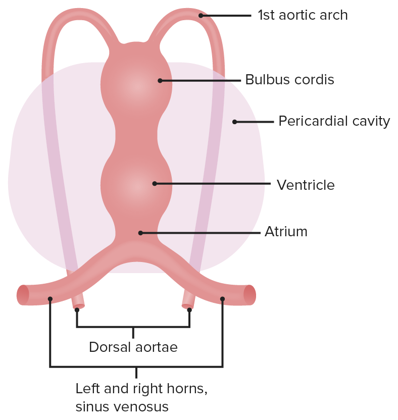
Derivatives of the Cardiac Tube
- First 3 weeks: primitive heart develops as a straight tube
- Truncus arteriosus → ascending aorta and pulmonary trunk
- Bulbus cordis
- Proximal 3rd of bulbus cordis → muscular right ventricle
- Conus cordis → smooth outflow portions of right and left ventricles
- Endocardial cushion → atrial septum, membranous interventricular septum, atrioventricular (AV) valves, and semilunar valves
- Derived from neural crest cells
- Endocardial cushion defects: common in Down syndrome
- Posterior, subcardinal, supracardinal veins → inferior vena cava (IVC)
- Primitive atrium → trabeculated parts of the left and right atria
- Primitive pulmonary vein → smooth left atrium
- Primitive ventricle → trabeculated parts of the left and right ventricles
- Right common cardinal vein (RCCV) and right anterior cardinal vein (RACV) → superior vena cava (SVC)
- Left horn of sinus venosus → coronary sinus
- Right horn of sinus venosus → posterior part of the right atrium (sinus venarum) with a relatively smooth surface
- Smooth portions are found in the areas where the heart connects with the vessels (e.g., below the pulmonic valve).
- Cardinal veins:
- Form superior and inferior venae cavae (that connect to the right atrium (RA))
- RCCV + RACV = SVC
- Posterior vein + subcardinal vein + supracardinal vein = IVC
The early heart is divided into five regions, each of which gives rise to different structures.
Image by Lecturio.Related videos
Cardiac Looping
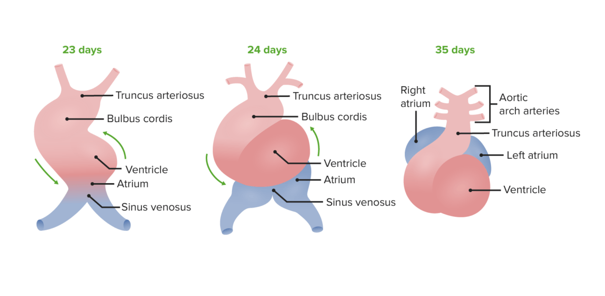
- Begins in 4th week: goal is to establish left-right polarity
- Cranial end bends ventrally and caudally.
- Then, rotates over to the right (24 days)
- Atrial end shifts dorso-cranially (posteriorly and cranially) and moves to the left (35 days).
- Requires cilia and dynein involvement
- Defect in dynein function → dextrocardia (heart on the right side, seen in Kartagener’s syndrome (primary ciliary dyskinesia))
Cardiac Septation
Atrial septum
The atrial septum begins with endocardial cushions at the base of the atrial chamber and septum primum.
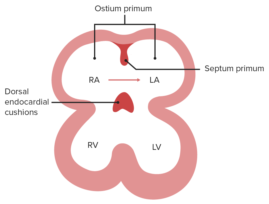
- Septum primum
- Grows toward the endocardial cushions
- Foramen (ostium) primum narrows → replaced by foramen (ostium) secundum (which forms in the septum primum as the ostium primum regresses)
- Oxygenated blood enters RA from IVC → passes into left atrium (LA) (through foramen secundum) → passes into developing ventricle
- Poorly oxygenated blood from SVC will be directed to the bulbus cordis (RV).
- Prenatal and postnatal physiology
- Septum secundum
- Develops on the right side of septum primum
- Foramen (ostium) secundum maintains right-to-left shunt.
- Expands and covers most of foramen (ostium) secundum → residual opening is the foramen ovale
- RA gets larger → pulls sinus venosus into its wall → moves SVC and IVC closer → one opening formed
- Foramen ovale
- Opening between septum primum and septum secundum (shunt between right and left atria)
- Blood continues to flow right to left, due to ↑↑ pressure in RA
- After birth: ↑↑ pressure in LA and decreased pressure in RA → septum primum pushed against septum secundum → foramen ovale closed
- Usually fused shut during infancy/early childhood, but in 70%–75% of the population it remains open as a small defect (patent foramen ovale).
Ventricular septum
- Muscular interventricular septum forms (growing from base of ventricle toward endocardial cushions).
- Aorticopulmonary septum rotates and fuses with the muscular ventricular septum to form the membranous interventricular septum (closing the interventricular foramen).
- Growth of endocardial cushions separates atria from ventricles → contributes to atrial septation and membranous portion of interventricular septum.
- Defect → membranous/muscular ventricular septal defect (VSD)
- Most common congenital cardiac defect
- Usually a defect in the membranous portion of the interventricular septum
Aorticopulmonary septum
- Spiral septum that divides the aorta from the pulmonary artery → forms outflow tract
- Neural crest cells migrate to truncal and bulbar ridges that spiral and fuse to form aorticopulmonary septum → forming ascending aorta and pulmonary trunk
- Also fuses with muscular portion of ventricular septum → forms membranous interventricular septum
- Defect (due to failure of migration of neural crest cells):
- Transposition of great vessels (TGV)
- Tetralogy of Fallot
- Persistent truncus arteriosus
Valve development
- Semilunar (aortic/pulmonic) valves: derived from endocardial cushions of outflow tract
- AV (mitral/tricuspid) valves: derived from fused endocardial cushions of AV canal
- Defects lead to stenotic, regurgitant, atretic (e.g., tricuspid atresia), or displaced (e.g., Ebstein anomaly) valves
The foramen (ostium) primum is the opening between the 2 embryonic atria.
Image by Lecturio.Endocardial cushions are derived from the neural crest. As the septum primum approaches the dorsal endocardial cushions, cells in the septum undergo apoptosis, which creates the foramen (ostium) secundum.
Image by Lecturio.The foramen (ostium) primum and secundum function together to deliver oxygenated blood to the left atrium, which will then flow to the left ventricle, whereas, poorly oxygenated blood delivered from the SVC will flow to the right ventricle.
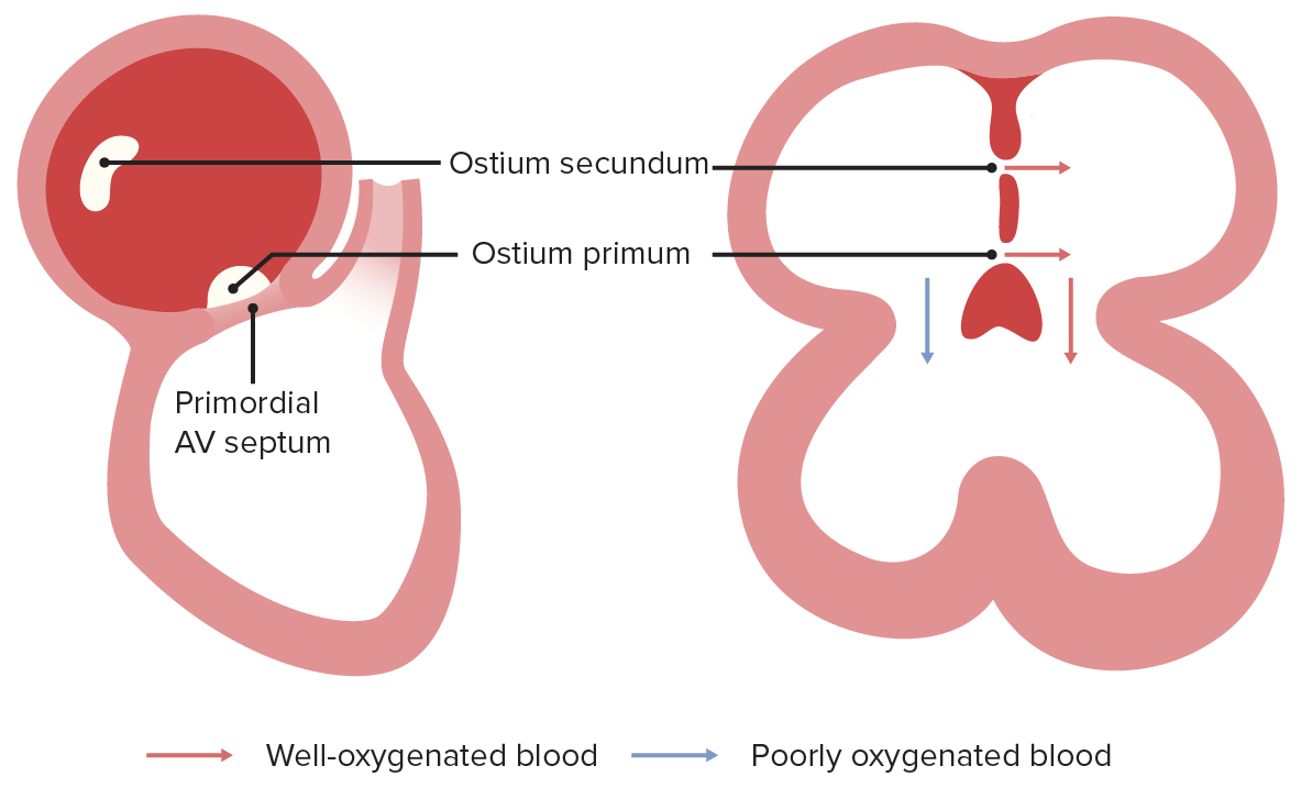
Image by Lecturio.The secundum will grow to narrow the foramen (ostium) secundum and form foramen ovale. In this case, the septum primum will function as a valve.
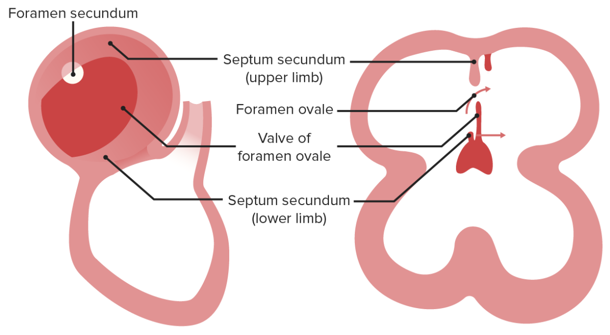
Image by Lecturio.Blood from the IVC pushes the septum primum (valve of foramen ovale) to move blood from the RA to the LA, after which blood will flow to the left ventricle. At birth, the pressure in the LA will increase, which will shut the foramen ovale.
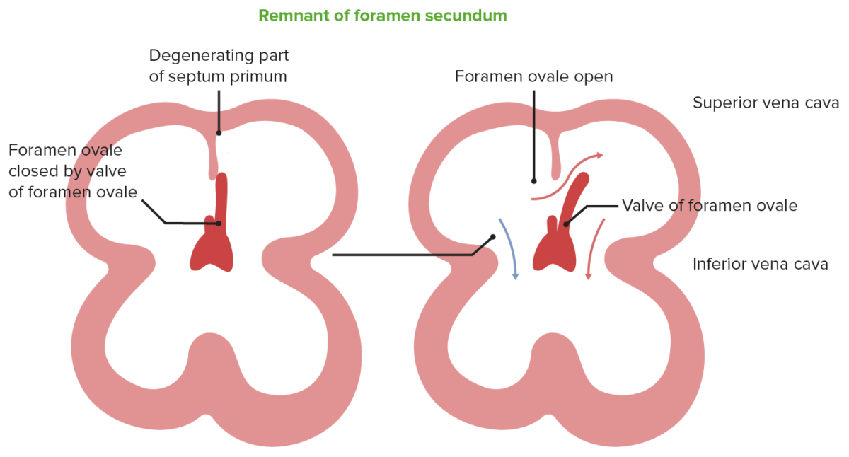
Image by Lecturio.Membranous portion of the interventricular septum grows upward to fuse with conotruncal ridges. Right and left ventricles, as well as the aorta and pulmonary tract, become separated from each other.
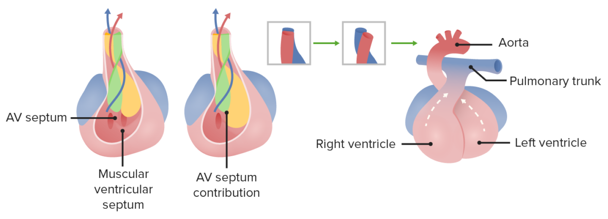
Image by Lecturio.
Fetal Circulation
- Placenta brings blood through the umbilical vein (80% oxygen saturation and PO2 of approximately 30 mm Hg)
- Umbilical arteries have low oxygen saturation
- Ductus venosus:
- Allows blood to bypass the liver
- Transports blood to IVC from the umbilical vein
- Blood is slightly deoxygenated due to mixing
- Pathway of oxygenated blood from IVC back to placenta:
- Blood hits atrial septum → pushes the valve of the foramen ovale (2/3 to LA, 1/3 to lungs) → LA → LV → aorta → common iliac arteries → umbilical arteries → placenta
- Mnemonic: Remember O for Oxygenated blood as well as for Ovale.
- Pathway of deoxygenated blood from SVC:
- SVC → RA → RV → pulmonary trunk → ductus arteriosus → descending aorta → common iliac arteries → umbilical arteries → placenta
- This shunt is due to high fetal pulmonary arterial resistance (partly because of low oxygen tension).
- Mnemonic: Remember D for deoxygenated blood as well as for Ductus arteriosus and Descending aorta.
- Transition from fetal to adult circulation:
- At birth, infant takes first breath → decreased resistance in pulmonary vasculature → increased left atrial pressure in relation to right atrial pressure, leading to the closing of the foramen ovale (becomes the fossa ovalis)
- ↑ In oxygen (from respiration) and ↓ in prostaglandins (from placental separation) → closure of ductus arteriosus
- Fetal-postnatal derivatives:
- Umbilical vein → ligamentum teres hepatis (round ligament – contained in falciform ligament)
- Umbilical arteries → medial umbilical ligaments
- Ductus venosus → ligamentum venosum
- Ductus arteriosus → ligamentum arteriosum (near left recurrent laryngeal nerve)
- Foramen ovale → Fossa ovalis
Mnemonics
- Indomethacin helps close the patent ductus arteriosus → ligamentum arteriosum (remnant of ductus arteriosus) → remember: “Come In and close the Door!”
- “Prostaglandins E1 and E2 kEEp PDA open!”
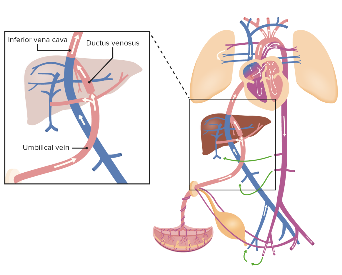
Oxygenated blood from the umbilical vein drains into the IVC, which also collects the blood from the organs and lower portion of the body; thus, the blood becomes slightly deoxygenated.
Image by Lecturio.Blood from the IVC pushes the valve of foramen ovale and flows into the left side of the heart, where it mixes in the LA with deoxygenated blood from the pulmonary veins. Blood then leaves the heart through the aorta and flows to the iliac arteries, which are connected to the placenta by the umbilical arteries. However, blood from the SVC flows directly into the RV and then to the pulmonary trunk, which is connected to the descending aorta through the ductus arteriosus.
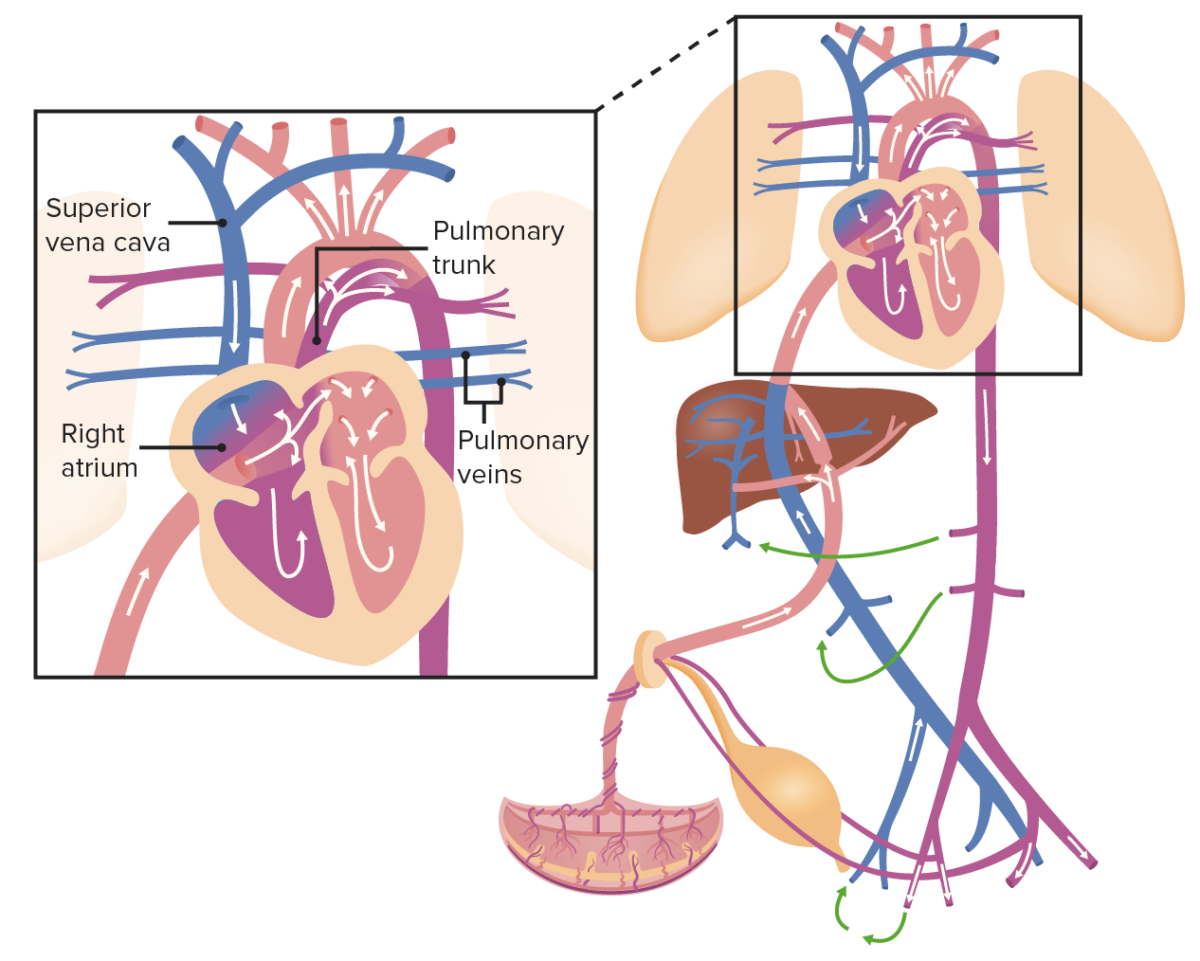
Image by Lecturio.After birth, the ductus arteriosus becomes the ligamentum arteriosum, as blood is able to enter the lungs to be oxygenated. The foramen ovale closes and becomes the fossa ovalis due to increased pressure in the LA.
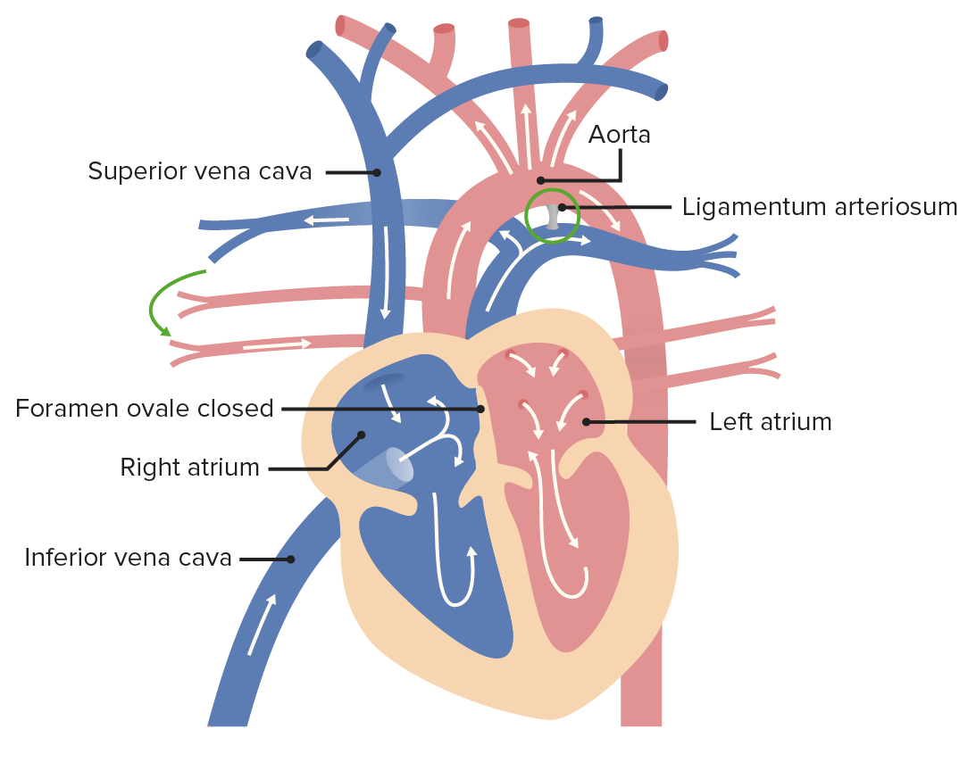
Image by Lecturio.After birth, the umbilical artery becomes the ligamentum teres hepatis, whereas the umbilical vein becomes the medial umbilical ligament.
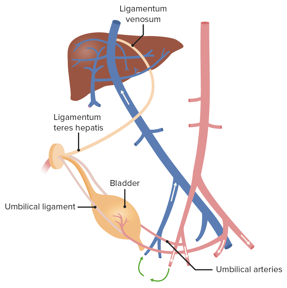
Image by Lecturio.
Congenital Abnormalities
Right-to-left shunts (cyanotic lesions)
- Truncus arteriosus: A single large vessel arises from both ventricles due to failure of formation of the aorticopulmonary septum to divide into the pulmonary trunk and aorta. Usually, it is accompanied by a VSD. Danger of irreversible pulmonary hypertension.
- TGV: Failure of the aorticopulmonary septum to spiral. Pulmonary artery arises from the LV (posterior), whereas the aorta comes out from the RV (anterior). Unsuitable for life without a shunt. In 65% of cases, TGV is accompanied by patent ductus arteriosus or patent foramen ovale. In 35% of cases, it is accompanied by a ventricular septal defect. Radiologically will have an “egg on string” appearance on chest X-ray due to narrowed mediastinum and globular heart outline. Surgical intervention is needed, otherwise most infants die within the first few months of life.
- Tricuspid atresia: The tricuspid valve is absent, which impedes the development of the right ventricle, leading to a hypoplastic right ventricle. Requires atrial septal defect (ASD) and VSD for viability. Blood will flow from the RA to the LA through the atrial septal defect or the patent foramen ovale. To get to the lungs, blood will flow from the RA to the LA through the patent foramen ovale, and then from the LV to the RV through the ventricular septal defect.
- Tetralogy of Fallot: Most common cause of early childhood cyanosis. Accompanied by pulmonary stenosis, right ventricular hypertrophy, overriding aorta, and a ventricular septal defect.
- Total anomalous pulmonary venous return (TAPVR): All pulmonary veins drain into the right heart circulation (SVC, coronary sinus, etc.). The defect is similar to the shunt from the LA to the RA. The RA and the RV cause oxygenated blood to mix with deoxygenated blood and spread throughout the body. Associated with an ASD and sometimes a PDA to allow for right-to-left shunting to maintain cardiac output.
Mnemonic: right-to-left shunts (cyanotic lesions) (the 5 Ts):
- Truncus arteriosus: 1 vessel
- Transposition of the great vessels: 2 switched vessels
- Tricuspid atresia: 3 = tri
- Tetralogy of Fallot: 4 = tetra
- Total anomalous pulmonary venous return (TAPVR): 5 letters in the name
Left-to-right shunts (acyanotic)
- VSD: Most common congenital anomaly, associated with fetal alcohol syndrome. The defect causes communication between LV and RV. Clinical presentation is harsh, holosystolic murmur that is heard best in the tricuspid area.
- ASD: Communication between LA and RA, can lead to pulmonary hypertension. Clinically characterized by wide splitting of S2. There are 4 types of ASD: primum, secundum, sinus venosus, and coronary sinus, the most common being a secundum type of ASD.
- Patent ductus arteriosus (PDA): Normally, ductus arteriosus shunts blood from the left pulmonary artery to the aorta. This should stop after birth due to decreased levels of prostaglandin E2. Clinical presentation of PDA is a continuous machinery-like murmur on auscultation, blue toes and normal fingers, and a widened pulse pressure.
Other congenital abnormalities
- Coarctation of the aorta: The aorta narrows in the area around the ductus arteriosus to a certain point and then widens to its normal diameter. Postductal coarctation (occurring after the ductus arteriosus) is clinically silent and only present in adults. Preductal coarctation (occurring before the ductus arteriosus) is an infantile type, where clinical presentation involves cyanosis in the toes.
- Patent foramen ovale (PFO): Present in 25% of adults. Failure of fusion of septum primum with septum secundum after increased left atrial pressure. Clinically detected with bubble test in evaluation of syncope and strokes.
- Hypoplastic left heart syndrome: High-grade stenosis or atresia of the aortic and/or mitral valve in addition to severe hypoplasia of the left ventricle, ascending aorta, and aortic arch.
- Ebstein anomaly: Defect of the tricuspid valve, which is displaced toward the right ventricle. Tethering of septal leaflet causes tricuspid regurgitation, which will result in right-sided heart failure.
- Double aortic arch: Embryonic vascular malformation resulting in a double aortic arch. This can lead to subsequent constriction of the trachea and esophagus.
- Endocardial cushion defect: Defect of the atrioventricular valves as well as the atrial septum and/or ventricular septum. Can present as a complete form (ASD, VSD, and severe abnormalities in the AV valves, leading to severe mixing of blood) or a partial form (only ASD and minor AV valve abnormalities). Commonly seen in patients with Down syndrome.