

Benign Benign Fibroadenoma or nonacute scrotal masses are represented by hydroceles, varicoceles, and spermatoceles. Key components to evaluation are physical exam and scrotal ultrasound. Hydroceles represent extra fluid in the tunica vaginalis Tunica vaginalis Testicles: Anatomy, leading to a swollen scrotum Scrotum A cutaneous pouch of skin containing the testicles and spermatic cords. Testicles: Anatomy. Varicoceles have a dilatation of the pampiniform venous plexus, giving the “bag of worms” appearance on exam. Spermatoceles present as an epididymal cyst, commonly arising from the head of the epididymis Epididymis The convoluted cordlike structure attached to the posterior of the testis. Epididymis consists of the head (caput), the body (corpus), and the tail (cauda). A network of ducts leaving the testis joins into a common epididymal tubule proper which provides the transport, storage, and maturation of spermatozoa. Testicles: Anatomy. Hydroceles, varicoceles, and spermatoceles are usually asymptomatic and do not require treatment unless they are causing pain Pain An unpleasant sensation induced by noxious stimuli which are detected by nerve endings of nociceptive neurons. Pain: Types and Pathways or other complications.
Last updated: Sep 30, 2026
Varicocele is the abnormal dilation and tortuosity of the pampiniform venous plexus within the spermatic cord Spermatic Cord Either of a pair of tubular structures formed by ductus deferens; arteries; veins; lymphatic vessels; and nerves. The spermatic cord extends from the deep inguinal ring through the inguinal canal to the testis in the scrotum. Testicles: Anatomy, which is connected to the internal spermatic or gonadal vein.
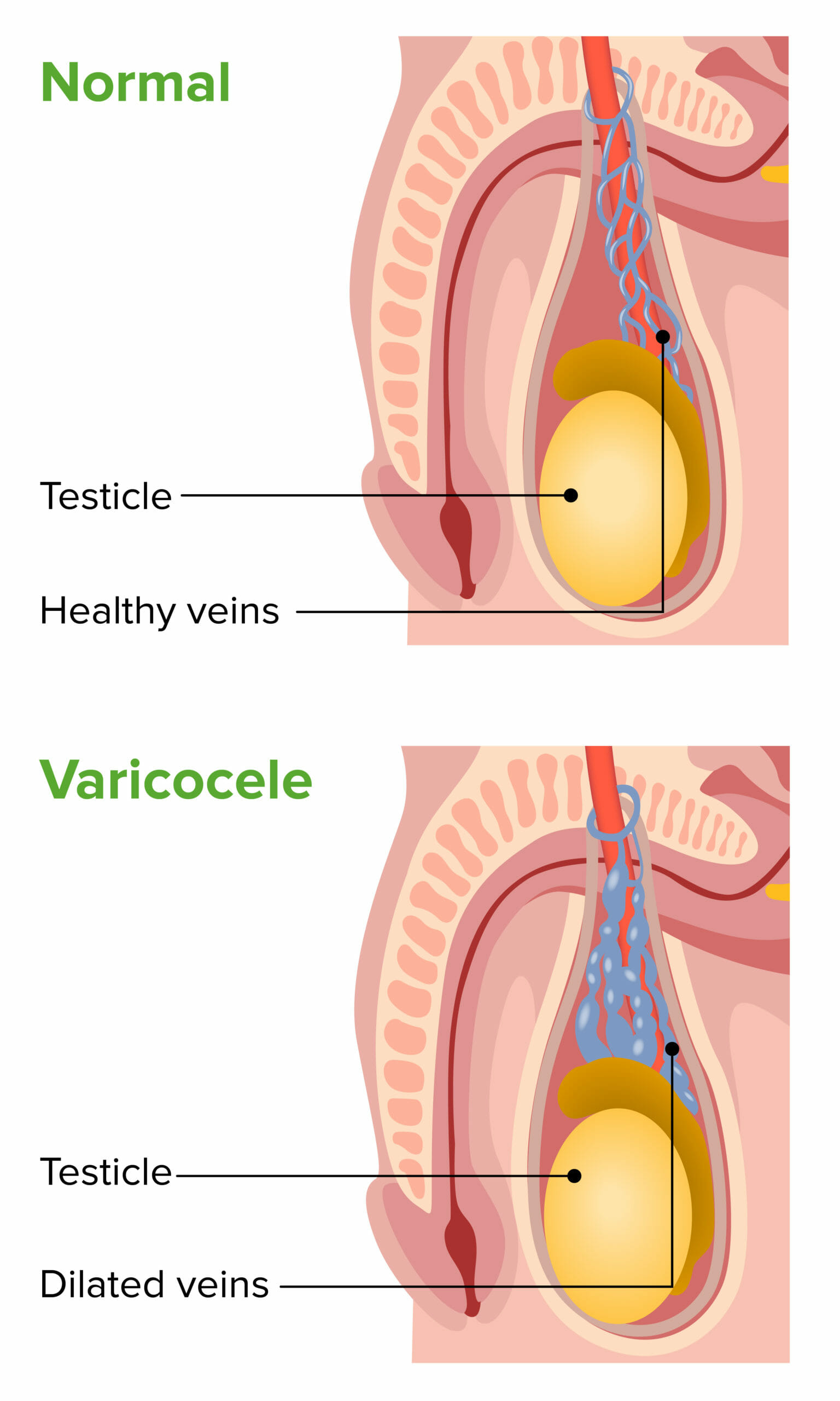
Varicocele: image showing left-sided dilated pampiniform venous plexus in the scrotum
Image by Lecturio.An isolated right-sided varicocele, sudden onset, failure to decompress, or new onset in an older patient raises concern for a renal or retroperitoneal Retroperitoneal Peritoneum: Anatomy mass Mass Three-dimensional lesion that occupies a space within the breast Imaging of the Breast causing venous obstruction.
Examination:
Imaging (scrotal ultrasound):
Treatment approach:
Clinical considerations:
Hydrocele is a collection of serous fluid between the parietal Parietal One of a pair of irregularly shaped quadrilateral bones situated between the frontal bone and occipital bone, which together form the sides of the cranium. Skull: Anatomy and visceral layers of the tunica vaginalis Tunica vaginalis Testicles: Anatomy surrounding the testis. If bloody, the condition is referred to as hematocele.
Multifactorial, consisting of anatomic and fluid factors:
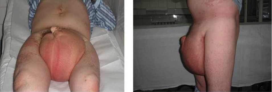
Hydrocele: enlarged scrotal mass filled with fluid
Image: “Hydrocele” by Dan Mischianu et al. License: CC BY 2.0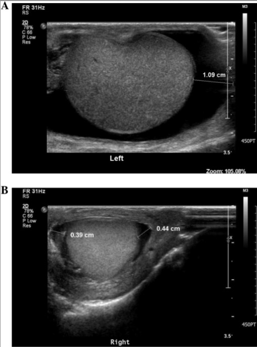
Transverse view of scrotal ultrasound showing hydrocele:
A: large left hydrocele as noted by the dark border delineating fluid surrounding the normal testicle in grey color
B: a smaller amount of fluid as a small hydrocele of the right testicle
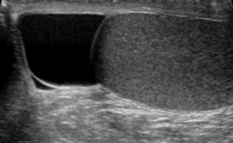
Transverse view of a scrotal ultrasound: Testicle is grey structure to the right and spermatocele is fluid-filled sac on the left (darker).
Image: “Spermatocele” by Iwak. License: Public DomainScrotal swelling Swelling Inflammation or mass Mass Three-dimensional lesion that occupies a space within the breast Imaging of the Breast has the following differential diagnoses: