The forearm is the region of the upper limb between the elbow and the wrist. The term “forearm” is used in anatomy to distinguish this area from the arm Arm The arm, or "upper arm" in common usage, is the region of the upper limb that extends from the shoulder to the elbow joint and connects inferiorly to the forearm through the cubital fossa. It is divided into 2 fascial compartments (anterior and posterior). Arm: Anatomy, a term that is commonly used to describe the entire upper limb. The forearm consists of 2 long bones Long bones Length greater than width. Bones: Structure and Types (the radius and the ulna), the interosseous membrane, and multiple arteries Arteries Arteries are tubular collections of cells that transport oxygenated blood and nutrients from the heart to the tissues of the body. The blood passes through the arteries in order of decreasing luminal diameter, starting in the largest artery (the aorta) and ending in the small arterioles. Arteries are classified into 3 types: large elastic arteries, medium muscular arteries, and small arteries and arterioles. Arteries: Histology, nerves, and muscles. The muscles are grouped into 2 compartments: anterior and posterior. The function of these muscles is flexion Flexion Examination of the Upper Limbs and extension Extension Examination of the Upper Limbs of the wrist and fingers, while also contributing to flexion Flexion Examination of the Upper Limbs of the elbow.
Last updated: Dec 15, 2025
The forearm contains 2 long bones Long bones Length greater than width. Bones: Structure and Types:
The bones of the forearm, the radius laterally and ulna medially, are held together by the fibrous Fibrous Fibrocystic Change interosseous membrane.
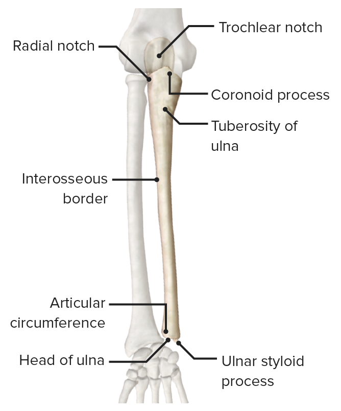
Anterior view of the ulna featuring its bony landmarks and articular surfaces
Image by BioDigital, edited by Lecturio.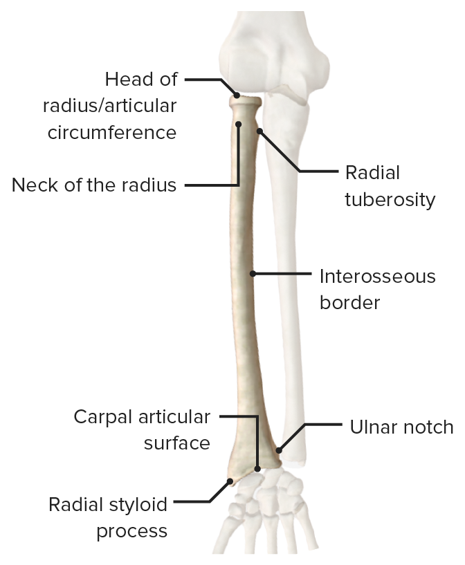
Anterior view of the radius featuring its bony landmarks and articular surfaces
Image by BioDigital, edited by Lecturio.
Distal radio-ulnar joint
Image by Lecturio.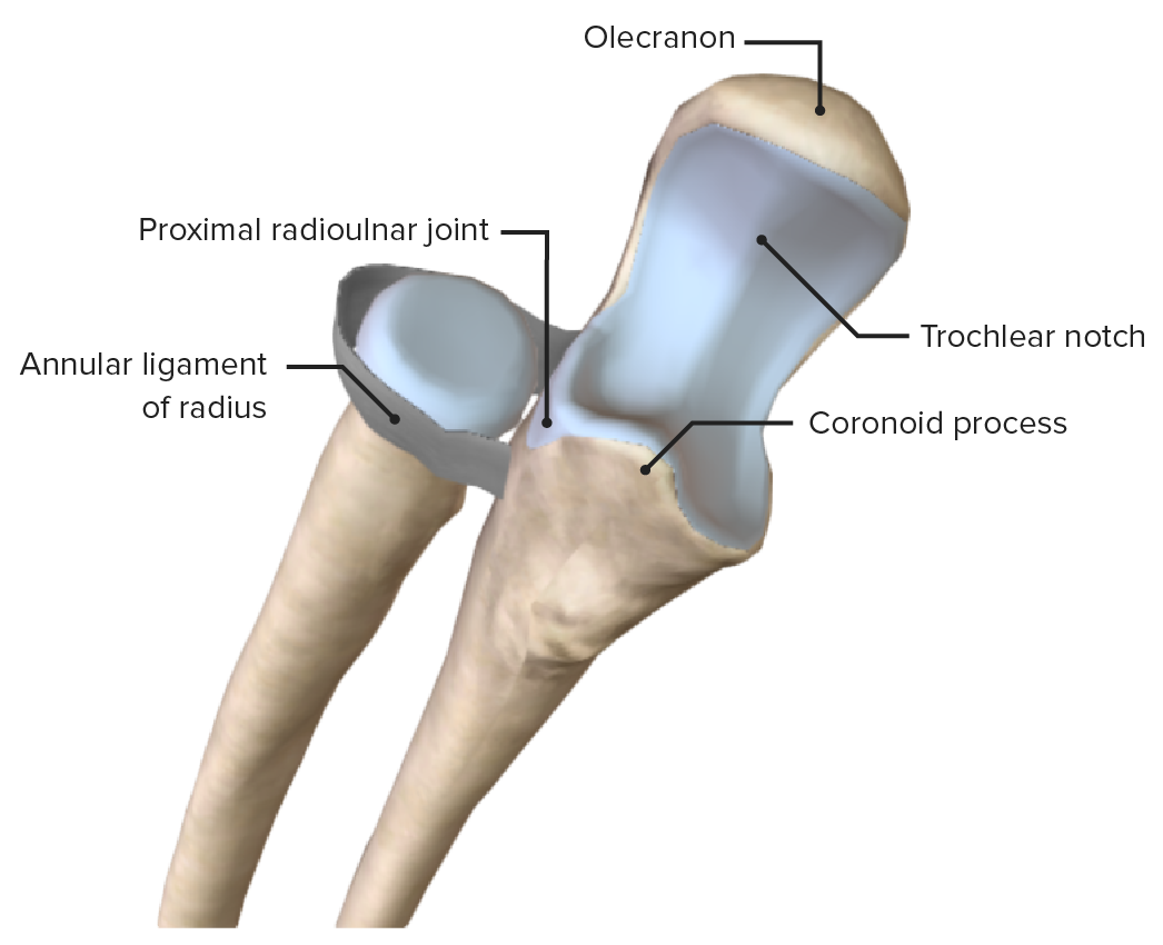
Proximal radioulnar joint, featuring its main supporting ligament, the annular ligament of the radius
Image by BioDigital, edited by Lecturio.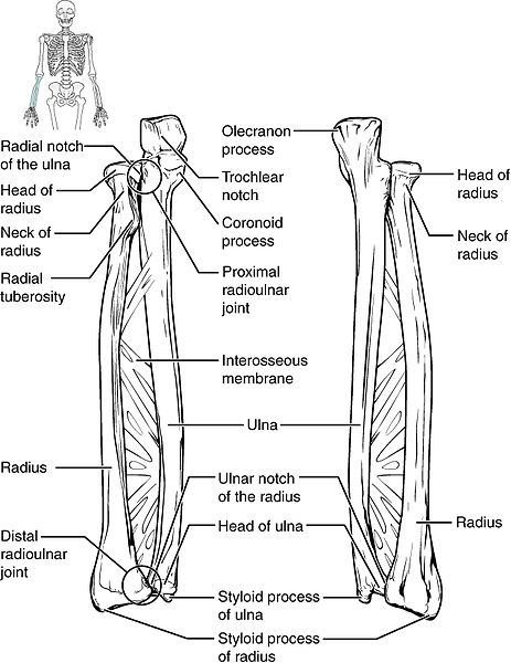
Interosseous membrane of the radius and ulna bones
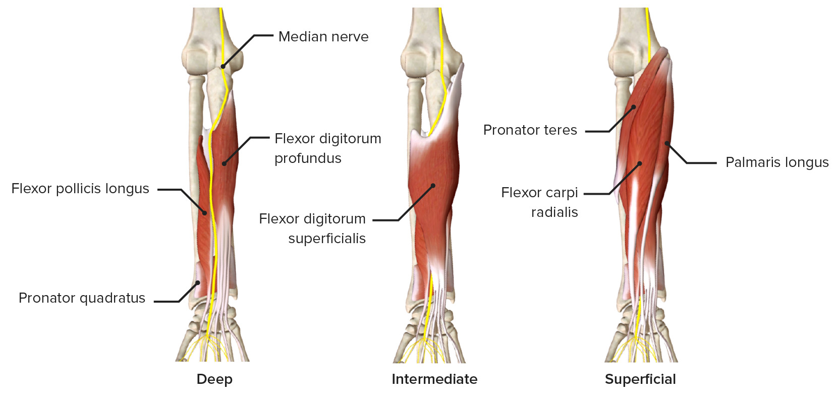
Image: “Ulna and Radius” by OpenStax College. License: CC BY 3.0The muscles of the anterior compartment of the forearm are often separated into superficial, intermediate, and deep layers.
| Muscle | Origin | Insertion | Innervation | Function |
|---|---|---|---|---|
| Pronator teres |
|
Middle of lateral surface of radius | Median nerve Median Nerve A major nerve of the upper extremity. In humans, the fibers of the median nerve originate in the lower cervical and upper thoracic spinal cord (usually C6 to T1), travel via the brachial plexus, and supply sensory and motor innervation to parts of the forearm and hand. Cubital Fossa: Anatomy (C7) | Pronates and flexes forearm |
| Flexor carpi radialis | Medial epicondyle Medial epicondyle Arm: Anatomy of humerus Humerus Bone in humans and primates extending from the shoulder joint to the elbow joint. Arm: Anatomy | Base of 2nd metacarpal | Flexes and abducts wrist | |
| Palmaris longus | Flexor retinaculum Flexor Retinaculum Ankle Joint: Anatomy and palmar aponeurosis Palmar aponeurosis Hand: Anatomy | Flexes wrist and tenses palmar aponeurosis Palmar aponeurosis Hand: Anatomy | ||
| Flexor carpi ulnaris | Olecranon Olecranon A prominent projection of the ulna that articulates with the humerus and forms the outer protuberance of the elbow joint. Arm: Anatomy and posterior ulna | Pisiform, hook of hamate, 5th metacarpal | Ulnar nerve Ulnar Nerve A major nerve of the upper extremity. In humans, the fibers of the ulnar nerve originate in the lower cervical and upper thoracic spinal cord (usually C7 to T1), travel via the medial cord of the brachial plexus, and supply sensory and motor innervation to parts of the hand and forearm. Axilla and Brachial Plexus: Anatomy (C8) | Flexes and adducts wrist |
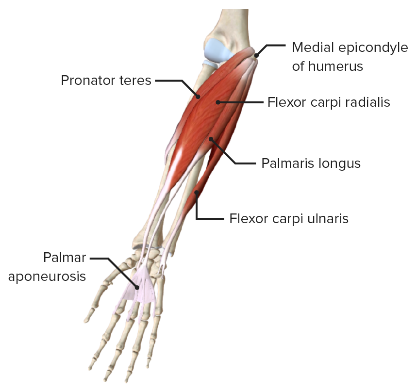
Anterior view of the right forearm, featuring the muscles of the superficial layer of the anterior compartment
Image by BioDigital, edited by Lecturio| Muscle | Origin | Insertion | Innervation | Function |
|---|---|---|---|---|
| Flexor digitorum superficialis |
|
Middle phalanges Phalanges Bones that make up the skeleton of the fingers, consisting of two for the thumb, and three for each of the other fingers. Hand: Anatomy of medial 4 digits | Median nerve Median Nerve A major nerve of the upper extremity. In humans, the fibers of the median nerve originate in the lower cervical and upper thoracic spinal cord (usually C6 to T1), travel via the brachial plexus, and supply sensory and motor innervation to parts of the forearm and hand. Cubital Fossa: Anatomy (C7, C8, T1) |
|
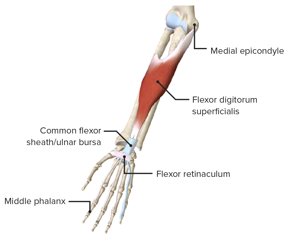
Anterior view of the right forearm, featuring the muscles of the intermediate layer of the anterior compartment
Image by BioDigital, edited by Lecturio| Muscle | Origin | Insertion | Innervation | Function |
|---|---|---|---|---|
| Flexor digitorum profundus | Proximal surfaces of medial and anterior surfaces of ulna and interosseous membrane | Distal phalanges Phalanges Bones that make up the skeleton of the fingers, consisting of two for the thumb, and three for each of the other fingers. Hand: Anatomy of medial 4 digits |
|
|
| Flexor pollicis longus | Anterior surface of radius and interosseous membrane | Distal phalanx of 1st digit | Anterior interosseous nerve Anterior Interosseous Nerve Supracondylar Fracture, branch of median (C8) |
|
| Pronator quadratus | Distal quarter of ulna | Distal quarter of radius | Pronates forearm |
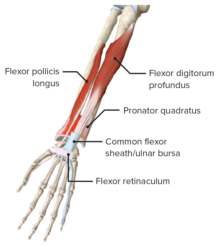
Anterior view of the right forearm, featuring the muscles of the deep layer of the anterior compartment
Image by BioDigital, edited by LecturioThe muscles of the posterior compartment of the forearm are separated into superficial and deep layers.
| Muscle | Origin | Insertion | Innervation | Function |
|---|---|---|---|---|
| Brachio-radialis | Proximal ⅔ of lateral supracondylar ridge | Lateral surface of distal radius and pre-styloid process | Radial nerve Radial Nerve A major nerve of the upper extremity. In humans the fibers of the radial nerve originate in the lower cervical and upper thoracic spinal cord (usually C5 to T1), travel via the posterior cord of the brachial plexus, and supply motor innervation to extensor muscles of the arm and cutaneous sensory fibers to extensor regions of the arm and hand. Axilla and Brachial Plexus: Anatomy (C6) | Weak flexor of elbow, strong flexor when midpronated |
| Extensor carpi radialis longus | Lateral supracondylar ridge | Dorsal aspect of 2nd metacarpal | Radial nerve Radial Nerve A major nerve of the upper extremity. In humans the fibers of the radial nerve originate in the lower cervical and upper thoracic spinal cord (usually C5 to T1), travel via the posterior cord of the brachial plexus, and supply motor innervation to extensor muscles of the arm and cutaneous sensory fibers to extensor regions of the arm and hand. Axilla and Brachial Plexus: Anatomy (C6, C7) | Extend and abduct wrist |
| Extensor carpi radialis brevis | Lateral epicondyle Lateral epicondyle Arm: Anatomy (common extensor origin) | Dorsal aspect of 3rd metacarpal | Deep branch of radial nerve Radial Nerve A major nerve of the upper extremity. In humans the fibers of the radial nerve originate in the lower cervical and upper thoracic spinal cord (usually C5 to T1), travel via the posterior cord of the brachial plexus, and supply motor innervation to extensor muscles of the arm and cutaneous sensory fibers to extensor regions of the arm and hand. Axilla and Brachial Plexus: Anatomy (C7) | |
| Extensor digitorum | Lateral epicondyle Lateral epicondyle Arm: Anatomy (common extensor origin) | Extensor expansion of medial 4 digits | Posterior interosseous nerve (C7; from deep radial nerve Radial Nerve A major nerve of the upper extremity. In humans the fibers of the radial nerve originate in the lower cervical and upper thoracic spinal cord (usually C5 to T1), travel via the posterior cord of the brachial plexus, and supply motor innervation to extensor muscles of the arm and cutaneous sensory fibers to extensor regions of the arm and hand. Axilla and Brachial Plexus: Anatomy) |
|
| Extensor digiti minimi | Extensor expansion of 5th digit | |||
| Extensor carpi ulnaris | Lateral epicondyle Lateral epicondyle Arm: Anatomy and posterior surface of ulna | Dorsal aspect of 5th metacarpal | Extends and adducts wrist | |
| Anconeus Anconeus Elbow Joint: Anatomy | Inserts on the posterior aspect of lateral epicondyle Lateral epicondyle Arm: Anatomy | Lateral surface of the olecranon Olecranon A prominent projection of the ulna that articulates with the humerus and forms the outer protuberance of the elbow joint. Arm: Anatomy | Radial nerve Radial Nerve A major nerve of the upper extremity. In humans the fibers of the radial nerve originate in the lower cervical and upper thoracic spinal cord (usually C5 to T1), travel via the posterior cord of the brachial plexus, and supply motor innervation to extensor muscles of the arm and cutaneous sensory fibers to extensor regions of the arm and hand. Axilla and Brachial Plexus: Anatomy (C7, C8) | Extends the elbow |
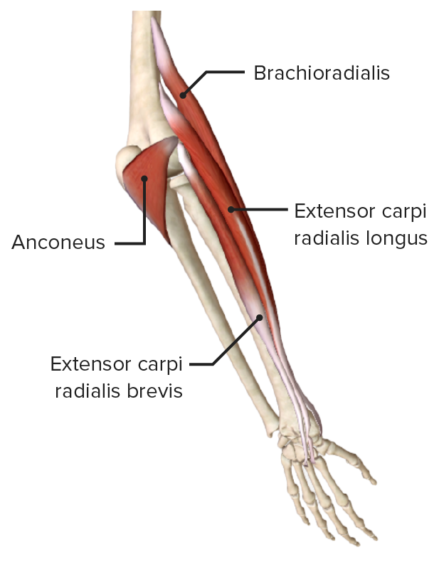
Lateroposterior view of the right forearm, featuring the muscles of the superficial layer of the posterior compartment (extensor digiti minimi, extensor digitorum, and extensor carpi ulnaris not pictured)
Image by BioDigital, edited by Lecturio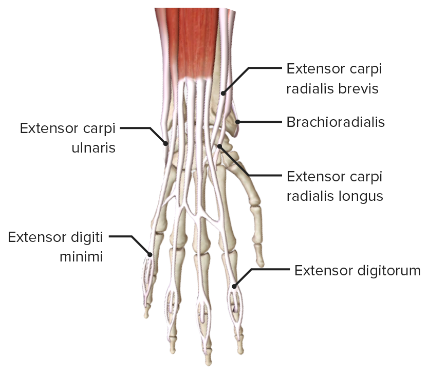
Close-up of the insertion of the extensor muscles of the forearm and digits, also known as the extensor expansions
Image by BioDigital, edited by Lecturio.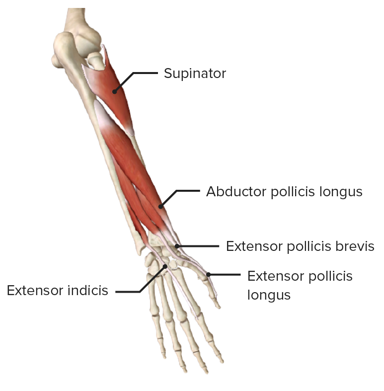
Posterior view of the right forearm, featuring the muscles of the deep layer of the posterior compartment
Image by BioDigital, edited by Lecturio.| Muscle | Origin | Insertion | Innervation | Function |
|---|---|---|---|---|
| Abductor pollicis longus | Posterior surface of radius and ulna and interosseous membrane | Metacarpal of 1st digit | Posterior interosseous nerve (C8) from deep radial nerve Radial Nerve A major nerve of the upper extremity. In humans the fibers of the radial nerve originate in the lower cervical and upper thoracic spinal cord (usually C5 to T1), travel via the posterior cord of the brachial plexus, and supply motor innervation to extensor muscles of the arm and cutaneous sensory fibers to extensor regions of the arm and hand. Axilla and Brachial Plexus: Anatomy |
|
| Extensor pollicis longus | Posterior surface of ulna and interosseous membrane | Dorsal surface of distal phalanx of 1st digit |
|
|
| Extensor pollicis brevis | Posterior surface of radius and interosseous membrane | Dorsal surface of proximal phalanx of 1st digit | ||
| Supinator | Lateral epicondyle Lateral epicondyle Arm: Anatomy of humerus Humerus Bone in humans and primates extending from the shoulder joint to the elbow joint. Arm: Anatomy, supinator fossa, and proximal ulna | Posterior/lateral/anterior surfaces of proximal radius | Deep branch of radial nerve Radial Nerve A major nerve of the upper extremity. In humans the fibers of the radial nerve originate in the lower cervical and upper thoracic spinal cord (usually C5 to T1), travel via the posterior cord of the brachial plexus, and supply motor innervation to extensor muscles of the arm and cutaneous sensory fibers to extensor regions of the arm and hand. Axilla and Brachial Plexus: Anatomy (C8) | Supination Supination Applies to movements of the forearm in turning the palm forward or upward. When referring to the foot, a combination of adduction and inversion movements of the foot. Examination of the Upper Limbs of forearm |
| Extensor indicis | Posterior surface of ulna and interosseous membrane | Extensor expansion of 2nd digit | Posterior interosseous nerve (C8) from deep radial nerve Radial Nerve A major nerve of the upper extremity. In humans the fibers of the radial nerve originate in the lower cervical and upper thoracic spinal cord (usually C5 to T1), travel via the posterior cord of the brachial plexus, and supply motor innervation to extensor muscles of the arm and cutaneous sensory fibers to extensor regions of the arm and hand. Axilla and Brachial Plexus: Anatomy |
|
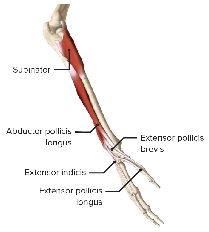
Lateral view of the right forearm, featuring the muscles of the deep layer of the posterior compartment
Image by BioDigital, edited by Lecturio.Posterior view of the right forearm, featuring the muscles of the deep layer of the posterior compartment
Image by BioDigital, edited by Lecturio.The 2 main arteries Arteries Arteries are tubular collections of cells that transport oxygenated blood and nutrients from the heart to the tissues of the body. The blood passes through the arteries in order of decreasing luminal diameter, starting in the largest artery (the aorta) and ending in the small arterioles. Arteries are classified into 3 types: large elastic arteries, medium muscular arteries, and small arteries and arterioles. Arteries: Histology of the forearm, the radial and ulnar arteries Arteries Arteries are tubular collections of cells that transport oxygenated blood and nutrients from the heart to the tissues of the body. The blood passes through the arteries in order of decreasing luminal diameter, starting in the largest artery (the aorta) and ending in the small arterioles. Arteries are classified into 3 types: large elastic arteries, medium muscular arteries, and small arteries and arterioles. Arteries: Histology, are branches of the brachial artery Brachial Artery The continuation of the axillary artery; it branches into the radial and ulnar arteries. Cubital Fossa: Anatomy. These 2 arteries Arteries Arteries are tubular collections of cells that transport oxygenated blood and nutrients from the heart to the tissues of the body. The blood passes through the arteries in order of decreasing luminal diameter, starting in the largest artery (the aorta) and ending in the small arterioles. Arteries are classified into 3 types: large elastic arteries, medium muscular arteries, and small arteries and arterioles. Arteries: Histology join in the hand Hand The hand constitutes the distal part of the upper limb and provides the fine, precise movements needed in activities of daily living. It consists of 5 metacarpal bones and 14 phalanges, as well as numerous muscles innervated by the median and ulnar nerves. Hand: Anatomy, forming an anastomosis, the superficial and deep palmar arteries Arteries Arteries are tubular collections of cells that transport oxygenated blood and nutrients from the heart to the tissues of the body. The blood passes through the arteries in order of decreasing luminal diameter, starting in the largest artery (the aorta) and ending in the small arterioles. Arteries are classified into 3 types: large elastic arteries, medium muscular arteries, and small arteries and arterioles. Arteries: Histology.
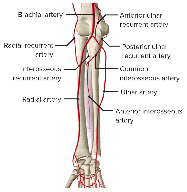
Arteries of the forearm
Image by BioDigital, edited by Lecturio.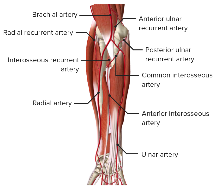
Anterior view of the right forearm, featuring the arteries of the forearm in the deep muscle layer
Image by BioDigital, edited by Lecturio.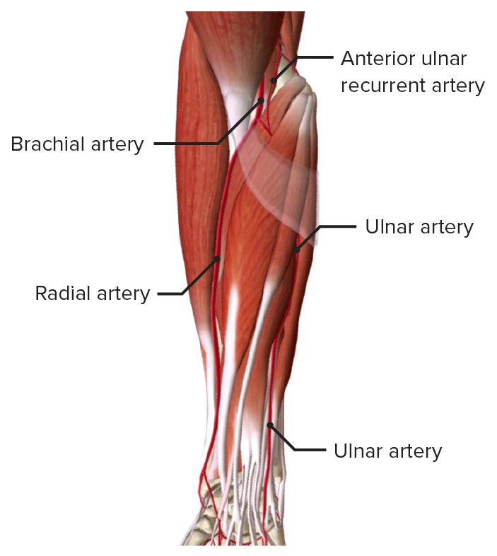
Anterior view of the right forearm, featuring the arteries of the forearm in the superficial muscle layer
Image by BioDigital, edited by Lecturio.The veins Veins Veins are tubular collections of cells, which transport deoxygenated blood and waste from the capillary beds back to the heart. Veins are classified into 3 types: small veins/venules, medium veins, and large veins. Each type contains 3 primary layers: tunica intima, tunica media, and tunica adventitia. Veins: Histology of the forearm are divided into superficial and deep:
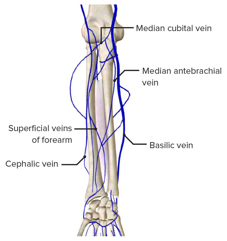
Superficial veins of the forearm
Image by BioDigital, edited by Lecturio.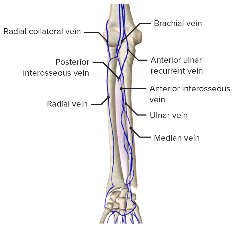
Deep veins of the forearm
Image by BioDigital, edited by Lecturio.The median, ulnar, and radial nerves pass through the forearm and innervate the muscles of both the anterior and posterior compartments.
The 3 main motor Motor Neurons which send impulses peripherally to activate muscles or secretory cells. Nervous System: Histology nerves of the forearm innervate the following muscles:
There are 3 main sensory Sensory Neurons which conduct nerve impulses to the central nervous system. Nervous System: Histology nerves of the forearm:
Median nerve as it passes through the anterior forearm
Image by BioDigital, edited by Lecturio.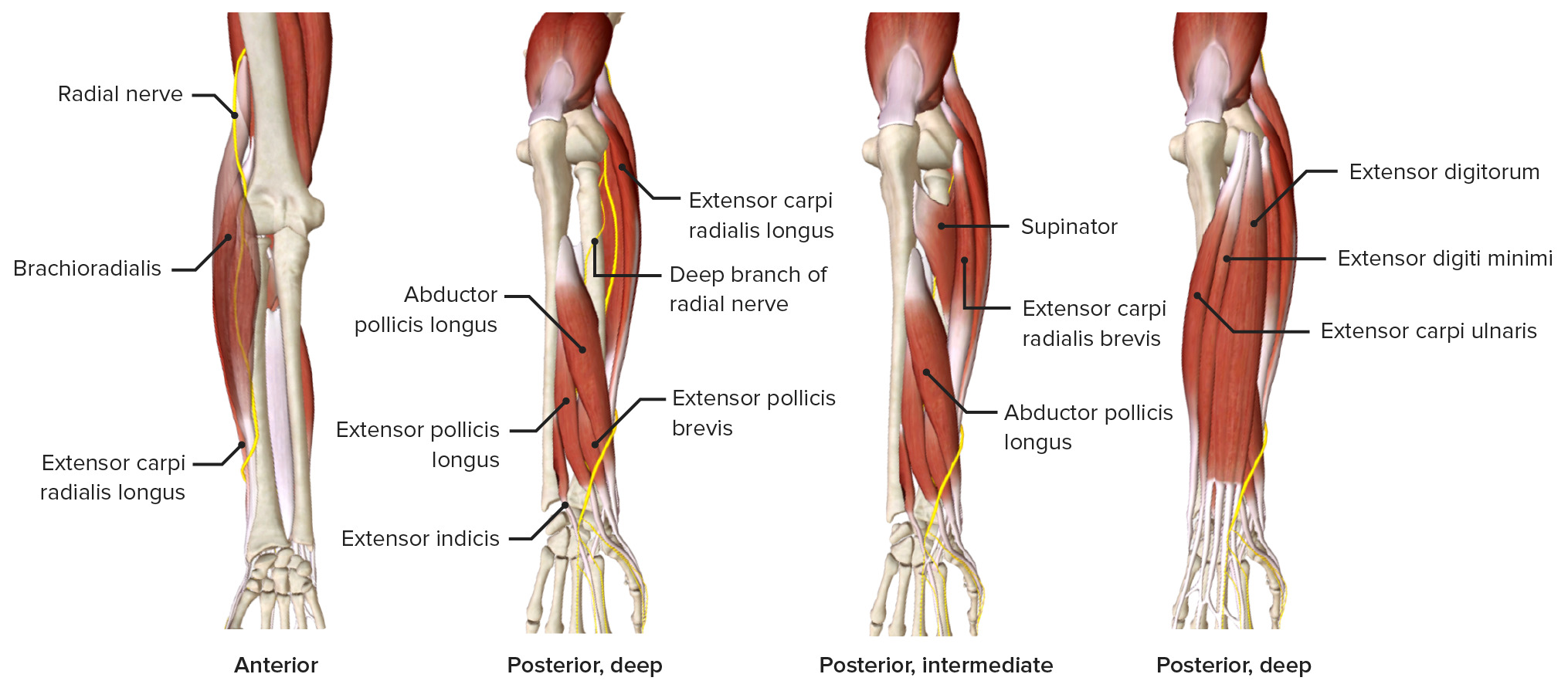
Radial nerve as it passes through the forearm, featuring the muscles it innervates
Image by BioDigital, edited by Lecturio.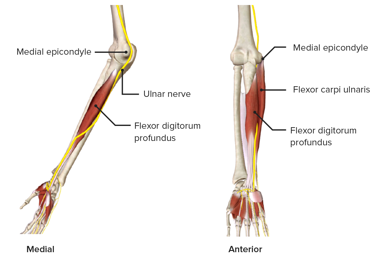
Ulnar nerve as it passes through the medial aspect of the forearm
Image by BioDigital, edited by Lecturio.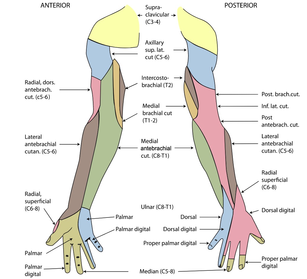
Cutaneous innervation of the right upper limb in its anterior (left) and posterior (right) views
Image: “Gray812and814” by Henry Vandyke Carter. License: Public DomainThe following are common clinical conditions related to the forearm: