Achieve Mastery of Medical Concepts

Study for medical school and boards with Lecturio

Chalazion (Clinical)

A chalazion is one of the most common inflammatory lesions of the eyelid. It is caused by obstruction of the meibomian or Zeis glands, leading to granulomatous inflammation and resulting in a firm, rubbery, slow-growing nodule that is typically non-tender. Diagnosis is based on history and physical exam findings. Most chalazia will resolve with conservative management.

Last updated: Mar 4, 2024

Editorial responsibility: Stanley Oiseth, Lindsay Jones, Evelin Maza

Epidemiology and Etiology

Epidemiology[2]

  • One of the most common inflammatory lesions of the eyelid
  • Exact incidence Incidence The number of new cases of a given disease during a given period in a specified population. It also is used for the rate at which new events occur in a defined population. It is differentiated from prevalence, which refers to all cases in the population at a given time. Measures of Disease Frequency and prevalence Prevalence The total number of cases of a given disease in a specified population at a designated time. It is differentiated from incidence, which refers to the number of new cases in the population at a given time. Measures of Disease Frequency in the United States are unknown.
  • More common in adults (especially those aged 30–50 years) than in children (possibly due to higher levels of androgenic hormones Hormones Hormones are messenger molecules that are synthesized in one part of the body and move through the bloodstream to exert specific regulatory effects on another part of the body. Hormones play critical roles in coordinating cellular activities throughout the body in response to the constant changes in both the internal and external environments. Hormones: Overview and Types and increased sebum Sebum The oily substance secreted by sebaceous glands. It is composed of keratin, fat, and cellular debris. Infectious Folliculitis viscosity)
  • Men and women are affected equally.

Etiology[2]

Occurs due to gland blockage, which can be associated with:

  • Lifestyle factors:
    • Poor lid hygiene
    • Stress (mechanism unknown)
  • Local factors:
    • Chronic blepharitis Blepharitis Blepharitis is an ocular condition characterized by eyelid inflammation. Anterior blepharitis involves the eyelid skin and eyelashes, while the posterior type affects the meibomian glands. Often, these conditions overlap. Blepharitis
    • Eyelid trauma or surgery
    • Viral conjunctivitis Viral Conjunctivitis Conjunctivitis (can cause recurrent chalazia in children) 
    • Hordeolum Hordeolum A hordeolum is an acute infection affecting the meibomian, Zeiss, or Moll glands of the eyelid. Stasis of the gland secretions predisposes to bacterial infection. Staphylococcus aureus is the most common pathogen. Hordeolum (Stye) ( chalazion Chalazion A chalazion is one of the most common inflammatory lesions of the eyelid. It is caused by obstruction of the Meibomian or Zeis glands, leading to granulomatous inflammation and resulting in a firm, rubbery, slow-growing nodule that is typically non-tender. Chalazion may arise afterward)
  • Systemic factors:
    • Seborrheic dermatitis Dermatitis Any inflammation of the skin. Atopic Dermatitis (Eczema) 
    • Rosacea Rosacea Rosacea is a chronic inflammatory disease of the skin that is associated with capillary hyperreactivity. This condition is predominantly seen in middle-aged women, and is more common in fair-skinned patients. Rosacea
    • Hyperlipidemia
    • Tuberculosis Tuberculosis Tuberculosis (TB) is an infectious disease caused by Mycobacterium tuberculosis complex bacteria. The bacteria usually attack the lungs but can also damage other parts of the body. Approximately 30% of people around the world are infected with this pathogen, with the majority harboring a latent infection. Tuberculosis spreads through the air when a person with active pulmonary infection coughs or sneezes. Tuberculosis
    • Immunodeficiency Immunodeficiency Chédiak-Higashi Syndrome
    • Malignancy Malignancy Hemothorax (can be disguised as recurrent chalazia, especially in the elderly)

Pathophysiology and Clinical Presentation

Pathophysiology[2-4]

  • A chalazion Chalazion A chalazion is one of the most common inflammatory lesions of the eyelid. It is caused by obstruction of the Meibomian or Zeis glands, leading to granulomatous inflammation and resulting in a firm, rubbery, slow-growing nodule that is typically non-tender. Chalazion forms due to obstruction of Meibomian or Zeis glands Zeis Glands Chalazion.  
  • Lipid breakdown products accumulate and leak into the surrounding tissue, causing a granulomatous inflammatory response. Sterile Sterile Basic Procedures inflammation Inflammation Inflammation is a complex set of responses to infection and injury involving leukocytes as the principal cellular mediators in the body’s defense against pathogenic organisms. Inflammation is also seen as a response to tissue injury in the process of wound healing. The 5 cardinal signs of inflammation are pain, heat, redness, swelling, and loss of function. Inflammation distinguishes a chalazion Chalazion A chalazion is one of the most common inflammatory lesions of the eyelid. It is caused by obstruction of the Meibomian or Zeis glands, leading to granulomatous inflammation and resulting in a firm, rubbery, slow-growing nodule that is typically non-tender. Chalazion from a hordeolum Hordeolum A hordeolum is an acute infection affecting the meibomian, Zeiss, or Moll glands of the eyelid. Stasis of the gland secretions predisposes to bacterial infection. Staphylococcus aureus is the most common pathogen. Hordeolum (Stye).
  • Location on the eyelid is dependent on which gland is obstructed:
    • Meibomian gland Meibomian Gland Chalazion inflammation Inflammation Inflammation is a complex set of responses to infection and injury involving leukocytes as the principal cellular mediators in the body’s defense against pathogenic organisms. Inflammation is also seen as a response to tissue injury in the process of wound healing. The 5 cardinal signs of inflammation are pain, heat, redness, swelling, and loss of function. Inflammation on the conjunctival portion 
    • Zeis gland → inflammation Inflammation Inflammation is a complex set of responses to infection and injury involving leukocytes as the principal cellular mediators in the body’s defense against pathogenic organisms. Inflammation is also seen as a response to tissue injury in the process of wound healing. The 5 cardinal signs of inflammation are pain, heat, redness, swelling, and loss of function. Inflammation along the lid margin
Sagittal cut of the upper lid featuring its internal structure

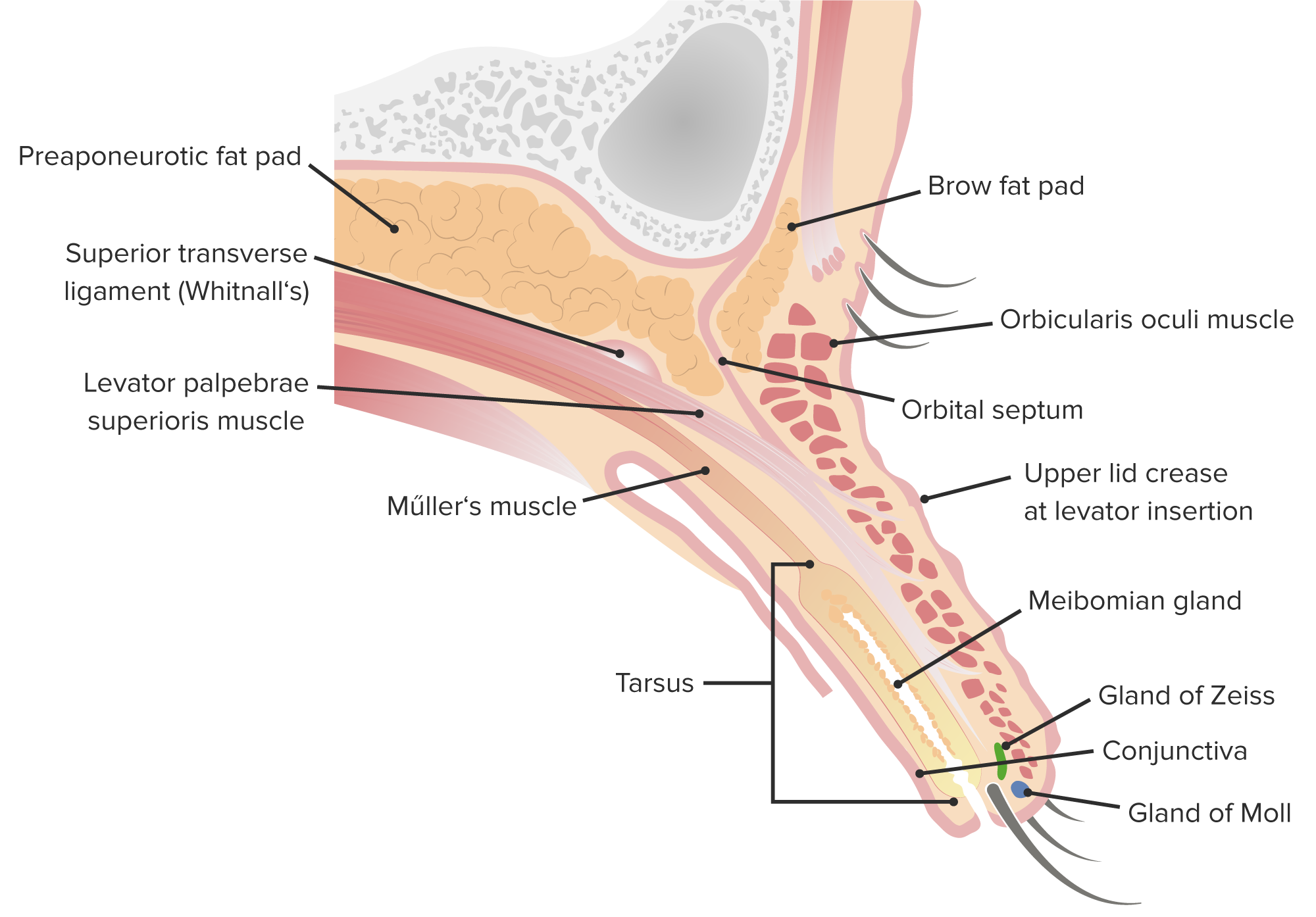
Anatomy of the eyelid: Note the locations of the Meibomian and Zeis glands, which are typically involved in the formation of a chalazion.

Image by Lecturio.
Chalazion causes

Visualization of a blocked Meibomian gland causing a chalazion

Image by BioDigital, edited by Lecturio

Clinical presentation[1,2]

  • Slow-growing, rubbery nodule Nodule Chalazion on the eyelid
    • If large enough, can compress the cornea Cornea The transparent anterior portion of the fibrous coat of the eye consisting of five layers: stratified squamous corneal epithelium; bowman membrane; corneal stroma; descemet membrane; and mesenchymal corneal endothelium. It serves as the first refracting medium of the eye. Eye: Anatomy and cause astigmatism Astigmatism Unequal curvature of the refractive surfaces of the eye. Thus a point source of light cannot be brought to a point focus on the retina but is spread over a more or less diffuse area. This results from the radius of curvature in one plane being longer or shorter than the radius at right angles to it. . Refractive Errors
    • More common on the upper lid due to the increased number and length of Meibomian glands
  • Normally painless, but may cause mild tenderness if the lesion progresses to a large size
  • Increased tearing
  • Eyelid heaviness
  • Conjunctival erythema Erythema Redness of the skin produced by congestion of the capillaries. This condition may result from a variety of disease processes. Chalazion
  • Swollen preauricular lymph nodes Lymph Nodes They are oval or bean shaped bodies (1 – 30 mm in diameter) located along the lymphatic system. Lymphatic Drainage System: Anatomy may be present in cases of secondary bacterial infection.
Chalazion

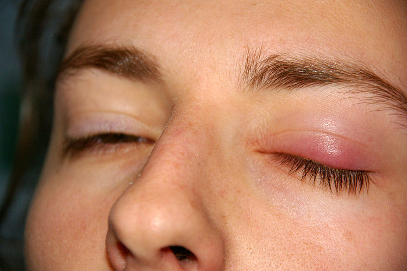
Patient presenting with a chalazion on the left eyelid with mild swelling

Image: “Chalazion” by jd. License: Public Domain

Diagnosis and Management

Diagnosis[4]

Diagnosis is clinical based on the history and physical exam.

  • Physical exam will show:
    • Presence of a non-tender, firm, palpable nodule Nodule Chalazion on the eyelid
    • Everting the eyelid may improve visualization.
  • Recurrent or persistent lesions should prompt further investigation: fine needle aspiration Fine Needle Aspiration Fibroadenoma cytology or biopsy Biopsy Removal and pathologic examination of specimens from the living body. Ewing Sarcoma to rule out malignancy Malignancy Hemothorax and other chronic conditions
    • Consider neoplasms Neoplasms New abnormal growth of tissue. Malignant neoplasms show a greater degree of anaplasia and have the properties of invasion and metastasis, compared to benign neoplasms. Benign Bone Tumors in recurrent lesions (particularly in the elderly):
      • Sebaceous gland Sebaceous Gland Small, sacculated organs found within the dermis. Each gland has a single duct that emerges from a cluster of oval alveoli. Each alveolus consists of a transparent basement membrane enclosing epithelial cells. The ducts from most sebaceous glands open into a hair follicle, but some open on the general surface of the skin. Sebaceous glands secrete sebum. Hordeolum (Stye) carcinoma
      • Basal cell Basal Cell Erythema Multiforme carcinoma
      • Squamous cell carcinoma Squamous cell carcinoma Cutaneous squamous cell carcinoma (cSCC) is caused by malignant proliferation of atypical keratinocytes. This condition is the 2nd most common skin malignancy and usually affects sun-exposed areas of fair-skinned patients. The cancer presents as a firm, erythematous, keratotic plaque or papule. Squamous Cell Carcinoma (SCC)
      • Merkel cell carcinoma
    • Leishmaniasis Leishmaniasis Leishmania species are obligate intracellular parasites that are transmitted by an infected sandfly. The mildest form is cutaneous leishmaniasis (CL), characterized by painless skin ulcers. The mucocutaneous type involves more tissue destruction, causing deformities. Visceral leishmaniasis (VL), the most severe form, presents with hepatosplenomegaly, anemia, thrombocytopenia, and fever. Leishmania/Leishmaniasis or tuberculosis Tuberculosis Tuberculosis (TB) is an infectious disease caused by Mycobacterium tuberculosis complex bacteria. The bacteria usually attack the lungs but can also damage other parts of the body. Approximately 30% of people around the world are infected with this pathogen, with the majority harboring a latent infection. Tuberculosis spreads through the air when a person with active pulmonary infection coughs or sneezes. Tuberculosis can present with masses that block meibomian gland Meibomian Gland Chalazion drainage.
      • Inquire about travel history and exposure
      • Other symptoms can include distant lymphadenopathy Lymphadenopathy Lymphadenopathy is lymph node enlargement (> 1 cm) and is benign and self-limited in most patients. Etiologies include malignancy, infection, and autoimmune disorders, as well as iatrogenic causes such as the use of certain medications. Generalized lymphadenopathy often indicates underlying systemic disease. Lymphadenopathy, ocular pain Pain An unpleasant sensation induced by noxious stimuli which are detected by nerve endings of nociceptive neurons. Pain: Types and Pathways, and limited ocular mobility.
    • Correlate with underlying conditions:
      • Large chalazia can be seen in hyper-IgE syndrome.
      • In patients Patients Individuals participating in the health care system for the purpose of receiving therapeutic, diagnostic, or preventive procedures. Clinician–Patient Relationship with HIV HIV Anti-HIV Drugs, Kaposi Kaposi A multicentric, malignant neoplastic vascular proliferation characterized by the development of bluish-red cutaneous nodules, usually on the lower extremities, most often on the toes or feet, and slowly increasing in size and number and spreading to more proximal areas. The tumors have endothelium-lined channels and vascular spaces admixed with variably sized aggregates of spindle-shaped cells, and often remain confined to the skin and subcutaneous tissue, but widespread visceral involvement may occur. Hhv-8 is the suspected cause. There is also a high incidence in AIDS patients. AIDS-defining Conditions sarcoma can manifest as eyelid nodules.
  • Visual acuity Visual Acuity Clarity or sharpness of ocular vision or the ability of the eye to see fine details. Visual acuity depends on the functions of retina, neuronal transmission, and the interpretative ability of the brain. Normal visual acuity is expressed as 20/20 indicating that one can see at 20 feet what should normally be seen at that distance. Visual acuity can also be influenced by brightness, color, and contrast. Ophthalmic Exam and field testing may be necessary if a chalazion Chalazion A chalazion is one of the most common inflammatory lesions of the eyelid. It is caused by obstruction of the Meibomian or Zeis glands, leading to granulomatous inflammation and resulting in a firm, rubbery, slow-growing nodule that is typically non-tender. Chalazion is large.

Management

Conservative management:[4,5]

  • Most will resolve without treatment.
  • Keep area clean.
  • Warm compresses Warm Compresses Chalazion (use for 10 minutes, 4x daily)
  • Lid massages and lid scrubs
  • If the chalazion Chalazion A chalazion is one of the most common inflammatory lesions of the eyelid. It is caused by obstruction of the Meibomian or Zeis glands, leading to granulomatous inflammation and resulting in a firm, rubbery, slow-growing nodule that is typically non-tender. Chalazion does not resolve within a couple of months, the patient may need a referral to ophthalmology. 
  • Systemic antibiotics are generally not necessary, but if a secondary infectious process is present, antibiotic options are:
    • Tetracycline Tetracycline A naphthacene antibiotic that inhibits amino Acyl tRNA binding during protein synthesis. Drug-Induced Liver Injury 500 mg twice daily
    • Doxycycline 100 mg daily
    • Minocycline Minocycline A tetracycline analog, having a 7-dimethylamino and lacking the 5 methyl and hydroxyl groups, which is effective against tetracycline-resistant staphylococcus infections. Tetracyclines 100 mg twice daily
    • Azithromycin Azithromycin A semi-synthetic macrolide antibiotic structurally related to erythromycin. It has been used in the treatment of Mycobacterium avium intracellulare infections, toxoplasmosis, and cryptosporidiosis. Macrolides and Ketolides 500 mg on day 1, then 250 mg daily

Invasive treatment (for a persistent or large, symptomatic chalazion Chalazion A chalazion is one of the most common inflammatory lesions of the eyelid. It is caused by obstruction of the Meibomian or Zeis glands, leading to granulomatous inflammation and resulting in a firm, rubbery, slow-growing nodule that is typically non-tender. Chalazion):[4,6]

  • Performed by an ophthalmologist
  • Steroid injection:
    • Intralesional injection of triamcinolone Triamcinolone A glucocorticoid given, as the free alcohol or in esterified form, orally, intramuscularly, by local injection, by inhalation, or applied topically in the management of various disorders in which corticosteroids are indicated. Glucocorticoids (4 mg used in the study) followed by lid massage: found to be as effective as incision and drainage Incision And Drainage Chalazion[7]
    • Possible steroid complications:
      • Intraocular penetration Penetration X-rays
      • Intraocular pressure Intraocular Pressure The pressure of the fluids in the eye. Ophthalmic Exam elevation
      • Visual loss
      • Subcutaneous fat Subcutaneous fat Fatty tissue under the skin throughout the body. Erythema Nodosum atrophy Atrophy Decrease in the size of a cell, tissue, organ, or multiple organs, associated with a variety of pathological conditions such as abnormal cellular changes, ischemia, malnutrition, or hormonal changes. Cellular Adaptation
      • Skin Skin The skin, also referred to as the integumentary system, is the largest organ of the body. The skin is primarily composed of the epidermis (outer layer) and dermis (deep layer). The epidermis is primarily composed of keratinocytes that undergo rapid turnover, while the dermis contains dense layers of connective tissue. Skin: Structure and Functions depigmentation Depigmentation Vitiligo
  • Incision and drainage Incision And Drainage Chalazion ( curettage Curettage A scraping, usually of the interior of a cavity or tract, for removal of new growth or other abnormal tissue, or to obtain material for tissue diagnosis. It is performed with a curet (curette), a spoon-shaped instrument designed for that purpose. Benign Bone Tumors)
    • Cases more amenable to incision and curettage Curettage A scraping, usually of the interior of a cavity or tract, for removal of new growth or other abnormal tissue, or to obtain material for tissue diagnosis. It is performed with a curet (curette), a spoon-shaped instrument designed for that purpose. Benign Bone Tumors:
      • Liquid chalazion Chalazion A chalazion is one of the most common inflammatory lesions of the eyelid. It is caused by obstruction of the Meibomian or Zeis glands, leading to granulomatous inflammation and resulting in a firm, rubbery, slow-growing nodule that is typically non-tender. Chalazion
      • Lesion with purulent component
      • Virally induced lesion
    • Lesions that are large and multiloculated benefit from a combination of incision and curettage Curettage A scraping, usually of the interior of a cavity or tract, for removal of new growth or other abnormal tissue, or to obtain material for tissue diagnosis. It is performed with a curet (curette), a spoon-shaped instrument designed for that purpose. Benign Bone Tumors, followed by steroid injection.

Differential Diagnosis

  • Hordeolum Hordeolum A hordeolum is an acute infection affecting the meibomian, Zeiss, or Moll glands of the eyelid. Stasis of the gland secretions predisposes to bacterial infection. Staphylococcus aureus is the most common pathogen. Hordeolum (Stye): a localized infection arising from the Zeis gland, Moll gland, or Meibomian gland Meibomian Gland Chalazion. Staphylococcus aureus Staphylococcus aureus Potentially pathogenic bacteria found in nasal membranes, skin, hair follicles, and perineum of warm-blooded animals. They may cause a wide range of infections and intoxications. Brain Abscess is a common cause. Examination findings of a tender, erythematous, pus-filled nodule Nodule Chalazion help establish the diagnosis. Management is generally conservative, though severe cases may require antibiotics or drainage. Chalazia, on the other hand Hand The hand constitutes the distal part of the upper limb and provides the fine, precise movements needed in activities of daily living. It consists of 5 metacarpal bones and 14 phalanges, as well as numerous muscles innervated by the median and ulnar nerves. Hand: Anatomy, are due to sterile Sterile Basic Procedures, granulomatous inflammation Granulomatous Inflammation Chalazion and are not painful.
  • Blepharitis Blepharitis Blepharitis is an ocular condition characterized by eyelid inflammation. Anterior blepharitis involves the eyelid skin and eyelashes, while the posterior type affects the meibomian glands. Often, these conditions overlap. Blepharitis: an inflammatory condition of the eyelid margins; classified as posterior or anterior blepharitis Anterior Blepharitis Blepharitis. Inflammation Inflammation Inflammation is a complex set of responses to infection and injury involving leukocytes as the principal cellular mediators in the body’s defense against pathogenic organisms. Inflammation is also seen as a response to tissue injury in the process of wound healing. The 5 cardinal signs of inflammation are pain, heat, redness, swelling, and loss of function. Inflammation is seen at the inner portion of the eyelid or at the base of the eyelashes. Patients Patients Individuals participating in the health care system for the purpose of receiving therapeutic, diagnostic, or preventive procedures. Clinician–Patient Relationship present with red, swollen, itchy eyelids Eyelids Each of the upper and lower folds of skin which cover the eye when closed. Blepharitis or vision Vision Ophthalmic Exam changes. Diagnosis is clinical, and management includes eyelid hygiene and conservative measures. Symptoms and physical exam differentiate this condition from a chalazion Chalazion A chalazion is one of the most common inflammatory lesions of the eyelid. It is caused by obstruction of the Meibomian or Zeis glands, leading to granulomatous inflammation and resulting in a firm, rubbery, slow-growing nodule that is typically non-tender. Chalazion.
  • Sebaceous carcinoma: a rare malignancy Malignancy Hemothorax of the sebaceous glands, such as the Meibomian and Zeis glands Zeis Glands Chalazion. Presents as a round, painless nodule Nodule Chalazion of the eyelid. Diagnosis is made by biopsy Biopsy Removal and pathologic examination of specimens from the living body. Ewing Sarcoma of the lesion, and management requires surgical removal of the tumor Tumor Inflammation. Distinguishing this from a chalazion Chalazion A chalazion is one of the most common inflammatory lesions of the eyelid. It is caused by obstruction of the Meibomian or Zeis glands, leading to granulomatous inflammation and resulting in a firm, rubbery, slow-growing nodule that is typically non-tender. Chalazion may be difficult, and biopsy Biopsy Removal and pathologic examination of specimens from the living body. Ewing Sarcoma should be pursued for persistent lesions.

References

  1. Gosh, C., Gosh, T. (2020). Eyelid lesions. UpToDate. Retrieved October 20, 2020, from https://www.uptodate.com/contents/eyelid-lesions
  2. Deschenes, J., You, J. Y. (2019). Chalazion. Medscape. Retrieved October 20, 2020, from https://emedicine.medscape.com/article/1212709-overview
  3. Horton J.C. (2022). Disorders of the eye. In Loscalzo, J., Fauci, A., Kasper, D., Hauser, S., Longo, D., Jameson, J. (Eds.), Harrison’s Principles of Internal Medicine (21st ed.). McGraw-Hill. https://accessmedicine.mhmedical.com/content.aspx?bookid=3095&sectionid=262790052
  4. Elsayed, M., Sultan, K. (2015). Chalazion management: evidence and questions. American Academy of Ophthalmology. Retrieved September 18, 2022, from https://www.aao.org/eyenet/article/chalazion-management-evidence-questions
  5. Kashkouli, M. B., Fazel, A. J., Kiavash, V., Nojomi, M., Ghiasian, L. (2015). Oral azithromycin versus doxycycline in meibomian gland dysfunction: a randomised double-masked open-label clinical trial. British Journal of Ophthalmology, 99(2), 199–204. https://doi.org/10.1136/bjophthalmol-2014-305410
  6. Goawalla, A., Lee, V. (2007). A prospective randomized treatment study comparing three treatment options for chalazia: triamcinolone acetonide injections, incision and curettage and treatment with hot compresses. Clinical & Experimental Ophthalmology, 35(8), 706–712. https://doi.org/10.1111/j.1442-9071.2007.01617.x
  7. Ben Simon, G. J., Rosen, N., Rosner, M., Spierer, A. (2011). Intralesional triamcinolone acetonide injection versus incision and curettage for primary chalazia: a prospective, randomized study. American Journal of Ophthalmology, 151(4), 714–718.e1. https://doi.org/10.1016/j.ajo.2010.10.026

Create your free account or log in to continue reading!

Sign up now and get free access to Lecturio with concept pages, medical videos, and questions for your medical education.

User Reviews

Details