The mediastinum is the thoracic area between the 2 pleural cavities. The mediastinum contains vital structures of the circulatory, respiratory, digestive, and nervous systems including the heart and esophagusEsophagusThe esophagus is a muscular tube-shaped organ of around 25 centimeters in length that connects the pharynx to the stomach. The organ extends from approximately the 6th cervical vertebra to the 11th thoracic vertebra and can be divided grossly into 3 parts: the cervical part, the thoracic part, and the abdominal part. Esophagus: Anatomy, and major thoracic vessels including the superior vena cava, inferior vena cava, pulmonary arteriesArteriesArteries are tubular collections of cells that transport oxygenated blood and nutrients from the heart to the tissues of the body. The blood passes through the arteries in order of decreasing luminal diameter, starting in the largest artery (the aorta) and ending in the small arterioles. Arteries are classified into 3 types: large elastic arteries, medium muscular arteries, and small arteries and arterioles. Arteries: Histology, pulmonary veinsPulmonary veinsThe veins that return the oxygenated blood from the lungs to the left atrium of the heart.Lungs: Anatomy, and aorta. The mediastinum extends from the upper thoracic aperture to the diaphragmDiaphragmThe diaphragm is a large, dome-shaped muscle that separates the thoracic cavity from the abdominal cavity. The diaphragm consists of muscle fibers and a large central tendon, which is divided into right and left parts. As the primary muscle of inspiration, the diaphragm contributes 75% of the total inspiratory muscle force.Diaphragm: Anatomy and is bordered by the lungsLungsLungs are the main organs of the respiratory system. Lungs are paired viscera located in the thoracic cavity and are composed of spongy tissue. The primary function of the lungs is to oxygenate blood and eliminate CO2. Lungs: Anatomy.
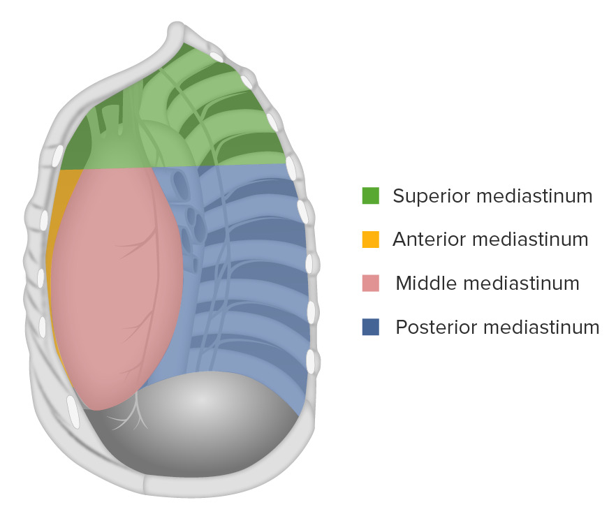
The mediastinum is the middle of the thoracic cavity, located between the lungsLungsLungs are the main organs of the respiratory system. Lungs are paired viscera located in the thoracic cavity and are composed of spongy tissue. The primary function of the lungs is to oxygenate blood and eliminate CO2. Lungs: Anatomy. The mediastinum is subdivided into the superior and inferior compartments, which are further divided into the anterior, middle, and posterior mediastina.
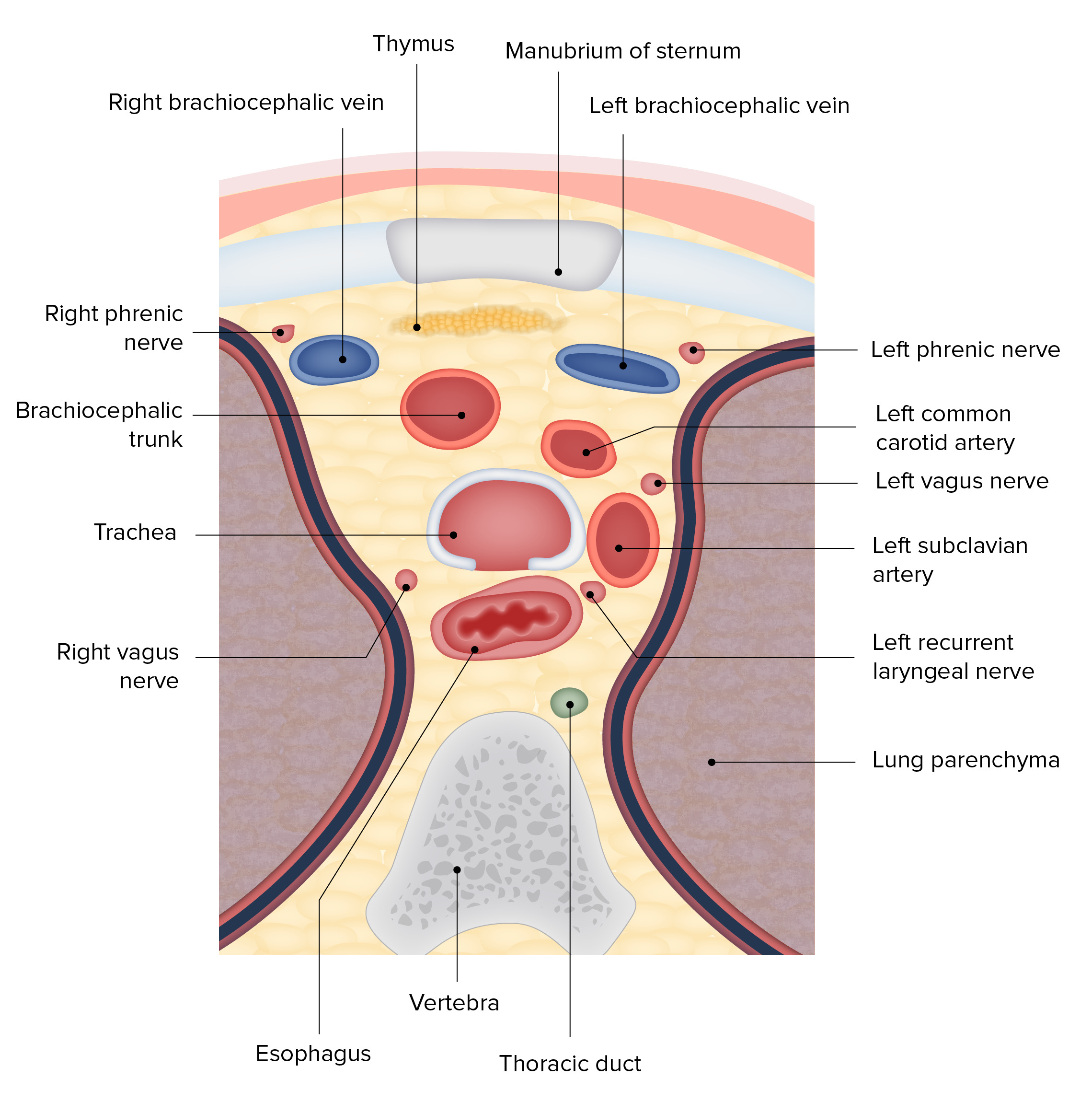
Inferior border of T4T4The major hormone derived from the thyroid gland. Thyroxine is synthesized via the iodination of tyrosines (monoiodotyrosine) and the coupling of iodotyrosines (diiodotyrosine) in the thyroglobulin. Thyroxine is released from thyroglobulin by proteolysis and secreted into the blood. Thyroxine is peripherally deiodinated to form triiodothyronine which exerts a broad spectrum of stimulatory effects on cell metabolism.Thyroid Hormones (thoracic plane) posteriorly
Contents:
TracheaTracheaThe trachea is a tubular structure that forms part of the lower respiratory tract. The trachea is continuous superiorly with the larynx and inferiorly becomes the bronchial tree within the lungs. The trachea consists of a support frame of semicircular, or C-shaped, rings made out of hyaline cartilage and reinforced by collagenous connective tissue. Trachea: Anatomy
EsophagusEsophagusThe esophagus is a muscular tube-shaped organ of around 25 centimeters in length that connects the pharynx to the stomach. The organ extends from approximately the 6th cervical vertebra to the 11th thoracic vertebra and can be divided grossly into 3 parts: the cervical part, the thoracic part, and the abdominal part. Esophagus: Anatomy
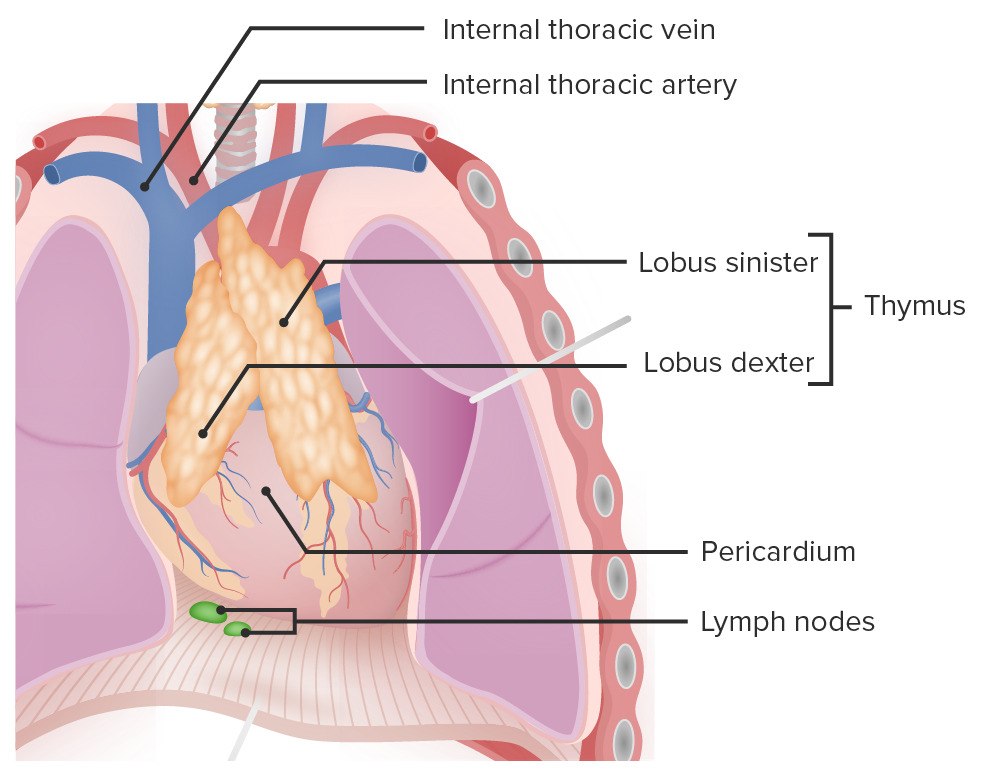
ThymusThymusA single, unpaired primary lymphoid organ situated in the mediastinum, extending superiorly into the neck to the lower edge of the thyroid gland and inferiorly to the fourth costal cartilage. It is necessary for normal development of immunologic function early in life. By puberty, it begins to involute and much of the tissue is replaced by fat.Lymphatic Drainage System: Anatomy (prepubertal)
Vessels:
Superior vena cava
Brachiocephalic veinsVeinsVeins are tubular collections of cells, which transport deoxygenated blood and waste from the capillary beds back to the heart. Veins are classified into 3 types: small veins/venules, medium veins, and large veins. Each type contains 3 primary layers: tunica intima, tunica media, and tunica adventitia. Veins: Histology
Aortic arch
Innominate artery
Thoracic portions of the left common carotid and left subclavian arteriesArteriesArteries are tubular collections of cells that transport oxygenated blood and nutrients from the heart to the tissues of the body. The blood passes through the arteries in order of decreasing luminal diameter, starting in the largest artery (the aorta) and ending in the small arterioles. Arteries are classified into 3 types: large elastic arteries, medium muscular arteries, and small arteries and arterioles. Arteries: Histology
Phrenic nervePhrenic nerveThe motor nerve of the diaphragm. The phrenic nerve fibers originate in the cervical spinal column (mostly C4) and travel through the cervical plexus to the diaphragm.Diaphragm: Anatomy
Vagus nerveVagus nerveThe 10th cranial nerve. The vagus is a mixed nerve which contains somatic afferents (from skin in back of the ear and the external auditory meatus), visceral afferents (from the pharynx, larynx, thorax, and abdomen), parasympathetic efferents (to the thorax and abdomen), and efferents to striated muscle (of the larynx and pharynx).Pharynx: Anatomy
Left recurrent laryngeal nerve
Diagram of a cross-section of the superior mediastinum featuring the spatial relations of the trachea
The inferior mediastinum is bordered superiorly by the thoracic plane and inferiorly by the diaphragmDiaphragmThe diaphragm is a large, dome-shaped muscle that separates the thoracic cavity from the abdominal cavity. The diaphragm consists of muscle fibers and a large central tendon, which is divided into right and left parts. As the primary muscle of inspiration, the diaphragm contributes 75% of the total inspiratory muscle force.Diaphragm: Anatomy, and is further subdivided into the anterior, middle, and posterior mediastina.
Anterior mediastinum:
Extends from the sternumSternumA long, narrow, and flat bone commonly known as breastbone occurring in the midsection of the anterior thoracic segment or chest region, which stabilizes the rib cage and serves as the point of origin for several muscles that move the arms, head, and neck.Chest Wall: Anatomy anteriorly to the anterior surface of the pericardiumPericardiumA conical fibroserous sac surrounding the heart and the roots of the great vessels (aorta; venae cavae; pulmonary artery). Pericardium consists of two sacs: the outer fibrous pericardium and the inner serous pericardium. The latter consists of an outer parietal layer facing the fibrous pericardium, and an inner visceral layer (epicardium) resting next to the heart, and a pericardial cavity between these two layers.Heart: Anatomy posteriorly
Remnants of thymusThymusA single, unpaired primary lymphoid organ situated in the mediastinum, extending superiorly into the neck to the lower edge of the thyroid gland and inferiorly to the fourth costal cartilage. It is necessary for normal development of immunologic function early in life. By puberty, it begins to involute and much of the tissue is replaced by fat.Lymphatic Drainage System: Anatomy
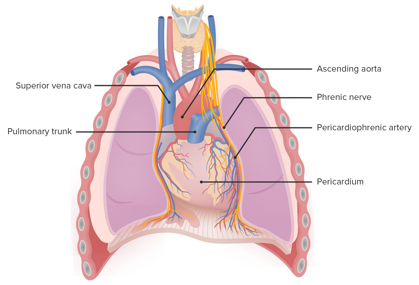
Middle mediastinum:
Extends between the anterior and posterior surfaces of the pericardiumPericardiumA conical fibroserous sac surrounding the heart and the roots of the great vessels (aorta; venae cavae; pulmonary artery). Pericardium consists of two sacs: the outer fibrous pericardium and the inner serous pericardium. The latter consists of an outer parietal layer facing the fibrous pericardium, and an inner visceral layer (epicardium) resting next to the heart, and a pericardial cavity between these two layers.Heart: Anatomy
Contains:
Heart and pericardiumPericardiumA conical fibroserous sac surrounding the heart and the roots of the great vessels (aorta; venae cavae; pulmonary artery). Pericardium consists of two sacs: the outer fibrous pericardium and the inner serous pericardium. The latter consists of an outer parietal layer facing the fibrous pericardium, and an inner visceral layer (epicardium) resting next to the heart, and a pericardial cavity between these two layers.Heart: Anatomy
Phrenic nervePhrenic nerveThe motor nerve of the diaphragm. The phrenic nerve fibers originate in the cervical spinal column (mostly C4) and travel through the cervical plexus to the diaphragm.Diaphragm: Anatomy
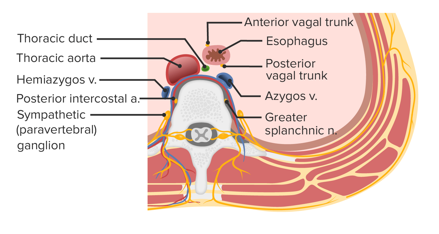
Posterior mediastinum:
Extends between the posterior surface of the pericardiumPericardiumA conical fibroserous sac surrounding the heart and the roots of the great vessels (aorta; venae cavae; pulmonary artery). Pericardium consists of two sacs: the outer fibrous pericardium and the inner serous pericardium. The latter consists of an outer parietal layer facing the fibrous pericardium, and an inner visceral layer (epicardium) resting next to the heart, and a pericardial cavity between these two layers.Heart: Anatomy and T4T4The major hormone derived from the thyroid gland. Thyroxine is synthesized via the iodination of tyrosines (monoiodotyrosine) and the coupling of iodotyrosines (diiodotyrosine) in the thyroglobulin. Thyroxine is released from thyroglobulin by proteolysis and secreted into the blood. Thyroxine is peripherally deiodinated to form triiodothyronine which exerts a broad spectrum of stimulatory effects on cell metabolism.Thyroid Hormones to T12
Contains:
EsophagusEsophagusThe esophagus is a muscular tube-shaped organ of around 25 centimeters in length that connects the pharynx to the stomach. The organ extends from approximately the 6th cervical vertebra to the 11th thoracic vertebra and can be divided grossly into 3 parts: the cervical part, the thoracic part, and the abdominal part. Esophagus: Anatomy
Azygos, hemiazygos, and accessory hemiazygos veinsVeinsVeins are tubular collections of cells, which transport deoxygenated blood and waste from the capillary beds back to the heart. Veins are classified into 3 types: small veins/venules, medium veins, and large veins. Each type contains 3 primary layers: tunica intima, tunica media, and tunica adventitia. Veins: Histology
Vagus nerveVagus nerveThe 10th cranial nerve. The vagus is a mixed nerve which contains somatic afferents (from skin in back of the ear and the external auditory meatus), visceral afferents (from the pharynx, larynx, thorax, and abdomen), parasympathetic efferents (to the thorax and abdomen), and efferents to striated muscle (of the larynx and pharynx).Pharynx: Anatomy
The aorta is the main artery of the body consisting of:
Ascending aorta:
Originates from the left ventricle
Gives rise to the right and left coronary arteriesArteriesArteries are tubular collections of cells that transport oxygenated blood and nutrients from the heart to the tissues of the body. The blood passes through the arteries in order of decreasing luminal diameter, starting in the largest artery (the aorta) and ending in the small arterioles. Arteries are classified into 3 types: large elastic arteries, medium muscular arteries, and small arteries and arterioles. Arteries: Histology
Located within the pericardiumPericardiumA conical fibroserous sac surrounding the heart and the roots of the great vessels (aorta; venae cavae; pulmonary artery). Pericardium consists of two sacs: the outer fibrous pericardium and the inner serous pericardium. The latter consists of an outer parietal layer facing the fibrous pericardium, and an inner visceral layer (epicardium) resting next to the heart, and a pericardial cavity between these two layers.Heart: Anatomy
Aortic arch:
Connects the ascending aorta and the descending aorta
Gives rise to the brachiocephalic trunk, left common carotid arteryCommon carotid arteryThe two principal arteries supplying the structures of the head and neck. They ascend in the neck, one on each side, and at the level of the upper border of the thyroid cartilage, each divides into two branches, the external and internal carotid arteries.Carotid Arterial System: Anatomy, and left subclavian artery
Located in the middle and posterior mediastinum
Runs to the left of the tracheaTracheaThe trachea is a tubular structure that forms part of the lower respiratory tract. The trachea is continuous superiorly with the larynx and inferiorly becomes the bronchial tree within the lungs. The trachea consists of a support frame of semicircular, or C-shaped, rings made out of hyaline cartilage and reinforced by collagenous connective tissue. Trachea: Anatomy
Descending aorta:
Starts at the T4T4The major hormone derived from the thyroid gland. Thyroxine is synthesized via the iodination of tyrosines (monoiodotyrosine) and the coupling of iodotyrosines (diiodotyrosine) in the thyroglobulin. Thyroxine is released from thyroglobulin by proteolysis and secreted into the blood. Thyroxine is peripherally deiodinated to form triiodothyronine which exerts a broad spectrum of stimulatory effects on cell metabolism.Thyroid Hormones level
Travels medially and ends anterior to the vertebral columnVertebral columnThe human spine, or vertebral column, is the most important anatomical and functional axis of the human body. It consists of 7 cervical vertebrae, 12 thoracic vertebrae, and 5 lumbar vertebrae and is limited cranially by the skull and caudally by the sacrum. Vertebral Column: Anatomy
Gives rise to the pericardial, bronchial, esophageal, mediastinal, intercostal, subcostal, and phrenic arteriesArteriesArteries are tubular collections of cells that transport oxygenated blood and nutrients from the heart to the tissues of the body. The blood passes through the arteries in order of decreasing luminal diameter, starting in the largest artery (the aorta) and ending in the small arterioles. Arteries are classified into 3 types: large elastic arteries, medium muscular arteries, and small arteries and arterioles. Arteries: Histology
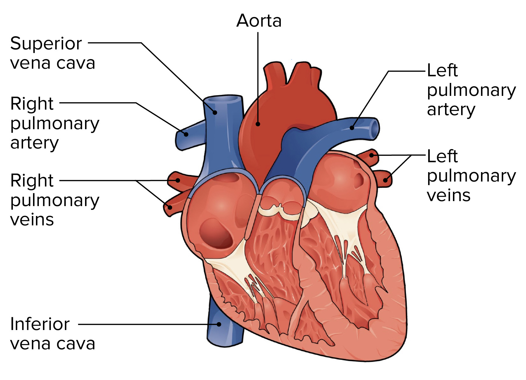
Superior vena cava
Originates from the confluence of the 2 brachiocephalic veinsVeinsVeins are tubular collections of cells, which transport deoxygenated blood and waste from the capillary beds back to the heart. Veins are classified into 3 types: small veins/venules, medium veins, and large veins. Each type contains 3 primary layers: tunica intima, tunica media, and tunica adventitia. Veins: Histology
Drains into the right atrium
Carries O2-poor blood from the upper body and head
Begins behind the 1st right costal cartilageCartilageCartilage is a type of connective tissue derived from embryonic mesenchyme that is responsible for structural support, resilience, and the smoothness of physical actions. Perichondrium (connective tissue membrane surrounding cartilage) compensates for the absence of vasculature in cartilage by providing nutrition and support. Cartilage: Histology and descends behind the 2nd and 3rd intercostal spacesIntercostal spacesChest Wall: Anatomy
Located in the superior and middle mediastina
The lower ½ is covered by the pericardiumPericardiumA conical fibroserous sac surrounding the heart and the roots of the great vessels (aorta; venae cavae; pulmonary artery). Pericardium consists of two sacs: the outer fibrous pericardium and the inner serous pericardium. The latter consists of an outer parietal layer facing the fibrous pericardium, and an inner visceral layer (epicardium) resting next to the heart, and a pericardial cavity between these two layers.Heart: Anatomy.
Inferior vena cava
Largest vein in the body
Originates from the right and left common iliac veinsVeinsVeins are tubular collections of cells, which transport deoxygenated blood and waste from the capillary beds back to the heart. Veins are classified into 3 types: small veins/venules, medium veins, and large veins. Each type contains 3 primary layers: tunica intima, tunica media, and tunica adventitia. Veins: Histology
Drains into the right atrium
Carries O2-poor blood from the lower half of the body
Enters the mediastinum through the caval foramen of the diaphragmDiaphragmThe diaphragm is a large, dome-shaped muscle that separates the thoracic cavity from the abdominal cavity. The diaphragm consists of muscle fibers and a large central tendon, which is divided into right and left parts. As the primary muscle of inspiration, the diaphragm contributes 75% of the total inspiratory muscle force.Diaphragm: Anatomy at the T8 level
Communicates with the superior vena cava through:
Azygos vein
Vertebral venous plexuses
Lumbar veinsVeinsVeins are tubular collections of cells, which transport deoxygenated blood and waste from the capillary beds back to the heart. Veins are classified into 3 types: small veins/venules, medium veins, and large veins. Each type contains 3 primary layers: tunica intima, tunica media, and tunica adventitia. Veins: Histology
Located in the middle mediastinum
Pulmonary arteriesArteriesArteries are tubular collections of cells that transport oxygenated blood and nutrients from the heart to the tissues of the body. The blood passes through the arteries in order of decreasing luminal diameter, starting in the largest artery (the aorta) and ending in the small arterioles. Arteries are classified into 3 types: large elastic arteries, medium muscular arteries, and small arteries and arterioles. Arteries: Histology
The main pulmonary arteryPulmonary arteryThe short wide vessel arising from the conus arteriosus of the right ventricle and conveying unaerated blood to the lungs.Lungs: Anatomy originates from the right ventricle.
Pulmonary arteriesArteriesArteries are tubular collections of cells that transport oxygenated blood and nutrients from the heart to the tissues of the body. The blood passes through the arteries in order of decreasing luminal diameter, starting in the largest artery (the aorta) and ending in the small arterioles. Arteries are classified into 3 types: large elastic arteries, medium muscular arteries, and small arteries and arterioles. Arteries: Histology carry deoxygenated blood to the lungsLungsLungs are the main organs of the respiratory system. Lungs are paired viscera located in the thoracic cavity and are composed of spongy tissue. The primary function of the lungs is to oxygenate blood and eliminate CO2. Lungs: Anatomy.
Located in the middle mediastinum
Divided into the right and left pulmonary arteriesArteriesArteries are tubular collections of cells that transport oxygenated blood and nutrients from the heart to the tissues of the body. The blood passes through the arteries in order of decreasing luminal diameter, starting in the largest artery (the aorta) and ending in the small arterioles. Arteries are classified into 3 types: large elastic arteries, medium muscular arteries, and small arteries and arterioles. Arteries: Histology
The right pulmonary arteryPulmonary arteryThe short wide vessel arising from the conus arteriosus of the right ventricle and conveying unaerated blood to the lungs.Lungs: Anatomy passes under the aortic arch.
Pulmonary veinsPulmonary veinsThe veins that return the oxygenated blood from the lungs to the left atrium of the heart.Lungs: Anatomy
4 pulmonary veinsPulmonary veinsThe veins that return the oxygenated blood from the lungs to the left atrium of the heart.Lungs: Anatomy: 2 on each side
Receive blood from the bronchial veinsVeinsVeins are tubular collections of cells, which transport deoxygenated blood and waste from the capillary beds back to the heart. Veins are classified into 3 types: small veins/venules, medium veins, and large veins. Each type contains 3 primary layers: tunica intima, tunica media, and tunica adventitia. Veins: Histology
The superior vena cava and right atrium lie anterior to the right pulmonary veinsPulmonary veinsThe veins that return the oxygenated blood from the lungs to the left atrium of the heart.Lungs: Anatomy.
The descending thoracic aorta lies posterior to the left pulmonary veinsPulmonary veinsThe veins that return the oxygenated blood from the lungs to the left atrium of the heart.Lungs: Anatomy.
The 5 great vessels: aorta, pulmonary artery, pulmonary veins, superior vena cava, and inferior vena cava
Coarctation of the aortaCoarctation of the aortaCoarctation of the aorta is a narrowing of the aorta between the aortic arch and the iliac bifurcation commonly around the point of insertion of the ductus arteriosus. Coarctation of the aorta is typically congenital and the clinical presentation depends on the age of the patient. Coarctation of the Aorta: narrowing in the aorta between the aortic arch and the iliac bifurcation. Coarctation of the aortaCoarctation of the aortaCoarctation of the aorta is a narrowing of the aorta between the aortic arch and the iliac bifurcation commonly around the point of insertion of the ductus arteriosus. Coarctation of the aorta is typically congenital and the clinical presentation depends on the age of the patient. Coarctation of the Aorta is most common around the point of insertion of the ductus arteriosusDuctus arteriosusA fetal blood vessel connecting the pulmonary artery with the descending aorta.Patent Ductus Arteriosus (PDA) and is congenital in the majority of cases. Neonates present with heart failureHeart FailureA heterogeneous condition in which the heart is unable to pump out sufficient blood to meet the metabolic need of the body. Heart failure can be caused by structural defects, functional abnormalities (ventricular dysfunction), or a sudden overload beyond its capacity. Chronic heart failure is more common than acute heart failure which results from sudden insult to cardiac function, such as myocardial infarction.Total Anomalous Pulmonary Venous Return (TAPVR) upon the closure of the ductus arteriosusDuctus arteriosusA fetal blood vessel connecting the pulmonary artery with the descending aorta.Patent Ductus Arteriosus (PDA), whereas children and adults present with symptoms of hypoperfusion and/or hypertensionHypertensionHypertension, or high blood pressure, is a common disease that manifests as elevated systemic arterial pressures. Hypertension is most often asymptomatic and is found incidentally as part of a routine physical examination or during triage for an unrelated medical encounter. Hypertension. The classic findings on physical exam include a radio- or brachiofemoral delayBrachiofemoral delayFemoral pulses are delayed compared to brachial pulses.Coarctation of the Aorta and lower blood pressure in the lower limbs. Affected individuals should be surgically managed as early as possible to avoid complications of hypertensionHypertensionHypertension, or high blood pressure, is a common disease that manifests as elevated systemic arterial pressures. Hypertension is most often asymptomatic and is found incidentally as part of a routine physical examination or during triage for an unrelated medical encounter. Hypertension. Close follow-up is required as the risk of hypertensionHypertensionHypertension, or high blood pressure, is a common disease that manifests as elevated systemic arterial pressures. Hypertension is most often asymptomatic and is found incidentally as part of a routine physical examination or during triage for an unrelated medical encounter. Hypertension and relapseRelapseRelapsing Fever remains.
Transposition of great vessels: a cyanotic congenital heart disease characterized by the “switching” of the great arteriesArteriesArteries are tubular collections of cells that transport oxygenated blood and nutrients from the heart to the tissues of the body. The blood passes through the arteries in order of decreasing luminal diameter, starting in the largest artery (the aorta) and ending in the small arterioles. Arteries are classified into 3 types: large elastic arteries, medium muscular arteries, and small arteries and arterioles. Arteries: Histology. Transposition of great vesselsaccounts for 3% of all cases of congenital heart disease and presents within the neonatal phase of life with cyanosisCyanosisA bluish or purplish discoloration of the skin and mucous membranes due to an increase in the amount of deoxygenated hemoglobin in the blood or a structural defect in the hemoglobin molecule.Pulmonary Examination that is unresponsive to O2 therapy. Diagnosis is confirmed by an echocardiogramEchocardiogramTransposition of the Great Arteries and verified by catheterization. A chest X-rayX-rayPenetrating electromagnetic radiation emitted when the inner orbital electrons of an atom are excited and release radiant energy. X-ray wavelengths range from 1 pm to 10 nm. Hard x-rays are the higher energy, shorter wavelength x-rays. Soft x-rays or grenz rays are less energetic and longer in wavelength. The short wavelength end of the x-ray spectrum overlaps the gamma rays wavelength range. The distinction between gamma rays and x-rays is based on their radiation source.Pulmonary Function Tests shows the classic “egg on a string” pattern. Treatment is eminently surgical, and the prognosisPrognosisA prediction of the probable outcome of a disease based on a individual’s condition and the usual course of the disease as seen in similar situations.Non-Hodgkin Lymphomas for surgically corrected cases is good.
Truncus arteriosusTruncus arteriosusTruncus arteriosus (TA) is a congenital heart defect characterized by the persistence of a common cardiac arterial trunk tract that fails to divide into the pulmonary artery and aorta during embryonic development. Truncus arteriosus is a rare congenital malformation with a high mortality rate within the 1st 5 weeks of life if not managed promptly. Truncus Arteriosus: a congenital heart defect characterized by the persistence of a common cardiac arterial trunkArterial TrunkTruncus Arteriosus tract that fails to divide into the pulmonary arteryPulmonary arteryThe short wide vessel arising from the conus arteriosus of the right ventricle and conveying unaerated blood to the lungs.Lungs: Anatomy and aorta during embryonic development. Truncus arteriosusTruncus arteriosusTruncus arteriosus (TA) is a congenital heart defect characterized by the persistence of a common cardiac arterial trunk tract that fails to divide into the pulmonary artery and aorta during embryonic development. Truncus arteriosus is a rare congenital malformation with a high mortality rate within the 1st 5 weeks of life if not managed promptly. Truncus Arteriosus is a rare congenital malformation with a high mortalityMortalityAll deaths reported in a given population.Measures of Health Status rate within the 1st 5 weeks of life if not managed promptly. Neonates may be asymptomatic at birth but will invariably develop respiratory distress and heart failureHeart FailureA heterogeneous condition in which the heart is unable to pump out sufficient blood to meet the metabolic need of the body. Heart failure can be caused by structural defects, functional abnormalities (ventricular dysfunction), or a sudden overload beyond its capacity. Chronic heart failure is more common than acute heart failure which results from sudden insult to cardiac function, such as myocardial infarction.Total Anomalous Pulmonary Venous Return (TAPVR). Diagnosis is commonly made prenatally based on screeningScreeningPreoperative Care and ultrasound. Treatment involves medical stabilization immediately after birth and is followed by definitive surgery.
Inflammatory disorders
MediastinitisMediastinitisMediastinitis refers to an infection or inflammation involving the mediastinum (a region in the thoracic cavity containing the heart, thymus gland, portions of the esophagus, and trachea). Acute mediastinitis can be caused by bacterial infection due to direct contamination, hematogenous or lymphatic spread, or extension of infection from nearby structures. Mediastinitis: an infection or inflammationInflammationInflammation is a complex set of responses to infection and injury involving leukocytes as the principal cellular mediators in the body’s defense against pathogenic organisms. Inflammation is also seen as a response to tissue injury in the process of wound healing. The 5 cardinal signs of inflammation are pain, heat, redness, swelling, and loss of function. Inflammation involving the mediastinum. Acute mediastinitisAcute MediastinitisMediastinitis can be caused by bacterial infection either due to direct contamination, hematogenousHematogenousHepatocellular Carcinoma (HCC) and Liver Metastases or lymphatic spread, or extensionExtensionExamination of the Upper Limbs of infection from nearby structures. Chronic mediastinitisChronic MediastinitisMediastinitis, also known as fibrosing mediastinitisFibrosing MediastinitisMediastinitis, is commonly related to chronic inflammatory conditions that cause connective tissueConnective tissueConnective tissues originate from embryonic mesenchyme and are present throughout the body except inside the brain and spinal cord. The main function of connective tissues is to provide structural support to organs. Connective tissues consist of cells and an extracellular matrix.Connective Tissue: Histology proliferation. MediastinitisMediastinitisMediastinitis refers to an infection or inflammation involving the mediastinum (a region in the thoracic cavity containing the heart, thymus gland, portions of the esophagus, and trachea). Acute mediastinitis can be caused by bacterial infection due to direct contamination, hematogenous or lymphatic spread, or extension of infection from nearby structures. Mediastinitis is treatable with supportive care, broad-spectrumBroad-SpectrumFluoroquinolones antibiotics, or surgery in severe cases. MortalityMortalityAll deaths reported in a given population.Measures of Health Status from this condition is high.
Degenerative disorders
Thoracic aortic aneurysmAortic aneurysmAn abnormal balloon- or sac-like dilatation in the wall of aorta.Thoracic Aortic Aneurysms (TAATAAThoracic aortic aneurysm (TAA) is the abnormal dilation of a segment of the thoracic aorta, usually the ascending aorta. Most TAAs are due to degenerative aortic disorders, commonly in patients > 65 years of age. Most TAAs are asymptomatic (incidentally found in imaging) but could present with symptoms from its effects on surrounding structures.Thoracic Aortic Aneurysms): dilatation of the arterial wall by > 50% of its diameter. The ascending aorta is most commonly involved. Most TAAs are due to degenerative aortic disorders, commonly occurring in individuals > 65 years of age. Genetic TAAs account for 20% of cases and are frequently found in younger individuals. Thoracic aortic aneurysmAortic aneurysmAn abnormal balloon- or sac-like dilatation in the wall of aorta.Thoracic Aortic Aneurysms may be associated with Marfan syndromeMarfan syndromeMarfan syndrome is a genetic condition with autosomal dominant inheritance. Marfan syndrome affects the elasticity of connective tissues throughout the body, most notably in the cardiovascular, ocular, and musculoskeletal systems. Marfan Syndrome. Among diagnostic imaging studies, CT angiographyAngiographyRadiography of blood vessels after injection of a contrast medium.Cardiac Surgery is the most widely used. Close monitoring is indicated for asymptomatic cases. Operative repair is recommended for symptomatic TAAs or in the case of increasing aortic diameter (criteria vary with location and the underlying condition).
Rizvi, S., Wehrle, C.J., Law, M.A. (2021). Anatomy, Thorax, Mediastinum Superior and Great Vessels. StatPearls. Treasure Island (FL): StatPearls Publishing. https://www.ncbi.nlm.nih.gov/books/NBK519576/