Leukoplakia is a potentially malignant lesion affecting the squamous epithelium, usually within the oral cavity. Leukoplakia can be associated with a history of chronic tobacco and alcohol use, both of which can synergistically damage the epithelium. Leukoplakia presents as a white plaque that cannot be scraped off. Diagnosis is confirmed with a biopsy. The lesion can be surgically treated, but close observation is always recommended owing to the risk of malignant transformation.
Last updated: Dec 15, 2025
Risk factors are similar to those for squamous cell carcinoma Squamous cell carcinoma Cutaneous squamous cell carcinoma (cSCC) is caused by malignant proliferation of atypical keratinocytes. This condition is the 2nd most common skin malignancy and usually affects sun-exposed areas of fair-skinned patients. The cancer presents as a firm, erythematous, keratotic plaque or papule. Squamous Cell Carcinoma (SCC) (SCC).
This form is less likely to be malignant and is characterized by:
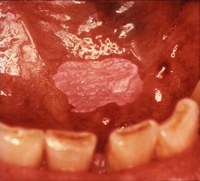
Homogeneous leukoplakia on the floor of the mouth in a smoker
Image: “Homogenous leukoplakia” by National Institutes of Health. License: Public Domain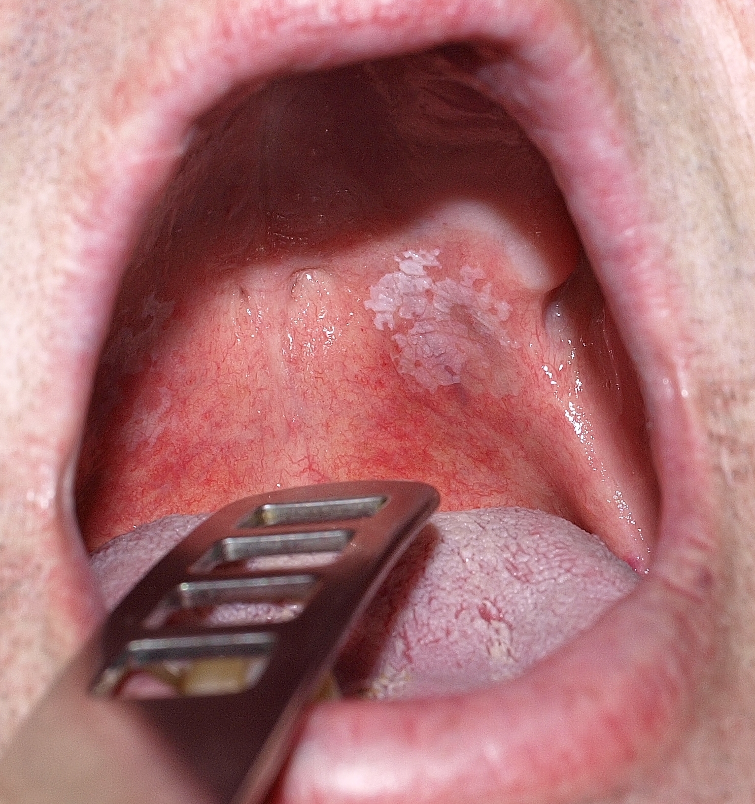
Homogeneous oral leukoplakia
Image: “Toxic leukoplakia of the oral mucosa in a 62-year-old man” by Klaus D. Peter. License: CC BY 3.0Nonhomogeneous leukoplakia Leukoplakia Leukoplakia is a potentially malignant lesion affecting the squamous epithelium usually within the oral cavity. Leukoplakia can be associated with a history of chronic tobacco and alcohol use, both of which can synergistically damage the epithelium. Leukoplakia presents a higher risk of malignant transformation Transformation Change brought about to an organism’s genetic composition by unidirectional transfer (transfection; transduction, genetic; conjugation, genetic, etc.) and incorporation of foreign DNA into prokaryotic or eukaryotic cells by recombination of part or all of that DNA into the cell’s genome. Bacteriology and may appear:
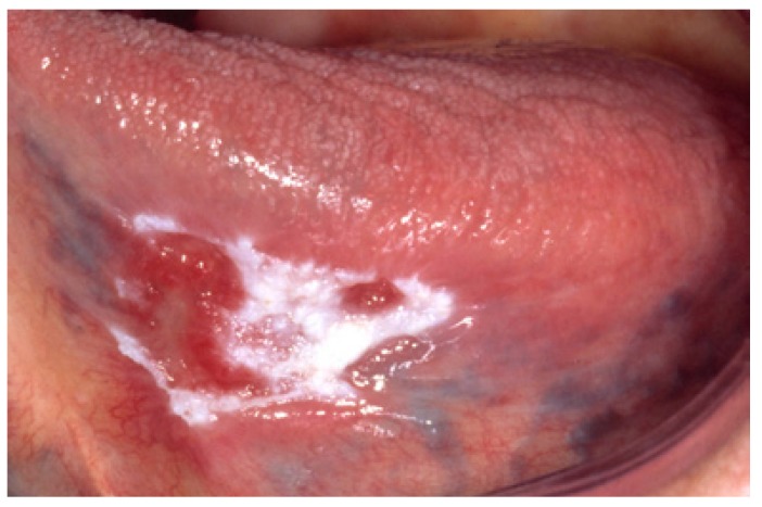
Nonhomogeneous leukoplakia of the tongue with white and red changes
Image: “Nonhomogeneous (white and red changes, also referred to as erythroleukoplakia) in an 88-year-old woman.” by Isaäc van der Waal. License: CC BY 2.5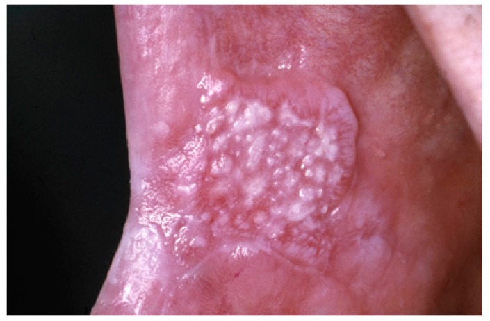
Nonhomogeneous oral leukoplakia with a nodular appearance
Image: “Nonhomogeneous, nodular, leukoplakia in a 61-year-old man” by Isaäc van der Waal. License: CC BY 2.5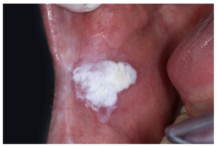
Nonhomogeneous oral leukoplakia with a verrucous texture
Image: “Nonhomogeneous, verrucous leukoplakia” by Isaäc van der Waal. License: CC BY 2.5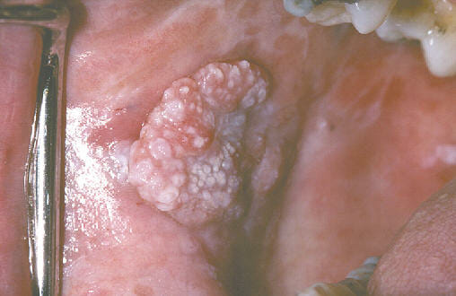
Nonhomogeneous exophytic leukoplakia
Image: “exophytic leukoplakia” by Aitor III. License: Public DomainLeukoplakia Leukoplakia Leukoplakia is a potentially malignant lesion affecting the squamous epithelium usually within the oral cavity. Leukoplakia can be associated with a history of chronic tobacco and alcohol use, both of which can synergistically damage the epithelium. Leukoplakia is a clinical term and diagnosis of exclusion. There are no specific histopathologic characteristics to define leukoplakia Leukoplakia Leukoplakia is a potentially malignant lesion affecting the squamous epithelium usually within the oral cavity. Leukoplakia can be associated with a history of chronic tobacco and alcohol use, both of which can synergistically damage the epithelium. Leukoplakia, but biopsy Biopsy Removal and pathologic examination of specimens from the living body. Ewing Sarcoma is performed to evaluate for dysplasia and to rule out other conditions. Potential findings may include:
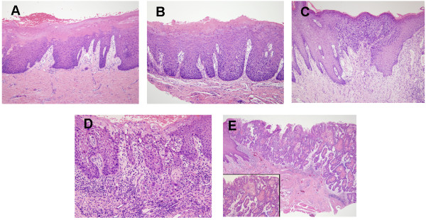
Histologic images of oral leukoplakia demonstrating progressive dysplasia and malignant transformation:
A: Hyperkeratosis with low-grade dysplasia present
B: Moderate dysplasia
C: High-grade dysplasia
D: Leukoplakia in the state of becoming invasive carcinoma
E: Invasive carcinoma
Medical therapy[9,14]
Medical therapy has very limited evidence available, and no reduction in the risk of SCC has been noted. The following have been used in an effort to heal lesions, but relapse Relapse Relapsing Fever is common:
Surgical management[3,8,14]
Photodynamic therapy Photodynamic Therapy Actinic Keratosis[3,8]
Prognosis Prognosis A prediction of the probable outcome of a disease based on a individual’s condition and the usual course of the disease as seen in similar situations. Non-Hodgkin Lymphomas[11‒14]
Diagnosis Codes:
This code is for
oral leukoplakia
Oral leukoplakia
A white patch seen on the oral mucosa. It is considered a premalignant condition and is often tobacco-induced. When evidence of Epstein-Barr virus is present, the condition is called hairy leukoplakia.
Oral Cancer, a condition characterized by thickened, white
patches
Patches
Vitiligo on the gums, inside of the
cheeks
Cheeks
The part of the face that is below the eye and to the side of the nose and mouth.
Melasma, or on the
tongue
Tongue
The tongue, on the other hand, is a complex muscular structure that permits tasting and facilitates the process of mastication and communication. The blood supply of the tongue originates from the external carotid artery, and the innervation is through cranial nerves.
Lips and Tongue: Anatomy that cannot be scraped off. It is considered a premalignant lesion.
| Coding System | Code | Description |
|---|---|---|
| ICD-10-CM | K13.21 | Leukoplakia Leukoplakia Leukoplakia is a potentially malignant lesion affecting the squamous epithelium usually within the oral cavity. Leukoplakia can be associated with a history of chronic tobacco and alcohol use, both of which can synergistically damage the epithelium. Leukoplakia of oral mucosa Oral mucosa Lining of the oral cavity, including mucosa on the gums; the palate; the lip; the cheek; floor of the mouth; and other structures. The mucosa is generally a nonkeratinized stratified squamous epithelium covering muscle, bone, or glands but can show varying degree of keratinization at specific locations. Stomatitis, including tongue Tongue The tongue, on the other hand, is a complex muscular structure that permits tasting and facilitates the process of mastication and communication. The blood supply of the tongue originates from the external carotid artery, and the innervation is through cranial nerves. Lips and Tongue: Anatomy |
| SNOMED CT | 95922000 | Leukoplakia Leukoplakia Leukoplakia is a potentially malignant lesion affecting the squamous epithelium usually within the oral cavity. Leukoplakia can be associated with a history of chronic tobacco and alcohol use, both of which can synergistically damage the epithelium. Leukoplakia of oral mucosa Oral mucosa Lining of the oral cavity, including mucosa on the gums; the palate; the lip; the cheek; floor of the mouth; and other structures. The mucosa is generally a nonkeratinized stratified squamous epithelium covering muscle, bone, or glands but can show varying degree of keratinization at specific locations. Stomatitis (disorder) |
Evaluation & Workup:
This CPT code is for a
biopsy
Biopsy
Removal and pathologic examination of specimens from the living body.
Ewing Sarcoma of the lesion, which is essential to rule out dysplasia (
precancerous
Precancerous
Pathological conditions that tend eventually to become malignant.
Barrett Esophagus changes) or invasive
squamous cell carcinoma
Squamous cell carcinoma
Cutaneous squamous cell carcinoma (cSCC) is caused by malignant proliferation of atypical keratinocytes. This condition is the 2nd most common skin malignancy and usually affects sun-exposed areas of fair-skinned patients. The cancer presents as a firm, erythematous, keratotic plaque or papule.
Squamous Cell Carcinoma (SCC).
| Coding System | Code | Description |
|---|---|---|
| CPT | 40808 | Biopsy Biopsy Removal and pathologic examination of specimens from the living body. Ewing Sarcoma, vestibule Vestibule An oval, bony chamber of the inner ear, part of the bony labyrinth. It is continuous with bony cochlea anteriorly, and semicircular canals posteriorly. The vestibule contains two communicating sacs (utricle and saccule) of the balancing apparatus. The oval window on its lateral wall is occupied by the base of the stapes of the middle ear. Ear: Anatomy of mouth |